shoulder ultrasound - bmus.org · shoulder ultrasound catherine kirkpatrick lincoln msk study day...
TRANSCRIPT
Shoulder Ultrasound
Catherine Kirkpatrick
Lincoln MSK Study Day
November 2017
Aims
• To revise ultrasound anatomy
• To revise some technique
• Identification of pathology
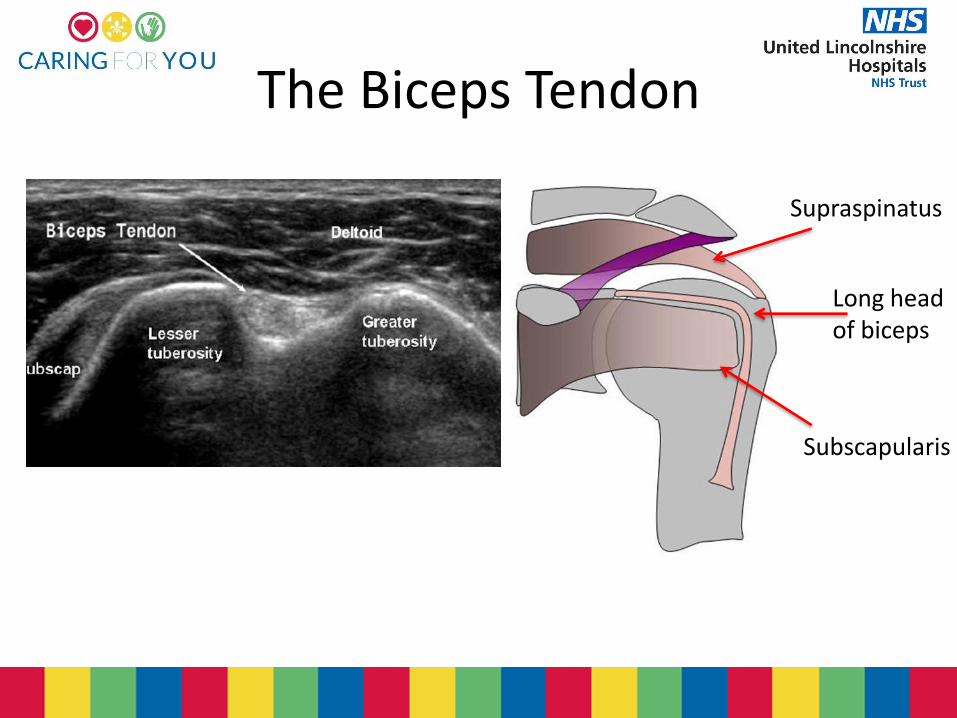
The Biceps Tendon
Supraspinatus
Long head of biceps
Subscapularis
SST
SSC
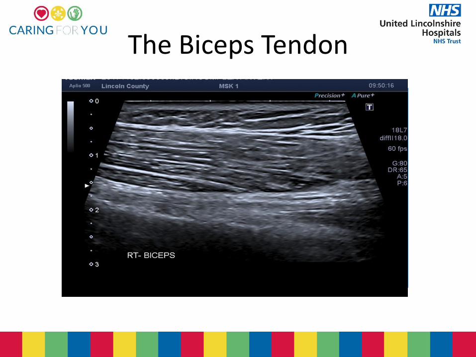
The Biceps Tendon
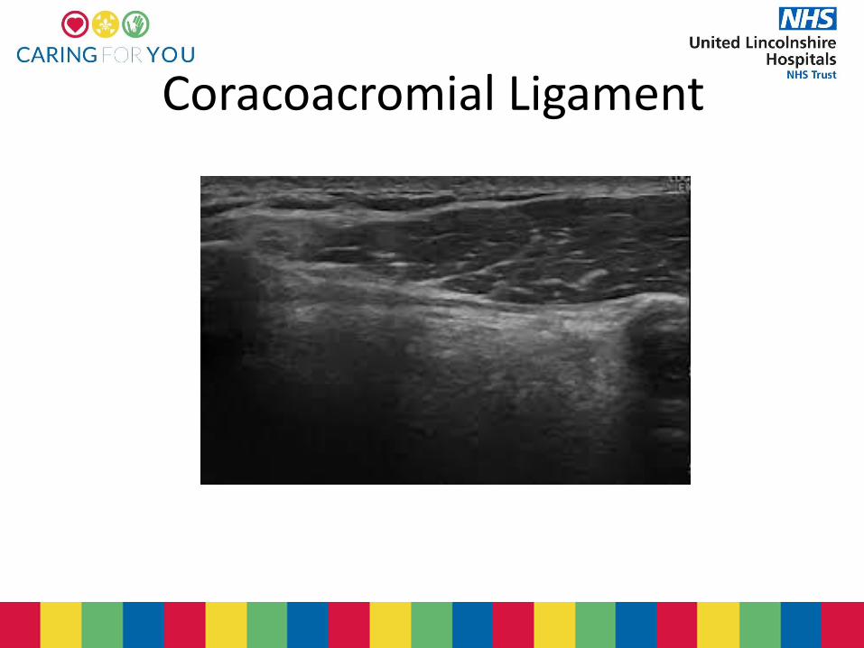
Coracoacromial Ligament
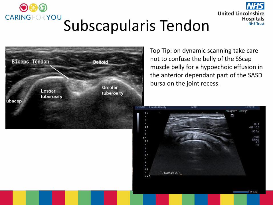
Subscapularis Tendon
Top Tip: on dynamic scanning take care not to confuse the belly of the SScap muscle belly for a hypoechoic effusion in the anterior dependant part of the SASD bursa on the joint recess.
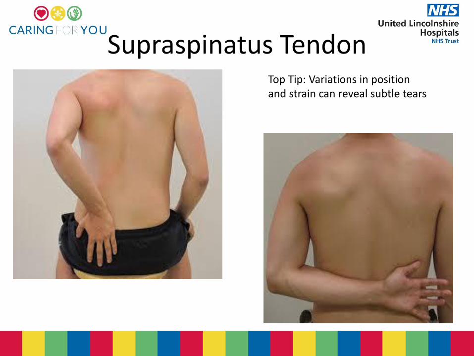
Supraspinatus Tendon Top Tip: Variations in position and strain can reveal subtle tears
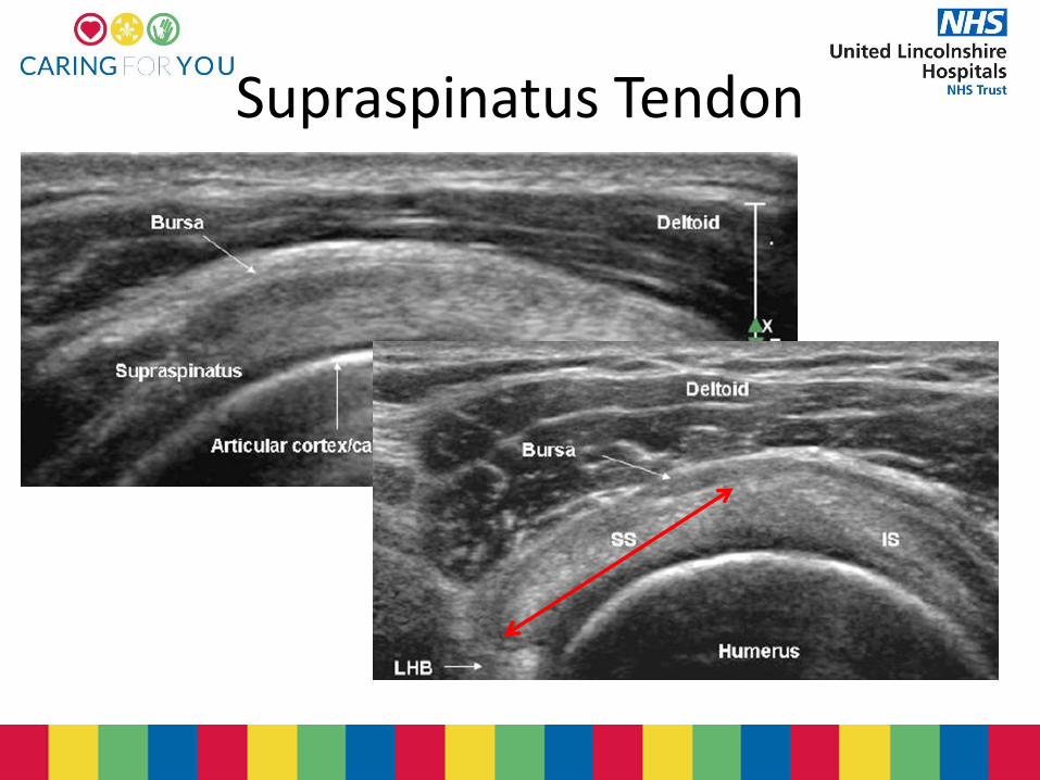
Supraspinatus Tendon
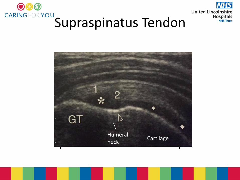
Supraspinatus Tendon
Humeral neck
Cartilage
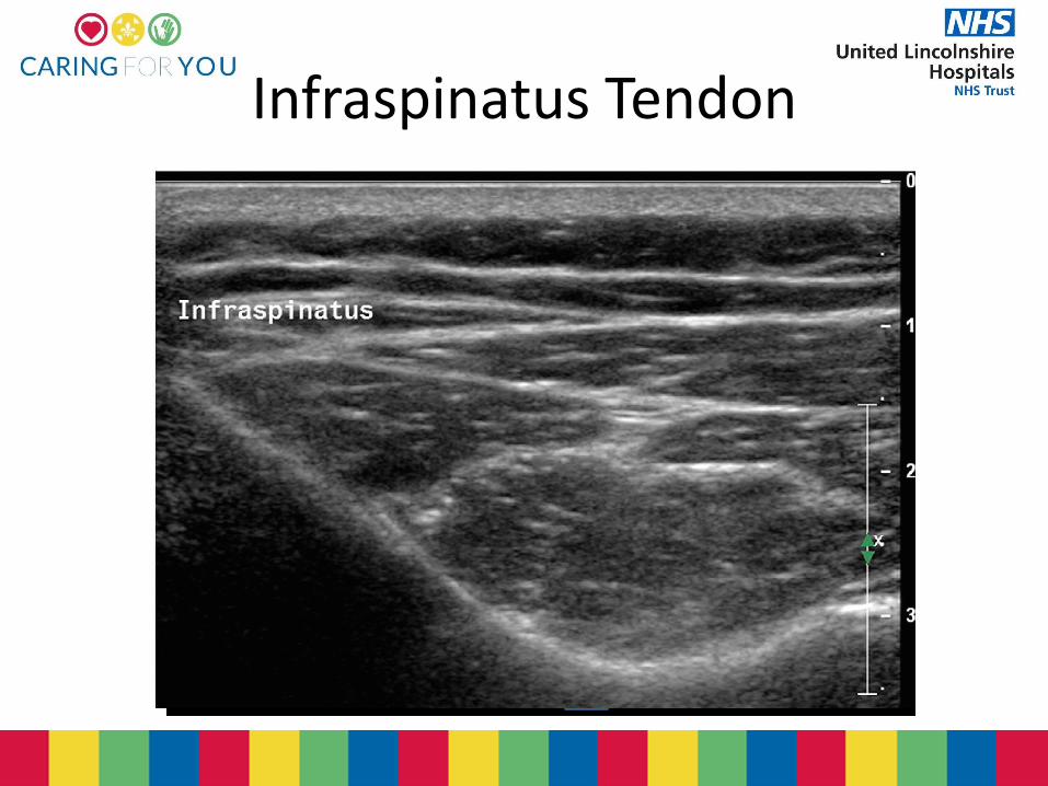
Infraspinatus Tendon
deltoid

Pathology
• Biceps Tendon
• Rotator Cuff
– Tears
– Tendinopathy
– Impingement
• Bursa
• The ACJ
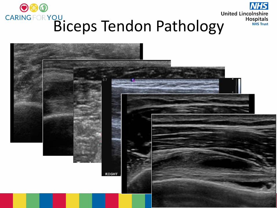
Biceps Tendon Pathology
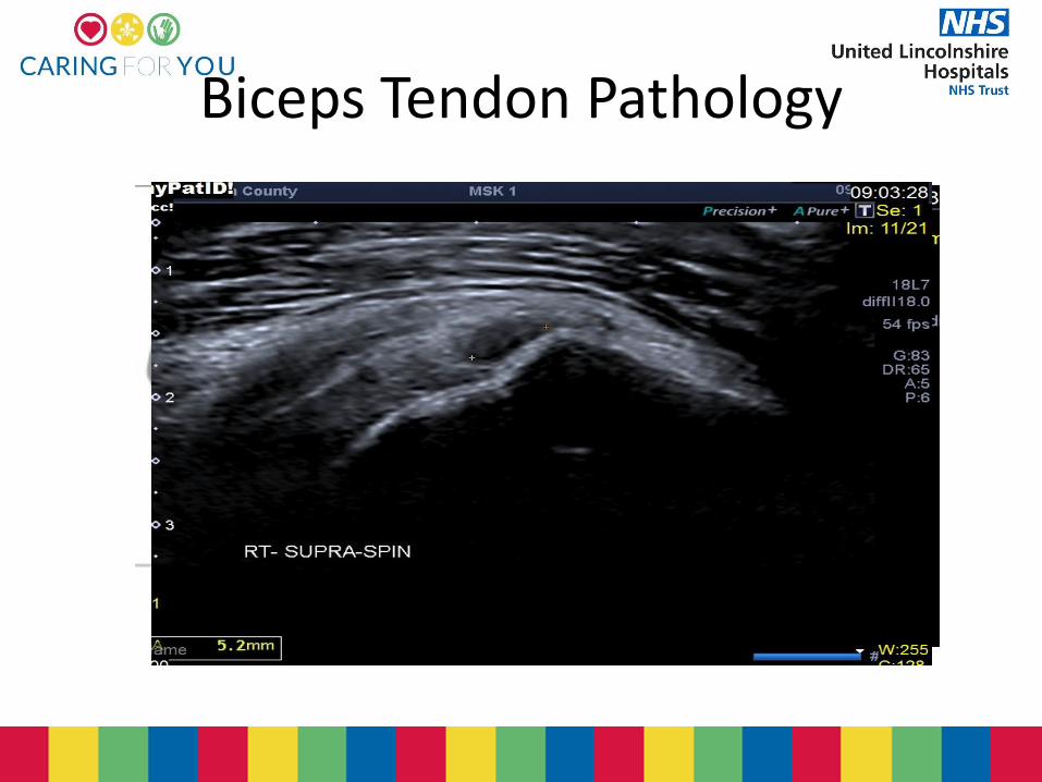
Biceps Tendon Pathology
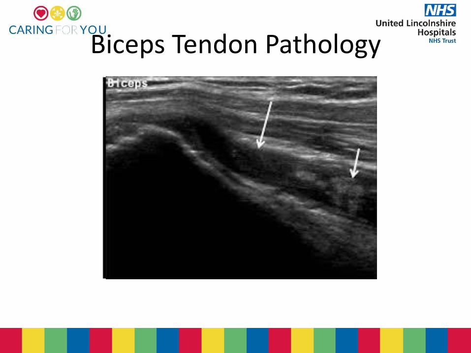
Biceps Tendon Pathology

Pathology of the Rotator Cuff
Prevalence of Cuff Tears
• 664 random people in a community – bilateral shoulders scanned
• 22.1% had full thickness tears
• Symptomatic rotator cuff tears accounted for 34.7% of all tears and asymptomatic tears for 65.3%
• The prevalence of asymptomatic rotator cuff tears was one-half of all tears in the 50s, whereas it accounted for two-thirds of those over the age of 60
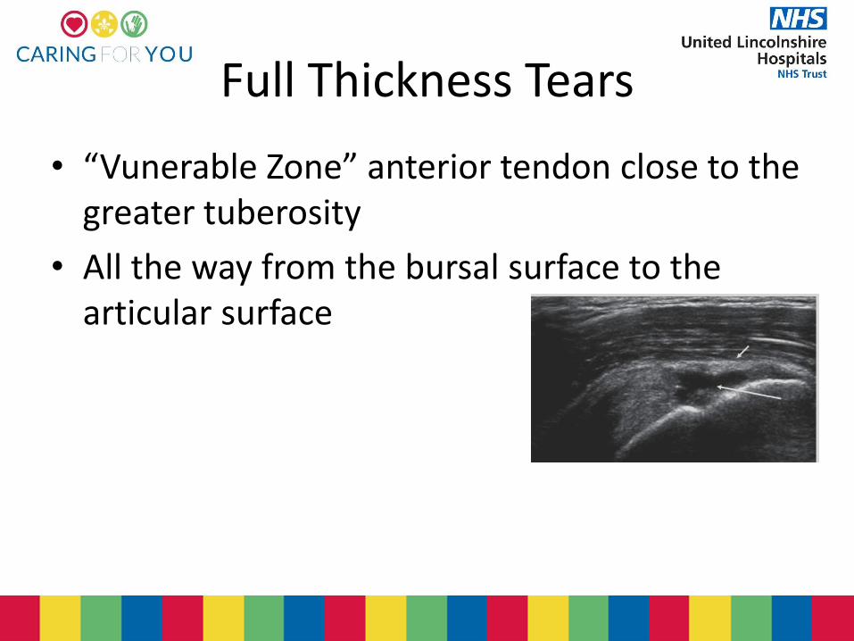
Full Thickness Tears
• “Vunerable Zone” anterior tendon close to the greater tuberosity
• All the way from the bursal surface to the articular surface
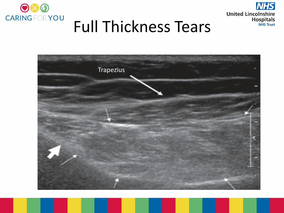
Full Thickness Tears
Trapezius
Full Thickness Tears
• GHJ effusion & hyperostosis of the GT has a PPV and Specificity of 100%
• Fluid in the SASD and GHJ has a PPV 95% & a specificity of 99%
• Fluid in the biceps tendon sheath & bursa PPV 54%
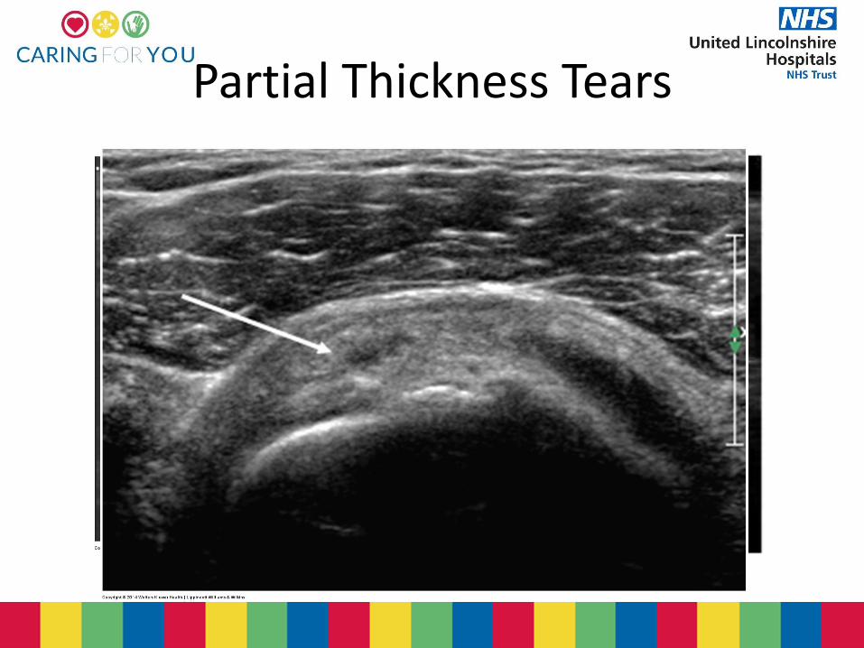
Partial Thickness Tears
• 13-18% of all rotator cuff tears
• Younger age group
• Small partial thickness tears & differentiation between from focal tendinopathy challenging
• However……..don’t have too many sleepless nights they are both treated conservatively
Partial Thickness Tears
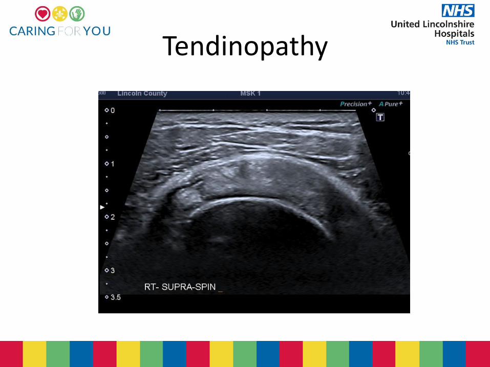
Tendinopathy
Tendinopathy
• Size difference between : a range from 1.5 to <2.5 mm difference to the contralateral side
• ≤8mm is considered abnormal
• Thickening and fluid within the SASD bursa
• Tendinosis can progress >intrasubstance>partial thickness>Full thickness
• Doppler??
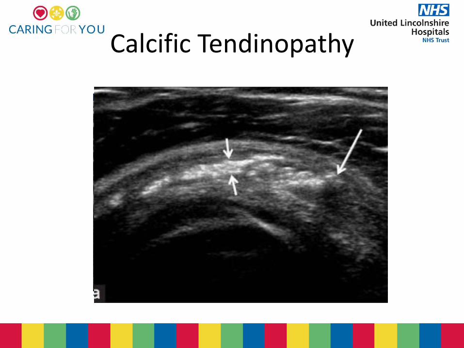
Calcific Tendinopathy
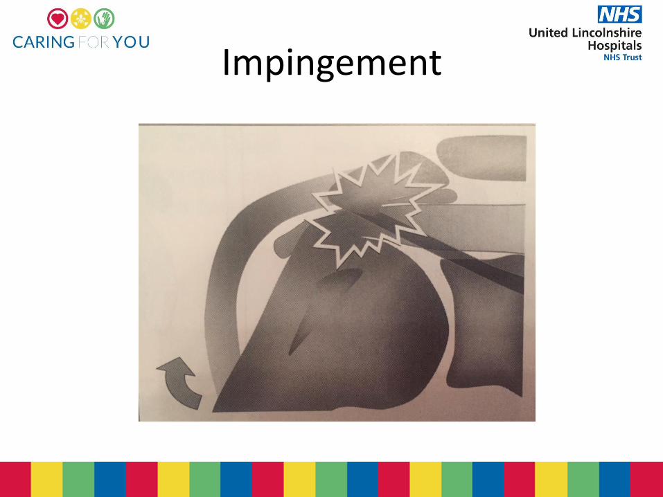
Impingement
Impingement
• Most often anterosuperior
• Involve the supraspinatus tendon and coracoacrominal arch
• Thickened or calcified Coracoacromial Ligament
• Thickened Bursa
• Tendinopathy
• Anatomic variations of the Acromion are a factor
Impingement
• Stage 1
Subacromial bursitis
Minor tendon changes
• Stage 2
Moderate to severe tendon changes
• Stage 3
Includes partial and full thickness tears
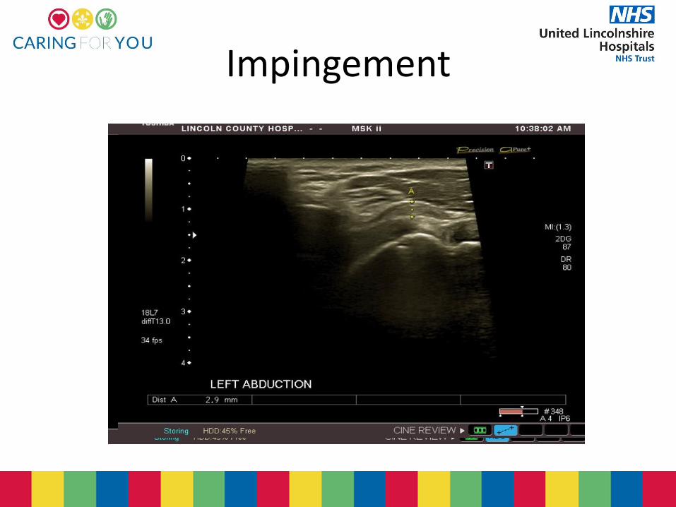
Impingement
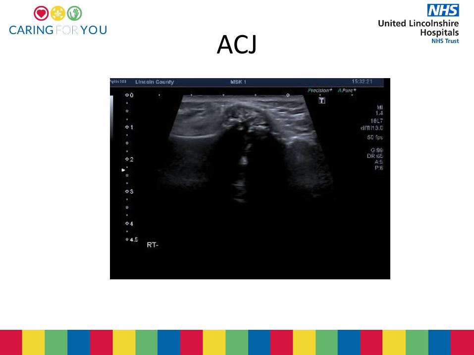
ACJ

So the point of this……
• The anatomy is the key to pathology and pitfalls
• A methodical approach to the cuff is essential
• The deeper our understanding of the anatomy the more advanced our work can be
• We can provide more useful and accurate reports for clinicians and the patient pathways

Enjoy the rest of the day
References
• http://pubs.rsna.org/doi/full/10.1148/radiol.11101082
• https://www.ncbi.nlm.nih.gov/pmc/articles/PMC3424700/
• https://www.ncbi.nlm.nih.gov/pmc/articles/PMC4799583/
• https://www.ncbi.nlm.nih.gov/pubmed/26666736
• http://www.journaloforthopaedicscience.com/article/S0949-2658(15)30226-8/fulltext
• http://ultrasoundcases.info/Slide-View.aspx?cat=322&case=4032
• https://www.ncbi.nlm.nih.gov/pmc/articles/PMC3768248/
• http://www.aafp.org/afp/1998/0215/p667.html
• https://www.ncbi.nlm.nih.gov/pmc/articles/PMC3785027/
• https://essr.org/content-essr/uploads/2016/10/shoulder.pdf
• Ferri et al. AJR 2005;184:180-184.
• Beggs I.2014. Musculoskeletal ultrasound. Lippincott Williams and Wilkins. London – IMAGES FROM HERE USED IN PRESENTATION
• Bianchi S, Martinoli C. 2007. Ultrasound of the Musculoskeletal System. Springer. New York -IMAGES FROM HERE USED IN PRESENTATION