severe bony ankylosis of the temporomandibular joint on ... · bony ankylosis of the...
TRANSCRIPT
- 103 -
Imaging Science in Dentistry 2015; 45: 103-8http://dx.doi.org/10.5624/isd.2015.45.2.103
Bony ankylosis of the temporomandibular joint (TMJ) is a fusion between the condyle and the temporal bone, which partially or completely obliterates the articular space and causes joint deformity. The fused bony frag-ment or a large bone mass may involve the condyle of the mandible, the temporal bone, and the zygomatic pro-cess. Thus, bony ankylosis can be easily diagnosed with conventional radiological tools such as panoramic view and computed tomography (CT).1-4 In 2002, El-Hakim and Metwalli4 reported a new classification of bony an-kylosis including surrounding vital structures (Table 1). This classification provides more information to surgeons during the pre-surgical period and during surgery.4 The common causes of ankylosis are trauma (31%-98%), in-
fection (10%-49%), and systemic diseases. TMJ ankylo-sis negatively affects the patient’s quality of life. Because the mouth opening is impaired, mastication and speech are difficult. Generalized caries, poor oral hygiene, fa-cial asymmetry, and obstructive sleep apnea may also occur.5-7 The pathogenesis of TMJ adhesion is not yet fully understood.8-10 Kaminishi and Davis8 suggested two mechanisms for TMJ adhesion. In the first mechanism, synovitis induces a fibrin layer on the articular surface; this layer becomes the fibrous tissue. In the second mech-anism, joint space hematomas become scar-like fibrous tissue during healing. This may eventually develop into fibrous adhesion. Since TMJ adhesion limits anterior and posterior TMJ movements, the mouth opening becomes impaired and is frequently associated with pain.9-11 To the best of our knowledge, only a few cases of TMJ ankylo-sis on one side and TMJ adhesion on the other side have been reported. In this paper, we report a rare case of TMJ ankylosis on one side and severe TMJ adhesion on the
Severe bony ankylosis of the temporomandibular joint on one side and contralateral adhesion: A case report
Ji-Young Song1,*, Seong-Gon Kim2, Hang-Moon Choi3, Hyun Jung Kim4
1Department of Oral and Maxillofacial Surgery, Jeju National University Hospital, Jeju National University School of Medicine, Jeju, Korea 2Department of Oral and Maxillofacial Surgery, School of Dentistry, Gangneung-Wonju National University, Gangneung, Korea 3Department of Oral and Maxillofacial Radiology, School of Dentistry, Gangneung-Wonju National University, Gangneung, Korea 4Department of Anesthesiology and Pain Medicine, Jeju National University School of Medicine, Jeju, Korea
AbStrAct
Bony fusion between the mandibular condyle and skull base involves temporomandibular joint (TMJ) bony ankylosis. This condition might originate from trauma, infection, or systemic disease. TMJ adhesion can develop after synovial damage. Both TMJ ankylosis and adhesion lead to functional impairment and pain. Here, we present a case of a 50-year-old female who had bony ankylosis of the right TMJ and adhesion of the left TMJ. She had otitis media in the right ear. A large mass in the right TMJ was observed on computed tomograph. Magnetic resonance image showed a large fused bone mass with normal bone marrow in the right TMJ and flattening of the condyle with a thin disk in the left TMJ. Gap arthroplasty with temporal fascia was performed on the right TMJ, and discectomy, high condylectomy, and coronoidectomy were performed on the left TMJ. During a 2-year follow-up after surgery, the patient had no recurrence. (Imaging Sci Dent 2015; 45: 103-8)
Key wordS: Temporomandibular Joint; Ankylosis; Arthroplasty; Mouth Rehabilitation
Copyright ⓒ 2015 by Korean Academy of Oral and Maxillofacial RadiologyThis is an Open Access article distributed under the terms of the Creative Commons Attribution Non-Commercial License (http://creativecommons.org/licenses/by-nc/3.0)
which permits unrestricted non-commercial use, distribution, and reproduction in any medium, provided the original work is properly cited.Imaging Science in Dentistry·pISSN 2233-7822 eISSN 2233-7830
Received January 9, 2015; Revised March 27, 2015; Accepted April 2, 2015 *Correspondence to : Prof. Ji-Young SongDepartment of Oral and Maxillofacial Surgery, Jeju National University Hospital, Aran 13 gil 15, Jeju-si, Jeju Special Self-Governing Province 690-767, KoreaTel) 82-64-717-1845, Fax) 82-64-717-1102, E-mail) [email protected]
*홀수페이지 시작.*표 선 두께=1/0.3pt
Severe bony ankylosis of the temporomandibular joint on one side and contralateral adhesion: A case report
- 104 -
opposite side and discuss the radiological diagnosis and treatment of the same.
case reportA 50-year-old female was referred to the Department
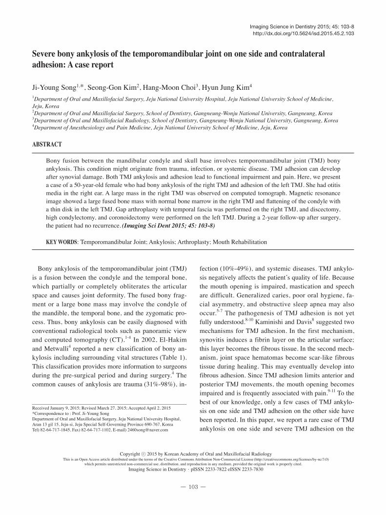
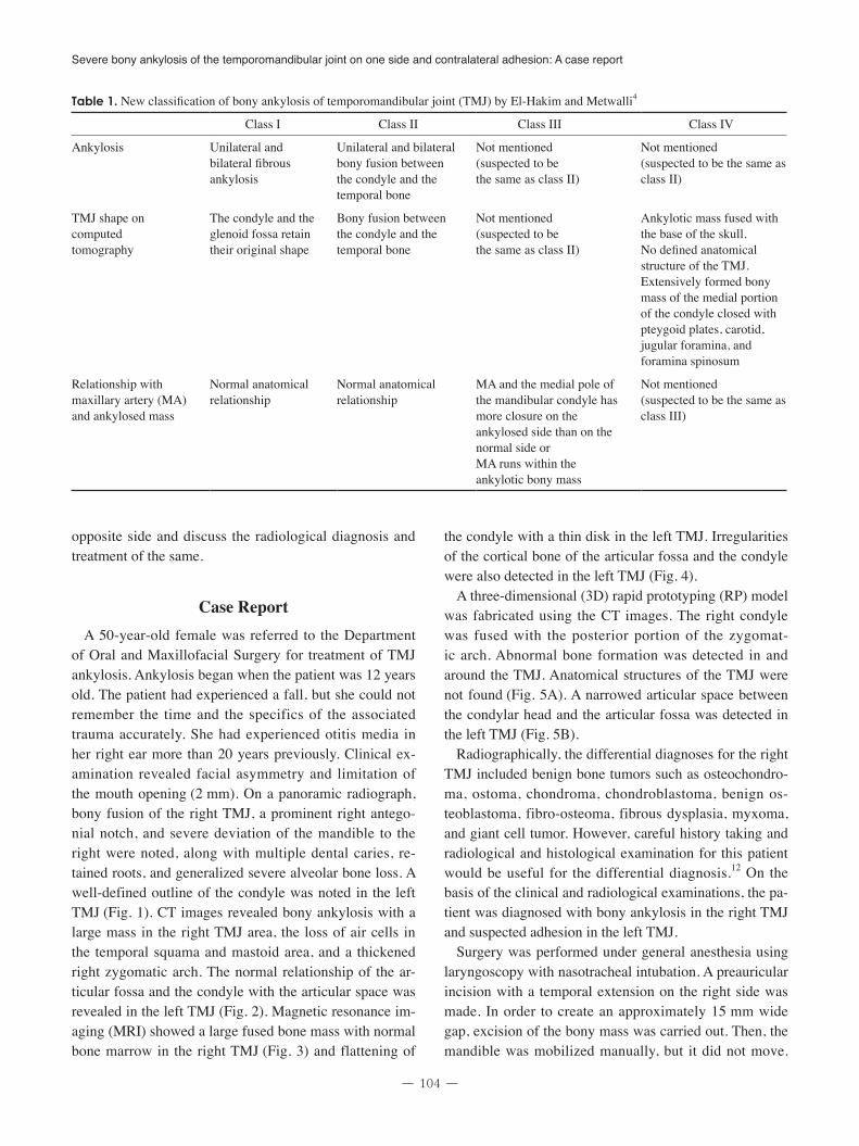
of Oral and Maxillofacial Surgery for treatment of TMJ ankylosis. Ankylosis began when the patient was 12 years old. The patient had experienced a fall, but she could not remember the time and the specifics of the associated trauma accurately. She had experienced otitis media in her right ear more than 20 years previously. Clinical ex-amination revealed facial asymmetry and limitation of the mouth opening (2 mm). On a panoramic radiograph, bony fusion of the right TMJ, a prominent right antego-nial notch, and severe deviation of the mandible to the right were noted, along with multiple dental caries, re-tained roots, and generalized severe alveolar bone loss. A well-defined outline of the condyle was noted in the left TMJ (Fig. 1). CT images revealed bony ankylosis with a large mass in the right TMJ area, the loss of air cells in the temporal squama and mastoid area, and a thickened right zygomatic arch. The normal relationship of the ar-ticular fossa and the condyle with the articular space was revealed in the left TMJ (Fig. 2). Magnetic resonance im-aging (MRI) showed a large fused bone mass with normal bone marrow in the right TMJ (Fig. 3) and flattening of
the condyle with a thin disk in the left TMJ. Irregularities of the cortical bone of the articular fossa and the condyle were also detected in the left TMJ (Fig. 4).
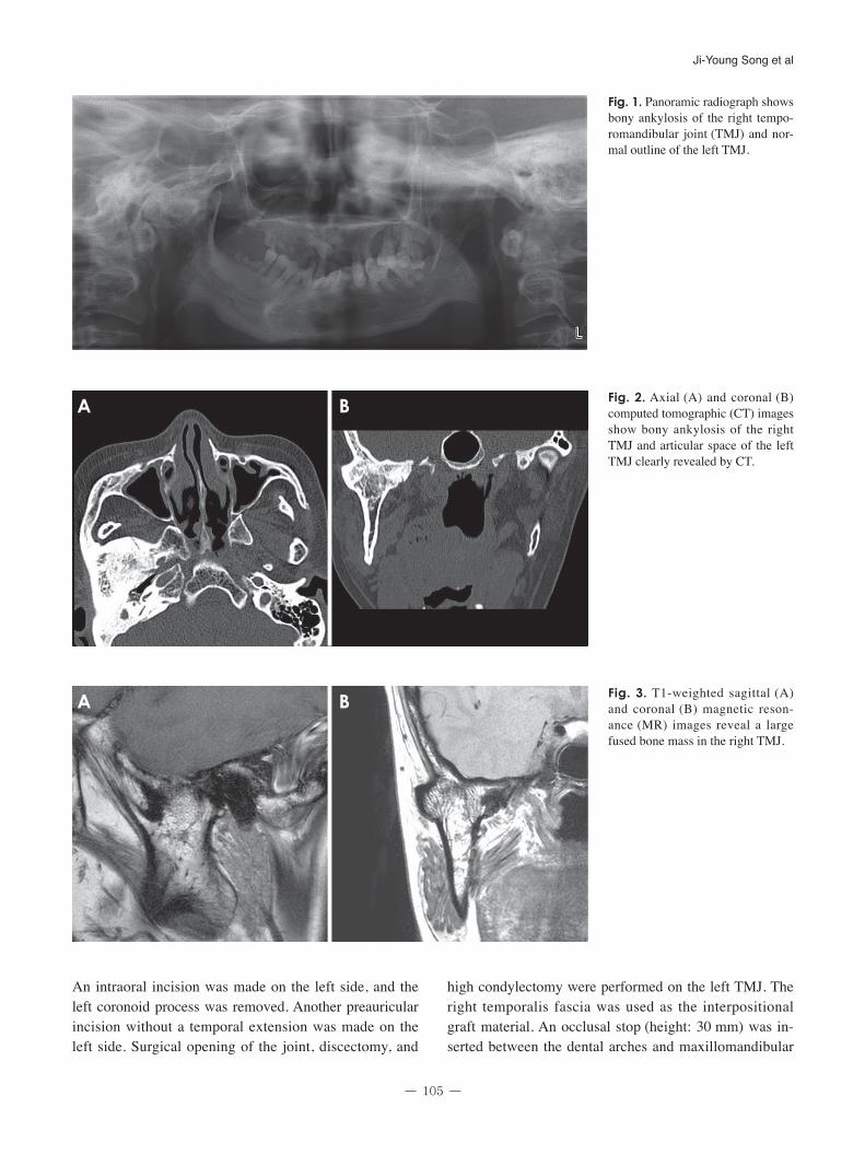
A three-dimensional (3D) rapid prototyping (RP) model was fabricated using the CT images. The right condyle was fused with the posterior portion of the zygomat-ic arch. Abnormal bone formation was detected in and around the TMJ. Anatomical structures of the TMJ were not found (Fig. 5A). A narrowed articular space between the condylar head and the articular fossa was detected in the left TMJ (Fig. 5B).
Radiographically, the differential diagnoses for the right TMJ included benign bone tumors such as osteochondro-ma, ostoma, chondroma, chondroblastoma, benign os-teoblastoma, fibro-osteoma, fibrous dysplasia, myxoma, and giant cell tumor. However, careful history taking and radiological and histological examination for this patient would be useful for the differential diagnosis.12 On the basis of the clinical and radiological examinations, the pa-tient was diagnosed with bony ankylosis in the right TMJ and suspected adhesion in the left TMJ.
Surgery was performed under general anesthesia using laryngoscopy with nasotracheal intubation. A preauricular incision with a temporal extension on the right side was made. In order to create an approximately 15 mm wide gap, excision of the bony mass was carried out. Then, the mandible was mobilized manually, but it did not move.
Table 1. New classification of bony ankylosis of temporomandibular joint (TMJ) by El-Hakim and Metwalli4
Class I Class II Class III Class IV
Ankylosis Unilateral andbilateral fibrous ankylosis
Unilateral and bilateral bony fusion between the condyle and the temporal bone
Not mentioned(suspected to be the same as class II)
Not mentioned(suspected to be the same as class II)
TMJ shape on computed tomography
The condyle and the glenoid fossa retain their original shape
Bony fusion between the condyle and the temporal bone
Not mentioned(suspected to be the same as class II)
Ankylotic mass fused with the base of the skull. No defined anatomical structure of the TMJ.Extensively formed bony mass of the medial portion of the condyle closed with pteygoid plates, carotid, jugular foramina, and foramina spinosum
Relationship with maxillary artery (MA) and ankylosed mass
Normal anatomical relationship
Normal anatomical relationship
MA and the medial pole of the mandibular condyle has more closure on the ankylosed side than on the normal side or MA runs within the ankylotic bony mass
Not mentioned(suspected to be the same as class III)
- 105 -
Ji-Young Song et al
An intraoral incision was made on the left side, and the left coronoid process was removed. Another preauricular incision without a temporal extension was made on the left side. Surgical opening of the joint, discectomy, and
high condylectomy were performed on the left TMJ. The right temporalis fascia was used as the interpositional graft material. An occlusal stop (height: 30 mm) was in-serted between the dental arches and maxillomandibular
Fig. 1. Panoramic radiograph shows bony ankylosis of the right tempo-romandibular joint (TMJ) and nor-mal outline of the left TMJ.
Fig. 2. Axial (A) and coronal (B) computed tomographic (CT) images show bony ankylosis of the right TMJ and articular space of the left TMJ clearly revealed by CT.
A B
Fig. 3. T1-weighted sagittal (A) and coronal (B) magnetic reson-ance (MR) images reveal a large fused bone mass in the right TMJ.
A B
Severe bony ankylosis of the temporomandibular joint on one side and contralateral adhesion: A case report
- 106 -
fixation (MMF) and was maintained for 5 days. The max-imum mouth opening (MMO) was 35 mm after surgery. Postoperative pain medications were administered, and vigorous postoperative physiotherapy was performed to preserve the range of motion and to prevent postoperative hypomobility due to recurrence.5,13
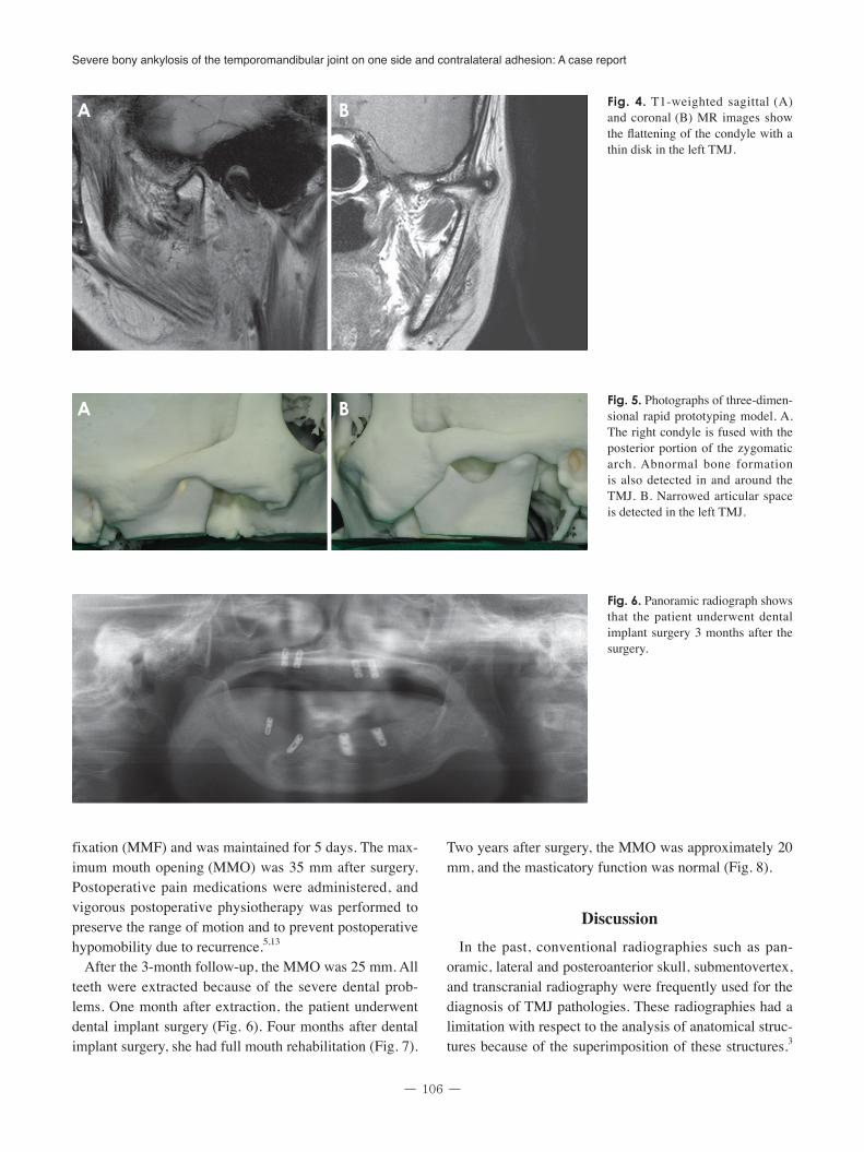
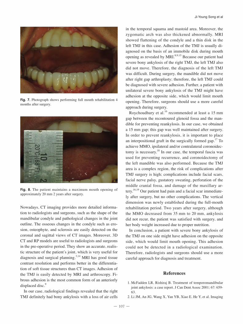
After the 3-month follow-up, the MMO was 25 mm. All teeth were extracted because of the severe dental prob-lems. One month after extraction, the patient underwent dental implant surgery (Fig. 6). Four months after dental implant surgery, she had full mouth rehabilitation (Fig. 7).
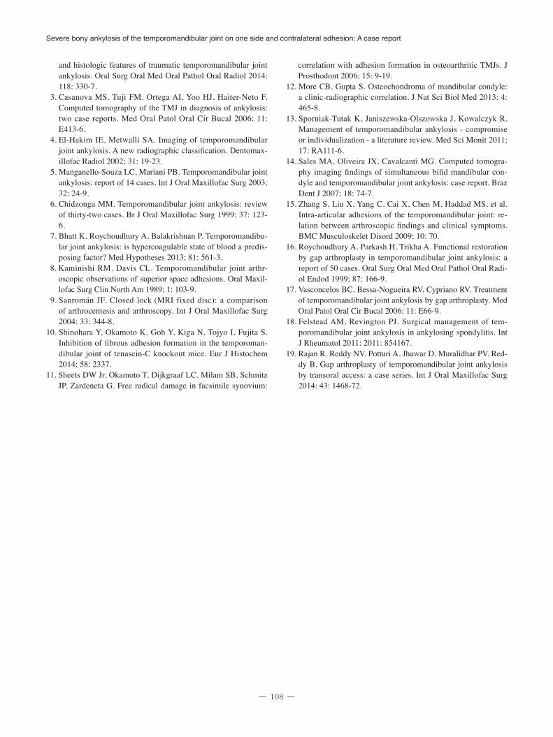
Two years after surgery, the MMO was approximately 20 mm, and the masticatory function was normal (Fig. 8).
discussionIn the past, conventional radiographies such as pan-
oramic, lateral and posteroanterior skull, submentovertex, and transcranial radiography were frequently used for the diagnosis of TMJ pathologies. These radiographies had a limitation with respect to the analysis of anatomical struc-tures because of the superimposition of these structures.3
Fig. 4. T1-weighted sagittal (A) and coronal (B) MR images show the flattening of the condyle with a thin disk in the left TMJ.
A B
Fig. 5. Photographs of three-dimen-sional rapid prototyping model. A. The right condyle is fused with the posterior portion of the zygomatic arch. Abnormal bone formation is also detected in and around the TMJ. B. Narrowed articular space is detected in the left TMJ.
A B
Fig. 6. Panoramic radiograph shows that the patient underwent dental implant surgery 3 months after the surgery.
- 107 -
Ji-Young Song et al
Nowadays, CT imaging provides more detailed informa-tion to radiologists and surgeons, such as the shape of the mandibular condyle and pathological changes in the joint outline. The osseous changes in the condyle such as ero-sion, osteophyte, and sclerosis are easily detected on the coronal and sagittal views of CT images. Moreover, 3D CT and RP models are useful to radiologists and surgeons in the pre-operative period. They show an accurate, realis-tic structure of the patient’s joint, which is very useful for diagnosis and surgical planning.3,14 MRI has good tissue contrast resolution and performs better in the differentia-tion of soft tissue structures than CT images. Adhesion of the TMJ is easily detected by MRI and arthroscopy. Fi-brous adhesion is the most common form of an anteriorly displaced disc.9
In our case, radiological findings revealed that the right TMJ definitely had bony ankylosis with a loss of air cells
in the temporal squama and mastoid area. Moreover, the zygomatic arch was also thickened abnormally. MRI showed flattening of the condyle and a thin disk in the left TMJ in this case. Adhesion of the TMJ is usually di-agnosed on the basis of an immobile disk during mouth opening as revealed by MRI.8,9,15 Because our patient had severe bony ankylosis of the right TMJ, the left TMJ also did not move. Therefore, the diagnosis of the left TMJ was difficult. During surgery, the mandible did not move after right gap arthroplasty; therefore, the left TMJ could be diagnosed with severe adhesion. Further, a patient with unilateral severe bony ankylosis of the TMJ might have adhesion at the opposite side, which would limit mouth opening. Therefore, surgeons should use a more careful approach during surgery.
Roychoudhury et al.16 recommended at least a 15 mm gap between the recontoured glenoid fossa and the man-dible for preventing reankylosis. In our case, we obtained a 15 mm gap; this gap was well maintained after surgery. In order to prevent reankylosis, it is important to place an interpositional graft in the surgically formed gap.17 To achieve MMO, ipsilateral and/or contralateral coronoidec-tomy is necessary.18 In our case, the temporal fascia was used for preventing recurrence, and coronoidectomy of the left mandible was also performed. Because the TMJ area is a complex region, the risk of complications after TMJ surgery is high; complications include facial scars, facial nerve palsy, gustatory sweating, perforation of the middle cranial fossa, and damage of the maxillary ar-tery.16,19 Our patient had pain and a facial scar immediate-ly after surgery, but no other complications. The vertical dimension was newly established during the full-mouth rehabilitation period. Two years after surgery, although the MMO decreased from 35 mm to 20 mm, ankylosis did not recur, the patient was satisfied with surgery, and her body weight increased due to proper nutrition.
In conclusion, a patient with severe bony ankylosis of the TMJ on one side might have adhesion on the opposite side, which would limit mouth opening. This adhesion could not be detected in a radiological examination. Therefore, radiologists and surgeons should use a more careful approach for diagnosis and treatment.
references
1. McFadden LR, Rishiraj B. Treatment of temporomandibular joint ankylosis: a case report. J Can Dent Assoc 2001; 67: 659-63.
2. Li JM, An JG, Wang X, Yan YB, Xiao E, He Y, et al. Imaging
Fig. 7. Photograph shows performing full mouth rehabilitation 4 months after surgery.
Fig. 8. The patient maintains a maximum mouth opening of approximately 20 mm 2 years after surgery.
Severe bony ankylosis of the temporomandibular joint on one side and contralateral adhesion: A case report
- 108 -
and histologic features of traumatic temporomandibular joint ankylosis. Oral Surg Oral Med Oral Pathol Oral Radiol 2014; 118: 330-7.
3. Casanova MS, Tuji FM, Ortega AI, Yoo HJ, Haiter-Neto F. Computed tomography of the TMJ in diagnosis of ankylosis: two case reports. Med Oral Patol Oral Cir Bucal 2006; 11: E413-6.
4. El-Hakim IE, Metwalli SA. Imaging of temporomandibular joint ankylosis. A new radiographic classification. Dentomax-illofac Radiol 2002; 31: 19-23.
5. Manganello-Souza LC, Mariani PB. Temporomandibular joint ankylosis: report of 14 cases. Int J Oral Maxillofac Surg 2003; 32: 24-9.
6. Chidzonga MM. Temporomandibular joint ankylosis: review of thirty-two cases. Br J Oral Maxillofac Surg 1999; 37: 123-6.
7. Bhatt K, Roychoudhury A, Balakrishnan P. Temporomandibu-lar joint ankylosis: is hypercoagulable state of blood a predis-posing factor? Med Hypotheses 2013; 81: 561-3.
8. Kaminishi RM, Davis CL. Temporomandibular joint arthr-oscopic observations of superior space adhesions. Oral Maxil-lofac Surg Clin North Am 1989; 1: 103-9.
9. Sanromán JF. Closed lock (MRI fixed disc): a comparison of arthrocentesis and arthroscopy. Int J Oral Maxillofac Surg 2004; 33: 344-8.
10. Shinohara Y, Okamoto K, Goh Y, Kiga N, Tojyo I, Fujita S. Inhibition of fibrous adhesion formation in the temporoman-dibular joint of tenascin-C knockout mice. Eur J Histochem 2014; 58: 2337.
11. Sheets DW Jr, Okamoto T, Dijkgraaf LC, Milam SB, Schmitz JP, Zardeneta G. Free radical damage in facsimile synovium:
correlation with adhesion formation in osteoarthritic TMJs. J Prosthodont 2006; 15: 9-19.
12. More CB, Gupta S. Osteochondroma of mandibular condyle: a clinic-radiographic correlation. J Nat Sci Biol Med 2013; 4: 465-8.
13. Sporniak-Tutak K, Janiszewska-Olszowska J, Kowalczyk R. Management of temporomandibular ankylosis - compromise or individualization - a literature review. Med Sci Monit 2011; 17: RA111-6.
14. Sales MA, Oliveira JX, Cavalcanti MG. Computed tomogra-phy imaging findings of simultaneous bifid mandibular con-dyle and temporomandibular joint ankylosis: case report. Braz Dent J 2007; 18: 74-7.
15. Zhang S, Liu X, Yang C, Cai X, Chen M, Haddad MS, et al. Intra-articular adhesions of the temporomandibular joint: re-lation between arthroscopic findings and clinical symptoms. BMC Musculoskelet Disord 2009; 10: 70.
16. Roychoudhury A, Parkash H, Trikha A. Functional restoration by gap arthroplasty in temporomandibular joint ankylosis: a report of 50 cases. Oral Surg Oral Med Oral Pathol Oral Radi-ol Endod 1999; 87: 166-9.
17. Vasconcelos BC, Bessa-Nogueira RV, Cypriano RV. Treatment of temporomandibular joint ankylosis by gap arthroplasty. Med Oral Patol Oral Cir Bucal 2006; 11: E66-9.
18. Felstead AM, Revington PJ. Surgical management of tem-poromandibular joint ankylosis in ankylosing spondylitis. Int J Rheumatol 2011; 2011: 854167.
19. Rajan R, Reddy NV, Potturi A, Jhawar D, Muralidhar PV, Red-dy B. Gap arthroplasty of temporomandibular joint ankylosis by transoral access: a case series. Int J Oral Maxillofac Surg 2014; 43: 1468-72.