serum estradiol measurement by solid-phase chemiluminescence immunoassay and direct radioimmunoassay
TRANSCRIPT

Analytica Chimicn Actu, 170 (1985) 117-123
Elsevler Science Publishers B.V., Amsterdam - Printed in The Netherlands
SERUM ESTRADIOL MEASUREMENT BY SOLID-PHASE CHEMILUMINESCENCE IMMUNOASSAY AND DIRECT RADIOIMMUNOASSAY
JOZEF DE BOEVER*, FORTUNA KOHENa, MARC DHONT and DIRK VANDEKERCKHOVE
Department of Obstetrics and Gynaecology, Academic Hospital, De Pmtelann 185, B-9000 Gent (Belgzum)
GEORGES VAN MAELE
Department of Medical Inform&on, Academic Hospital, De Pmtelaan 185, B-9000 Gent (Belgium)
(Received September 5th 1984)
SUMMARY
Estradlol-170 is determined in serum extracts by solid-phase chemilummescence immunoassay The results are compared with those obtained from unextracted serum in routine conditions with a commercial radioimmunoassay (r.1.a.) kit. For the chemi- luminescence procedure, a purified monoclonal antibody to estradiol-6-carboxymethyl- oxnne/bovine serum albumin and the homologous chemiluminescent marker conlugate estradiol-6-carboxymethyloxime aminobutylethylisoluminol are used. Bound and free ligand are separated by washing and simple centrifugation. Results obtained by the chemilummescence assay (y) and by r.1.a. (x) on 170 serum specimens from women durmg ovulation induction showed good correlation (y = 1.01x - 16 with r = 0 95). The methods are similar m selectivity, detection limit (ca. 10 ng 1-l) and precision (interassay relative standard deviation, 8-13%).
Successful monitoring of ovulation induction requires the sequential analysis of one or several hormones in peripheral blood or in urine [l-3]. Daily measurement of estrogens (e.g., estradiol) in serum or plasma has become the method of choice in many laboratories [4-S]. This demands rapid and reliable assays, which have indeed been developed; for some years, commercial reagent kits for the direct measurement of estradiol by radio- immunoassay (r.i.a.) have been available.
Radioimmunoassay, however, is associated with health hazards, the short half-life and radiolysis of labelled reagents and radioactive waste disposal. Recent studies from several laboratories have indicated that chemiluminescent markers can be a feasible alternative to radiolabelling. Such markers have been used in developing reliable solid-phase chemiluminescence immuno-
aPermanent address. Department of Hormone Research, The Weizmann Institute of Science, Rehovot 76100, Israel.
0003-2670/85/$03.30 0 1985 Elsevier Science Publishers B V.
118
assays for steroids in serum or plasma [ 7-91. This paper compares the results of estradiol measurement by a solid-phase chemilummescence immunoassay after extraction from serum, with those obtained routinely with direct r.1.a. without extraction.
EXPERIMENTAL
Sample collection and extraction Blood samples were obtained by venipuncture from patients during treat-
ment for ovulation inducation with menopausal gonadotropins and chorio- gonadotropin. Sera were stored frozen at -20°C until assayed. A l-ml portion of serum was extracted with 7.5 ml of diethyl ether. The mean efficiency of extraction, as found by measuring the recovery of tritiated estradiol added to the serum samples, was 96.0% (s.d. = 2.6%). The ether phase was evaporated under nitrogen and the dry residue was dissolved in 0.5 ml of assay buffer. Duplicate O.l-ml aliquots were analysed by chemi- luminescence immunoassay.
Procedures Chemllummescence immunoassay. Serum levels of estradiol were assayed
by the solid-phase chemiluminescence assay technique described by De Boever et al. [9] . The assay uses a monoclonal antibody to estradiol-6- carboxymethyloxime/bovine serum albumin and the chemiluminescent marker conjugate, estradiol-6-carboxymethyloxime aminobutylethyl- isoluminol (E,-ABEI). Portions (0.1 ml) of solid-phase antibody, reconstituted serum extract or standard and steroid-marker conjugate (i.e., 100 pg of E,- ABEI) are added to lumacuvettes (12 X 50 mm polystyrene cuvettes; Lumac Systems, Basel, Switzerland). After incubation for 1 h or overnight at 4°C bound and free ligand are separated by adding 0.9 ml of wash solution, centrifugation (10 min, 2OOOg, room temperature), and decantation of the supernate. To the precipitate, 0.2 ml of 2 mol 1-l sodium hydroxide is added. The mixture is incubated for 30 min at 60°C and then cooled to room tem- perature. The amount of bound ligand marker is measured by adding 0.1 ml of diluted (10 pg/ml) microperoxidase and 0.1 ml of x% = 0.2% hydrogen peroxide solution and integrating the light emitted for 10 s with a luminom- eter (Biocounter M-2000; Lumac).
Dzrect radioimmunoassay. The commercial EIR “‘1-estradiol direct r.1.a. kit (code ER-155, EIR, Radio-isotopen Service, 5303 Wiirenlingen, Switzerland) was used. The assay has been described in detail [lo]. Briefly, duplicate 50-~1 amounts of standards or unknown serum samples are added to glass tubes and incubated with 100 ~1 of ‘251-labelled estradiol and 100 ~1 of primary antiserum for 2 h at 37°C. Incubation with 0.5 ml of solid-phase second antibody for 30 min at 37” C is followed by centrifugation (2OOOg, 10 min, 4”C), and decantation to separate the bound and free hgand. The radioactivity in the pellet was measured with a model 1270 Rackgamma- counter (LKB Wallac, Bromma, Sweden).

119
Data procesmg. Non-specific blank values were subtracted from the counts obtained in the bound fraction. Calibration graphs were constructed by plotting B/B, values vs. log dose of the standards (B = net signal at a particular estradiol concentration, I?,, = net signal in the absence of estradiol). The extraction efficiency data were used to calculate estradiol concentrations in serum as determined by the chemiluminescence immunoassay. Best-fit linear regression lines were calculated as described by Davies [ll] . The method described by Jeffcoate [ 121 was used to construct precision profiles; data from more than 100 duplicate measurements in consecutive assays were used to calculate the linear least-squares fit of the mean counts from the bound material (response) and the standard deviation (s.d.) of the duplicates. Response replicates whose s.d. was 7% or more were rejected. The mean dose/ response curve and the response/s.d. relationship (i.e., the error-envelope) was constructed. With this curve, the error in dose over the entire dose range was calculated and precision profiles were constructed by plotting the error in dose (corrected for the use of duplicates) vs. the dose.
RESULTS
Senshvlty and selectivity The least amount of estradiol that could be distinguished from zero (mean
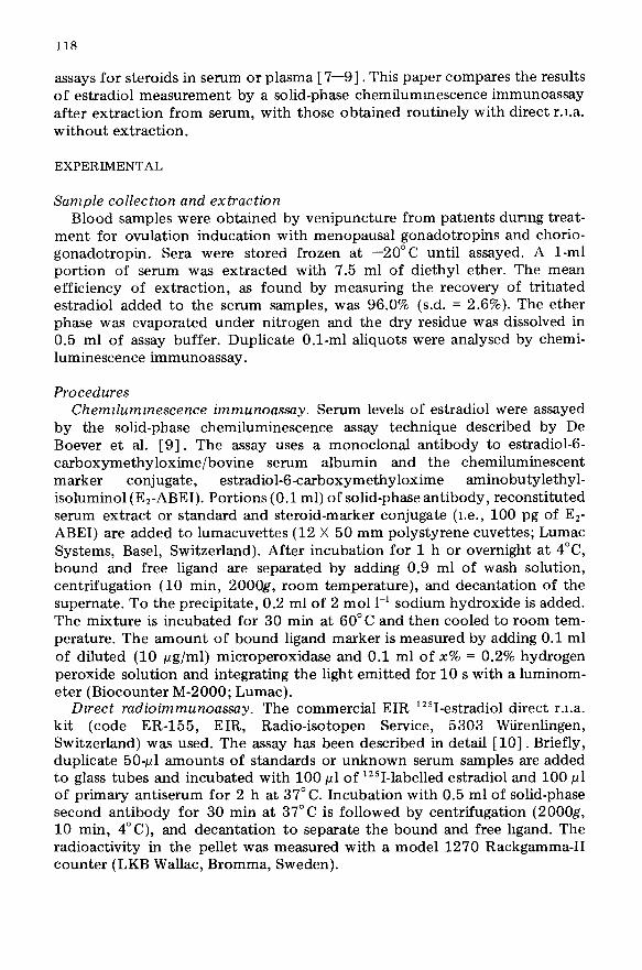
- 2s.d.) was calculated from five chemiluminescence immunoassay and six- teen r.i.a. consecutive calibration curves prepared in duplicate (Fig. 1). The values were 12.5 ng 1-l for chemiluminescence assay and 7.4 ng 1-l for direct r.i.a. Similar values were obtained from the precision profiles.
For both methods, cross-reactivity data are given for 50% inhibition of binding of labelled ligand. For r.i.a., the cross-reactivities as indicated by the kit manufacturer were (estradiol = 100%): estrone and estriol, 2%; ethinyl- estradiol, progesterone, testosterone, and rostanediol, <O.l%; estradiol-3- glucuronide, 0.04%; estradiol-l7-glucuronide, 0.07%. The cross-reactivitles found in the solid-phase chemiluminescence immunoassay were (estradiol = 100%): estrone, 0.7%; estriol, 45%; ethinylestradiol, 0.5%; progesterone, test- osterone, 5c+androstane-3a,l7P diol, estrone-3-sulphate and estradiol-17/3- glucuronide, < 0 .O 1%.
Precisslon and accuracy Figure 2 shows within-batch precision profiles for both assays. At relative
standard deviations of <lo%, the working range is 12-625 ng 1-l for the chemiluminescence procedure and 15-1000 ng 1-l for the r.i.a. Withm-batch precisions for the chemiluminescence assay estimated from 9 replicate samples analysed in duplicate were 7.2%, 9.4% and 8.4% at 150, 700 and 1800 ng l-l, respectively, within a single assay. The within-batch precision of the r.i.a. calculated from the variation of duplicate means in 30 consecutive assays was 8.9% (51 pairs) and 11.9% (76 pairs) over the ranges 23-145 and 151-850 ng 1-l. Between-batch precision, calculated from replicate deter- minations in consecutive assays were, for the chemiluminescence immuno-
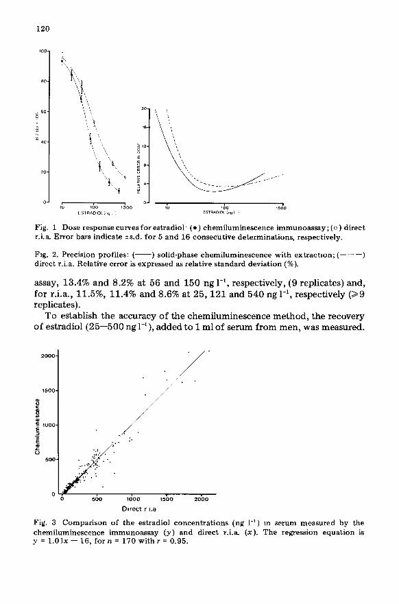
Fig. 1 Dose response curves for estradiol. (0) chemiluminescence immunoassay; (0) direct r.i.a. Error bars indicate +s.d. for 5 and 16 consecutive determinations, respectively.
Fig. 2. Precision profiles: (-) solid-phase chemiluminescence with extractron; (---) direct r.i.a. Relative error is expressed as relative standard deviation (%).
assay, 13.4% and 8.2% at 56 and 150 ng l-l, respectively, (9 replicates) and, for r.i.a., 11.5%, 11.4% and 8.6% at 25,121 and 540 ng l-l, respectively (29 replicates).
To establish the accuracy of the chemiluminescence method, the recovery of estradiol (25-500 ng l-l), added to 1 ml of serum from men, was measured. . . .
1500 : i 2ooom ; 1000 . 3
/ .
1000 1500 2000
Darect r ~.a
Fig. 3 Comparison of the estradiol concentrations (ng 1-l) m serum measured by the chemiluminescence immunoassay (y) and direct r.i.a. (x). The regression equation is y = 1.01.x - 16, for n = 170 with I = 0.95.

121
600
-L F
600
d E 400
i?
L-l w 200
0 hCG June 14--23
1350 I”, ““114 hMG i lS00 l”l ““l,S llMG T --
19 21 23 19 24
June
l
06 l J . . . . . . . . . . . .f
April m-May 18 hCG
4300 I”, un,ts llMG i I I I I I I
4 7 10 13 16 20
May
Fig. 4 Estradiol concentrations in serum from women (VA, OB) undergoing ovulation mduction therapy: (-) chemiluminescence immunoassay; (. * .) direct r.i a. The patients received various doses of human menopausal gonadotropins (hMG) and a single dose
(5000 int units) of human choriogonadotropm (hCG) at the times indicated.
The mean recovery was 97.9%. For direct r.i.a., recoveries stated by the kit manufacturer and by Mertens et al. [lo] average 109% and 101.9%, respec- tively.
Sera containing high concentrations of estradiol were assayed at various dilutions (l+ 1 to 1+7) prepared in assay buffer (chemiluminescence proce- dure) or in steroid-free serum (r.i.a.). The correlations between the expected (x) and found (y) estradiol concentrations (ng 1-l) were, for chemilumines- cence immunoassay (n = 15 sera), y = 1.03~ + 24 (r = 0.99), and for r.i.a. (n = 21 sera), y = 0.91x + 14 (r = 0.94).
Serum estradiol values obtained by the solid-phase chemiluminescence immunoassay correlated well with those from direct r.i.a. (Fig. 3). When both methods were used, increasing e&radio1 concentrations in the serum of women were observed during ovulation induction therapy with human menopausal gonadotropins and human choriogonadotropin (Fig. 4).
DISCUSSION
Both the solid-phase chemiluminescence immunoassay and the direct r.i.a. provide satisfactory accuracy and precision. Significant bias between results is not observed, as evidenced by the correlation between the estradiol con- centrations determined by both methods. Both procedures have similar sensitivities and detection limits, and their lower working ranges with relative standard deviations <lo% show no differences (15 ng 1-l (r.i.a.), 12 ng 1-l (chemiluminescence)). Both calibration curves can thus be used over the complete range. At 10 ng l-‘, the concentration of the lowest standard used,

122
the relative errors in dose derived from the precision profiles were 16% (r.i.a.) and 12.5% (chemiluminescence).
The direct r.i.a. has the advantage of a shorter total assay time, requirmg about 6 h vs. 8 h for the chemiluminescence assay (with extraction of sera) for 15-20 patients. The difference is partly due to the extraction procedure and evaporation of the organic phase prior to the chemiluminescence assay, and partly to the washing step. The time required for the latter up to the heating with sodium hydroxide is considerable, being 2 h for samples from 20 patients. However, the measurement of luminescence is very fast (15-20 s per sample). Readings of duplicate standards and unknowns are thus com- pleted within 20 min.
Chemiluminescence immunoassays that do not require separation of bound and free hormone (homogeneous assays) have been developed for progesterone and estriol in plasma [13]. These assays, however, are affected by interfer- ence from luminescent compounds present in plasma extracts. Another approach is to shorten the time required for the enhancement of chemi- luminescence. The use of luminol or isoluminol as a chemiluminescent label requires the addition of sodium hydroxide to all samples and heating for 30 min. The use of acridinium ester derivatives [14, 151 that do not require a catalyst would obviate the need of heating with sodium hydroxide. It is uncertain, however, if these esters could be used as chemiluminescent labels for steroid hormones. A thud possibility would be to work out a direct chemiluminescence immunoassay for estradiol in serum or plasma. Recently, such direct methods for steroid hormones in serum have been described for cortisol (detection limit 5.4 1.18 1-l) [16], estriol (0.2 pg 1-l) [17], and progesterone (0.17 pg 1-l) [ 181.
For the determination of estradiol in unextracted serum, the sensitivity of the present method (ca. 11 ng 1-l) is adequate but the amount of serum required per assay tube would by far exceed the lo-20 ~1 that have been used for direct r.i.a. for estriol, progesterone or cortisol [ 16-181.
Despite the longer working time required, the solid-phase chemilumines- cence assay with extraction of sera can be done within acceptable time limits, i.e., one working day. The assay is simple and stable and the results show that it can be used for clinical application.
We thank Dr. P. Stanley and Mr. J. Vossen of Lumac 3M, Schaesberg, the Netherlands for the loan of a Biocounter M-2000 luminometer. This work was supported by grants to F. K. from the Bosch Foundation and the Bina- tional Science Foundation (Jerusalem).
REFERENCES
1 C. H. Wu, Obstet Gynecol 49 , (1977) 308. 2 WHO, Am. J. Obstet. Gynecol , 138 (1980) 383. 3 C M. Branch, P 0. Collins and W. P. Collms, J. Steroid Blochem., 16 (1982) 345. 4M M. Shaaban and A. Klopper, J Obstet Gynaecol Br. Commonw., 80 (1973) 783.

123
5 D R. Tredway, U Goebelsmann, I. H. Thorneycroft and D. R. Mishell, Jr., Am. J. Obstet. Gynecol., 120 (1974) 1035.
6 J. S. G. Biggs, J. Hennessey and I. Jones, Obstet. Gynecol., 51 (1978) 10. 7 F. Kohen, M. Pazzagli, M. Serio, J. De Boever and D. Vandekerckhove, in W. P. Collins
(Ed.), Alternative Immunoassays, Wiley, London, 1985, p. 103. 8 F. Kohen, J. B. Kim, H R. Lindner and W. P. Collins, Steroids, 38 (1981) 73. 9 J De Boever, F. Kohen and D. Vandekerckhove, Clin. Chem., 29 (1983) 2068.
10R. Mertens, R. J. Liedtke and J. D BatIer, Clin. Chem., 29 (1983) 1961. 11 R. G. Davies, Computer Programmmg m Quantitative Biology, Academic Press, London,
1981, p. 208. 12 S. L. Jeffcoate, Efficiency and Effectiveness m the Endocrine Laboratory, Academic
Press, London, 1981, p 72. 13 F Kohen, J. B. Kim and H. R. Lindner, in M. A. Deluca and W. D. McElroy (Eds.),
Bioluminescence and Chemrhrminescence, Academic Press, New York, 1981, p. 357. 14 F. McCapra, Act. Chem. Res., 9 (1976) 201. 15 I. Weeks, I. Beheshti, F. McCapra, A. K. Campbell and J. S. Woodhead, Clin. Chem.,
29 (1983) 1474. 16 L. Lindstrom, L. Meurlmg and T. Ldvgren, J. Steroid Blochem., 16 (1982) 577. 17 W Klingler, 0. Haupt, G. v. Postel and R. Knuppen, Steroids, 42 (1983) 123. 18 J. De Boever, F. Kohen, D. Vandekerckhove and G. Van Maele, Clin. Chem., 30 (1984)
1637.