sensory system organs of hearing, equilibrium and taste part 2 lecture for medical students...
TRANSCRIPT
SENSORY SYSTEM
Organs of Hearing, Equilibrium
and
Taste
Part 2Lecture for medical students
Department of histology,cytology and embryology KhNMU
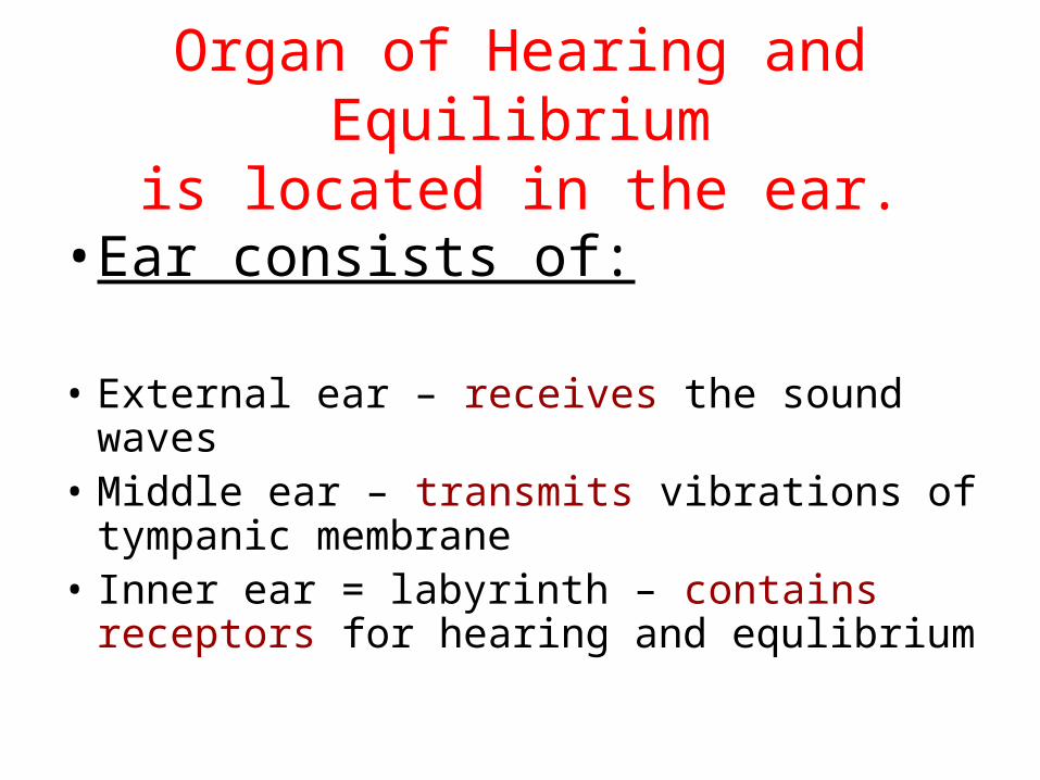
Organ of Hearing and Equilibriumis located in the ear.
• Ear consists of:
• External ear – receives the sound waves • Middle ear – transmits vibrations of
tympanic membrane • Inner ear = labyrinth – contains receptors
for hearing and equlibrium
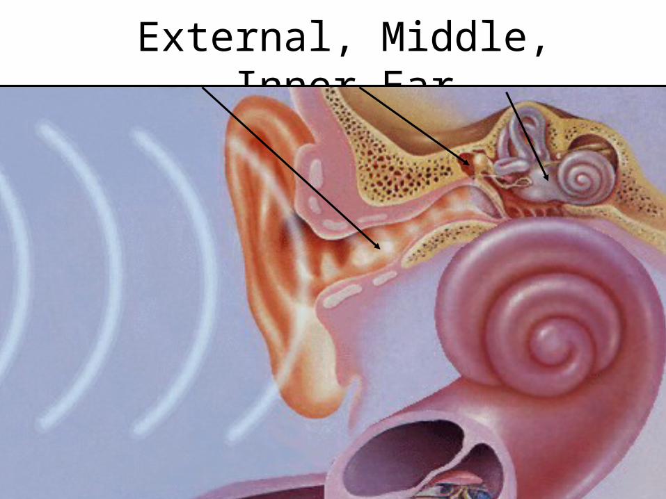
External, Middle, Inner Ear
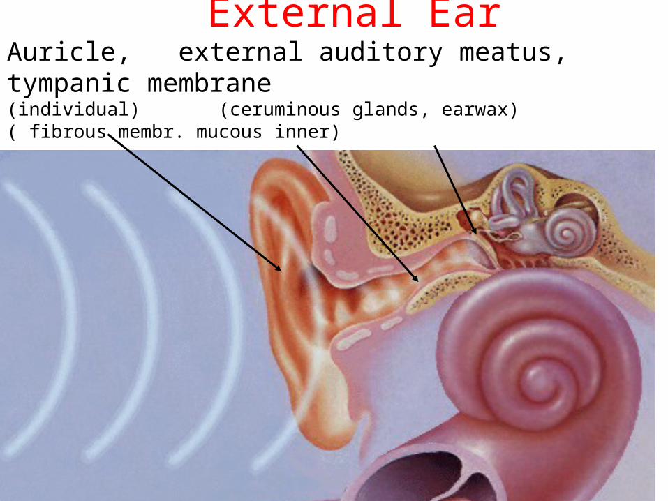
External EarAuricle, external auditory meatus, tympanic membrane(individual) (ceruminous glands, earwax) ( fibrous membr. mucous inner)
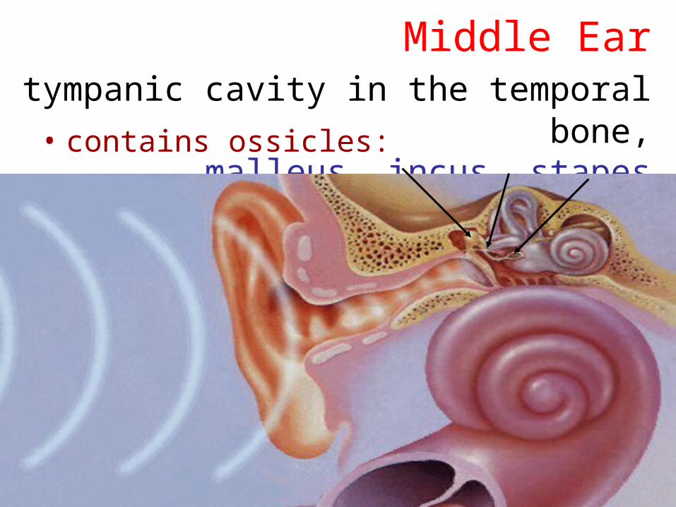
Middle Earis the tympanic cavity in the temporal bone,
malleus, incus, stapes• contains ossicles:
Middle Ear
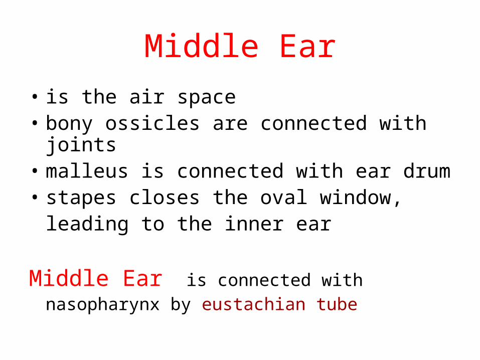
• is the air space • bony ossicles are connected with joints • malleus is connected with ear drum• stapes closes the oval window,
leading to the inner ear
Middle Ear is connected with nasopharynx by eustachian tube
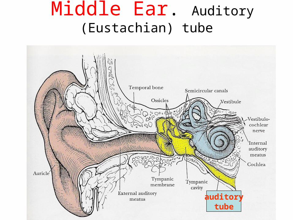
Middle Ear. Auditory (Eustachian) tube
auditorytube
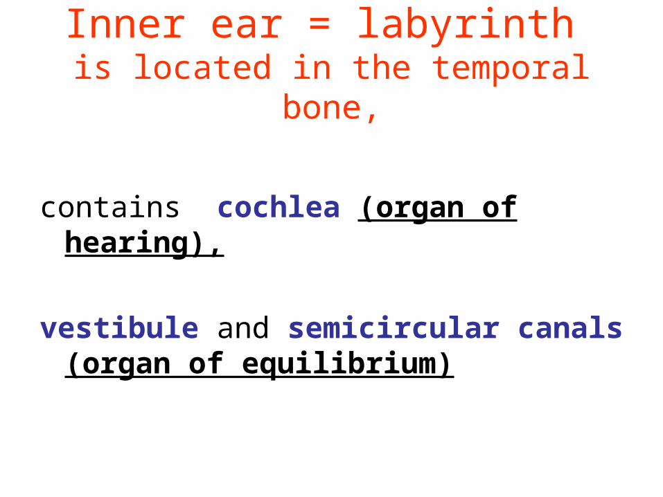
Inner ear = labyrinth is located in the temporal bone,
contains cochlea (organ of hearing),
vestibule and semicircular canals (organ of equilibrium)
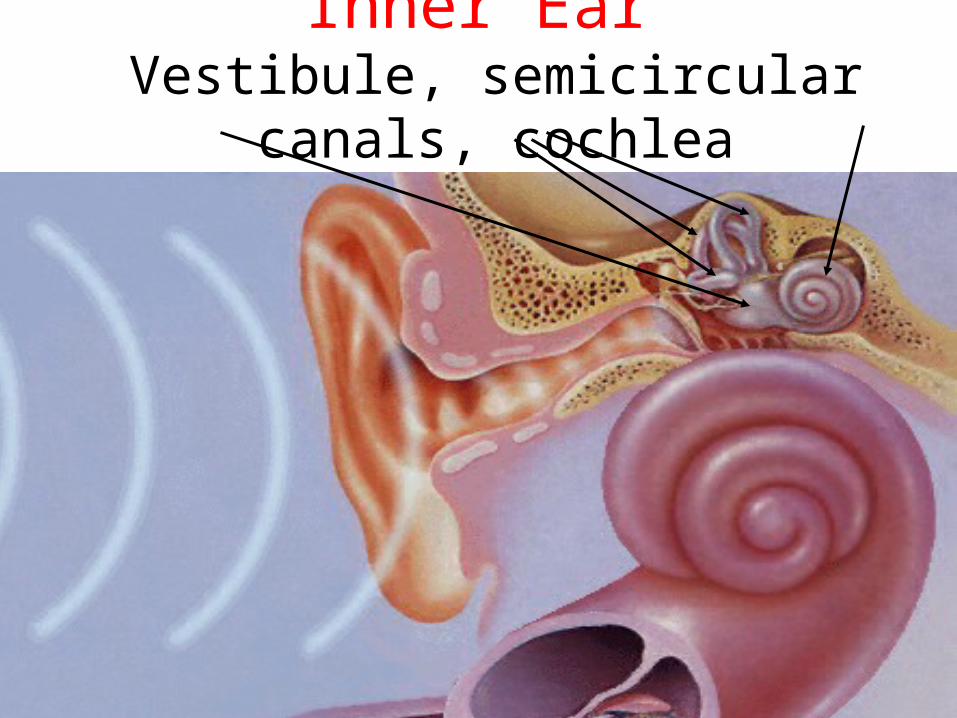
Inner Ear Vestibule, semicircular canals, cochlea
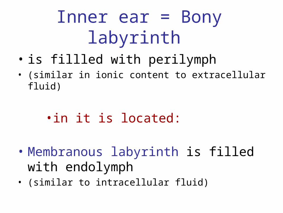
Inner ear = Bony labyrinth
• is fillled with perilymph• (similar in ionic content to extracellular fluid)
• in it is located:
• Membranous labyrinth is filled with endolymph
• (similar to intracellular fluid)
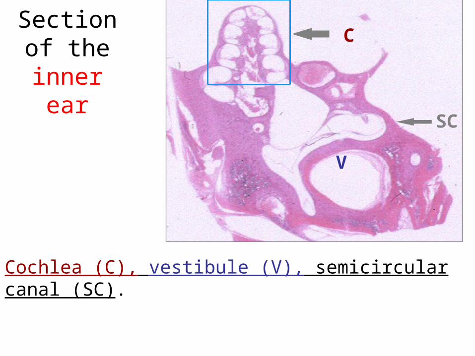
Section of the inner
ear
Cochlea (C), vestibule (V), semicircular canal (SC).
C
V
SC
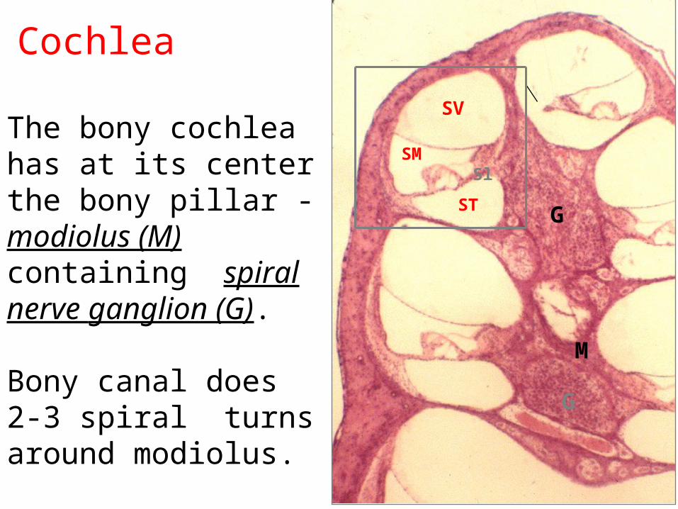
Cochlea
G
M
SM Sl
SV
ST
G
The bony cochlea has at its center the bony pillar - modiolus (M) containing spiral nerve ganglion (G).
Bony canal does 2-3 spiral turns around modiolus.
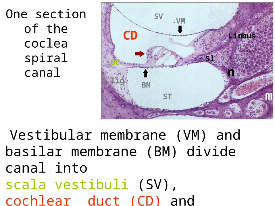
One section of the coclea
spiral canal
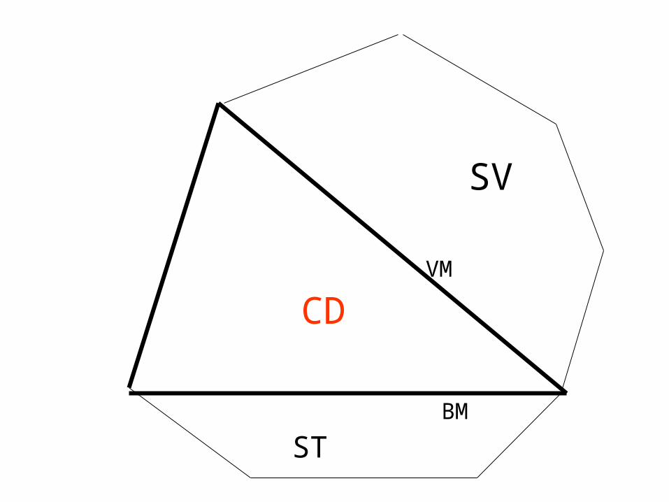
Vestibular membrane (VM) and basilar membrane (BM) divide canal into scala vestibuli (SV), cochlear duct (CD) and scala tympani (ST).
SVVM
CD
ST
BM
OC
S lig
Limbus
Sl n
m
SV
CD
ST
VM
BM
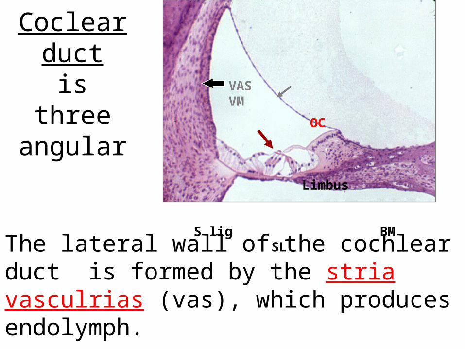
Coclear duct
is three angular
The lateral wall of the cochlear duct is formed by the stria vasculrias (vas), which produces endolymph.
OC Limbus S lig BM SL
VAS VM
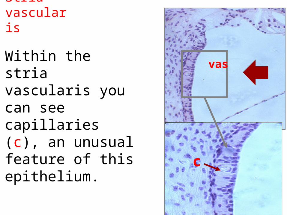
Stria vascularis
Within the stria vascularis you can see capillaries (c), an unusual feature of this epithelium.
vas
c
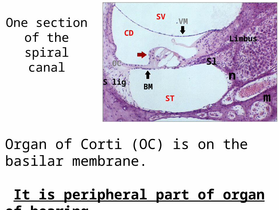
One section of the spiral canal
Organ of Corti (OC) is on the basilar membrane.
It is peripheral part of organ of hearing
SVVM
CD
ST
BM
OC
S lig
Limbus
Sl n
m
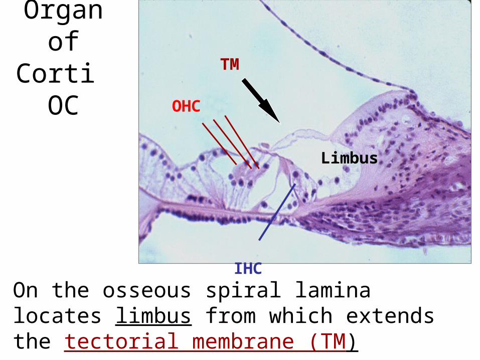
Organ of Corti OC
On the osseous spiral lamina locates limbus from which extends the tectorial membrane (TM)
TM
OHC
Limbus
IHC
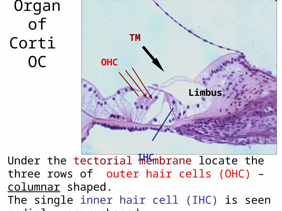
Organ of Corti OC
Under the tectorial membrane locate the three rows of outer hair cells (OHC) – columnar shaped. The single inner hair cell (IHC) is seen medial - pear shaped.
TM
OHC
Limbus
IHC
Organ of Corti OC
tmIHC
OHC
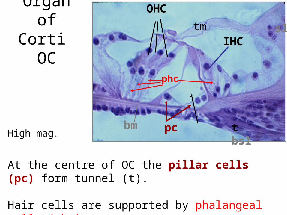
High mag.
At the centre of OC the pillar cells (pc) form tunnel (t).
Hair cells are supported by phalangeal cells (phc).
sl
pc t bsl
bm
phc
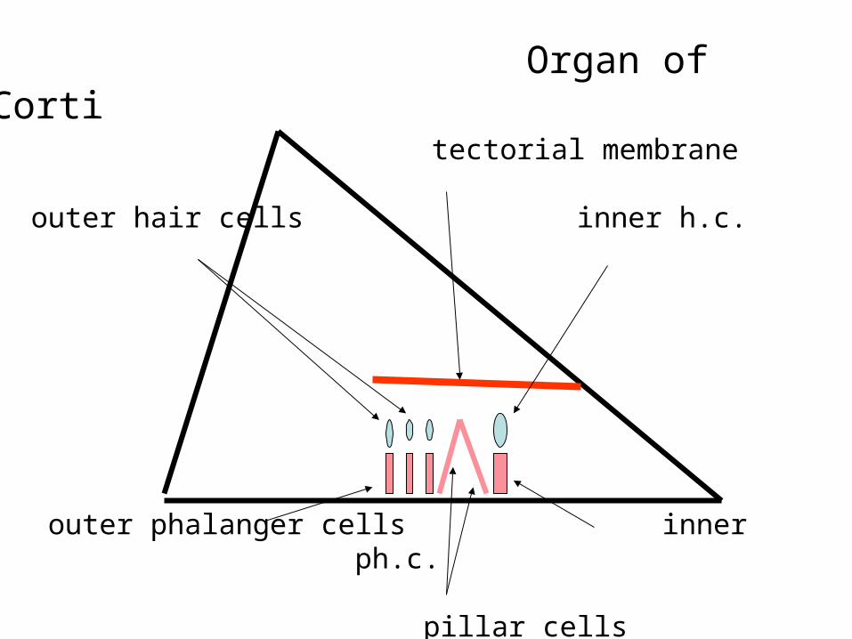
tectorial membrane
outer hair cells inner h.c.
outer phalanger cells inner ph.c.
pillar cells
Organ of Corti
G
M
CD
SV
ST
H
G
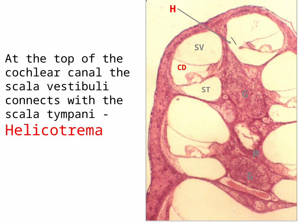
At the top of the cochlear canal thescala vestibuli connects with the scala tympani -
Helicotrema
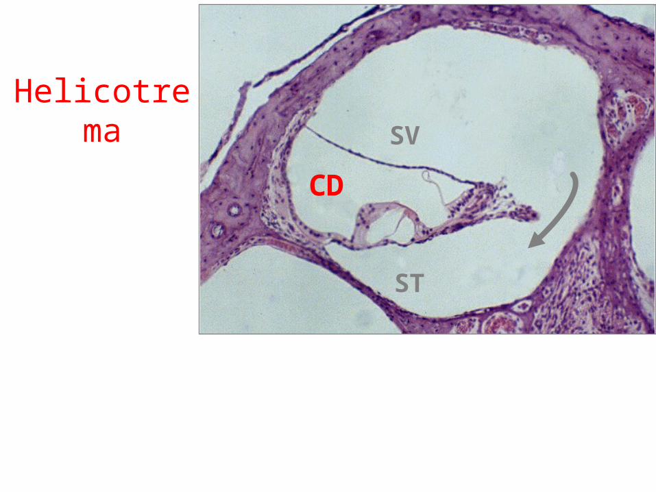
HelicotremaSV
ST
CD
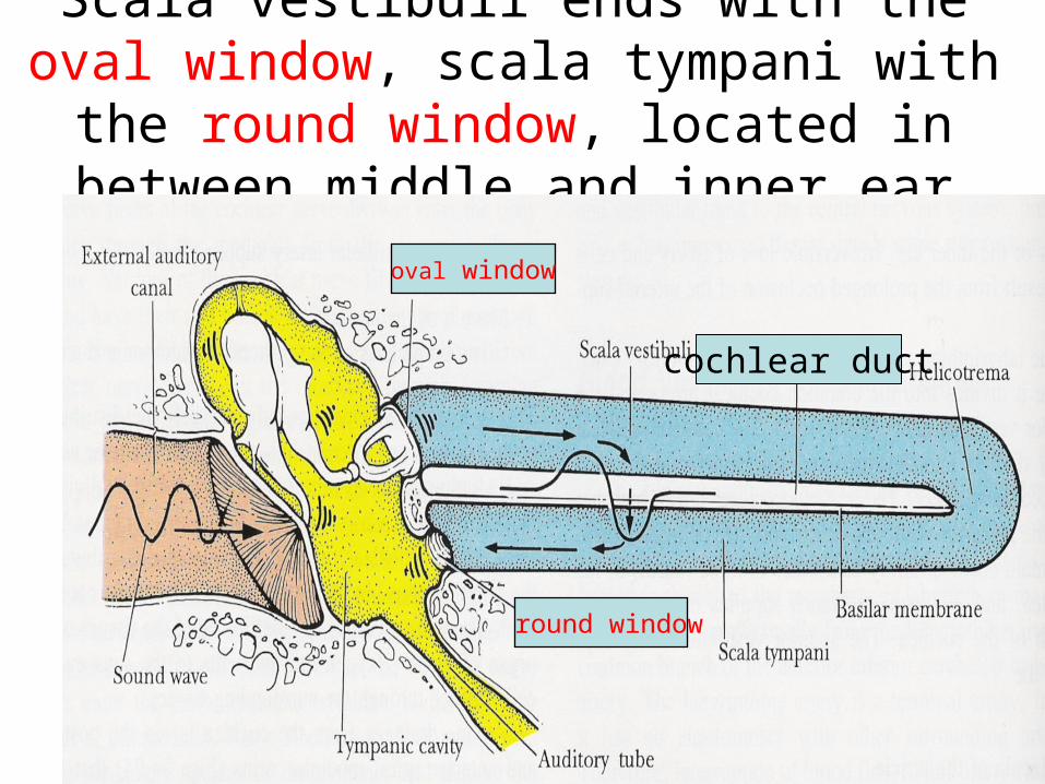
Scala vestibuli ends with the oval window, scala tympani with the round window,
located in between middle and inner ear
cochlear duct
oval window
round window
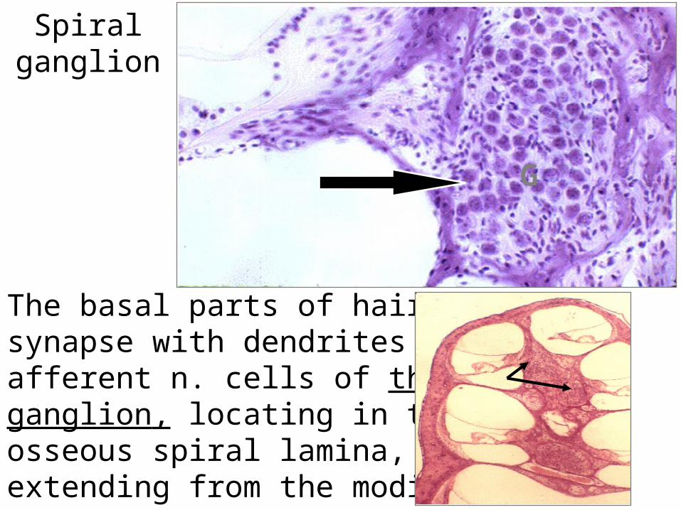
Spiral ganglion
G
The basal parts of hair cells synapse with dendrites of afferent n. cells of the spiralganglion, locating in the osseous spiral lamina, extending from the modiolus.
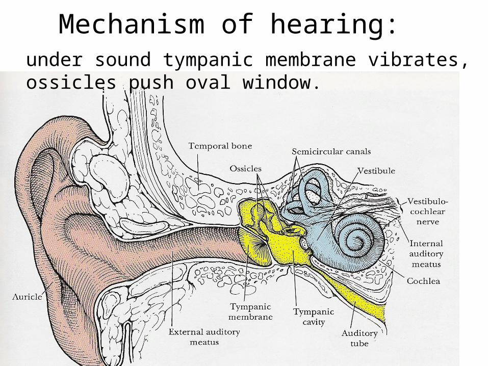
Mechanism of hearing: under sound tympanic membrane vibrates, ossicles push oval window.
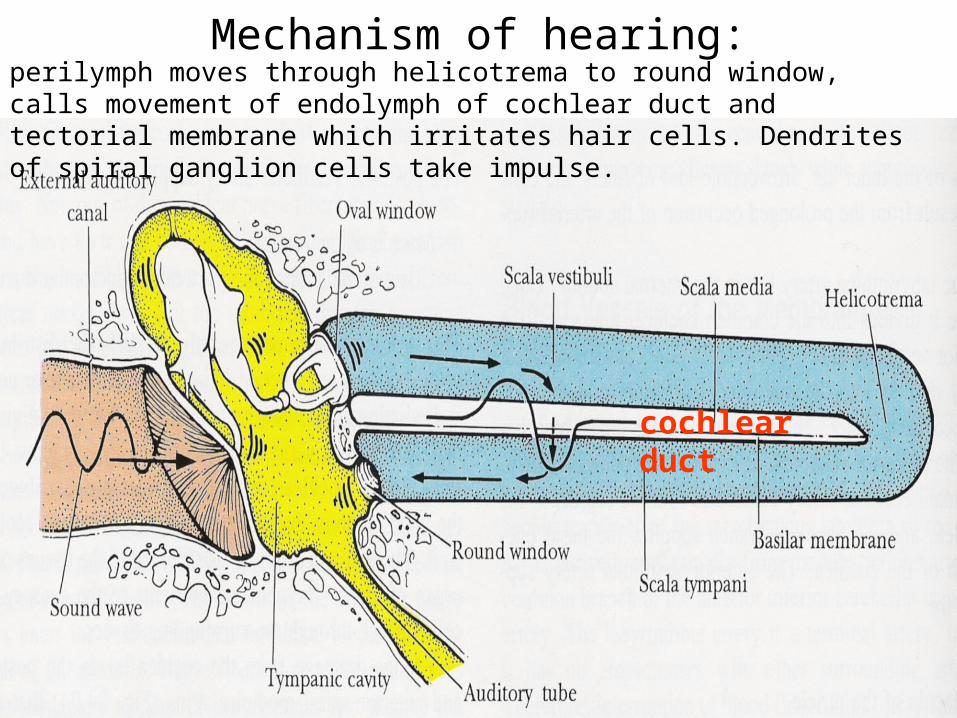
Mechanism of hearing:
cochlear duct
perilymph moves through helicotrema to round window, calls movement of endolymph of cochlear duct and tectorial membrane which irritates hair cells. Dendrites of spiral ganglion cells take impulse.
G
M
SV
CD
SV
ST
H
G
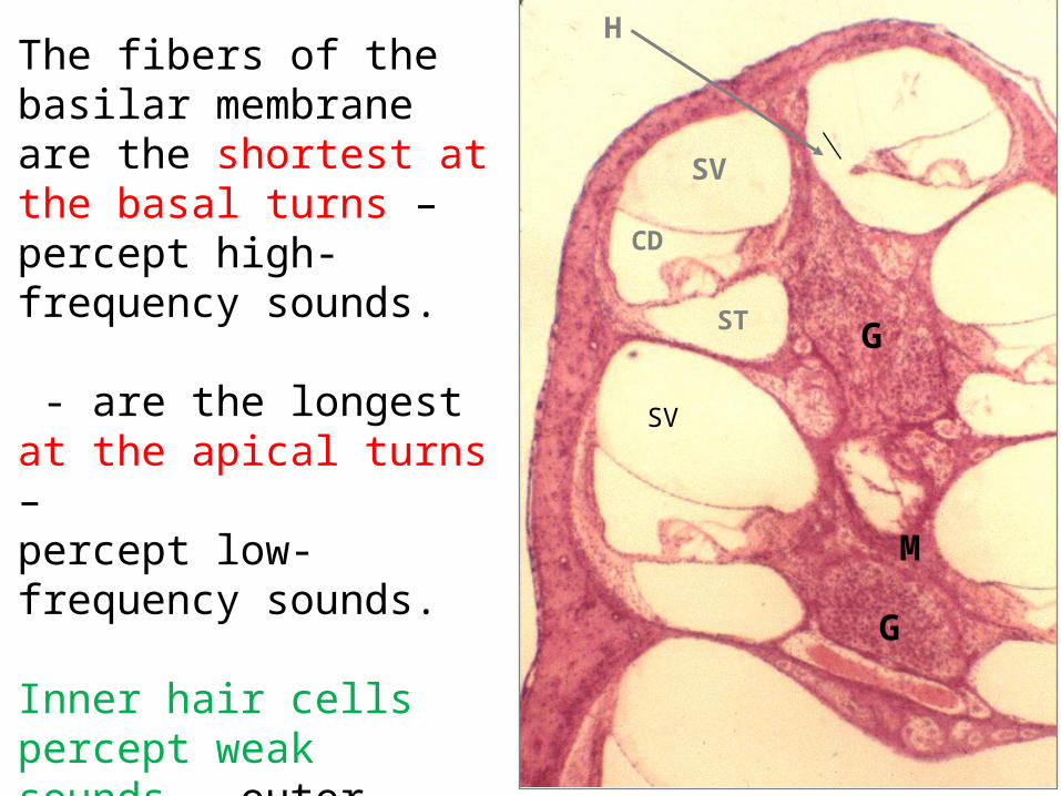
The fibers of the basilar membrane are the shortest at the basal turns – percept high-frequency sounds.
- are the longest at the apical turns –percept low-frequency sounds.
Inner hair cells percept weak sounds, outer - intense sounds.

Pathology• Hearing loss: conductive
sensorineural
• Otosclerosis
Ogran of equilibrium
ACSemicircular canal
M
Utricle
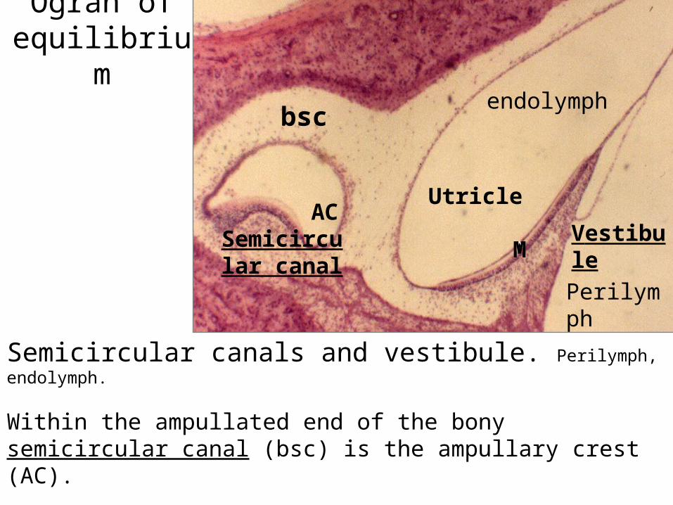
Semicircular canals and vestibule. Perilymph, endolymph.
Within the ampullated end of the bony semicircular canal (bsc) is the ampullary crest (AC).
The flat macula (M) is in the utricle and saccule of the vestibule.
bsc
Vestibule
endolymph
Perilymph
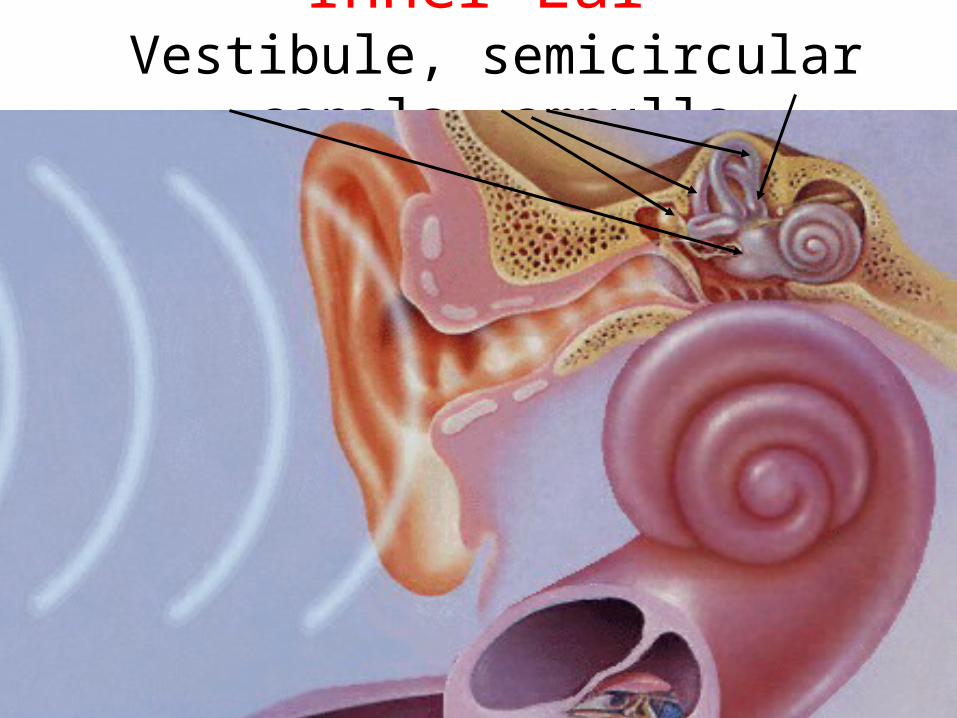
Inner Ear Vestibule, semicircular canals, ampulla
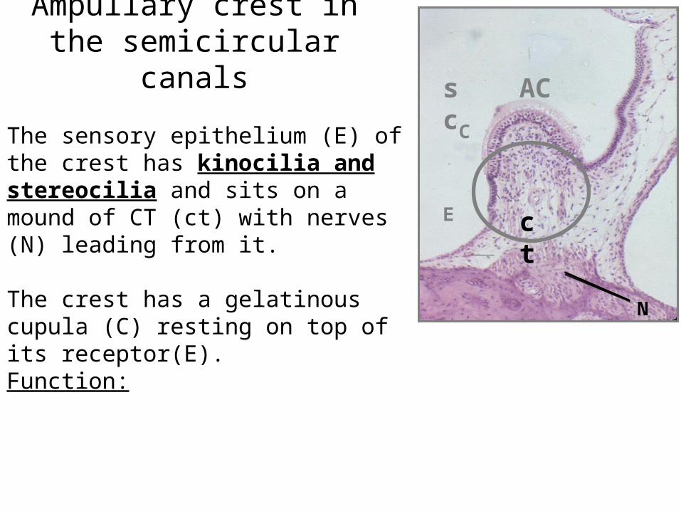
Ampullary crest in the semicircular canals
C
E
N
The sensory epithelium (E) of the crest has kinocilia and stereocilia and sits on a mound of CT (ct) with nerves (N) leading from it.
The crest has a gelatinous cupula (C) resting on top of its receptor(E). Function:
sc AC
ct
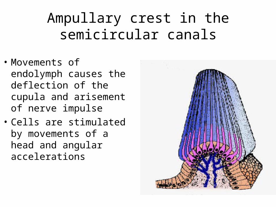
Ampullary crest in the semicircular canals
• Movements of endolymph causes the deflection of the cupula and arisement of nerve impulse
• Cells are stimulated by movements of a head and angular accelerations
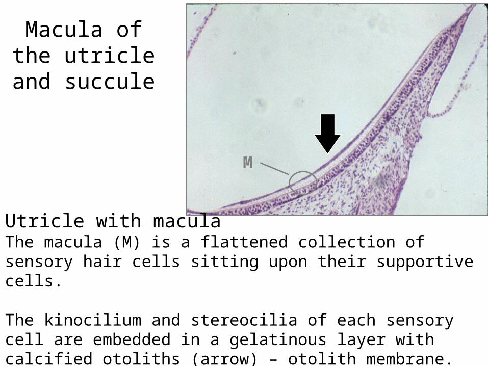
Macula of the utricle and
succule
Utricle with macula The macula (M) is a flattened collection of sensory hair cells sitting upon their supportive cells.
The kinocilium and stereocilia of each sensory cell are embedded in a gelatinous layer with calcified otoliths (arrow) – otolith membrane.
M
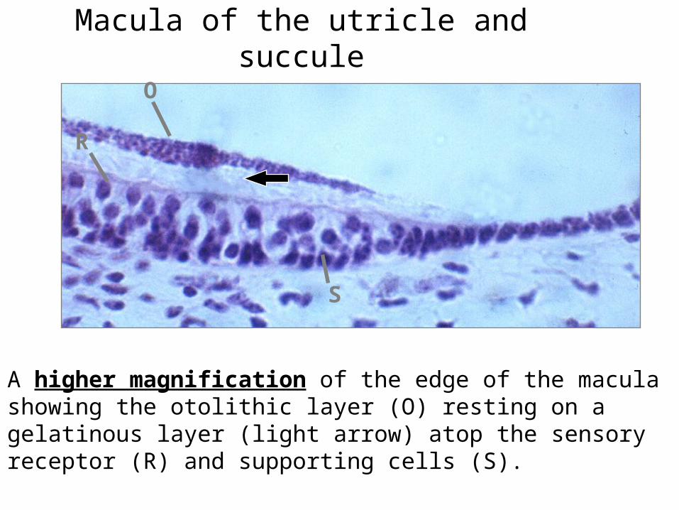
Macula of the utricle and succule
O
R
S
A higher magnification of the edge of the macula showing the otolithic layer (O) resting on a gelatinous layer (light arrow) atop the sensory receptor (R) and supporting cells (S).
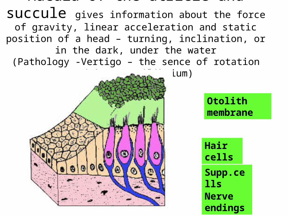
Macula of the utricle and succule gives information about the force of gravity, linear acceleration and static position of a head – turning, inclination, or in the dark,
under the water(Pathology -Vertigo – the sence of rotation without equilibrium)
Otolith membrane
Hair cells
Supp.cells
Nerve endings
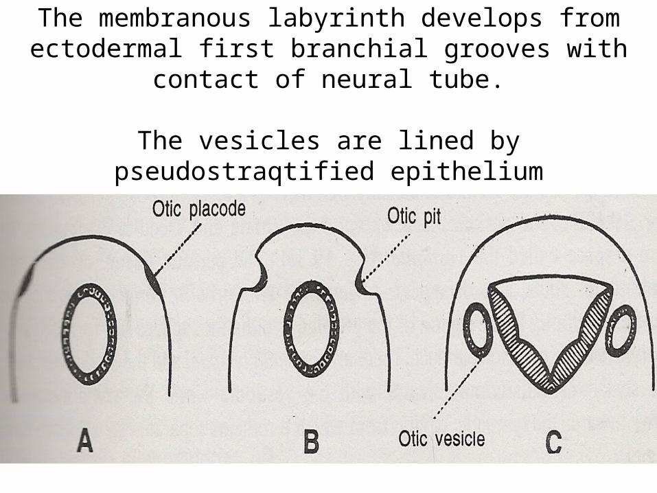
The membranous labyrinth develops from ectodermal first branchial grooves with contact of neural tube.
The vesicles are lined by pseudostraqtified epithelium
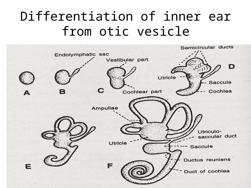
Differentiation of inner ear from otic vesicle
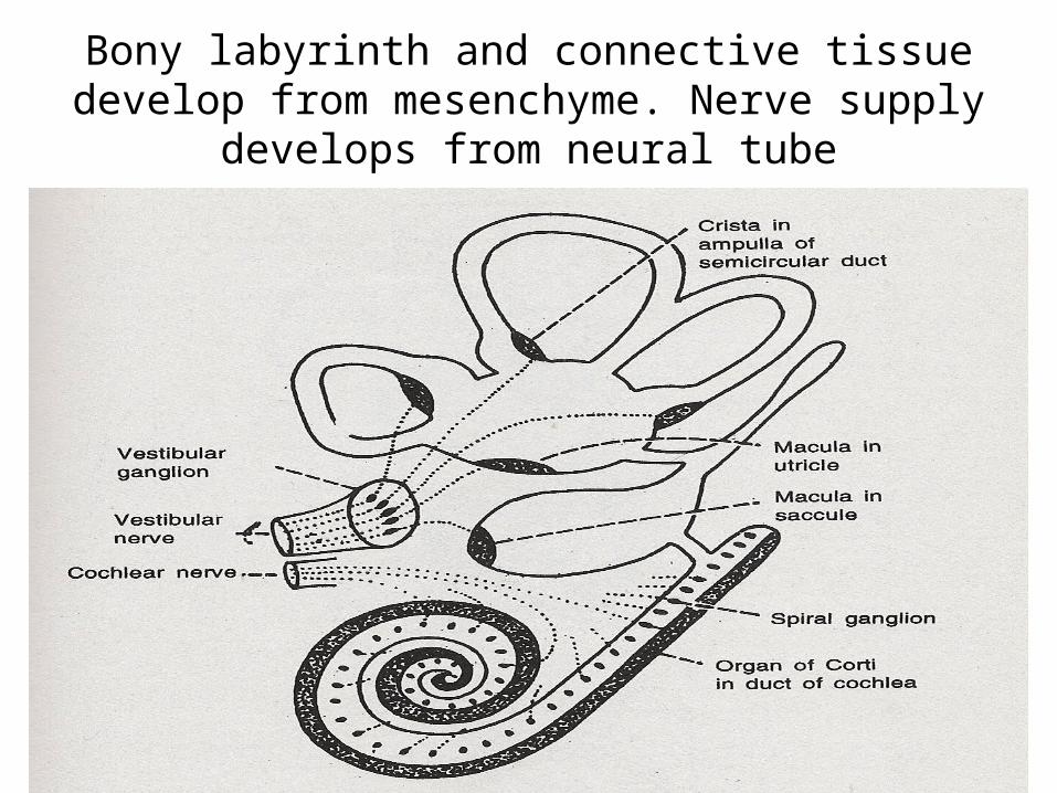
Bony labyrinth and connective tissue develop from mesenchyme. Nerve supply develops from neural
tube
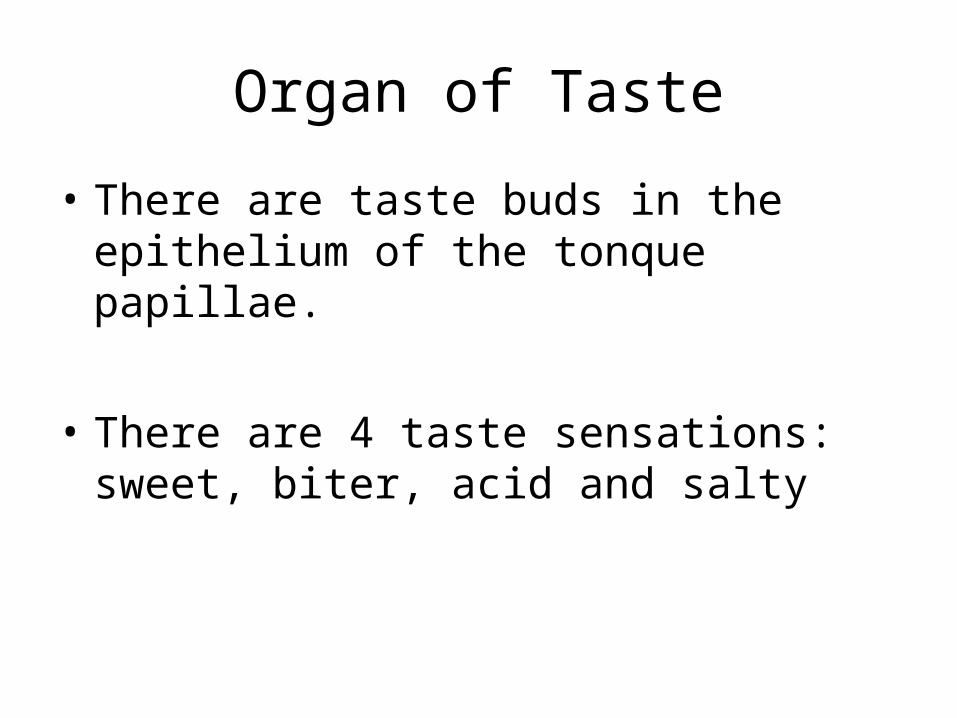
Organ of Taste
• There are taste buds in the epithelium of the tonque papillae.
• There are 4 taste sensations: sweet, biter, acid and salty
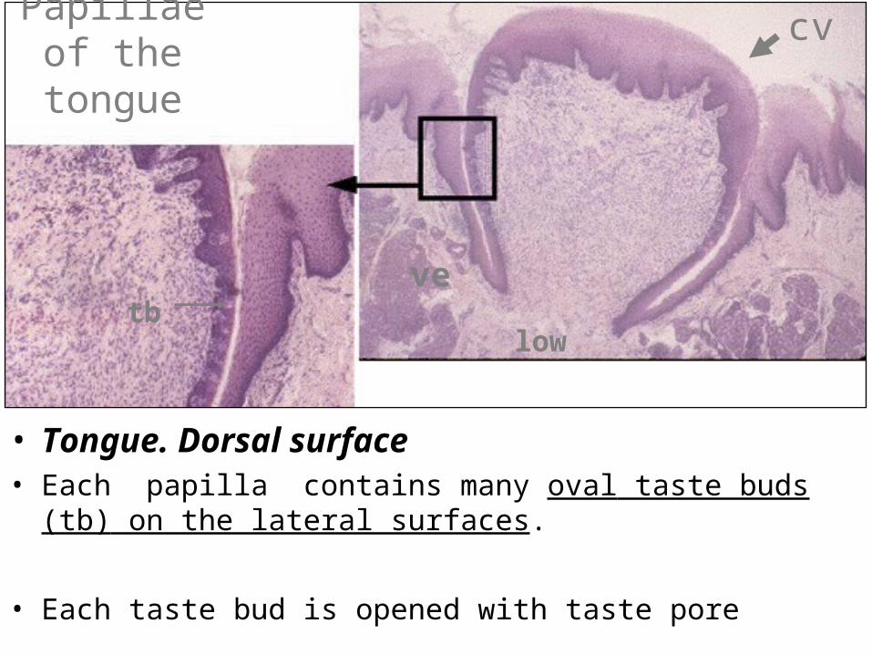
Papillae of the tongue
• Tongue. Dorsal surface• Each papilla contains many oval taste buds (tb) on the lateral
surfaces.
• Each taste bud is opened with taste pore
cv
tbve
low
Taste bud
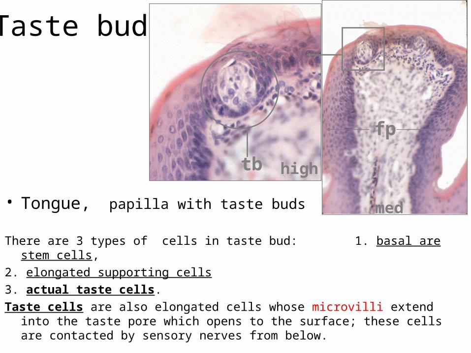
• Tongue, papilla with taste buds
There are 3 types of cells in taste bud: 1. basal are stem cells,
2. elongated supporting cells
3. actual taste cells.
Taste cells are also elongated cells whose microvilli extend into the taste pore which opens to the surface; these cells are contacted by sensory nerves from below.
high
med
fp
tb
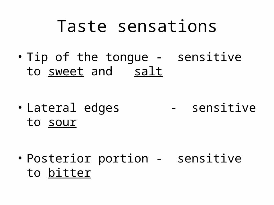
Taste sensations
• Tip of the tongue - sensitive to sweet and salt
• Lateral edges - sensitive to sour
• Posterior portion - sensitive to bitter