sensory system lecture for medical students part 1 (organ of smell, eye) department of histology,...
TRANSCRIPT
Sensory System
Lecture for medical students
Part 1 (organ of smell, eye)
Department of histology,cytology and embryology KhNMU
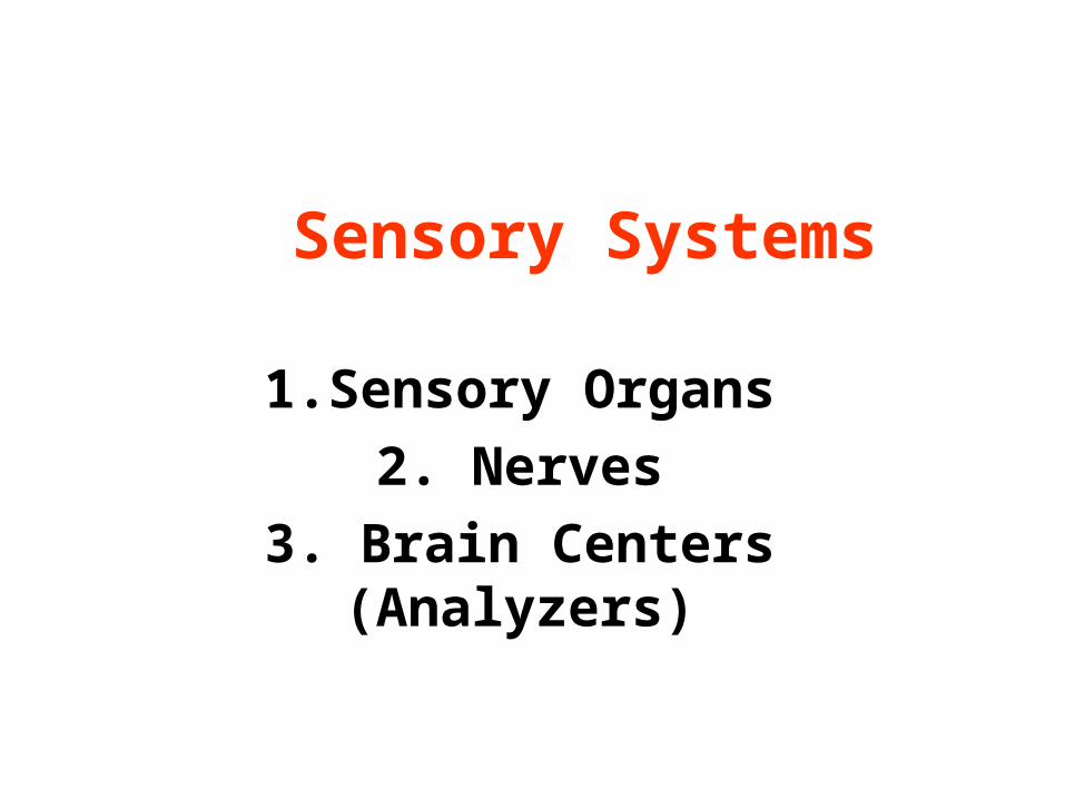
Sensory Systems
1.Sensory Organs2. Nerves
3. Brain Centers (Analyzers)
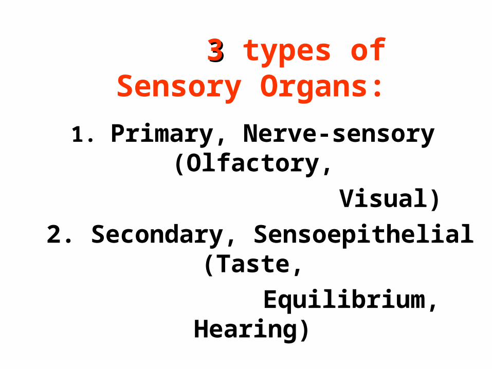
33 types of Sensory Organs:
1. Primary, Nerve-sensory (Olfactory,
Visual) 2. Secondary, Sensoepithelial
(Taste,Equilibrium,
Hearing)
3. Sensory Nerve Endings
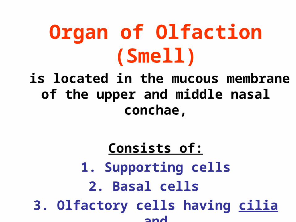
Organ of Olfaction (Smell)
is located in the mucous membrane of the upper and middle nasal
conchae,
Consists of:1. Supporting cells2. Basal cells
3. Olfactory cells having cilia and
axons, forming nerve
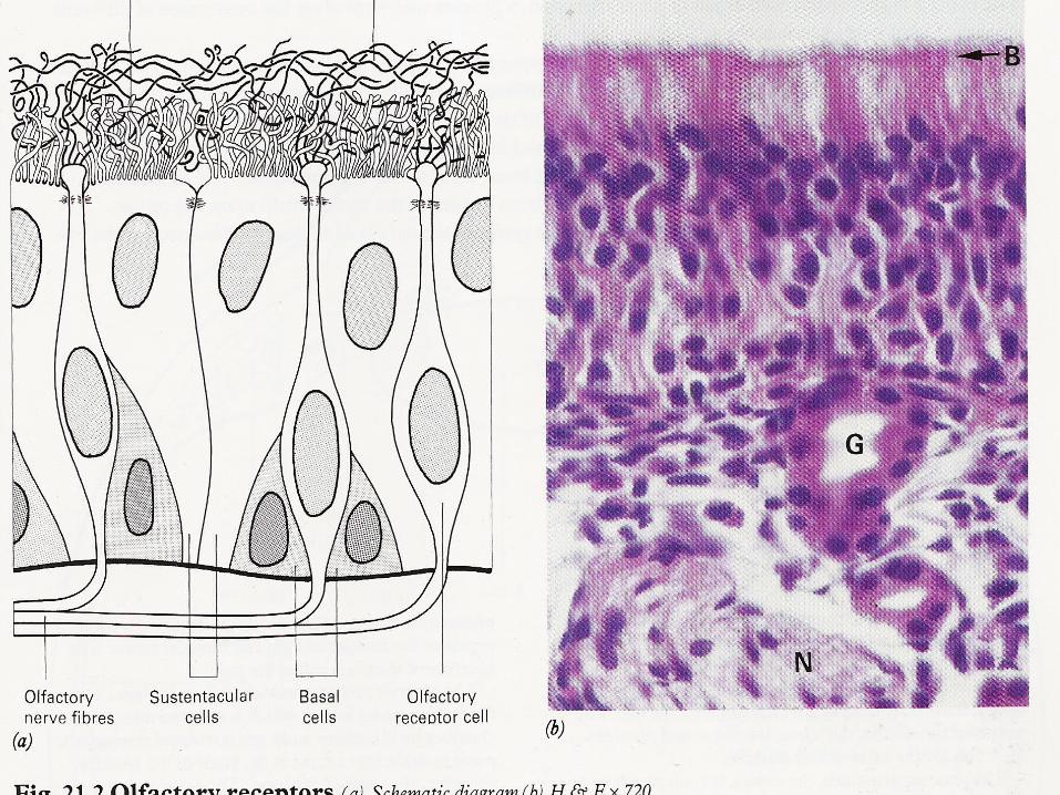
Cilia of olfactory cells are irritated by odorous substances.
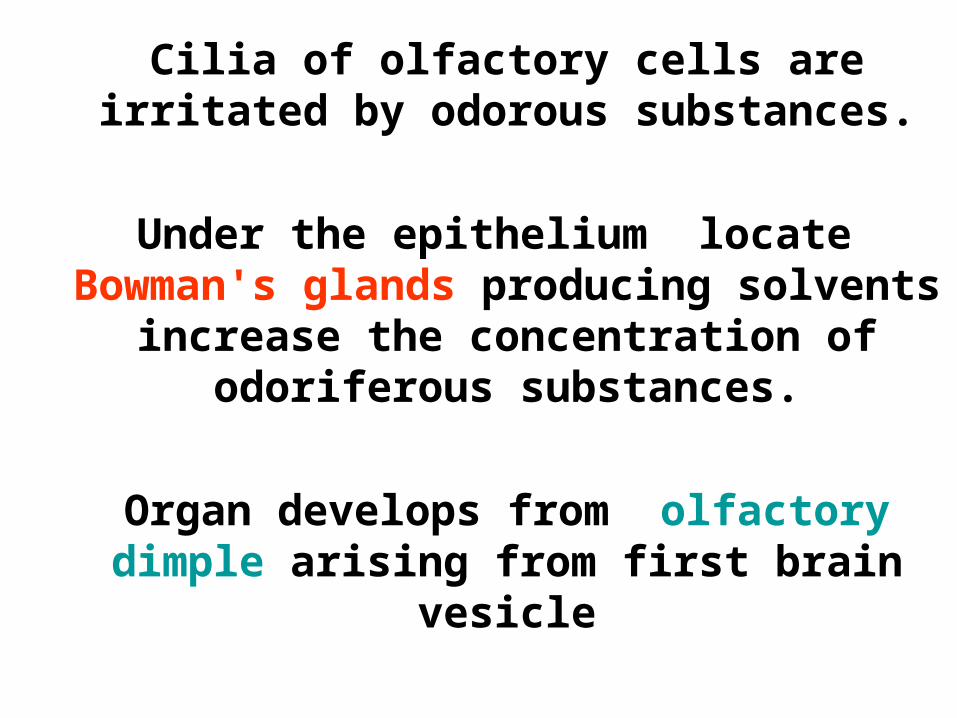
Under the epithelium locate Bowman's glands producing solvents
increase the concentration of odoriferous substances.
Organ develops from olfactory dimple arising from first brain
vesicle
Organ of olfaction gets 10000 smells,
1000 genes are responsible for smell
• It is the most ancient sensory organ
• Wet weather increases smell
• Without this organ a person becomes anxious
• Smell of lavender keeps anxiety away
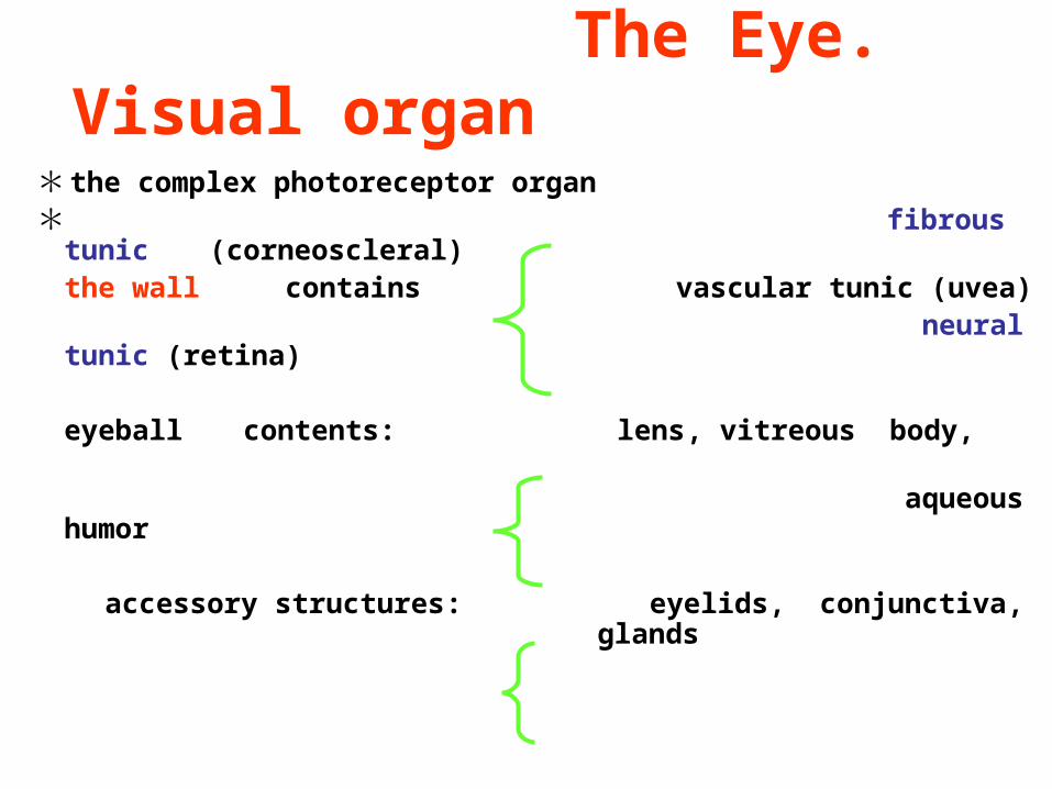
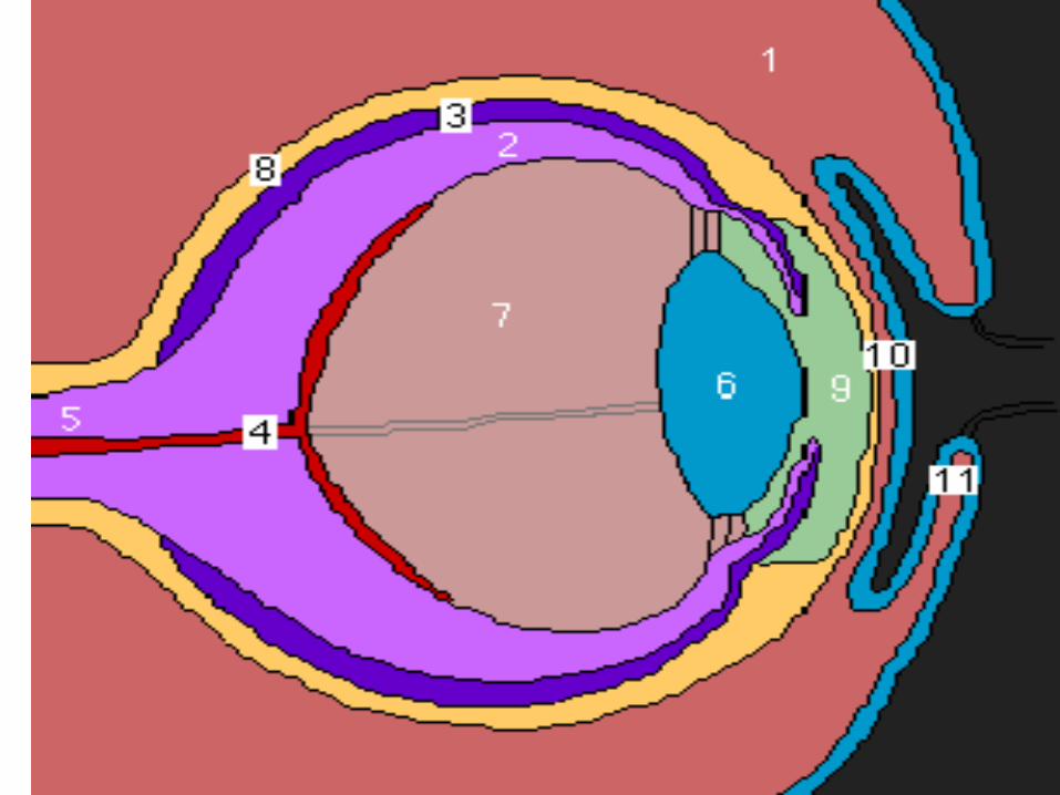
The Eye. Visual organ
* the complex photoreceptor organ* fibrous tunic
(corneoscleral) the wall contains vascular tunic (uvea)
neural tunic (retina)
eyeball contents: lens, vitreous body, aqueous humor accessory structures: eyelids, conjunctiva,
glands
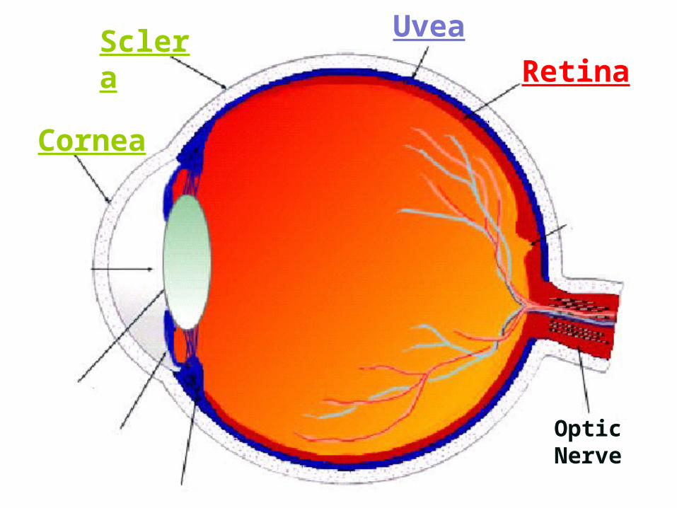
Optic Nerve
Cornea
ScleraRetina
Uvea
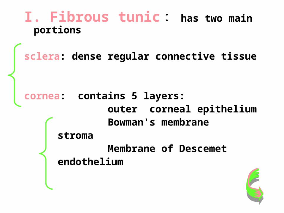
I. Fibrous tunic : has two main portions
sclera: dense regular connective tissue
cornea: contains 5 layers: outer corneal epithelium Bowman's membrane
stroma Membrane of Descemet
endothelium
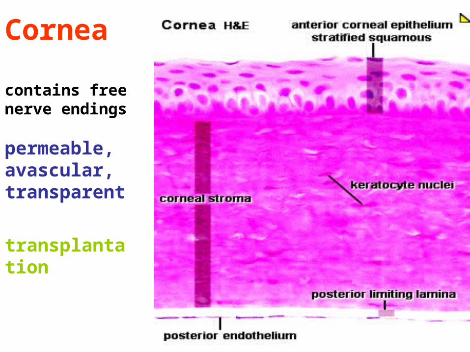
Cornea
contains free nerve endings
permeable,avascular, transparent
transplantation
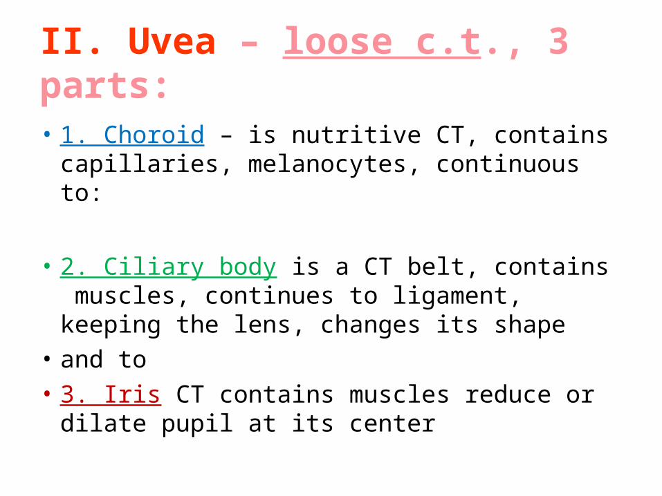
II. Uvea – loose c.t., 3 parts:
• 1. Choroid – is nutritive CT, contains capillaries, melanocytes, continuous to:
• 2. Ciliary body is a CT belt, contains muscles, continues to ligament, keeping the lens, changes its shape
• and to• 3. Iris CT contains muscles reduce or dilate
pupil at its center
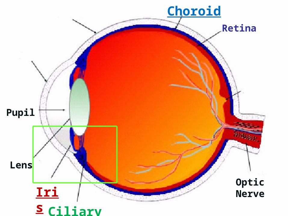
Pupil
Lens
Iris
Ciliary Body
Optic Nerve
Cornea
ScleraRetina
Choroid
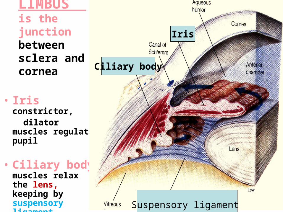
LIMBUS is the junction between sclera and cornea
• Iris constrictor,
dilator muscles regulate pupil
• Ciliary body muscles relax the lens, keeping by suspensory ligament
Iris
Ciliary body
Suspensory ligament
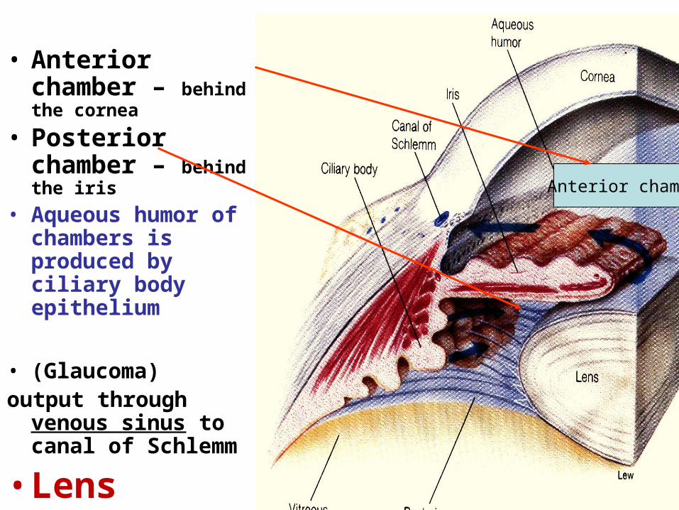
• Anterior chamber – behind the cornea
• Posterior chamber – behind the iris
• Aqueous humor of chambers is produced by ciliary body epithelium
• (Glaucoma)output through venous
sinus to canal of Schlemm
• Lens
Anterior chamber
• LENS is transperent elastic body, covered by flattened epithelium, consists of fibers
• VITREOUS BODY is transperent, colorless, contains 99% of water, hyaluronic acid, collagen
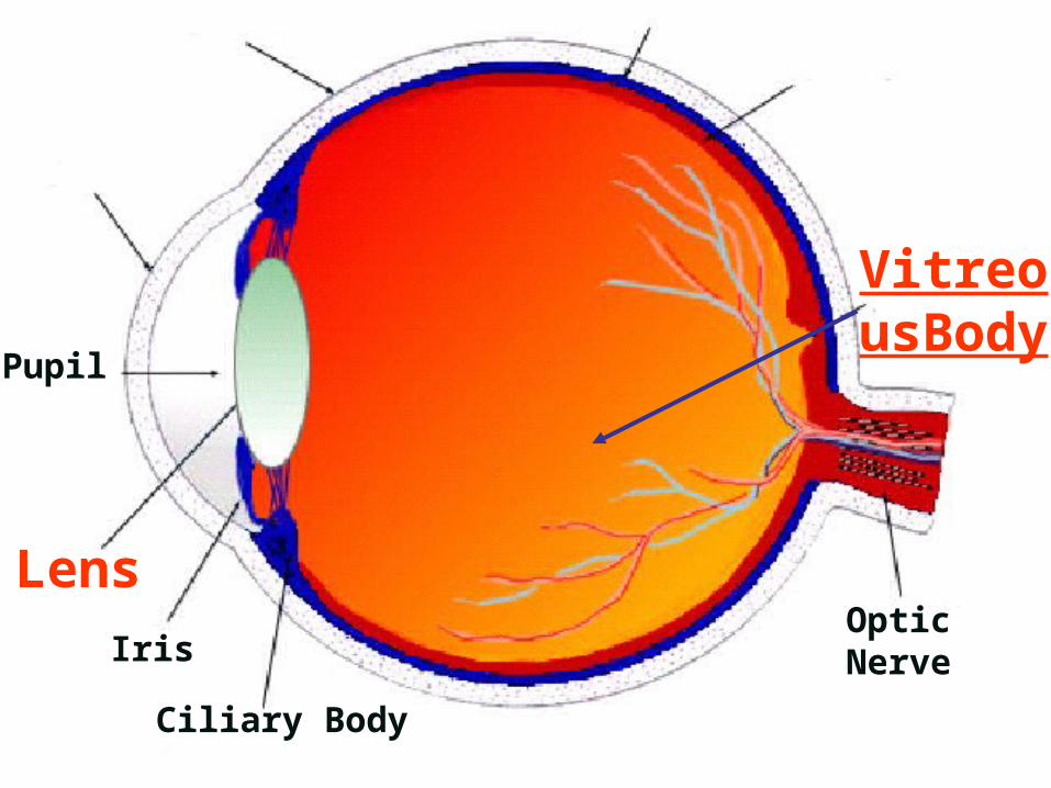
Pupil
LensIris
Ciliary Body
Optic Nerve
VitreousBody
Cornea
ScleraRetina
Choroid
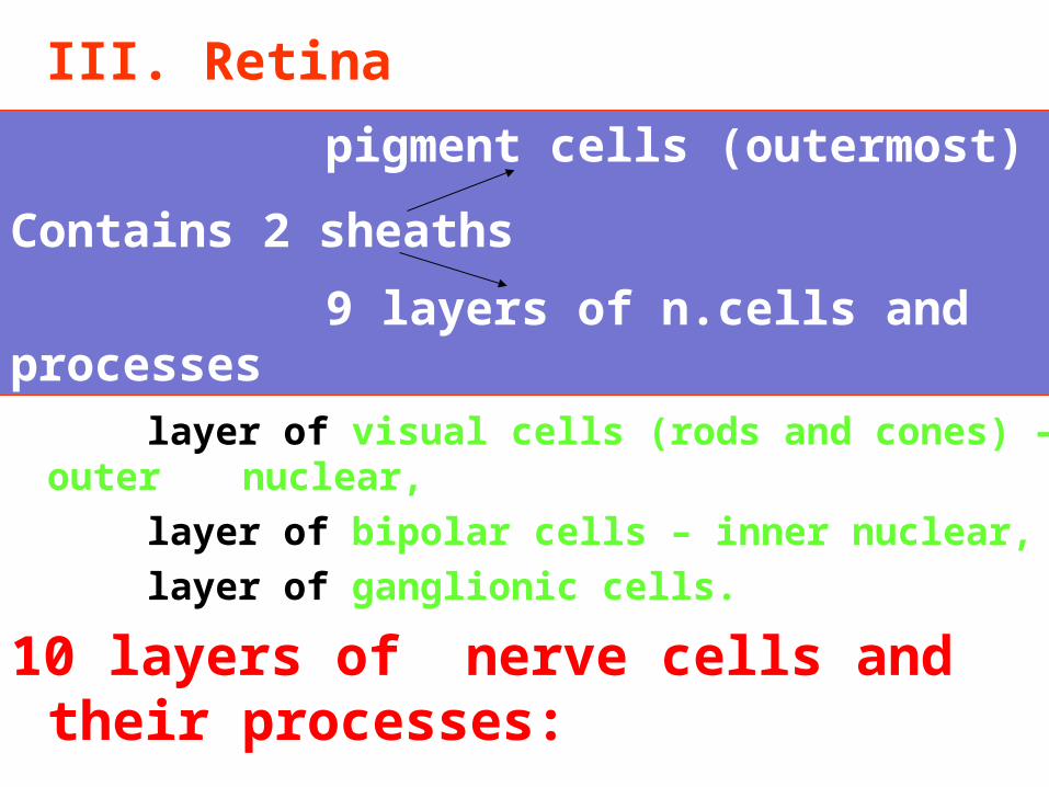
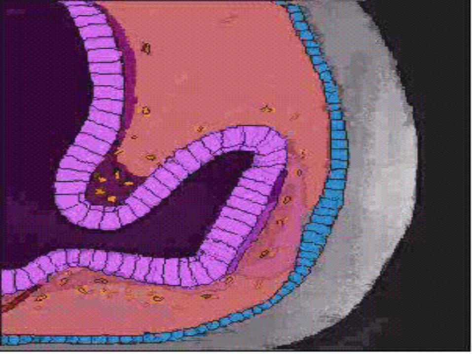
III. Retina
• chain of 3 neurons:
layer of visual cells (rods and cones) – outer nuclear,
layer of bipolar cells – inner nuclear,
layer of ganglionic cells.
10 layers of nerve cells and their processes:
pigment cells (outermost)
Contains 2 sheaths
9 layers of n.cells and processes
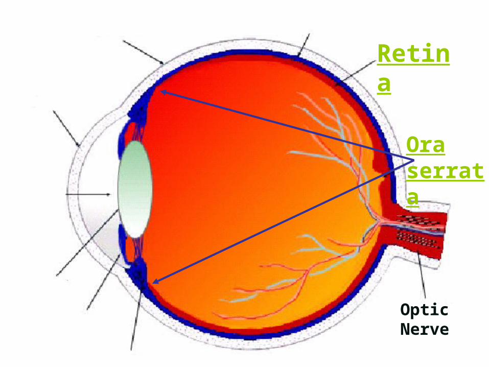
Optic Nerve
Ora serrata
Cornea
Sclera
Retina
Choroid
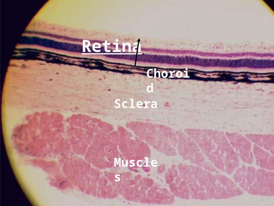
Sclera
Muscles
Choroid
Retina

Sclera
Choroid
Pigment epith..
Inner nuc.
Ganglionic l.
Outer nu.
Ganglionic layer
Inner nuclear layer
Outer nuclear layer
Pigment epithelium
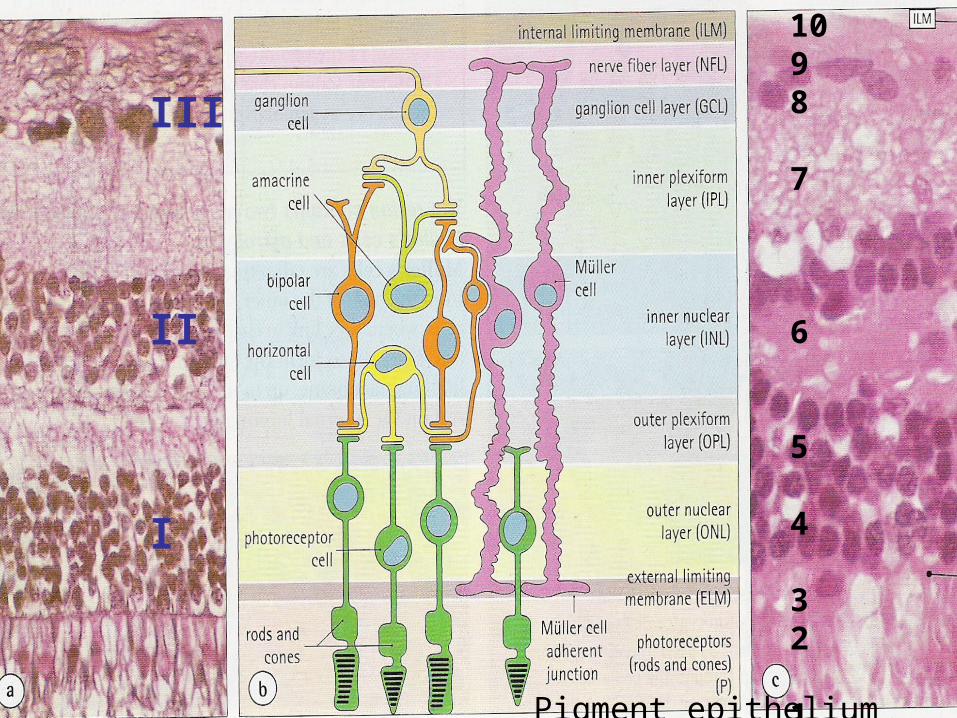
Pigment epithelium
1098
7
6
5
4
32
1
III
II
I

Ultrastructure of rod and cone cells Rod Cone
Outer slender conical, wider segment cylinder shorter
Membrane closed opened, continue
Limiting to cell membrane
disks
Visual rhodopsin iodopsin pigment Sensitive to black, white blue, green, red lights twilight vision (krok)
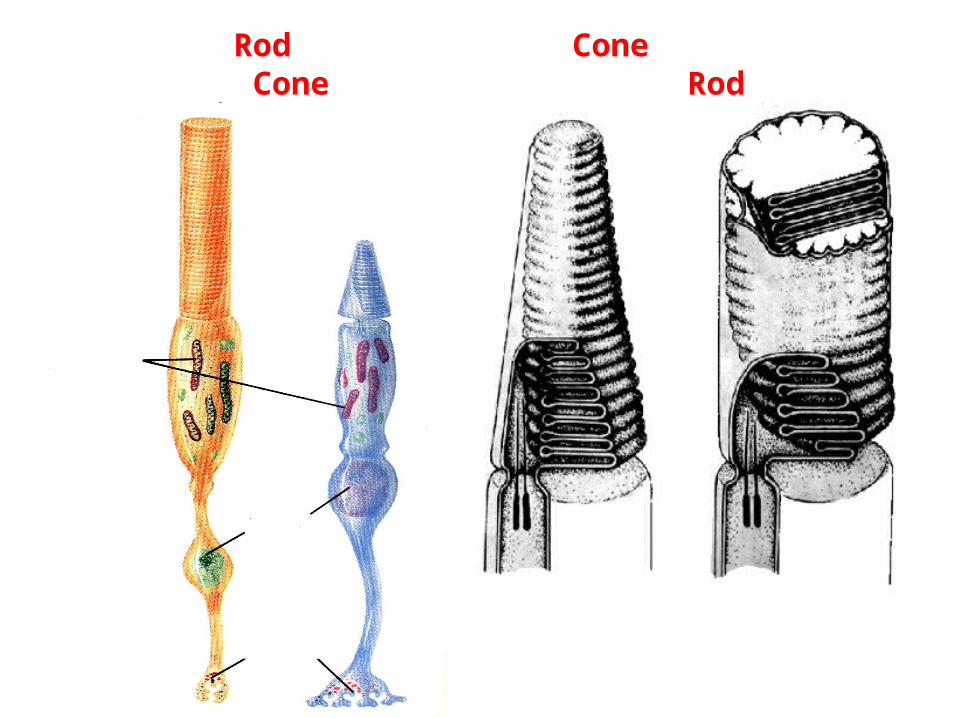
Cone Rod Rod Cone
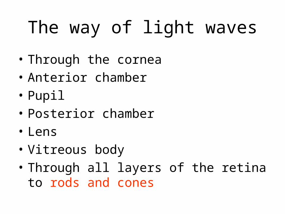
The way of light waves
• Through the cornea
• Anterior chamber
• Pupil
• Posterior chamber
• Lens
• Vitreous body
• Through all layers of the retina to rods and cones
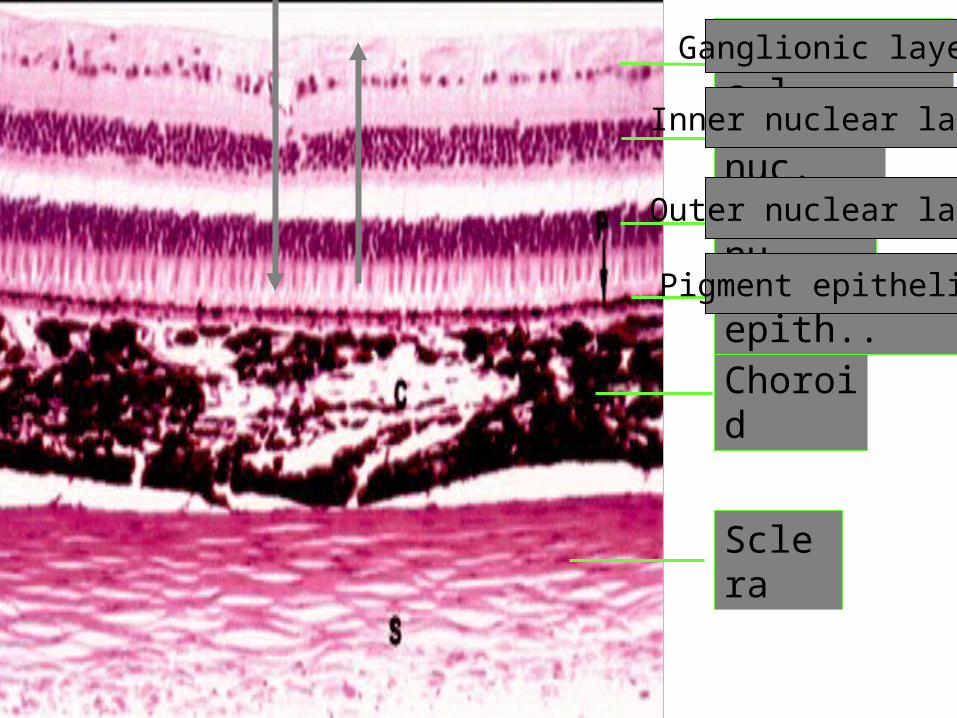
Sclera
Choroid
Pigment epith..
Inner nuc.
Ganglionic l.
Outer nu.
Ganglionic layer
Inner nuclear layer
Outer nuclear layer
Pigment epithelium
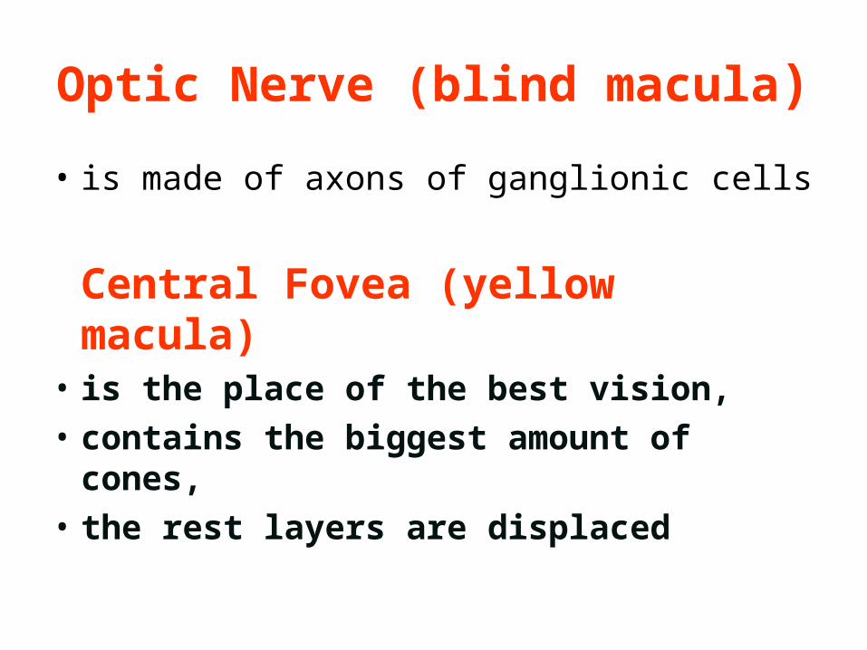
Optic Nerve (blind macula)
• is made of axons of ganglionic cells
Central Fovea (yellow macula)• is the place of the best vision,
• contains the biggest amount of cones,
• the rest layers are displaced

• 3 functional apparatuses of the eye
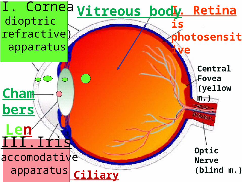
ChambersLens
Iris- Ciliary body
Optic Nerve (blind m.)
Central Fovea (yellow m.)
Sclera I. Retina is photosensitive
Vitreous body II. Corneadioptric
(refractive) apparatus
III.Iris accomodative
apparatus
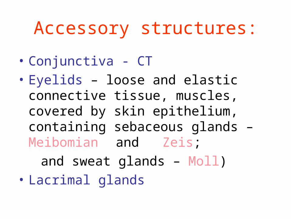
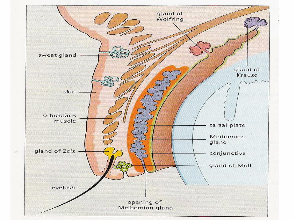
Accessory structures:
• Conjunctiva - CT
• Eyelids – loose and elastic connective tissue, muscles, covered by skin epithelium, containing sebaceous glands – Meibomian and Zeis;
and sweat glands – Moll)
• Lacrimal glands
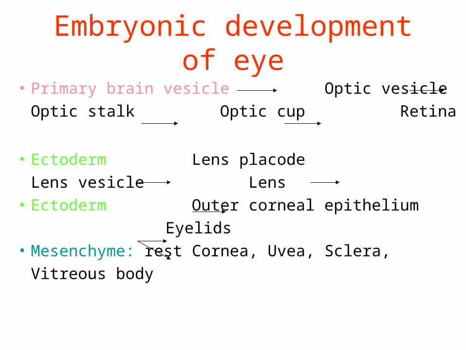
Embryonic development of eye
• Primary brain vesicle Optic vesicle
Optic stalk Optic cup Retina
• Ectoderm Lens placode
Lens vesicle Lens• Ectoderm Outer corneal epithelium
Eyelids• Mesenchyme: rest Cornea, Uvea, Sclera,
Vitreous body
Chapter 9 Eye and Ear
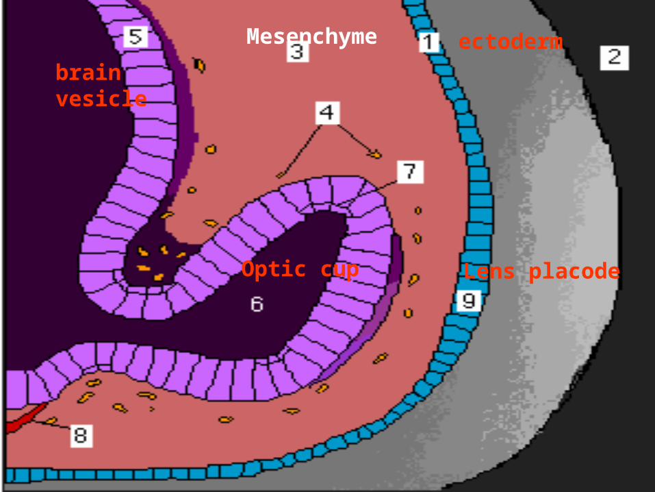
ectoderm
brain vesicle
Optic cup Lens placode
Mesenchyme
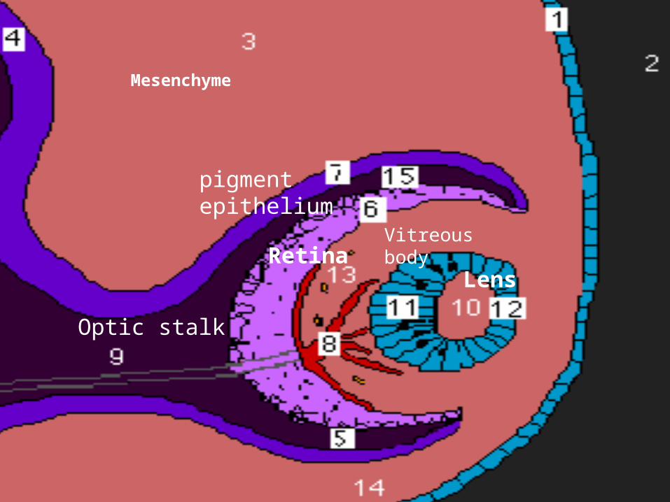
Retina
pigment epithelium
Vitreous body
Lens
Optic stalk
Mesenchyme