senile plaque neurites in alzheimer disease accumulate ... · apphas beenlocalized to cell...
TRANSCRIPT
Proc. Nadl. Acad. Sci. USAVol. 88, pp. 7552-7556, September 1991Medical Sciences
Senile plaque neurites in Alzheimer disease accumulate amyloidprecursor protein
(P-amyloid protein/T/ubiquitin/neurofflaments/dystrophic neurites)
PATRICK CRAS*t, MITSURU KAWAI*, DAVID LOWERYf, PATTY GONZALEZ-DEWHIT-t, BARRY GREENBERGt,AND GEORGE PERRY**Institute of Pathology, Case Western Reserve University, 2085 Adelbert Road, Cleveland, OH 44106; and tThe Upjohn Company, 301 Henrietta Street,Kalamazoo, MI 49001
Communicated by Mortimer Mishkin, June 3, 1991 (received for review August 20, 1990)
ABSTRACT Senile plaques are polymorphous P-amyloidprotein deposits found in the brain in Alzheimer disease andnormal aging. This .3-amyloid protein is derived from a largerprecursor molecule of which neurons are the principal pro-ducers in brain. We found that amyloid precursor protein(APP)-immunoreactive neurites were involved in senile plaquesand that only a subset of these neurites showed markers for theabnormal filaments characteristic ofneurofibrillary pathology.In the neocortex of nondemented individuals with senileplaques but spared of neurofibrillary pathology, dystrophicneurites in senile plaques showed only APP accumulation. Incontrast, in the brains of Alzheimer patients, virtually allAPP-immunoreactive neurites also showed immunoreactivitywith ubiquitin, x, and phosphorylated neurofilaments. Thepresence of Xand neurofrlament epitopes in dystrophic neuritesin senile plaques was correlated with the extent of neurofibril-lary pathology in the surrounding brain tissue. AccumulationofAPP and the formation of neurofibrillary pathology in senileplaque neurites are therefore distinct phenomena. Our findingssuggest that APP accumulation in senile plaque neurites occursprior to X accumulation and is therefore more closely related toappearance of neuritic dystrophy.
Senile plaques (SP) are extracellular amyloid deposits foundin the brain most prominently in Alzheimer disease (AD) butalso in normal aging (1). Their 6- to 10-nm-wide filamentsconsist ofa 39- to 42-amino acid f3-amyloid protein (f3-AP) (2),which is derived from a much larger transmembrane amyloidprecursor protein (APP) (3-5). The morphology and size ofSP are highly variable and are regionally dependent (6).Recent studies have addressed the pathogenetic role ofneurons, astrocytes, microglia, and capillaries in the devel-opment of SP (7-11). Another prominent lesion of AD is theaccumulation of straight and paired helical filaments in neu-ronal cell bodies and in neurites (12). Studies of Downsyndrome patients, who invariably develop pathology likethat in AD, have shown that amyloid is probably depositedbefore any neurofibrillary pathology occurs (13). The mostfrequent form of dystrophic neurites in AD are neuropilthreads that are not confined to the SP (14). Furthermore,neuropil threads are also found in progressive supranuclearpalsy (15) and subacute sclerosing panencephalitis (16) in theabsence of 8-AP deposits. Neuritic SP contain dystrophicneurites that accumulate paired helical filaments and mem-branous dense bodies (17). It is not established whether thesedystrophic neurites are a reactive phenomenon or if they areactually contributing to the amyloid deposit. The primary roleof dystrophic neurites in producing the amyloid deposit hasbeen questioned because r-immunoreactive dystrophic neu-
rites are rare in diffuse-type SP, which may be the earlieststage of SP (18). Recently, however, 'r-negative, but synap-tophysin- and ubiquitin-immunoreactive, neurites have beendemonstrated in preamyloid deposits (19).APP has been localized to cell processes that were tenta-
tively identified as neurites in AD brains (20-23). We re-ported previously that APP-positive cell processes occur inthe absence ofM-AP deposits, in diffuse SP, as well as in fullydeveloped f3-AP core-containing plaques (24). These findingssuggested that APP accumulation in cell processes couldcontribute to the deposition of f3-AP in SP.The present study was undertaken to define the cellular
origin of APP-immunostained cell processes in SP and todefine their relationship to neurofibrillary pathology in ADand normal aging.
MATERIAL AND METHODSWe studied the hippocampus and the temporal and frontalcortex of 24 AD patients (mean age 77, range 65-87, meanpostmortem interval 2.8 hr, range 2-9 hr) and 18 aged andclinically normal controls (mean age 66, range 31-82, meanpostmortem interval 10 hr, range 2.5-48 hr). Clinical andpathological diagnoses were made according to establishedcriteria (25), which included a documented progressive cog-nitive decline in the AD patients. Brain tissue of mentallyintact controls was obtained from the Cuyahoga CountyCoroner's Office. Most of these subjects died as a result of amotor vehicle accident or from cardiovascular disease. In nocase was there any history of mental impairment.Coronal slices of hippocampus and temporal cortex at the
level of the corpora mamillaria and of the middle third of thesuperior frontal gyrus were fixed in methacarn for 16 hr.Six-micrometer paraffin sections were bleached in 3% hy-drogen peroxide in methanol for 30 min, washed in 0.05 MTris-buffered saline with 1% normal goat serum, and, afterblocking with 10o normal goat serum, incubated with pri-mary antibodies (Table 1) at 40C for 16 hr. We used theunlabeled antibody bridge technique (26) to detect immuno-reactivity.We determined the density of neurofibrillary tangles (NFT)
and SP in different regions (frontal cortex, entorhinal cortex,subiculum) in AD and controls. For this, sections wereimmunostained with Alz-50 and the number of immuno-stained NFT, SP, or both in three fields of 0.198 mm2 wasdetermined at x250.To study the relation ofAPP accumulation and cytoskeletal
abnormalities in SP neurites, we selected three AD patients
Abbreviations: AD, Alzheimer disease; f-AP, P3-amyloid protein;APP, amyloid precursor protein; NFT, neurofibrillary tangles;phNF-H, heavy molecular weight subunit of phosphorylated neuro-filaments; SP, senile plaques.tTo whom reprint requests should be addressed.
7552
The publication costs of this article were defrayed in part by page chargepayment. This article must therefore be hereby marked "advertisement"in accordance with 18 U.S.C. §1734 solely to indicate this fact.

Proc. NatL. Acad. Sci. USA 88 (1991) 7553
Table 1. Antibodies
Antibody Antigen* Dilution Ref.Anti-Bac695 APP695 1:300 24Anti-Bac770 APP770 1:300 24RGP-3 APP-(45-62) 1:100 20RGP-8 APP-(638-658) 1:100 20RGP-9 APP-(597-638) 1:500 24597 APP-(597-609) 1:20 272A1/1OB1O APP-(597-606) 1:20 t5E2 T 1:5 28Alz-50 T 1:50 29R26 NF 1:500 301.1.1 NF-H 1:200 31SMI-34 phNF-H 1:1000 f4.2D8 Ubiquitin 1:100 32
*APP695 and APP770 are full-length recombinant APP; APP-(45-62)is residues 45-62 of APP, etc. NF, neurofilament; NF-H, heavymolecular weight subunit of NF; phNF-H, phosphorylated NFH.
tGift of G. Glenner (University of California, San Diego) and J.Zuckermann (DuPont, Newark, DE).tFrom Sternberger Monoclonals (Baltimore).
and three controls that had at least some SP in hippocampusand neocortex. We first immunolabeled with a rabbit anti-body to APP by using the avidin-biotinylated alkaline phos-phatase technique with naphthol AS-MX phosphate (Sigma)and fast red TR salt (Sigma). We photographed the sectionsand removed the phosphatase reaction product with ethanoland xylene. The sections were then incubated with a secondantibody (to ubiquitin, T, or phNF-H) and subjected to theimmunoperoxidase technique with diaminobenzidine (Sigma)as cosubstrate. This result was also photographed, so thatpairs of photographs could be compared. In this way, wecould study the colocalization of several antigens in the sameneurites. We counted the SP neurites that were immunore-active for APP alone and for T, ubiquitin, or phNF-H aloneor together with APP. In total, we studied 106 pairs ofphotographs and approximately 2500 neurites in three con-trols and three AD patients. As a control, we reversed theorder ofthe immunoreactions to exclude blocking of epitopesand interference with subsequent antibody binding.
Characterization and absorption controls for the APPantibodies have been described (24). The references in Table1 contain the data on characterization of the other antibodiesused in this study.
RESULTSAPP and 13-AP Immunostaining. All the APP antibodies
directed to regions outside the 3-AP domain (anti-Bac695,anti-Bac770, RGP-3, RGP-8) recognized fine granular struc-tures in neurons and neurites (Fig. LA). In the subiculum ofthe AD patients, there were also rare pyramidal neurons (Fig.1C) that showed intense and homogeneous cytoplasmicimmunoreactivity for APP. Some of these neurons carriedNFT and many were shrunken, with their nucleus no longerdetectable. In several control brains and in all the AD brains,we found small neuritic clusters and single APP-immunore-active neurites (Fig. 1B). By double immunostaining withAPP and /8-AP antibodies, we demonstrated that some ofthese clusters and single neurites were surrounded by smallamyloid deposits, while others were unrelated to amyloid(results not shown). In AD patients as well as in controlsabout one-half of the diffuse and virtually all neuritic andcore-containing SP (Fig. 1D) contained APP-immunoreactiveneurites. Microvessel walls, astrocytes, oligodendrocytes,and microglia remained unlabeled. We found no qualitativedifference in immunostaining with antibodies to recombinantAPP695, APP770, synthetic peptides APP-(45-62) (sequence
5o-9~
.,..
t ..
!tN I.-
' W
B
4,-Ab.
A.f
..20 *
qW~k
.C DFIG. 1. (A) Normal neurons and neurites show a granular immu-
nostain with anti-Bac695. (B) Neurites of normal caliber and neuriticclusters are immunostained with anti-Bac695. (C) In addition tonormal neuronal staining, rare degenerating neurons are intenselyimmunoreactive for APP (anti-Bac695). APP-immunoreactive dys-trophic neurites surround a core containing SP (anti-Bac695). (D) TheSP core itself is not stained. (All x580.)
as in ref. 3) and APP-(638-658) (Table 1). However, the lattertwo stained neurites less intensely.Using antibodies directed to the /3-AP region (RGP-9, 597,
and 2A1/1OB10), we found numerous SP in 24/24 (100%) ofAD brains and in the hippocampus of 9/18 (50%) of thecontrol patients. The youngest controls did not show anyamyloid deposits and no abnormal neurites were found.Variable numbers of amyloid deposits were present in theneocortex of 5/16 (31%) of the controls. Of the controls over70 years old, 8/9 (89o) showed P-AP deposits in the hippo-campus and 5/8 (63%) in the temporal and frontal neocortex.In the controls, these SP were predominantly of the diffusetype, especially when present in neocortical areas.
x, Ubiquitin, and Neurofflament Immunostaining. Normalimmunostaining of X (5E2) and neurofilaments (SMI-34,1.1.1) was present in all brains: X immunolabeling consistedof a fine network of immunoreactive neurites in layers I andless in II and in subcortical white matter. Normal immuno-staining for neurofilaments was present as an extensiveneuritic network and also as white matter axonal staining.Ubiquitin (4.2D8) immunostaining was present in all thebrains as intense labeling of SP neurites and NFT whenpresent, diffuse vessel staining, and focally as nuclear stain-ing. Also, corpora amylacea and small rounded corpuscles ingray and less in white matter were intensely stained.
In all the AD patients numerous r-immunoreactive neuro-pil threads were present in the hippocampus. They weremuch less numerous but were present in 9/18 (50%0o) of thecontrol hippocampi. Also, variable numbers of NFT were
Medical Sciences: Cras et al.
IW
?I'i
-1. d'ON,-.;SAW

7554 Medical Sciences: Cras et al.
150-
cq 100-
C) 50-
0.
0 U
0 50 100 150 200NFr/MM2
FIG. 2. Presence of T immunoreactivity in SP is correlated withT-immunoreactive NFT in the same region (Pearson correlationcoefficient 0.680, P < 0.001). n, Control subiculum; m, AD subicu-lum; o, control temporal cortex; 0, AD temporal cortex; and A,control frontal cortex.
immunostained in the frontal cortex of all AD patients and5/16 (31%) of the controls. Of the controls 70 years or older,8/9 (89%) had some NFT- and r-positive SP neurites in thehippocampus, and 5/8 (63%) had such neurites in the neo-cortex. There was a significant difference between the groupof controls and AD patients with respect to the density ofr-immunoreactive SP and NFT (Hotelling's test, F = 7.08, P= 0.02). In controls as well as AD patients, there was asignificant correlation between the density of x-positive SPand NFT (Fig. 2) in the same brain region (Pearson correla-tion coefficient 0.680, P < 0.01).
Relationship Between APP and Cytoskeletal Markers in SPNeurites. In the hippocampus of AD patients, virtually allAPP-immunoreactive SP neurites also showed X immunore-activity (Fig. 3). On the other hand, not all T-immunoreactiveneurites were also APP-immunoreactive. In particular, manyT-immunoreactive neuropil threads in the corona of SP re-mained unlabeled by APP antibodies. In contrast, in brains ofmentally intact controls, the proportion of neurites that wasAPP positive and X negative was much higher (Fig. 4). Whenno NFT were present in the surrounding cortex, the SPneurites showed only APP accumulation and no X positivity(Fig. 5). The presence of X in SP neurites varied with the
degree of neurofibrillary pathology. Most of the v-negativeneurites in control brains also remained unlabeled withantibodies to ubiquitin or phosphorylated neurofilaments.There were two remarkable types of abnormal neurites,
both prominent in AD brains: first, swollen and solelyAPP-immunoreactive neurites that were not related to amy-loid deposits (not shown); second, the numerous neuropilthreads that were mostly X immunoreactive but APP negative(Fig. 3).
DISCUSSIONSeveral important conclusions can be drawn from our study:First, by the presence of normal and altered neuronal cytoskel-etal components, APP-immunoreactive cell processes could beunequivocally identified as neurites. Second, the presence ofcytoskeletal alterations in APP-immunoreactive SP neurites isrelated to neurofibrillary pathology in the surrounding brain.
It is well established that in Down syndrome, amyloiddeposits are found in a younger age group than NFT andT-immunoreactive neurites (33, 34). A similar phenomenonoccurs in a cross-sectional autopsy study that includes men-tally intact elderly people and Alzheimer patients. As theincidence of AD in the control group is high, some of thecontrol patients could have developed dementia had they livedlonger. Also, mild dementia in some of the control patientscould have gone unnoticed (35). Therefore, in this population,different degrees of pathology would be expected, somecorresponding to normal aged brains, others to preclinical AD,still others to fully developed Alzheimer pathology. It isreasonable to assume that these different degrees of abnor-mality would reflect the development of lesions in a singleindividual over time. At one end of this spectrum, we foundthat r-immunoreactive SP neurites and NFT were most prom-inent in AD patients and that APP-immunoreactive neurites inthese cases consistently displayed X immunoreactivity. Incontrols, SP neurites were always APP positive, but only asmall proportion were also X immunoreactive. In this group,there was a tendency for SP neurites to show T immunoreac-tivity, but only when NFT were present in the neurons of thesame brain region. These results confirm and expand ont- Z -I
* Ho _ . F
_ 4_iiShtF f _ v
.. fw Jan At _ _]Svt#l
,; As., 1tC¢ - 7 *s And . .Aft e ^ E
Ross A;- In -to - - -
He A. -.X. -w it ..o r ;_
.t'* An. + .R o., *
. . ,, 0 w .J. w. z Z * *
Z - scz: + .w*,-*w: {.tt
* *_ e se .__^,,J =
..^. 's
^_ ala.. f#.- . .,> *tIIF -.j* > *. i_ w
'. .....Be w2'F. [ _ _.
_'n _
A ,. .., -.* n''
?'~~~~~~~~~~~1
*A7
VON,
Io
" v Lt{A:
II
_...*
0. P. I
* %-IbI I
V -
E-
't #
V.
Vf
. 0
* Ar:
4.I-'
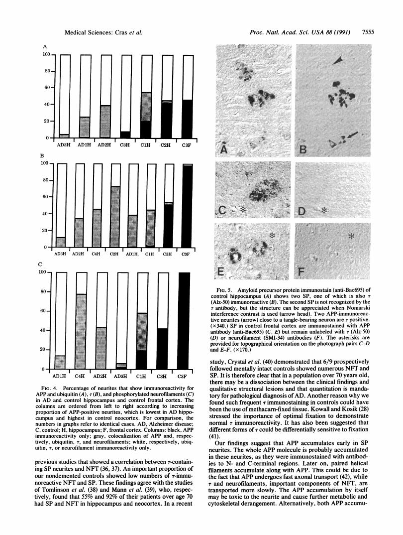
FFIG. 3. Three serial sections of SP in the dentate gyrus in AD hippocampus. First, the sections were immunostained with APP antibody
(anti-Bac695) (A, C, E), then we removed the chromogen and immunostained for ubiquitin (42D8) (B), T (Alz-50) (D), or phosphorylatedneurofilaments (SMI-34) (F). All the APP-immunoreactive neurites also show positivity for the second marker. In addition, some structures areimmunoreactive only for ubiquitin, a, or neurofilaments (arrowheads). D also shows many APP-negative, T-immunoreactive neuropil threads.(All x250.)
Proc. Nati. Acad Sci. USA 88 (1991)
Proc. Natl. Acad. Sci. USA 88 (1991) 7555
I '. . . . ... A.. i
.Se, *@#: 4
A,...;, *e
*.
%.* of. ..
A 5:;f
Bo
.B
C
E r
.70
4f,.
-A
4:
I
w }
, 4
_ .: :, i,
. f
.6 ., $ J as
_
_
|If# row
AD1H C4H AD2H AD3H C1H C3H C3F
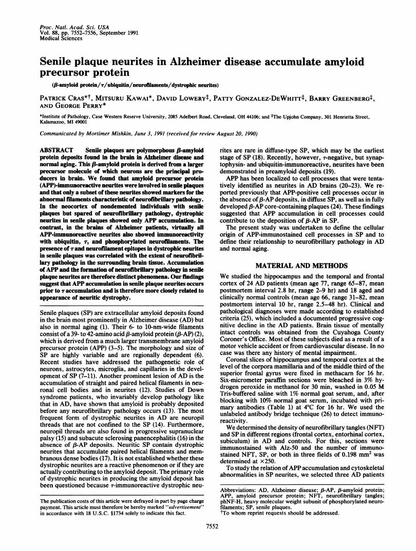
FIG. 4. Percentage of neurites that show immunoreactivity forAPP and ubiquitin (A), T (B), and phosphorylated neurofilaments (C)in AD and control hippocampus and control frontal cortex. Thecolumns are ordered from left to right according to increasingproportion of APP-positive neurites, which is lowest in AD hippo-campus and highest in control neocortex. For comparison, thenumbers in graphs refer to identical cases. AD, Alzheimer disease;C, control; H, hippocampus; F, frontal cortex. Columns: black, APPimmunoreactivity only; gray, colocalization of APP and, respec-tively, ubiquitin, T, and neurofilaments; white, respectively, ubiq-uitin, T, or neurofilament immunoreactivity only.
previous studies that showed a correlation between T-contain-ing SP neurites and NFT (36, 37). An important proportion ofour nondemented controls showed low numbers of T-immu-noreactive NFT and SP. These findings agree with the studiesof Tomlinson et al. (38) and Mann et al. (39), who, respec-tively, found that 55% and 92% of their patients over age 70had SP and NFT in hippocampus and neocortex. In a recent
FIG. 5. Amyloid precursor protein immunostain (anti-Bac695) ofcontrol hippocampus (A) shows two SP, one of which is also(Alz-50) immunoreactive (B). The second SP is not recognized by theT antibody, but the structure can be appreciated when Nomarskiinterference contrast is used (arrow head). Two APP-immunoreac-tive neurites (arrow) close to a tangle-bearing neuron are r positive.(x340.) SP in control frontal cortex are immunostained with APPantibody (anti-Bac69S) (C, E) but remain unlabeled with T (Alz-50)(D) or neurofilament (SMI-34) antibodies (F). The asterisks areprovided for topographical orientation on the photograph pairs C-Dand E-F. (x170.)
study, Crystal et al. (40) demonstrated that 6/9 prospectivelyfollowed mentally intact controls showed numerous NFT andSP. It is therefore clear that in a population over 70 years old,there may be a dissociation between the clinical findings andqualitative structural lesions and that quantitation is manda-tory for pathological diagnosis ofAD. Another reason why wefound such frequent r immunostaining in controls could havebeen the use of methacarn-fixed tissue. Kowall and Kosik (28)stressed the importance of optimal fixation to demonstratenormal r immunoreactivity. It has also been suggested thatdifferent forms of r could be differentially sensitive to fixation(41).Our findings suggest that APP accumulates early in SP
neurites. The whole APP molecule is probably accumulatedin these neurites, as they were immunostained with antibod-ies to N- and C-terminal regions. Later on, paired helicalfilaments accumulate along with APP. This could be due tothe fact that APP undergoes fast axonal transport (42), whiler and neurofilaments, important components of NFT, are
transported more slowly. The APP accumulation by itselfmay be toxic to the neurite and cause further metabolic andcytoskeletal derangement. Alternatively, both APP accumu-
A100.
B100
Medical Sciences: Cras et al.
's9 ao0ol .-
op0

7556 Medical Sciences: Cras et al.
lation and the formation of straight and paired helical fila-ments could be reactions to the extracellular amyloid deposit.The occurrence of APP-immunoreactive neuritic clustersunrelated to amyloid deposits would argue against this hy-pothesis. The development of paired helical filaments alsoseems to depend on the brain region, as dystrophic neuritesin the cerebellum do not show T immunoreactivity (43) but doshow membranous dense bodies that can be immunolabeledwith ubiquitin antibodies (44). In ultrastructural studies, wehave found APP accumulation in similar membranous densebodies in SP neurites (M.K., unpublished results).
It is not known whether the APP in SP neurites contributesto the extracellular amyloid deposits and if so, how the APPis released so that the ,3-AP region remains intact (45). On thebasis ofprevious reports and our present findings, we suggestthe following pathogenetic mechanism: A neuron or neuriteaccumulates APP and eventually degenerates, thereby re-leasing APP together with numerous enzymes (46, 47). ThisAPP would then be locally processed, presumably by micro-glia, and amyloid fibers would be formed in close proximityto these cells. This amyloid protein would then exert itsgrowth-promoting and neurotoxic activities (48, 49) on sur-rounding neurites that are recruited into the lesion, wherethese neurites would show signs of degeneration and regen-eration (50) and in their turn start to accumulate APP anddevelop neurofibrillary pathology.
The help of Drs. E. Balraj, R. C. Challener, P. S. Murthy, C.Santoscoy, S. Seligmann, and K. Jiraki of the Cuyahoga CountyCoroner's Office in collecting control brains is gratefully acknowl-edged. This study was supported by a Fogarty International Fellow-ship to P.C. and M.K. and by National Institutes of Health GrantsK04-AG00415 and AG-007552.
1. Terry, R. D. (1985) in Textbook ofNeuropathology, eds. Davis,R. L. & Robertson, D. M. (Williams & Wilkins, Baltimore), pp.824-841.
2. Glenner, G. & Wong, C. (1984) Biochem. Biophys. Res. Com-mun. 120, 885-890.
3. Kang, J., Lemaire, H.-G., Unterbeck, A., Salbaum, J. M.,Masters, C. L., Grzeschik, K.-H., Multhaup, G., Beyreuther,K. & Muller-Hill, B. (1987) Nature (London) 325, 733-736.
4. Tanzi, R. E., Gusella, J. F., Watkins, P. C., Bruns, G. A., St.George Hyslop, P. H., Van Keuren, M. L., Patterson, D.,Pagan, S., Kurnit, D. M. & Neve, R. L. (1987) Science 235,880-884.
5. Ponte, P., Gonzalez-DeWhitt, P., Schilling, J., Miller, D., Hsu,Greenberg, B., Davis, K., Wallace, W., Lieberburg, I., Fuller,F. & Cordell, B. (1988) Nature (London) 331, 525-527.
6. Wisniewski, H. M., Bancher, C., Barcikowska, M., Wen,G. Y. & Currie, J. (1989) Acta Neuropathol. (Berlin) 78,337-347.
7. Allsop, D., Haga, S.-I., Haga, C., Ikeda, S.-I., Mann, D. M. A.& Ishii, T. (1989) Neuropathol. Appl. Neurobiol. 15, 531-542.
8. Itagaki, S., McGeer, P. L., Akiyama, H., Zhu, S. & Selkoe, D.(1989) J. Neuroimmunol. 24, 173-182.
9. Wisniewski, H. M., Wegiel, J., Wang, K. C., Kujawa, M. &Lach, B. (1989) Can. J. Neurol. Sci. 16, 535-542.
10. Miyakawa, T., Shimoji, A., Kuramoto, R. & Higuchi, Y. (1982)Virchows Arch. B 40, 121-129.
11. Kawai, M., Kalaria, R. N., Harik, S. & Perry, G. (1990) Am.J. Pathol. 137, 1435-1446.
12. Terry, R. D. & Wisniewski, H. M. (1970) in Alzheimer's Dis.Rel. Cond. Ciba Found. Symp. 145-168.
13. Giaccone, G., Tagliavini, F., Linoli, G., Bouras, C., Frigerio,L., Frangione, B. & Bugiani, 0. (1989) Neurosci. Lett. 97,232-238.
14. Braak, H., Braak, E., Ohm, T. & Bohl, J. (1989) Neurosci. Lett.103, 24-28.
15. Probst, A., Langui, D., Lautenschlager, C., Ulrich, J., Brion,J. P. & Anderton, B. H. (1988) Acta Neuropathol. (Berlin) 77,61-68.
16. Tabaton, M., Mandybur, T. I., Perry, G., Onorato, M., Autilio-Gambetti, L. & Gambetti, P. (1989) Ann. Neurol. 26, 771-778.
17. Wisniewski, H. M. & Terry, R. D. (1973) in Progress in Neu-ropathology, ed. Zimmerman, H. M. (Grune & Stratton, NewYork), pp. 1-28.
18. Shin, R.-W., Ogomori, K., Kitamoto, T. & Tateishi, J. (1989)Am. J. Pathol. 134, 1365-1371.
19. Bugiani, O., Giaccone, G., Verga, L., Pollo, B., Ghetti, B.,Frangione, B. & Tagliavini, F. (1990) Neurosci. Lett. 119,56-59.
20. Perry, G., Lipphardt, S., Mulvihill, P., Kancherla, M., Mijares,M., Gambetti, P., Sharma, S., Maggiora, L., Cornette, J., Lobl,T. & Greenberg, B. (1988) Lancet H, 746.
21. Ishii, T., Kametani, F., Haga, S. & Satoh, M. (1989) Prog. Clin.Biol. Res. 317, 965-970.
22. Shoji, M., Hirai, S., Yamaguchi, H., Harigaya, Y.-& Kawara-bayashi, T. (1990) Brain Res. 512, 164-168.
23. Joachim, C., Games, D., Morris, J., Ward, P., Frenkel, D. &Selkoe, D. (1991) Am. J. Pathol. 138, 373-384.
24. Cras, P., Kawai, M., Siedlak, S., Mulvihill, P., Gambetti, P.,Lowery, D., Gonzalez-DeWhitt, P., Greenberg, B. & Perry, G.(1990) Am. J. Pathol. 137, 241-246.
25. Khachaturian, Z. S. (1985) Arch. Neurol. 42, 1097-1105.26. Sternberger, L. A. (1986) Immunocytochemistry (Wiley, New
York), 3rd Ed., pp. 125-127.27. Shelton, E. R., Cohn, R., Fish, L., Obernolte, R., Tahibra-
mani, R., Nestor, J. J. & Chan, H. (1990) J. Neurochem. 55,60-69.
28. Kowall, N. & Kosik, K. (1987) Ann. Neurol. 22, 639-643.29. Wolozin, B., Pruchnicki, A., Dickson, D. & Davies, P. (1986)
Science 232, 648-650.30. Mulvihill, P. & Perry, G. (1989) Brain Res. 484, 150-156.31. Autilio-Gambetti, L., Crane, R. C. & Gambetti, P. (1986) J.
Neurochem. 46, 366-379.32. Perry, G., Friedman, R., Shaw, G. & Chau, V. (1987) Proc.
Natl. Acad. Sci. USA 84, 3033-3036.33. Motte, J. & Williams, R. S. (1989) Acta Neuropathol. 77,
535-546.34. Mann, D. M. A., Yates, P. O., Marcyniuk, B., Ravindra, C. R.
(1986) Neuropathal. Appl. Neurobiol. 12, 447-457.35. Morris, J. C., McKeel, D. W., Storandt, M., Rubin, E. H.,
Price, J. L., Grant, E. A., Ball, M. J. & Berg, L. (1991)Neurology 41, 469-478.
36. Probst, A., Anderton, B. H., Brion, J.-P. & Ulrich, J. (1989)Acta Neuropathol. (Berlin) 77, 430-436.
37. Barcikowska, M., Wisniewski, H. M., Bancher, C., Grundke-Iqbal, I. (1989) Acta Neuropathol. (Berlin) 78, 225-231.
38. Tomlinson, B. E., Blessed, G. & Roth, M. (1968) J. Neurol.Sci. 7, 331-356.
39. Mann, D. M. A., Brown, A. M. T., Prinja, D., Jones, D. &Davies, C. A. (1990) Neuropathol. Appl. Neurobiol. 16, 17-25.
40. Crystal, H., Dickson, D., Fuld, P., Scott, R., Mehler, M.,Masdue, J., Kawas, C., Aronson, M. & Wolfson, L. (1988)Neurology 38, 1682-1687.
41. Pollock, N. J. & Wood, J. G. (1988) J. Histochem. Cytochem.36, 1117-1121.
42. Koo, E. H., Sisodia, S. S., Archer, D. R., Martin, L. J.,Weidemann, A., Beyreuther, K., Fischer, P., Masters, C. L. &Price, D. L. (1990) Proc. Natl. Acad. Sci. USA 87, 1561-1565.
43. Suenaga, T., Hirano, A., Llena, J. F., Ksiezak, R. H., Yen,S. H. & Dickson, D. W. (1990) J. Neuropathol. Exp. Neurol.49, 31-40.
44. Dickson, D. W., Wertkin, A., Mattiace, L. A., Fier, E., Kress,Y., Davies, P. & Yen, S. H. (1990) Acta Neuropathol. (Berlin)79, 486-493.
45. Sisodia, S. S., Koo, E. H., Beyreuther, K., Unterbeck, A. &Price, D. L. (1990) Science 248, 492-495.
46. Cataldo, A. M., Thayer, C. Y., Bird, E. D., Wheelock, T. R.& Nixon, R. A. (1990) Brain Res. 513, 181-192.
47. Cataldo, A. M. & Nixon, R. A. (1990) Proc. Natl. Acad. Sci.USA 87, 3861-3865.
48. Whitson, J. S., Selkoe, D. J. & Cotman, C. W. (1989) Science243, 1488-1490.
49. Yankner, B. A., Dawes, L. R., Fisher, S., Villa-Komaroff, L.,Oster-Granite, M. L. & Neve, R. L. (1989) Science 245, 417-420.
50. Lampert, P. (1967) J. Neuropathol. Appl. Neurobiol. 26, 345-368.
Proc. NatL Acad Sci. USA 88 (1991)