selective lignin and polysaccharide removal in …digital.csic.es/bitstream/10261/34499/1/selective...
TRANSCRIPT

Selective lignin and polysaccharide removal in naturalfungal decay of wood as evidenced by in situstructural analysesemi_2312 96..107
Angel T. Martínez,1* Jorge Rencoret,2† Lidia Nieto,1
Jesús Jiménez-Barbero,1 Ana Gutiérrez2 andJosé C. del Río2**1Centro de Investigaciones Biológicas, CSIC, Ramiro deMaeztu 9, E-28040 Madrid, Spain.2Instituto de Recursos Naturales y Agrobiología deSevilla, CSIC, P.O. Box 1052, E-41080-Seville, Spain.
Summary
Selective modification/degradation of the main plantpolymers (cellulose, hemicelluloses and lignin) wasinvestigated in a hardwood after white and brown-rotfungal decay under environmental conditions. Thechemical changes produced in the plant cell wall wereanalysed in situ, by nuclear magnetic resonance(NMR) at the gel state, and analytical pyrolysis. Two-dimensional (2D) NMR of the white-rotted woodshowed only cellulose and (deacetylated) hemicellu-lose, and the complete removal of lignin. On the otherhand, the brown-rotted wood showed the nearly com-plete absence of polysaccharides, while the main fea-tures of lignin structure, as revealed by 2D-NMR, couldbe observed. These included well-resolved aromaticand side-chain cross-signals, although the intensity ofthe latter signals was lowered indicating a reduction inthe number of side-chain linkages (b-O-4� and b-b�) peraromatic unit (their relative abundances remainingunchanged). These results contrast with a recentstudy concluding that the aromatic polymer afterbrown-rot decay is not longer recognized as lignin.Some oxidative alteration of lignin during brown-rotdecay was evidenced and, more interesting, severalcompounds with 3-methoxycatechol skeleton werereleased upon pyrolysis. Lignin demethylation isconsistent with recent brown-rot transcriptomic/secretomic studies showing overexpression of metha-nol oxidase, which could use lignin-derived methanol
to generate the peroxide required for cellulose depo-lymerization via Fenton chemistry.
Introduction
The major constituents of wood are cellulose, hemicellu-loses and lignin (Higuchi, 1997). The latter is a recalcitrantaromatic polymer, which protects the plant structuralpolysaccharides against microbial attack, and also playsother roles in wood tissues (Gellerstedt and Henriksson,2008). Wood-rotting fungi play a central ecological role inforest ecosystems enabling the cycling of the carbon fixedby plant photosynthesis, since other microorganisms arenot able to remove or circumvent the lignin barrier (Mar-tínez et al., 2005). In a similar way, the access to plantpolysaccharides represents a central issue for the indus-trial use of plant biomass (e.g. in paper pulp manufactureor bioethanol production).
White-rot fungi are the only organisms being able todegrade (mineralize) lignin in wood, thus opening up theplant cell wall. The white-rotted wood often presents awhitish colour, hence the name ‘white-rot fungi’. In mostcases, lignin removal by white-rot fungi parallels somedegradation of polysaccharides, mainly hemicelluloses, inthe so-called simultaneous degradation pattern (Otjenand Blanchette, 1986). However, a few white-rot speciesare able to remove lignin without causing a significant lossof polysaccharides, in a selective degradation pattern thathas the highest biotechnological interest. On the otherhand, brown-rot fungi have developed a different decaystrategy being able to degrade most of the cellulose andhemicelluloses leaving the lignin polymer. The brown-rotted wood becomes therefore enriched in lignin andpresents a dark reddish-brown to golden colour, and thusits name.
Due to the above characteristics, both white-rot(Watanabe, 2007) and brown-rot basidiomycetes (Schill-ing et al., 2009) are attracting considerable attention asmodels for two different strategies of wood attack. Thesebiotechnological models are of interest in lignocellulosebiorefineries, for the sustainable production of chemicals,materials and fuels from renewable plant resources(Ragauskas et al., 2006). Interestingly, the model white-rot fungus Phanerochaete chrysosporium was the first
Received 16 April, 2010; accepted 16 June, 2010. For correspon-dence. *E-mail [email protected]; Tel. (+34) 918373112 (ext4407); Fax (+34) 915360432; or **E-mail [email protected]; Tel.(+34) 954624711 (ext 119); Fax (+34) 954624002. †Present address:Great Lakes Bioenergy Research Center, University of Wisconsin-Madison, WI, USA.
Environmental Microbiology (2011) 13(1), 96–107 doi:10.1111/j.1462-2920.2010.02312.x
© 2010 Society for Applied Microbiology and Blackwell Publishing Ltd

basidiomycete whose genome was sequenced (Martínezet al., 2004), and the Postia placenta genome has beenrecently sequenced as a model brown-rot fungus (Mar-tínez et al., 2009a). The mechanism of lignin removal bywhite-rot basidiomycetes has been extensively investi-gated during last years, mainly with the aim of developingbiotechnological applications for the pulp and paperindustry (Martínez et al., 2009b). It has been found to beconstituted by unique high redox-potential peroxidasesand H2O2-generating oxidases, among other enzymes(Ruiz-Dueñas and Martínez, 2009). In contrast, themechanism of wood attack by brown-rot fungi is morepoorly understood. In particular, the way used by thesefungi to circumvent the lignin barrier, and be able todegrade cellulose is still under investigation (Baldrian andValaskova, 2008). Such a mechanism is of biotechnologi-cal interest when simple sugars are to be obtained fromwood, e.g. for fermentation and production of ethanol orother chemical compounds.
Elucidating the mechanisms of biological degradationor modification of lignin is a difficult task due to its complexmacromolecular structure (a branched polymer includingdifferent units and inter-unit bonds) and intimate associa-tion to cell-wall polysaccharides (Gellerstedt and Henriks-son, 2008). For many years, simple model compoundswere used to establish the main reactions caused bylignin-modifying organisms (Kishimoto, 2009). However,some modern analytical techniques, such as two-dimensional (2D) or three-dimensional (3D) nuclear mag-netic resonance (NMR), provided new tools for theanalysis of complex macromolecules, like lignin. In fact,several new lignin substructures were for the first timedetected using 2D-NMR (Karhunen et al., 1995; Zhanget al., 2006). Moreover, studies with ionic liquids and otherstrong solvents have recently shown the possibility toanalyse plant polymers in situ (Lu and Ralph, 2003; Kil-pelainen et al., 2007; Yelle et al., 2008a), overcoming theisolation problem that, in the case of lignin, always resultsin low recovery and chemical modifications (Fujimotoet al., 2005).
In the present work, previously characterized samplesof Eucryphia cordifolia wood extensively degraded by thewhite-rot fungus Ganoderma australe and by an uniden-tified brown-rot basidiomycete under environmental con-ditions in the Austral rainforest (Martínez et al., 1991)were analysed using modern techniques. For the NMRanalyses, the chemical modifications were analysed insitu by heteronuclear single quantum (HSQC) experi-ments on the whole wood sample using a method thatconsists in swelling finely ground samples in deuterateddimethylsulfoxide (DMSO-d6) and forming a gel in theNMR tube (Kim et al., 2008; Rencoret et al., 2009).Pyrolysis coupled to gas chromatography/mass spec-trometry (Py-GC/MS) is a convenient tool for the rapid
analysis of lignin, and has been already used to charac-terize different patterns of wood decay by fungi (del Ríoet al., 2001; 2002). In this work, pyrolysis of the wholewood was performed both in the absence and in thepresence of tetrabutylammonium hydroxide (Py/TBAH).The latter results in alkaline degradation (thermochemoly-sis) and simultaneous butylation (of acid and phenolicgroups) facilitating products analysis by GC/MS (del Ríoet al., 1996).
The aim of the study is to provide new information onthe wood-rotting processes by the use of the above ana-lytical techniques, with special emphasis of the fungalalterations of the recalcitrant lignin polymer. Although2D-NMR of wood gels after in vitro brown-rot fungal decayhas been reported (Yelle et al., 2008b), this is the first timethat this analytical approach is applied on wood decayedunder environmental conditions in the field. The presentstudy also tries to clarified some discrepancies with thegenerally accepted wood-rot patterns, raised during the invitro study mentioned above, which claimed that the aro-matic polymer remaining after brown rot cannot be longerrecognized as lignin. Finally, the changes observed in thecomposition and structure of the decayed wood polymersare discussed taking into account the biochemical infor-mation available, as well as the data provided by recentgenomic, transcriptomic and secretomic studies on wood-rotting basidiomycetes.
Results and discussion
General characteristics of the rotted wood samples
White- and brown-rot decay of E. cordifolia wood wererevealed by the characteristic visual aspect of wood (seeFig. S1a and b), together with microscopic examination(Fig. S1c and d), and chemical analyses. The latterrevealed a selective removal of lignin (94% of the initialamount) and conservation of cellulose (only 24% of thetotal glucose, including hemicellulose glucans, wasremoved) by the white-rot fungus; in contrast with theselective removal of cellulose (92% of the initial glucan)and conservation of lignin (only 1% of its initial amountwas removed) after the brown-rot decay (xylan waslargely removed, 96–99%, in both cases). Interestingly,the weight losses caused by the two fungi on the E.cordifolia wood (estimated from the decrease of its bulkdensity) were very similar (61–62%) facilitating thepresent comparison of the two decay patterns.
Divergent white- and brown-rot patterns as shown bywood NMR at the gel state
Figure 1a–c shows the HSQC 2D-NMR spectra of thewhole sound, white-rotted and brown-rotted woods
Fungal removal of wood lignin or polysaccharides 97
© 2010 Society for Applied Microbiology and Blackwell Publishing Ltd, Environmental Microbiology, 13, 96–107

A
O
O
HO
HO
H3CO
1 6
5 4
3
2
2 '
6 ' 5 '
4 ' 3 '
1 '
OCH3
H3CO OCH3
B
O
O
O
O
'
'
'
OCH3
OCH3
H3CO
OCH3
5 '
3 ' 2 '
1 ' 6 '
4 '
6 5
4 3
2
1
C
O
HO
O
OCH3
OCH3
H3CO
3 '
1 ' 6 '
5 ' 4 '
2 '
6
5
4 3
2 1
D
O
OCH3
O
OAr
OH HO
O
1 ' 6 '
5 ' 4 '
3 '
2 '
'
'
1 6
5
4 3 2
OCH3
H3CO
H3CO
'
E
O
O
HO
O
H3CO
1 6
5 4
3
2
2 '
6 ' 5 '
4 ' 3 '
1 '
OCH3
H3CO OCH3
G
O
OCH3
OH
1 6
5 4
3
2
S
H3CO
O
OCH3
OH
1 6
5 4
3
2
7 6 5 4 3
120
1
10
100
9
0 8
0 7
0 6
0 δC
[ppm
]
δH [ppm]
b
X5 X5
X2
X4
Gl1
X1
X3
δH [ppm] 7 6 5 4 3
δC [p
pm]
120
1
10
100
9
0 8
0 7
0 6
0
-OMeCβ
Bβ
Aγ
Dβ
Aβ(S)
Aβ(G)
Dα
Cα
Aα
BαDα’
Bγ
M’2
Cγ
X5 X5
X2
X3X4
U4
U1X’1
Gl1
X1/X’1S2,6
G2
G5
G6
X1(R)
M1
S’2,6
X’3X’2
a
7 6 5 4 3
120
1
10
100
9
0 8
0 7
0 6
0
δH [ppm]
δC [p
pm]
c
S2,6
G2
G5
G6
S’2,6
-OMeBβ
Aγ
Aβ(S)
Aβ(G)
Bα
Bγ
Eβ
MC’6
MC6
Aα
S'
OR’
O
OCH3 H3CO
1 6
5 4 3
2
R
MC' OR’’
O
OCH3 RO
1 6
5 4 3
2
R’
MC
RO
O
OCH3
OH
1 6
5 4
3
2
Fig. 1. Two-dimensional (2D) NMR spectra (HSQC experiments) at the gel state of: (a) sound E. cordifolia wood; (b) E. cordifolia wood afterselective white-rot fungal decay under environmental conditions (by G. australe); and (c) E. cordifolia wood after selective brown-rot fungaldecay under environmental conditions. The lignin structures identified are also shown: (A) b-O-4′ substructure; (B) resinol substructure; (C)phenylcoumaran substructure; (D) spirodienone substructure; (E) Ca-oxidized substructure A; (G) guaiacyl unit; (S) syringyl unit; (S′)Ca-oxidized S unit (R, lignin or OH; R′, H or lignin); (MC) 5-hydroxyguaiacyl (R, H) or 5-aryl-ether-guaiacyl unit (R, lignin aromatic ring); (MC′)Ca-oxidized MC unit (R′, lignin or OH; R″, H or lignin). See Table 1 for signal assignment.
98 A.T. Martínez et al.
© 2010 Society for Applied Microbiology and Blackwell Publishing Ltd, Environmental Microbiology, 13, 96–107
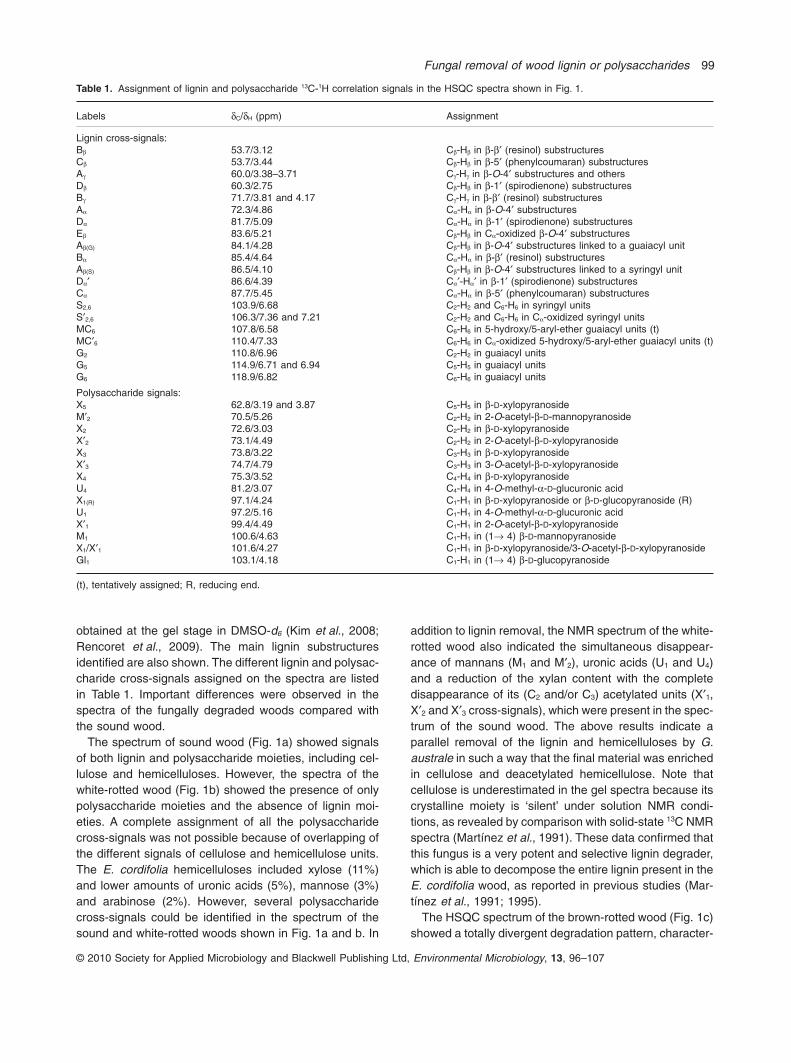
obtained at the gel stage in DMSO-d6 (Kim et al., 2008;Rencoret et al., 2009). The main lignin substructuresidentified are also shown. The different lignin and polysac-charide cross-signals assigned on the spectra are listedin Table 1. Important differences were observed in thespectra of the fungally degraded woods compared withthe sound wood.
The spectrum of sound wood (Fig. 1a) showed signalsof both lignin and polysaccharide moieties, including cel-lulose and hemicelluloses. However, the spectra of thewhite-rotted wood (Fig. 1b) showed the presence of onlypolysaccharide moieties and the absence of lignin moi-eties. A complete assignment of all the polysaccharidecross-signals was not possible because of overlapping ofthe different signals of cellulose and hemicellulose units.The E. cordifolia hemicelluloses included xylose (11%)and lower amounts of uronic acids (5%), mannose (3%)and arabinose (2%). However, several polysaccharidecross-signals could be identified in the spectrum of thesound and white-rotted woods shown in Fig. 1a and b. In
addition to lignin removal, the NMR spectrum of the white-rotted wood also indicated the simultaneous disappear-ance of mannans (M1 and M′2), uronic acids (U1 and U4)and a reduction of the xylan content with the completedisappearance of its (C2 and/or C3) acetylated units (X′1,X′2 and X′3 cross-signals), which were present in the spec-trum of the sound wood. The above results indicate aparallel removal of the lignin and hemicelluloses by G.australe in such a way that the final material was enrichedin cellulose and deacetylated hemicellulose. Note thatcellulose is underestimated in the gel spectra because itscrystalline moiety is ‘silent’ under solution NMR condi-tions, as revealed by comparison with solid-state 13C NMRspectra (Martínez et al., 1991). These data confirmed thatthis fungus is a very potent and selective lignin degrader,which is able to decompose the entire lignin present in theE. cordifolia wood, as reported in previous studies (Mar-tínez et al., 1991; 1995).
The HSQC spectrum of the brown-rotted wood (Fig. 1c)showed a totally divergent degradation pattern, character-
Table 1. Assignment of lignin and polysaccharide 13C-1H correlation signals in the HSQC spectra shown in Fig. 1.
Labels dC/dH (ppm) Assignment
Lignin cross-signals:Bb 53.7/3.12 Cb-Hb in b-b′ (resinol) substructuresCb 53.7/3.44 Cb-Hb in b-5′ (phenylcoumaran) substructuresAg 60.0/3.38–3.71 Cg-Hg in b-O-4′ substructures and othersDb 60.3/2.75 Cb-Hb in b-1′ (spirodienone) substructuresBg 71.7/3.81 and 4.17 Cg-Hg in b-b′ (resinol) substructuresAa 72.3/4.86 Ca-Ha in b-O-4′ substructuresDa 81.7/5.09 Ca-Ha in b-1′ (spirodienone) substructuresEb 83.6/5.21 Cb-Hb in Ca-oxidized b-O-4′ substructuresAb(G) 84.1/4.28 Cb-Hb in b-O-4′ substructures linked to a guaiacyl unitBa 85.4/4.64 Ca-Ha in b-b′ (resinol) substructuresAb(S) 86.5/4.10 Cb-Hb in b-O-4′ substructures linked to a syringyl unitDa′ 86.6/4.39 Ca′-Ha′ in b-1′ (spirodienone) substructuresCa 87.7/5.45 Ca-Ha in b-5′ (phenylcoumaran) substructuresS2,6 103.9/6.68 C2-H2 and C6-H6 in syringyl unitsS′2,6 106.3/7.36 and 7.21 C2-H2 and C6-H6 in Ca-oxidized syringyl unitsMC6 107.8/6.58 C6-H6 in 5-hydroxy/5-aryl-ether guaiacyl units (t)MC′6 110.4/7.33 C6-H6 in Ca-oxidized 5-hydroxy/5-aryl-ether guaiacyl units (t)G2 110.8/6.96 C2-H2 in guaiacyl unitsG5 114.9/6.71 and 6.94 C5-H5 in guaiacyl unitsG6 118.9/6.82 C6-H6 in guaiacyl units
Polysaccharide signals:X5 62.8/3.19 and 3.87 C5-H5 in b-D-xylopyranosideM′2 70.5/5.26 C2-H2 in 2-O-acetyl-b-D-mannopyranosideX2 72.6/3.03 C2-H2 in b-D-xylopyranosideX′2 73.1/4.49 C2-H2 in 2-O-acetyl-b-D-xylopyranosideX3 73.8/3.22 C3-H3 in b-D-xylopyranosideX′3 74.7/4.79 C3-H3 in 3-O-acetyl-b-D-xylopyranosideX4 75.3/3.52 C4-H4 in b-D-xylopyranosideU4 81.2/3.07 C4-H4 in 4-O-methyl-a-D-glucuronic acidX1(R) 97.1/4.24 C1-H1 in b-D-xylopyranoside or b-D-glucopyranoside (R)U1 97.2/5.16 C1-H1 in 4-O-methyl-a-D-glucuronic acidX′1 99.4/4.49 C1-H1 in 2-O-acetyl-b-D-xylopyranosideM1 100.6/4.63 C1-H1 in (1→ 4) b-D-mannopyranosideX1/X′1 101.6/4.27 C1-H1 in b-D-xylopyranoside/3-O-acetyl-b-D-xylopyranosideGl1 103.1/4.18 C1-H1 in (1→ 4) b-D-glucopyranoside
(t), tentatively assigned; R, reducing end.
Fungal removal of wood lignin or polysaccharides 99
© 2010 Society for Applied Microbiology and Blackwell Publishing Ltd, Environmental Microbiology, 13, 96–107

ized by the absence of practically all the polysaccharidemoieties, while only the lignin polymer was present. Theobtained data indicated that this polymer was not drasti-cally modified by the brown-rot fungus, all the typicalcross-signals of lignin remaining visible. This includedwell-resolved signals of b-O-4′ (A) and b-b′ (resinol-type,B) side-chain linkages together with strong aromaticsignals of syringyl (S) and guaiacyl (G) lignin units, whoserelative intensities are apparently not strongly affected. Toverify this point, the relative abundance of the main inter-unit linkages (given as per aromatic units, and as percent-age of total side-chains involved) were calculated from theHSQC spectra (Table 2). These data indicated that brown-rot decay only slightly increased the relative abundancesof the b-O-4′ substructures (from 84% to 86% of totalside-chains) but did not modify that of the b-b′ substruc-tures (12% of total side-chains). Minor spirodienones (D)found in sound wood (2% of side-chains) were notdetected after the brown-rot decay.
On the other hand, a small increase of oxygenatedmoieties in the brown-rotted wood was evidenced in theHSQC spectrum by the small signal corresponding toaliphatic 13Cb-1Hb correlation in Ca-oxidized b-O-4′ sub-structures (E). The aromatic 13C2,6-1H2,6 correlation signalin Ca-oxidized S units (S′) also showed comparativelyhigher intensity. An estimation of 12% oxidized S units inthe sound wood, and 20% in the brown-rotted wood wasobtained. The existence of carbonyl and carboxyl groupsin brown-rotted wood has been also shown by solid-state13C NMR and other techniques (Martínez et al., 1991; Sunet al., 2009; Koenig et al., 2010). This indicates that someoxidative alterations in the lignin structure occurred duringbrown-rot decay, in agreement with the pyrolysis resultsdescribed below. Moreover, two new aromatic cross-signals (MC and MC′) were found in the spectrum of thebrown-rotted wood (Fig. 1c). The same signals have beenreported in HSQC spectra of brown-rotted oak wood,obtained using high-resolution magic-angle spinning(HRMAS) NMR (Koenig et al., 2010). We assigned thesetwo signals to aromatic structures derived from the
demethylation/demethoxylation of S units (yielding5-hydroxyguaiacyl units that can form new 5-O-aryl etherbonds), a modification reaction that was further confirmedby pyrolysis (yielding 3-methoxycatechols). However,more studies are necessary for the unambiguous assign-ment of the two new signals. In addition to the presence ofthese new aromatic units, which represented 14% oflignin units (Table 3), and the absence of p-hydroxyphenyl(H) units, the spectrum of the brown-rotted wood alsoshowed a small increase of the S/G ratio (from 3.2 insound wood to 4.2 after brown-rot decay).
Side-chain linkages and methoxyl removal inbrown-rot decay
The relative abundances of the main lignin linkagesremained largely unchanged after brown-rot decay, asshown in the previous section. However, a comparison ofthe lignin side-chain (involved in b-O-4′, b-b′ and otherlinkages) and methoxyl cross-signals on an aromatic unitbasis indicates around 45% depletion of the above link-ages and near 20% depletion of methoxyls. This agreeswith the Yelle and colleagues (2008b) hypothesis sug-gesting that lignin attack by hydroxyl-free radical, the mainwood decay agent in brown rot (Suzuki et al., 2006; Mar-tínez et al., 2009a), should result in simultaneous break-down of both methoxyl groups and inter-unit etherlinkages. Taking into account the decrease of methoxylgroups, the depletion of side-chain linkages in E. cordifo-lia lignin, on a methoxyl basis, amounts to only 30%. Thisobservation contrasts with Yelle and colleagues (2008b)who reported a depletion of more than 70% of the ligninside-chain linkages (on a methoxyl basis) in white spruce(Picea glauca) wood degraded in vitro with the brown-rotfungus Gloeophyllum trabeum. In fact, some of the ligninside-chain signals that are clearly visible in the HSQCspectrum of the brown-rotted E. cordifolia wood (Fig. 1c)were hardly detectable in the HSQC spectrum of the G.trabeum rotted P. glauca wood. Therefore, these authorsconcluded that extensive ligninolysis (i.e. degradation oflignin inter-unit linkages) was produced by brown-rot fungi
Table 2. Abundances of lignin side-chain linkages and methoxylgroups, as per aromatic unit (and as percentage of total side-chains,in parenthesis), in the sound and brown-rotted E. cordifolia wood(calculated from the 2D-NMR spectra of Fig. 1A and C).
Sound wood Brown rot
Linkage abundance:b-O-4′ aryl ethers (A) 0.82 (83) 0.44 (80)b-b′ resinols (B) 0.11 (12) 0.07 (12)b-5′ phenylcoumarans (C) 0.02 (2) 0.01 (2)b-1′ spirodienones (D) 0.02 (2) 0.00 (0)Ca-oxidized b-O-4′ substructures (E) 0.01 (1) 0.03 (6)
Methoxyl content 2.77 2.24
Table 3. Ratio of unmodified (H, G and S) and S-type demethylated(MC) lignin units in the sound and brown-rotted E. cordifolia wood, asestimated from 2D-NMR, Py-GC/MS and Py/TBAH analyses (MC/[MC+S] is shown in parentheses).
H : G : S : MC ratio (S-demethylation as %)
Sound wood Brown-rotted wood
2D-NMR 0:24:76:0 (0%) 0:17:69:14 (18%)Py-GC/MS 3:25:68:4a (5%) 3:21:62:12a (16%)Py/TBAH 2:25:72:0 (1%) 2:23:69:6 (8%)
a. Small amounts of demethylated G units were also found byPy-GC/MS of sound (1%) and brown-rotted (2%) woods.
100 A.T. Martínez et al.
© 2010 Society for Applied Microbiology and Blackwell Publishing Ltd, Environmental Microbiology, 13, 96–107
yielding a material that is not longer recognized aslignin. This contrasts with the typical lignin spectrumreported here (Fig. 1c), as well as with the recentlyreported HRMAS analysis of naturally brown-rottedoak wood (Koenig et al., 2010). Moreover, no signalsassigned to new (aryl-aryl or side-chain) linkages, whichmay be formed to replace the > 70% degraded side-chainlinkages in lignin, were detected in the HSQC spectrum ofthe in vitro degraded wood (Yelle et al., 2008b). In con-trast, the two new aromatic signals (MC and MC′) found inthe HSQC spectrum of the E. cordifolia wood after naturalbrown-rot decay could include correlations of newaryl-O-aryl ether linkages formed after demethylation/demethoxylation of S units (as discussed in the nextsection).
The atypical decay pattern described by Yelle and col-leagues (2008b) cannot be due to a more extensivedecay, since the weight losses were similar to those foundon E. cordifolia wood (62–64%). Interestingly, the typicallignin spectra reported by Koenig and colleagues (2010)for brown-rotted oak by Laetiporus sulphureus were alsoobtained from naturally decayed wood. This suggest thatdifferences between laboratory and field conditions, thelatter resulting in more selective brown-rot removal ofpolysaccharides (as shown by NMR) could explain theabove discrepancies, more than the type of wood or thefungal species. A similar situation was produced when theauthors failed to reproduce the selective white-rot decaypattern by G. australe under laboratory conditions, usingAustral (E. cordifolia) and European (Fagus sylvatica)woods (Barrasa et al., 1992; Bechtold et al., 1993).
Confirmation of lignin demethylation in brown-rot decay:Py-GC/MS and Py/TBAH analyses
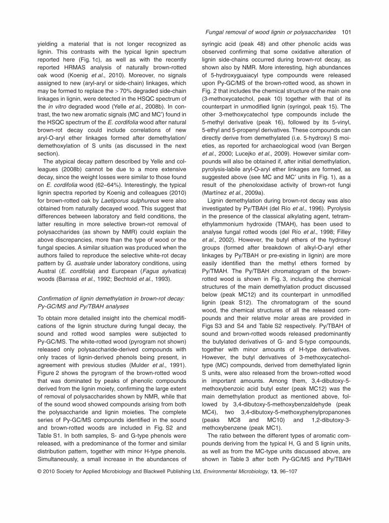
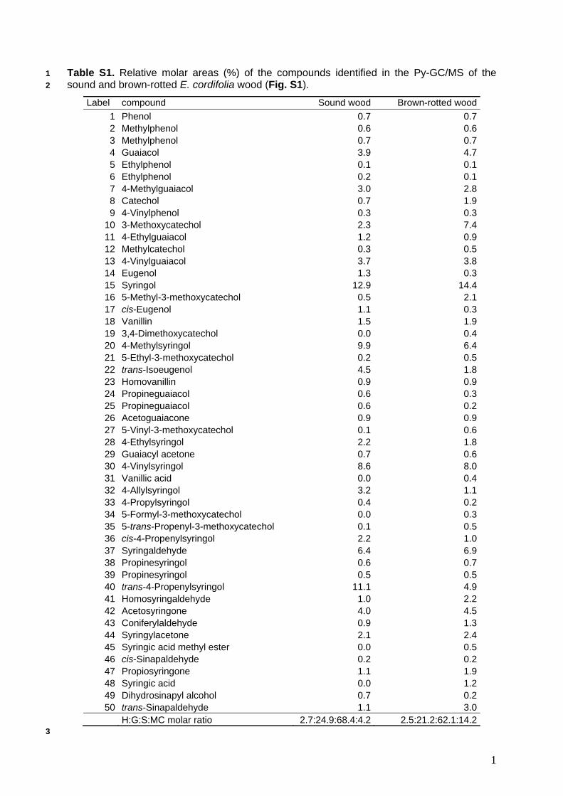
To obtain more detailed insight into the chemical modifi-cations of the lignin structure during fungal decay, thesound and rotted wood samples were subjected toPy-GC/MS. The white-rotted wood (pyrogram not shown)released only polysaccharide-derived compounds withonly traces of lignin-derived phenols being present, inagreement with previous studies (Mulder et al., 1991).Figure 2 shows the pyrogram of the brown-rotted woodthat was dominated by peaks of phenolic compoundsderived from the lignin moiety, confirming the large extentof removal of polysaccharides shown by NMR, while thatof the sound wood showed compounds arising from boththe polysaccharide and lignin moieties. The completeseries of Py-GC/MS compounds identified in the soundand brown-rotted woods are included in Fig. S2 andTable S1. In both samples, S- and G-type phenols werereleased, with a predominance of the former and similardistribution pattern, together with minor H-type phenols.Simultaneously, a small increase in the abundances of
syringic acid (peak 48) and other phenolic acids wasobserved confirming that some oxidative alteration oflignin side-chains occurred during brown-rot decay, asshown also by NMR. More interesting, high abundancesof 5-hydroxyguaiacyl type compounds were releasedupon Py-GC/MS of the brown-rotted wood, as shown inFig. 2 that includes the chemical structure of the main one(3-methoxycatechol, peak 10) together with that of itscounterpart in unmodified lignin (syringol, peak 15). Theother 3-methoxycatechol type compounds include the5-methyl derivative (peak 16), followed by its 5-vinyl,5-ethyl and 5-propenyl derivatives. These compounds candirectly derive from demethylated (i.e. 5-hydroxy) S moi-eties, as reported for archaeological wood (van Bergenet al., 2000; Lucejko et al., 2009). However similar com-pounds will also be obtained if, after initial demethylation,pyrolysis-labile aryl-O-aryl ether linkages are formed, assuggested above (see MC and MC′ units in Fig. 1), as aresult of the phenoloxidase activity of brown-rot fungi(Martínez et al., 2009a).
Lignin demethylation during brown-rot decay was alsoinvestigated by Py/TBAH (del Río et al., 1996). Pyrolysisin the presence of the classical alkylating agent, tetram-ethylammonium hydroxide (TMAH), has been used toanalyse fungal rotted woods (del Río et al., 1998; Filleyet al., 2002). However, the butyl ethers of the hydroxylgroups (formed after breakdown of alkyl-O-aryl etherlinkages by Py/TBAH or pre-existing in lignin) are moreeasily identified than the methyl ethers formed byPy/TMAH. The Py/TBAH chromatogram of the brown-rotted wood is shown in Fig. 3, including the chemicalstructures of the main demethylation product discussedbelow (peak MC12) and its counterpart in unmodifiedlignin (peak S12). The chromatogram of the soundwood, the chemical structures of all the released com-pounds and their relative molar areas are provided inFigs S3 and S4 and Table S2 respectively. Py/TBAH ofsound and brown-rotted woods released predominantlythe butylated derivatives of G- and S-type compounds,together with minor amounts of H-type derivatives.However, the butyl derivatives of 3-methoxycatechol-type (MC) compounds, derived from demethylated ligninS units, were also released from the brown-rotted woodin important amounts. Among them, 3,4-dibutoxy-5-methoxybenzoic acid butyl ester (peak MC12) was themain demethylation product as mentioned above, fol-lowed by 3,4-dibutoxy-5-methoxybenzaldehyde (peakMC4), two 3,4-dibutoxy-5-methoxyphenylpropanones(peaks MC8 and MC10) and 1,2-dibutoxy-3-methoxybenzene (peak MC1).
The ratio between the different types of aromatic com-pounds deriving from the typical H, G and S lignin units,as well as from the MC-type units discussed above, areshown in Table 3 after both Py-GC/MS and Py/TBAH
Fungal removal of wood lignin or polysaccharides 101
© 2010 Society for Applied Microbiology and Blackwell Publishing Ltd, Environmental Microbiology, 13, 96–107

analyses of the brown-rotted and the sound wood. Thesedata agree with those obtained from 2D-NMR, alsoincluded in Table 3, although the minor H units are prob-ably below the detection level of 2D-NMR. Py-GC/MSshowed some demethylation products in the sound wood(that could be formed during thermal depolymerizationsince they were not detected after alkaline depolymeriza-tion in Py/TBAH nor in the NMR spectrum). However, theyare much less abundant than in the brown-rotted wood.Interestingly, a preferential demethylation of the S unitswas observed after the different analyses, with values thatranged 8–18% depending of the estimation procedure.Methoxycatechol had been previously detected afterPy-GC/MS of both sound and brown-rotted E. cordifoliawood (Mulder et al., 1991), but no information was pro-vided on its increase during wood decay. However, theincreased occurrence of 3-methoxycatechol typePy-GC/MS compounds, and the detection of similar diag-nostic compounds after Py/TBAH (being absent from thesound wood) clearly indicates that some demethylation oflignin occurred during brown-rot decay, in agreement with
the classical work of Kirk (1975) and other studies (Filleyet al., 2002) including radiolabelling demonstration(Niemenmaa et al., 2008). As mentioned above, lignindemethylation would result in new phenolic units thatcould experiment new oxidation reactions during fungalattack resulting in new aryl-O-aryl ether linkages byradical coupling. It is interesting that Klason lignin estima-tion in the sound and brown-rotted E. cordifolia wood(62% weight loss) revealed no significant lignin degrada-tion (only ~1% of the initial lignin) indicating that thechanges revealed by the 2D-NMR and pyrolysis analysesdiscussed above are due to fungal reworking (modifica-tion) of the polymer (including the new aryl-O-aryl etherlinkages) more than to complete degradation of somelignin moieties.
Wood rotting in the light of new genomic/transcriptomicinformation
The recent sequencing of the first brown-rot fungalgenome (Martínez et al., 2009a) has provided new clues
2 4 6 8 10 12 14 16 18 20 22 24
Retention time (min)
4
7
8
10
13
15
1618
20
22 28
30
32
3740
42
43
44
47
5041
48
36
HO
OH
O
OH
O O
Fig. 2. Py-GC/MS of E. cordifolia wood after brown-rot fungal decay under environmental conditions. The chemical structures of the mainlignin demethylation product (peak 10) and its counterpart in unmodified lignin (peak 15) are shown. Peak identification (relative molar area �1%): 4, guaiacol; 7, 4-methylguaiacol; 8, catechol; 10, 3-methoxycatechol; 13, 4-vinylguaiacol; 15, syringol; 16, 5-methyl-3-methoxycatechol;18, vanillin; 20, 4-methylsyringol; 22, trans-isoeugenol; 28, 4-ethylsyringol; 30, 4-vinylsyringol; 32, 4-allylsyringol; 36, cis-4-propenylsyringol; 37,syringaldehyde; 40, trans-4-propenylsyringol; 41, homosyringaldehyde; 42, acetosyringone; 43, coniferaldehyde; 44, syringylacetone; 47,propiosyringone; 48, syringic acid; and 50, trans-sinapaldehyde. For Py-GC/MS of the control wood, and identification of minor peaks, seeFig. S2 and Table S1.
102 A.T. Martínez et al.
© 2010 Society for Applied Microbiology and Blackwell Publishing Ltd, Environmental Microbiology, 13, 96–107

about how this decay process takes place, although dif-ferences between brown-rot fungi probably exist (asrevealed by P. placenta and G. trabeum genome and ESTinformation currently available in GenBank). In thiscontext, the above discussed lignin demethylation couldplay a central role since it releases methanol, and thesecretomic and transcriptomic studies associated to thebrown-rot genome have shown that methanol oxidasegenes are overexpressed when the fungus degrades cel-lulose.
In the light of the information currently available, it istherefore possible to infer that brown-rot fungi most prob-ably produces H2O2 using methanol as the reducing sub-strate of methanol oxidase, an enzyme that has beeninvestigated by Daniel and colleagues (2007) in relation tobrown rot. Then, H2O2 would be reduced to hydroxylradical by ferrous iron formed in hydroquinone redox-cycling (Suzuki et al., 2006) or by other enzymatic mecha-nisms. The hydroxyl-free radical would be the main agent
responsible for the oxidative depolymerization of cellu-lose, since a reduced number of cellulases (but a varietyof hemicellulases) has been found in the brown-rot fungalgenome (Martínez et al., 2009a). At the same time it pro-motes partial demethylation of lignin, a reaction that hasbeen proved using 13C-TMAH thermochemolysis (Aranteset al., 2009).
A very recent study (Vanden Wymelenberg et al., 2010)has shown that methanol oxidase is also overexpressedin the white-rot fungus P. chrysosporium. Demethylation isalso a common side-reaction after one-electron oxidationof lignin units to their cation radicals by white-rot fungi andtheir ligninolytic peroxidases (Ander et al., 1992; Martínezet al., 2005). These results suggest that several oxidases(such as glyoxal oxidase and methanol oxidase) areinvolved in H2O2 production by P. chrysosporium usingdifferent lignocellulose-derived compounds as substrates.Most probably, the same happens in brown-rot fungi,since expression of aryl-alcohol oxidase (and other H2O2-
S1
S4
S8 S10
S12
S14MC12
12 14 16 18 20 22 24 26 28 30 3210
Retention time (min)
MC4
OO
O O
O
OO
O
O
O
MC8
MC10MC1
MC14
Fig. 3. Py/TBAH of E. cordifolia wood after brown-rot fungal decay under environmental conditions. The structures of the main lignindemethylation product (MC12) and its counterpart in unmodified lignin (S12) are depicted, and those of other compounds are shown inFig. S4. Identification of peaks from demethylated lignin and their unmodified counterparts: S1, 1-butoxy-2,6-dimethoxybenzene; MC1,1,2-dibutoxy-3-methoxybenzene; S4, 3,5-dimethoxy-4-butoxybenzaldehyde; S8, 3,5-dimethoxy-4-butoxyphenylacetone; S10,1-(3,5-dimethoxy-4-butoxyphenyl)propan-1-one; MC4, 3,4-dibutoxy-5-methoxybenzaldehyde; S12, 3,5-dimethoxy-4-butoxybenzoic acid butylester; MC8, 3,4-dibutoxy-5-methoxyphenylacetone; MC10, 1-(3,4-dibutoxy-5-methoxyphenyl)-propan-1-one; S14,3,5-dimethoxy-4-butoxy-benzenepropanoic acid butyl ester; MC12, 3,4-dibutoxy-5-methoxybenzoic acid butyl ester; and MC14,3,4-dibutoxy-5-methoxybenzenepropanoic acid butyl ester. For Py/TBAH of the control wood, and identification of minor peaks, see Fig. S3and Table S2.
Fungal removal of wood lignin or polysaccharides 103
© 2010 Society for Applied Microbiology and Blackwell Publishing Ltd, Environmental Microbiology, 13, 96–107

producing oxidases) has been found in P. placenta, inaddition to the main methanol oxidase (Martínez et al.,2009a). In this context, it is interesting to remark that H2O2
production is a common step in both white and brown-rotdecay of wood by fungi: in the first case as the oxidizingsubstrate of ligninolytic peroxidases, and for hydroxylradical generation in the second case.
Experimental procedures
Materials and general analyses
Samples of E. cordifolia (Chilean ‘ulmo’; order Oxalidales,family Cunoniaceae) wood naturally degraded by white andbrown-rot basidiomycetes were collected at two locations onChiloe Island, in the Chilean rain forest (Martínez et al.,1991). The samples include: (i) sound wood, (ii) extremelydelignified wood by the white-rot fungus G. australe and (iii)wood decayed by an unidentified brown-rot basidiomycete.Sound and decayed wood samples were dried, milled in aJanke and Kunkel mill and passed through a 20-mesh sieve.In addition to their macroscopic aspect (such as colour andtexture) and eventual presence of fruit bodies, microscopicexamination using the selective safranin-Astra blue staining(Srebotnik and Messner, 1994) and chemical analysesincluding Klason lignin content, polysaccharide composition(neutral sugars by GC as alditol acetates, and uronic acids bythe carbazole method), and other wood constituents (ashes,extractives and water-soluble material) (Martínez et al.,1991) were used to confirm and characterize the brown andwhite-rot decay patterns of the samples analysed.
2D-NMR
Roughly 100 mg of finely ball-milled wood was suspended in0.75 ml of DMSO-d6 in the NMR tube and sonicated for10 min in an ultrasonic bath (Ultrasons JS 3000513 fromSelecta, with a frequency of 40 kHz and 150 W power con-sumption), until a homogeneous gel was formed. 2D-NMRspectra were recorded at 25°C on a Bruker AVANCE500 MHz equipped with a z-gradient triple resonance probe.For the HSQC experiments, the spectral widths were5000 Hz and 25 000 Hz for the 1H- and 13C-dimensionsrespectively. The number of collected complex points was2048 for 1H-dimension with a recycle delay of 5 s. Thenumber of transients was 64, and 256 time increments werealways recorded in 13C-dimension. The 1JCH used was140 Hz. The J-coupling evolution delay was set to 3.2 ms.Squared cosine-bell apodization function was applied in bothdimensions. Prior to Fourier transform, the data matrixeswere zero filled up to 1024 points in the 13C-dimension. Thecentral DMSO peak (dC 39.5; dH 2.50) was used as chemicalshift reference. HSQC cross-signals of lignin were assignedby comparing them with previously reported data (Ralphet al., 1999; 2004; Liitiä et al., 2003; Capanema et al., 2004;2005; Ibarra et al., 2007a; 2007b; del Río et al., 2008; Mar-tínez et al., 2008; Rencoret et al., 2008). Some woodpolysaccharide signals were also assigned (Yelle et al.,2008a; Rencoret et al., 2009; Kim and Ralph, 2010).
A semiquantitative analysis of the intensities of the HSQCcross-signal intensities was performed (Heikkinen et al.,2003; Liitiä et al., 2003; Zhang and Gellerstedt, 2007). First,cross-signal integration was performed separately for thedifferent regions of the spectra, which contain chemicallyanalogous carbon–proton pairs. In the aliphatic oxygenatedregion, the relative abundance of the different inter-unit link-ages were estimated from Ca-Ha correlations, and the relativeabundance of side-chains involved in the different interunitlinkages were calculated. In the aromatic region, C2,6-H2,6
correlations from S units, C2-H2 plus C6-H6 correlations fromG units, and C6-H6 correlations from MC units were used toestimate the G : S : MC ratio of lignin. Finally, the abun-dances of the different interunit linkages were referred to thenumber of aromatic units, to obtain a comparative estimationof their removal during fungal decay.
Py-GC/MS and Py/TBAH
Py-GC/MS of wood (approximately 1 mg) was performed witha 2020 micro-furnace pyrolyser (Frontier Laboratories) con-nected to an Agilent 6890 GC/MS system equipped with aDB-5MS (Agilent J&W) fused-silica capillary column(30 m ¥ 0.25 mm i.d., 0.25 mm film thickness) and an Agilent5973 mass selective detector (EI at 70 eV). The pyrolysis wasperformed at 500°C. The oven temperature was programmedfrom 40°C (1 min) to 300°C at 6°C min-1 (10 min). Heliumwas used as carrier gas (1 ml min-1). For Py/TBAH, 1 mg ofwood sample was mixed with approximately 5 ml of TBAH(25%, w/w, methanol solution) and the pyrolysis was carriedout as described above. The compounds were identified byfragmentography and by comparing their mass spectra withthose of the Wiley and NIST libraries and reported in theliterature (Faix et al., 1990; Ralph and Hatfield, 1991; del Ríoet al., 1996). Peak molar areas were calculated for the lignin-derived products, the summed areas were normalized to 100,and the data for two repetitive analyses were averaged andexpressed as percentages.
Acknowledgements
This study has been supported by the BIORENEWEU-project (NMP2-CT-2006-026456), the CSIC project201040E075, and the Spanish projects ELLE (AGL2008-00709) and RAPERO (BIO2008-01533). The authors thankAldo E. González (CBM, CSIC, Madrid, Spain) and RafaelVicuña (Pontificia University, Santiago de Chile) for his help inthe collection and identification of wood samples; and JoséM. Barrasa (Alcalá University, Madrid, Spain) for his help themicroscopic examinations.
References
Ander, P., Hatakka, A.I., Lundell, T.K., Pettersson, B., Stal-masek, M., and Volc, J. (1992) Demethoxylation of lignin bylignin peroxidases from Phlebia radiata and Phanerocha-ete chrysosporium. In Ligno-Cellulosics. Science, Technol-ogy, Development and Use. Kennedy, J.F., Phillips, G.O.,and Williams, P.A. (eds). New York, NY, USA: EllisHorwood, pp. 109–119.
104 A.T. Martínez et al.
© 2010 Society for Applied Microbiology and Blackwell Publishing Ltd, Environmental Microbiology, 13, 96–107

Arantes, V., Qian, Y.H., Kelley, S.S., Milagres, A.M.F., Filley,T.R., Jellison, J., and Goodell, B. (2009) Biomimetic oxida-tive treatment of spruce wood studied by pyrolysis-molecular beam mass spectrometry coupled withmultivariate analysis and 13C-labeled tetramethylammo-nium hydroxide thermochemolysis: implications for fungaldegradation of wood. J Biol Inorg Chem 14: 1253–1263.
Baldrian, P., and Valaskova, V. (2008) Degradation of cellu-lose by basidiomycetous fungi. FEMS Microbiol Rev 32:501–521.
Barrasa, J.M., González, A.E., and Martínez, A.T. (1992)Ultrastructural aspects of fungal delignification of Chileanwoods by Ganoderma australe and Phlebia chrysocrea: astudy of natural and in vitro degradation. Holzforschung 46:1–8.
Bechtold, R., González, A.E., Almendros, G., Martínez, M.J.,and Martínez, A.T. (1993) Lignin alteration by Ganodermaaustrale and other white-rot fungi after solid-state fermen-tation of beech wood. Holzforschung 47: 91–96.
van Bergen, P.F., Poole, I., Ogilvie, T.M.A., Caple, C., andEvershed, R.P. (2000) Evidence for demethylation ofsyringyl moieties in archaeological wood using pyrolysis-gas chromatography/mass spectrometry. Rapid CommunMass Spectrom 14: 71–79.
Capanema, E.A., Balakshin, M.Y., and Kadla, J.F. (2004) Acomprehensive approach for quantitative lignin character-ization by NMR spectroscopy. J Agric Food Chem 52:1850–1860.
Capanema, E.A., Balakshin, M.Y., and Kadla, J.F. (2005)Quantitative characterization of a hardwood milled woodlignin by nuclear magnetic resonance spectroscopy.J Agric Food Chem 53: 9639–9649.
Daniel, G., Volc, J., Filonova, L., Plihal, O., Kubátová, E., andHalada, P. (2007) Characteristics of Gloeophyllum trabeumalcohol oxidase, an extracellular source of H2O2 in brownrot decay of wood. Appl Environ Microbiol 73: 6241–6253.
Faix, O., Meier, D., and Fortmann, I. (1990) Thermal degra-dation products of wood. Holz Roh Werkst 48: 281–285.
Filley, T.R., Cody, G.D., Goodell, B., Jellison, J., Noser, C.,and Ostrofsky, A. (2002) Lignin demethylation and poly-saccharide decomposition in spruce sapwood degraded bybrown rot fungi. Org Geochem 33: 111–124.
Fujimoto, A., Matsumoto, Y., Chang, H.M., and Meshitsuka,G. (2005) Quantitative evaluation of milling effects on ligninstructure during the isolation process of milled wood lignin.J Wood Sci 51: 89–91.
Gellerstedt, G., and Henriksson, G. (2008) Lignins: majorsources, structure and properties. In Monomers, Polymersand Composites from Renewable Resources. Belgacem,M., and Gandini, A. (eds). Amsterdam, the Netherlands:Elsevier, pp. 201–224.
Heikkinen, S., Toikka, M.M., Karhunen, P.T., and Kilpeläinen,I.A. (2003) Quantitative 2D HSQC (Q-HSQC) via suppres-sion of J-dependence of polarization transfer in NMR spec-troscopy: application to wood lignin. J Am Chem Soc 125:4362–4367.
Higuchi, T. (1997) Biochemistry and Molecular Biology ofWood. London, UK: Springer Verlag.
Ibarra, D., Chávez, M.I., Rencoret, J., del Río, J.C., Gutiérrez,A., Romero, J., et al. (2007a) Lignin modification duringEucalyptus globulus kraft pulping followed by totally chlo-
rine free bleaching: a two-dimensional nuclear magneticresonance, Fourier transform infrared, and pyrolysis-gaschromatography/mass spectrometry study. J Agric FoodChem 55: 3477–3499.
Ibarra, D., Chávez, M.I., Rencoret, J., del Río, J.C., Gutiérrez,A., Romero, J., et al. (2007b) Structural modification ofeucalypt pulp lignin in a totally chlorine free bleachingsequence including a laccase-mediator stage. Holzfors-chung 61: 634–646.
Karhunen, P., Rummakko, P., Sipila, J., Brunow, G., andKilpeläinen, I. (1995) Dibenzodioxocins – a novel type oflinkage in softwood lignins. Tetrahedron Lett 36: 169–170.
Kilpelainen, I., Xie, H., King, A., Granstrom, M., Heikkinen, S.,and Argyropoulos, D.S. (2007) Dissolution of wood in ionicliquids. J Agric Food Chem 55: 9142–9148.
Kim, H., and Ralph, J. (2010) Solution-state 2D NMR ofball-milled plant cell wall gels in DMSO-d6/pyridine-d5. OrgBiomol Chem 8: 576–591.
Kim, H., Ralph, J., and Akiyama, T. (2008) Solution-state 2DNMR of ball-milled plant cell wall gels in DMSO-d6. Bioen-erg Res 1: 56–66.
Kirk, T.K. (1975) Effects of brown-rot fungus Lenzites trabeaon lignin of spruce wood. Holzforschung 29: 99–107.
Kishimoto, T. (2009) Synthesis of lignin model compoundsand their application to wood research. Mokuzai Gakkaishi55: 187–197.
Koenig, A.B., Sleighter, R.L., Salmon, E., and Hatcher, P.G.(2010) NMR structural characterization of Quercus alba(white oak) degraded by the brown rot fungus, Laetiporussulphureus. J Wood Chem Technol 30: 61–85.
Liitiä, T.M., Maunu, S.L., Hortling, B., Toikka, M., and Kil-peläinen, I. (2003) Analysis of technical lignins by two- andthree-dimensional NMR spectroscopy. J Agric Food Chem51: 2136–2143.
Lu, F.C., and Ralph, J. (2003) Non-degradative dissolutionand acetylation of ball-milled plant cell walls: high-resolution solution-state NMR. Plant J 35: 535–544.
Lucejko, J.J., Modugno, F., Ribechini, E., and del Río, J.C.(2009) Characterisation of archaeological waterloggedwood by pyrolytic and mass spectrometric techniques.Anal Chim Acta 654: 26–34.
Martínez, A.T., González, A.E., Valmaseda, M., Dale, B.E.,Lambregts, M.J., and Haw, J.F. (1991) Solid-state NMRstudies of lignin and plant polysaccharide degradation byfungi. Holzforschung 45 (Suppl.): 49–54.
Martínez, A.T., Barrasa, J.M., Martínez, M.J., Almendros, G.,Blanco, M., and González, A.E. (1995) Ganoderma aus-trale: a fungus responsible for extensive delignification ofsome Austral hardwoods. In Ganoderma. Systematics,Phytopathology and Pharmacology. Buchanan, P.K., Hseu,R.S., and Moncalvo, J.M. (eds). Taipei, Taiwan: NationalTaiwan University, pp. 67–77.
Martínez, D., Larrondo, L.F., Putnam, N., Gelpke, M.D.,Huang, K., Chapman, J., et al. (2004) Genome sequenceof the lignocellulose degrading fungus Phanerochaetechrysosporium strain RP78. Nat Biotechnol 22: 695–700.
Martínez, A.T., Speranza, M., Ruiz-Dueñas, F.J., Ferreira, P.,Camarero, S., Guillén, F., et al. (2005) Biodegradation oflignocellulosics: microbiological, chemical and enzymaticaspects of fungal attack to lignin. Intern Microbiol 8: 195–204.
Fungal removal of wood lignin or polysaccharides 105
© 2010 Society for Applied Microbiology and Blackwell Publishing Ltd, Environmental Microbiology, 13, 96–107

Martínez, A.T., Rencoret, J., Marques, G., Gutiérrez, A.,Ibarra, D., Jiménez-Barbero, J., and del Río, J.C. (2008)Monolignol acylation and lignin structure in some non-woody plants: a 2D NMR study. Phytochemistry 69: 2831–2843.
Martínez, A.T., Ruiz-Dueñas, F.J., Martínez, M.J., del Río,J.C., and Gutiérrez, A. (2009a) Enzymatic delignification ofplant cell wall: from nature to mill. Curr Opin Biotechnol 20:348–357.
Martínez, D., Challacombe, J., Morgenstern, I., Hibbett, D.S.,Schmoll, M., Kubicek, C.P., et al. (2009b) Genome, tran-scriptome, and secretome analysis of wood decay fungusPostia placenta supports unique mechanisms of lignocel-lulose conversion. Proc Natl Acad Sci USA 106: 1954–1959.
Mulder, M., Pureveen, J.B.M., Boon, J.J., and Martínez, A.T.(1991) An analytical pyrolysis mass spectrometry study ofEucryphia cordifolia wood decayed by white-rot and brown-rot fungi. J Anal Appl Pyrolysis 19: 175–191.
Niemenmaa, O., Uusi-Rauva, A., and Hatakka, A. (2008)Demethoxylation of [O14CH3]-labelled lignin model com-pounds by the brown-rot fungi Gloeophyllum trabeum andPoria (Postia) placenta. Biodegradation 19: 555–565.
Otjen, L., and Blanchette, R.A. (1986) A discussion of micro-structural changes in wood during decomposition by whiterot basidiomycetes. Can J Bot 64: 905–911.
Ragauskas, A.J., Williams, C.K., Davison, B.H., Britovsek,G., Cairney, J., Eckert, C.A., et al. (2006) The path forwardfor biofuels and biomaterials. Science 311: 484–489.
Ralph, J., and Hatfield, R.D. (1991) Pyrolysis-GC-MS char-acterization of forage materials. J Agric Food Chem 39:1426–1437.
Ralph, J., Marita, J.M., Ralph, S.A., Hatfield, R.D., Lu, F.,Ede, R.M., et al. (1999) Solution-state NMR of lignin. InAdvances in Lignocellulosics Characterization. Argyropou-los, D.S. (ed.). Atlanta, GA, USA: Tappi Press, pp. 55–108.
Ralph, S.A., Ralph, J., and Landucci, L. (2004) NMR data-base of lignin and cell wall model compounds. US ForestProd. Lab., One Gifford Pinchot Dr., Madison, WI [WWWdocument]. URL http://ars.usda.gov/Services/docs.htm?docid=10491.
Rencoret, J., Marques, G., Gutiérrez, A., Ibarra, D., Li, J.,Gellerstedt, G., et al. (2008) Structural characterization ofmilled wood lignin from different eucalypt species. Holzfor-schung 62: 514–526.
Rencoret, J., Marques, G., Gutiérrez, A., Nieto, L., Santos, I.,Jiménez-Barbero, J., et al. (2009) HSQC-NMR analysis oflignin in woody (Eucalyptus globulus and Picea abies) andnon-woody (Agave sisalana) ball-milled plant materials atthe gel state. Holzforschung 63: 691–698.
del Río, J.C., Martín, F., and González-Vila, F.J. (1996) Ther-mally assisted hydrolysis and alkylation as a novel pyrolyticapproach for the structural characterization of naturalbiopolymers and geomacromolecules. Trends Anal Chem15: 70–79.
del Río, J.C., McKinney, D.E., Knicker, H., Nanny, M.A.,Minard, R.D., and Hatcher, P.G. (1998) Structural charac-terization of bio- and geo-macromolecules by off-line ther-mochemolysis with tetramethylammonium hydroxide.J Chromatogr A 823: 433–448.
del Río, J.C., Gutiérrez, A., Martínez, M.J., and Martínez, A.T.(2001) Py-GC-MS study of Eucalyptus globulus woodtreated with different fungi. J Anal Appl Pyrolysis 58/59:441–453.
del Río, J.C., Speranza, M., Gutiérrez, A., Martínez, M.J., andMartínez, A.T. (2002) Lignin attack during eucalypt wooddecay by selected basidiomycetes: a Py-GC/MS study.J Anal Appl Pyrolysis 64: 421–431.
del Río, J.C., Rencoret, J., Marques, G., Gutiérrez, A., Ibarra,D., Santos, J.I., et al. (2008) Highly acylated (acetylatedand/or p-coumaroylated) native lignins from diverse herba-ceous plants. J Agric Food Chem 56: 9525–9534.
Ruiz-Dueñas, F.J., and Martínez, A.T. (2009) Microbial deg-radation of lignin: how a bulky recalcitrant polymer is effi-ciently recycled in nature and how we can take advantageof this. Microb Biotechnol 2: 164–177.
Schilling, J.S., Tewalt, J.P., and Duncan, S.M. (2009) Synergybetween pretreatment lignocellulose modifications andsaccharification efficiency in two brown rot fungal systems.Appl Microbiol Biotechnol 84: 465–475.
Srebotnik, E., and Messner, K. (1994) A simple method thatuses differential staining and light microscopy to assessthe selectivity of wood delignification by white rot fungi.Appl Environ Microbiol 60: 1383–1386.
Sun, Q.N., Qin, T.F., and Li, G.Y. (2009) Chemical groups andstructural characterization of brown-rotted Pinus massoni-ana lignin. Int J Polym Anal Charact 14: 19–33.
Suzuki, M.R., Hunt, C.G., Houtman, C.J., Dalebroux, Z.D.,and Hammel, K.E. (2006) Fungal hydroquinones contributeto brown rot of wood. Environ Microbiol 8: 2214–2223.
Vanden Wymelenberg, A., Gaskell, J., Mozuch, M., Sabat,G., Ralph, J., Skyba, O., et al. (2010) Comparative tran-scriptome and secretome analysis of wood decay fungiPostia placenta and Phanerochaete chrysosporium. ApplEnviron Microbiol 76: 3599–3610.
Watanabe, T. (2007) Trends in biorefinery and pretreatmentsof lignocellulosics by white rot fungi. Mokuzai Gakkaishi53: 1–13.
Yelle, D.J., Ralph, J., and Frihart, C.R. (2008a) Characteriza-tion of nonderivatized plant cell walls using high-resolutionsolution-state NMR spectroscopy. Magn Reson Chem 46:508–517.
Yelle, D.J., Ralph, J., Lu, F., and Hammel, K.E. (2008b)Evidence for cleavage of lignin by a brown rot basidi-omycete. Environ Microbiol 10: 1844–1849.
Zhang, L.M., and Gellerstedt, G. (2007) Quantitative 2DHSQC NMR determination of polymer structures by select-ing suitable internal standard references. Magn ResonChem 45: 37–45.
Zhang, L.M., Gellerstedt, G., Ralph, J., and Lu, F.C. (2006)NMR studies on the occurrence of spirodienone structuresin lignins. J Wood Chem Technol 26: 65–79.
Supporting information
Additional Supporting Information may be found in the onlineversion of this article:
Fig. S1. Macroscopic (top) and microscopic (bottom)aspects of wood (E. cordifolia) after white rot (left) and brownrot (right) under environmental conditions in the Austral rain-
106 A.T. Martínez et al.
© 2010 Society for Applied Microbiology and Blackwell Publishing Ltd, Environmental Microbiology, 13, 96–107

forest (Chiloe island, Chile). Wood after the selective andextensive removal of lignin (by G. australe) showed a softfibrous texture and white colour being consumed by cattle asa natural feed (a); while the same wood after brown-rot decay(by an unidentified basidiomycete) exhibited a cubic appear-ance and reddish-brown colour (b). Light microscopyrevealed selective lignin staining with safranin (d) while cel-lulose was stained with the Astra blue contrast dye (c). Thebars (in c and d) correspond to 250 mm.Fig. S2. Py-GC/MS of: (a) sound E. cordifolia wood; and(b) E. cordifolia wood degraded by a brown-rot fungus.The numbers refer to the lignin-derived compounds listedin Table S1. Letters refer to the polysaccharide-derivedcompounds: a, 2-methylfuran; b, hydroxyacetaldehyde;c, (3H)-furan-2-one; d, (2H)-furan-3-one; e, furfural; f,2-hydroxymethylfuran; g, 2-acetylfuran; h, 2-methyl-2-cyclopenten-1-one; i, (5H)-furan-2-one; j, 2,3-dihydro-5-methylfuran-2-one; k, 5-methylfurfural; l, 4-hydroxy-5,6-dihydro-(2H)-pyran-2-one; m, 2-hydroxy-3-methyl-2-cyclopenten-1-one; n, 3-hydroxy-2-methyl-(4H)-pyran-4-one;o, 3,5-dihydroxy-2-methyl-(4H)-pyran-4-one; p, 5-hydroxymethyl-2-furfural; and q, levoglucosane.
Fig. S3. Py/TBAH of: (a) sound E. cordifolia wood; and (b) E.cordifolia wood degraded by a brown-rot fungus. The labelsrefer to the H-, G-, S- and MC-type lignin-derived compoundslisted in Table S2, whose structures are depicted in Fig. S4.Fig. S4. Structures of the p-hydroxyphenyl (H), guaiacyl (G),syringyl (S) and methoxycatechol (MC) lignin-derived com-pounds released after Py/TBAH of the sound and brown-rotted E. cordifolia wood (Fig. S3).Table S1. Relative molar areas (%) of the compounds iden-tified in the Py-GC/MS of the sound and brown-rotted E.cordifolia wood (Fig. S1).Table S2. Relative molar areas (%) of the p-hydroxyphenyl(H), guaiacyl (G), syringyl (S) and methoxycatechol(MC) type lignin-derived compounds identified afterPy/TBAH of sound and brown-rotted E. cordifolia wood(Fig. S2).
Please note: Wiley-Blackwell are not responsible for thecontent or functionality of any supporting materials suppliedby the authors. Any queries (other than missing material)should be directed to the corresponding author for thearticle.
Fungal removal of wood lignin or polysaccharides 107
© 2010 Society for Applied Microbiology and Blackwell Publishing Ltd, Environmental Microbiology, 13, 96–107

Fig. S1. Macroscopic (top) and microscopic (bottom) aspects of wood (E. cordifolia) after white rot (left) and brown rot (right) under environmental conditions in the Austral rainforest
(Chiloe island, Chile). Wood after the selective and extensive removal of lignin (by G. australe) showed a soft fibrous texture and white colour being consumed by cattle as a natural
feed (a); while the same wood after brown-rot decay (by an unidentified basidiomycete) exhibited a cubic appearance and reddish-brown colour (b). Light microscopy revealed
selective lignin staining with safranin (d) while cellulose was stained with the Astra blue contrast dye (c). The bars (in c and d) correspond to 250 μm.
Fig. S2. Py-GC/MS of: (a) sound E. cordifolia wood; and (b) E. cordifolia wood degraded by a brown-rot fungus. The numbers refer to the lignin-derived compounds listed in Table
S1. Letters refer to the polysaccharide-derived compounds: a, 2-methylfuran; b, hydroxyacetaldehyde; c, (3H)-furan-2-one; d, (2H)-furan-3-one; e, furfural; f, 2-hydroxymethylfuran;
g, 2-acetylfuran; h, 2-methyl-2-cyclopenten-1-one; i, (5H)-furan-2-one; j, 2,3-dihydro-5-methylfuran-2-one; k, 5-methylfurfural; l, 4-hydroxy-5,6-dihydro-(2H)-pyran-2-one; m, 2-
hydroxy-3-methyl-2-cyclopenten-1-one; n, 3-hydroxy-2-methyl-(4H)-pyran-4-one; o, 3,5-dihydroxy-2-methyl-(4H)-pyran-4-one; p, 5-hydroxymethyl-2-furfural; and q, levoglucosane.
Fig. S3. Py/TBAH of: (a) sound E. cordifolia wood; and (b) E. cordifolia wood degraded by a brown-rot fungus. The labels refer to the H-, G-, S- and MC-type lignin-derived
compounds listed in Table S2, whose structures are depicted in Fig. S4.
Fig. S4. Structures of the p-hydroxyphenyl (H), guaiacyl (G), syringyl (S) and methoxycatechol (MC) lignin-derived compounds released after Py/TBAH of the sound and brown-
rotted E. cordifolia wood (Fig. S3).
Table S1. Relative molar areas (%) of the compounds identified in the Py-GC/MS of the sound and brown-rotted E. cordifolia wood (Fig. S1).
Table S2. Relative molar areas (%) of the p-hydroxyphenyl (H), guaiacyl (G), syringyl (S) and methoxycatechol (MC) type lignin-derived compounds identified after Py/TBAH of
sound and brown-rotted E. cordifolia wood (Fig. S2).
Please note: Wiley-Blackwell are not responsible for the content or functionality of any supporting materials supplied by the authors. Any queries (other than missing material)
should be directed to the corresponding author for the article.
Please note: Wiley-Blackwell are not responsible for the content or functionality of any supporting materials supplied by the authors. Any queries (other than missing material)
should be directed to the corresponding author for the article.
Selective lignin and polysaccharide removal in natural fungal decay of wood as evidenced by in situ structural analyses
Article first published online: 1 AUG 2010
DOI: 10.1111/j.1462-2920.2010.02312.x
© 2010 Society for Applied Microbiology and Blackwell Publishing Ltd
Angel T. Martínez1,*, Jorge Rencoret2,†, Lidia
Nieto1, Jesús Jiménez-Barbero1, Ana
Gutiérrez2, José C. del Río2,*
Issue
Environmental Microbiology
Volume 13, Issue 1, pages 96–107, January 2011
Additional Information (Show All)
How to Cite Author Information Publication History
Abstract Article References Supporting Information Cited By
View Full Article with Supporting Information (HTML) Get PDF (933K) Servicio de Enlaces CSIC
Filename Format Size Description
EMI_2312_sm_fS1.eps 4240K Supporting info item
EMI_2312_sm_fS2.eps 652K Supporting info item
EMI_2312_sm_fS3.eps 378K Supporting info item
EMI_2312_sm_fS4.eps 112K Supporting info item
EMI_2312_sm_tS1-2.doc 140K Supporting info item
View Full Article with Supporting Information (HTML) Get PDF (933K) Servicio de Enlaces CSIC
Find more content: like this article
Find more content written by: Angel T. Martínez Jorge Rencoret Lidia Nieto Jesús Jiménez-Barbero Ana Gutiérrez José C. del Río All Authors
More content like this
Página 1 de 1Selective lignin and polysaccharide removal in natural fungal decay of wood as evi...
1/12/2011http://onlinelibrary.wiley.com/doi/10.1111/j.1462-2920.2010.02312.x/suppinfo

Fig S1
ba
dc

2 4 6 8 10 12 14 16 18 20 22 24
Retention time (min)
1 2 3
4
56
7
89
1110
12
13
15
1617
18
20
22
232425
26
28
29
30
32
33
36
37
38
40
42
43
44
46
47
4950
1 2 3
4
56
7
8
9
11
10
12
13
15
16
17
18
20
22
2324
26
28
29
30
3236
37
38
40
42
43
44
46
47
49
50
19
21
27 35
41
45
48
a
b
313334
ab
c
d
e
fg+h
i
j
k
l
m
no
p
q
bcd
e
i j klm p
q

G1
G2S1
G3
S2 G4S3
G5 G6
G7
S4
S5
S7
S8 S9S10
S6
G12
S12
S13G14
S14
G15
MC12
G1
G2S1 G3
S2 G4S3G5 G6
G7
S4
S5
S7
S8 S9S10
S6
G12
S12
S13 G14
S14
G15MC12
MC1MC2
MC3
MC4
MC7 MC8MC10
S15
S15
12 14 16 18 20 22 24 26 28 30 3210
Retention time (min)
G10
G10MC14
G11S11
G11 S11
a
b
H3
H4 H7H12
H3H4
H7
H12

G1 R= HS1 R= OMeMC1 R= OBu
OMe
OBu
R
G2 R= HS2 R= OMeMC2 R= OBu
OMe
OBu
R
H3 R1= H, R2= HG3 R1= H, R2= OMeS3 R1= OMe, R2= OMeMC3 R1= OBu, R2= OMe
R2
OBu
R1
H4 R1= H, R2= HG4 R1= H, R2= OMeS4 R1= OMe, R2= OMeMC4 R1= OBu, R2= OMe
R2
OBu
R1
O H
G5 R= HS5 R= OMe
OMe
OBu
R
G6 R= HS6 R= OMe
R
OH
OMe
O OBu
H7 R1= H, R2= HG7 R1= H, R2= OMeS7 R1= OMe, R2= OMeMC7 R1= OBu, R2= OMe
R2
OBu
R1
O
S8 R= OMeMC8 R= OBu
OMe
OBu
R
O
H12 R1= H, R2= HG12 R1= H, R2= OMeS12 R1= OMe, R2= OMeMC12 R1= OBu, R2= OMe
R2
OBu
R1
OBuO
G11 R= HS11 R= OMe
OBu
OMeR
OBu
G10 R= HS10 R= OMeMC10 R= OBu
OMe
OBu
R
O
S9 R= OMe
OMe
OBu
R
OO
S13 R= OMe
OMe
OBu
R
OBu
G14 R= HS14 R= OMeMC14 R= OBu
OBu
OMeR
OBuO
G15 R= HS15 R= OMe
OBu
OMeR
OBu
O
1
Table S1. Relative molar areas (%) of the compounds identified in the Py-GC/MS of the 1
sound and brown-rotted E. cordifolia wood (Fig. S1). 2
Label compound Sound wood Brown-rotted wood1 Phenol 0.7 0.72 Methylphenol 0.6 0.63 Methylphenol 0.7 0.74 Guaiacol 3.9 4.75 Ethylphenol 0.1 0.16 Ethylphenol 0.2 0.17 4-Methylguaiacol 3.0 2.88 Catechol 0.7 1.99 4-Vinylphenol 0.3 0.3
10 3-Methoxycatechol 2.3 7.411 4-Ethylguaiacol 1.2 0.912 Methylcatechol 0.3 0.513 4-Vinylguaiacol 3.7 3.814 Eugenol 1.3 0.315 Syringol 12.9 14.416 5-Methyl-3-methoxycatechol 0.5 2.117 cis-Eugenol 1.1 0.318 Vanillin 1.5 1.919 3,4-Dimethoxycatechol 0.0 0.420 4-Methylsyringol 9.9 6.421 5-Ethyl-3-methoxycatechol 0.2 0.522 trans-Isoeugenol 4.5 1.823 Homovanillin 0.9 0.924 Propineguaiacol 0.6 0.325 Propineguaiacol 0.6 0.226 Acetoguaiacone 0.9 0.927 5-Vinyl-3-methoxycatechol 0.1 0.628 4-Ethylsyringol 2.2 1.829 Guaiacyl acetone 0.7 0.630 4-Vinylsyringol 8.6 8.031 Vanillic acid 0.0 0.432 4-Allylsyringol 3.2 1.133 4-Propylsyringol 0.4 0.234 5-Formyl-3-methoxycatechol 0.0 0.335 5-trans-Propenyl-3-methoxycatechol 0.1 0.536 cis-4-Propenylsyringol 2.2 1.037 Syringaldehyde 6.4 6.938 Propinesyringol 0.6 0.739 Propinesyringol 0.5 0.540 trans-4-Propenylsyringol 11.1 4.941 Homosyringaldehyde 1.0 2.242 Acetosyringone 4.0 4.543 Coniferylaldehyde 0.9 1.344 Syringylacetone 2.1 2.445 Syringic acid methyl ester 0.0 0.546 cis-Sinapaldehyde 0.2 0.247 Propiosyringone 1.1 1.948 Syringic acid 0.0 1.249 Dihydrosinapyl alcohol 0.7 0.250 trans-Sinapaldehyde 1.1 3.0
H:G:S:MC molar ratio 2.7:24.9:68.4:4.2 2.5:21.2:62.1:14.23

2
Table S2. Relative molar areas (%) of the p-hydroxyphenyl (H), guaiacyl (G), syringyl (S) and 1
methoxycatechol (MC) type lignin-derived compounds identified after Py/TBAH of sound and 2
brown-rotted E. cordifolia wood (Fig. S2). 3
4
Label Compound Sound wood
Brown-rottedwood
H3 4-Butoxystyrene 0.4 0.2H4 4-Butoxybenzaldehyde 0.7 0.9H7 4-Butoxyacetophenone 0.6 0.2H12 4-Butoxybenzoic acid butyl ester 0.7 0.7G1 1-Butoxy-2-methoxybenzene 1.4 1.0G2 3-Methoxy-4-butoxytoluene 3.9 3.0G3 3-Methoxy-4-butoxystyrene 2.9 1.7G4 3-Methoxy-4-butoxybenzaldehyde 3.1 3.4G5 1-(3-Methoxy-4-butoxyphenyl)prop-2-ene 0.8 0.5G6 3-Methoxy-4-hydroxybenzoic acid butyl ester 4.3 2.7G7 3-Methoxy-4-butoxyacetophenone 1.6 1.3G8 3-Methoxy-4-butoxyphenylacetone 0.0 0.0G9 3-methoxy-4-butoxybenzoic acid methyl ester 0.0 0.0G10 1-(3-Methoxy-4-butoxyphenyl)propan-1-one 0.4 0.2G11 3-Methoxy-4-butoxybenzenepropanol butyl ether 1.2 0.4G12 3-Methoxy-4-butoxybenzoic acid butyl ester 4.8 5.9G13 3-Methoxy-4-butoxybenzenemethanol butyl ether 0.0 0.0G14 3-Methoxy-4-butoxybenzenepropanoic acid butyl ester 0.6 1.0G15 1-(3-Methoxy-4-butoxyphenyl)-3-butoxyprop-2-en-1-one (t) 0.3 1.7S1 1-Butoxy-2,6-dimethoxybenzene 3.1 2.6S2 3,5-Dimethoxy-4-butoxytoluene 4.4 3.3S3 3,5-Dimethoxy-4-butoxystyrene 4.7 2.4S4 3,5-Dimethoxy-4-butoxybenzaldehyde 12.4 15.4S5 1-(3,5-Dimethoxy-4-butoxyphenyl)prop-2-ene 1.8 1.2S6 3,5-Dimethoxy-4-hydroxybenzoic acid butyl ester 15.8 9.6S7 3,5-Dimethoxy-4-butoxyacetophenone 3.6 4.2S8 3,5-Dimethoxy-4-butoxyphenylacetone 1.7 1.5S9 3,5-Dimethoxy-4-butoxybenzoic acid methyl ester 1.6 1.0S10 1-(3,5-Dimethoxy-4-butoxyphenyl)propan-1-one 2.3 1.7S11 3,5-Dimethoxy-4-butoxybenzenepropanol butyl ether 0.6 0.7S12 3,5-Dimethoxy-4-butoxybenzoic acid butyl ester 17.6 22.0S13 3,5-Dimethoxy-4-butoxybenzenemethanol butyl ether 0.4 0.7S14 3,5-Dimethoxy-4-butoxybenzenepropanoic acid butyl ester 1.5 2.3S15 1-(3,5-Dimethoxy-4-butoxyphenyl)-3-butoxyprop-2-en-1-one 0.2 0.6MC1 1,2-Dibutoxy-3-methoxybenzene 0.0 0.5MC2 3,4-Dibutoxy-5-methoxytoluene 0.0 0.1MC3 3,4-Dibutoxy-5-methoxystyrene 0.0 0.2MC4 3,4-Dibutoxy-5-methoxybenzaldehyde 0.0 1.1MC7 3,4-Dibutoxy-5-methoxyacetophenone 0.0 0.4MC8 3,4-Dibutoxy-5-methoxyphenylacetone 0.0 0.5MC10 1-(3,4-Dibutoxy-5-methoxyphenyl)-propan-1-one 0.0 0.5MC12 3,4-Dibutoxy-5-methoxybenzoic acid butyl ester 0.5 2.0MC14 3,4-Dibutoxy-5-methoxybenzenepropanoic acid butyl ester 0.0 0.3 H:G:S:MC molar ratio 2.4:25.2:71.9:0.5 2.1:23.0:69.4:5.6
(t) tentatively assigned. 5
6 7