selective degradation of mitochondria by mitophagy
TRANSCRIPT
7/28/2019 Selective degradation of mitochondria by mitophagy
http://slidepdf.com/reader/full/selective-degradation-of-mitochondria-by-mitophagy 1/9
Minireview
Selective degradation of mitochondria by mitophagy q
Insil Kim a,b, Sara Rodriguez-Enriquez c, John J. Lemasters a,*
a Center for Cell Death, Injury and Regeneration, Departments of Pharmaceutical Sciences and Biochemistry & Molecular Biology,
Medical University of South Carolina, QF308 Quadrangle Building, 280 Calhoun Street, P.O. Box 250140, Charleston, SC 29425, USAb Department of Cell and Developmental Biology, University of North Carolina at Chapel Hill, Chapel Hill, NC, USA
c Instituto Nacional de Cardiologia Ignacio Chavez, Juan Badiano No 1, Colonia Seccio n 16, Tlalpan CP 14080, Mexico
Received 5 February 2007Available online 12 April 2007
Abstract
Mitochondria are the essential site of aerobic energy production in eukaryotic cells. Reactive oxygen species (ROS) are an inevitableby-product of mitochondrial metabolism and can cause mitochondrial DNA mutations and dysfunction. Mitochondrial damage can alsobe the consequence of disease processes. Therefore, maintaining a healthy population of mitochondria is essential to the well-being of cells. Autophagic delivery to lysosomes is the major degradative pathway in mitochondrial turnover, and we use the term mitophagyto refer to mitochondrial degradation by autophagy. Although long assumed to be a random process, increasing evidence indicates thatmitophagy is a selective process. This review provides an overview of the process of mitophagy, the possible role of the mitochondrialpermeability transition in mitophagy and the importance of mitophagy in turnover of dysfunctional mitochondria.Ó 2007 Elsevier Inc. All rights reserved.
Keywords: Aging; Apoptosis; Autophagy; LC3; Mitochondrial permeability transition; mtDNA; Necrosis; Photodamage; Reactive oxygen species
Mitochondria are the site of oxidative phosphorylation,which generates ATP coupled to electron transfer fromrespiratory substrates to oxygen by a series of oxidation– reduction reactions that pump protons across the mito-chondrial inner membrane from the matrix space [1]. Inmitochondria and submitochondrial particles as well as inintact cells, respiration produces reactive oxygen species(ROS)1 like H2O2 and superoxide anion ðO2
ÅÀÞ, especially
if respiration is inhibited or otherwise disordered [2–5].ROS derived from mitochondria can promote cytotoxicityand cell death [4,5].
As a major source of ROS production, mitochondria areespecially prone to ROS damage. Such damage can inducethe mitochondrial permeability transition (MPT) caused byopening of non-specific high conductance permeabilitytransition (PT) pores in the mitochondrial inner membrane(see below). ATP depletion from uncoupling of oxidativephosphorylation then promotes necrotic cell death,
whereas release of cytochrome c after mitochondrial swell-ing activates caspases and onset of apoptotic cell death[6,7].
ROS also attack nucleic acids and are thus genotoxic. Alack of histones in mitochondrial DNA (mtDNA)accounts, at least in part, for a 10- to 20-fold higher muta-tion rate of mtDNA compared to nuclear DNA [8].Although oxidative stress and various disease processescause mitochondrial damage and dysfunction, even normalmitochondria will accumulate enough oxidative ‘‘hits’’ overtime to become damaged and possibly dangerous to the
0003-9861/$ - see front matter Ó 2007 Elsevier Inc. All rights reserved.
doi:10.1016/j.abb.2007.03.034
q This work was supported, in part, by Grants 2-R01 DK37034, 1 P01
DK59340 and C06 RR015455 from the National Institutes of Health.* Corresponding author. Fax: +1 843 792 1617.E-mail address: [email protected] (J.J. Lemasters).
1 Abbreviations used: ROS, reactive oxygen species; MPT, mitochondrialpermeability transition, PT, permeability transition; mtDNA, mitochon-drial DNA; ER, rough endoplasmic reticulum; PI3K, phosphatidylcholine-3-kinase; GFP, green fluorescent protein; PP2A, protein phos-phatase 2A; CsA, cyclosporin A; CypD, cyclophilin D; ULK1, Unc-51-like kinase; PI3Ks, phosphoinositide 3-kinases; GAIP, G
a-interacting
protein; ATPase, ATP synthase operating in reverse; VDAC, voltagedependent anion channel; ANT, adenine nucleotide translocator; TMRM,tetramethylrhodamine methylester; LTR, LysoTracker Red; 3MA,3-methyladenine; mTor, mammalian target of rapamycin.
www.elsevier.com/locate/yabbi
ABBArchives of Biochemistry and Biophysics 462 (2007) 245–253
7/28/2019 Selective degradation of mitochondria by mitophagy
http://slidepdf.com/reader/full/selective-degradation-of-mitochondria-by-mitophagy 2/9
cell. Such defective mitochondria have the potential forfutile ATP hydrolysis, accelerated production of ROSand release of proapoptotic proteins. Timely eliminationof aged and dysfunctional mitochondria is essential to pro-tect cells from the harm of disordered mitochondrialmetabolism and release of proapoptotic proteins. The
mechanism of mitochondrial turnover is predominantlyautophagic sequestration and delivery to lysosomes forhydrolytic degradation, a process also called mitophagy.Here, we discuss recent the evidence that suggestsmitophagy is a selective process that can specifically targetdysfunctional mitochondria.
General features of autophagy
Autophagy is the process by which organelles and bits of cytoplasm are sequestered and subsequently delivered tolysosomes for hydrolytic digestion [9]. Autophagy is ongo-ing in nucleated cells and is typically activated by fasting
and nutrient deprivation. In the liver particularly, glucagonpromotes autophagy, whereas insulin negatively regulatesit [10,11]. During fasting, autophagy is important for gen-erating amino acids, fueling the tricarboxylic cycle, andmaintaining ATP energy production. Autophagy alsoremoves toxic protein aggregates and unneeded organelles.Both insufficient and excess autophagy seem capable of promoting cell injury [12]. Appropriate regulation of autophagy is thus essential for cellular well-being.
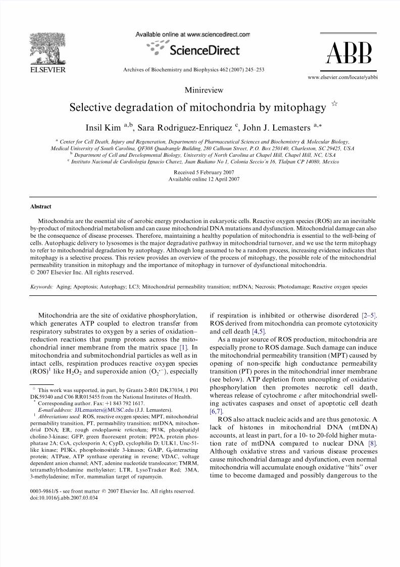
During autophagy, an isolation membrane forms a cup-shaped membranous structure called a phagophore or pre-autophagosome that eventually envelopes the autophagic
target (Fig. 1) [13–15]. The origin of isolation membranesis controversial. One proposed source is ribosome-freeregions of the rough endoplasmic reticulum (ER), but oth-ers suggest that isolation membranes are novel structuresdevoid of Golgi and ER markers [14]. As isolation mem-branes envelop and seal around their targets, double-mem-brane vesicles called autophagosomes form. Theseautophagosomes then fuse with lysosomes to form auto-lysosomes, and their sequestered contents are degradedby lysosomal hydrolases and recycled.
As first characterized in yeast, a machinery of geneticallyconserved autophagy-related proteins regulate and partici-pate in autophagy [16]. These Atg proteins are groupedinto different categories depending on their function,including (1) a pair of novel ubiquitin-like protein conju-gating systems, the ATG12 and Atg8 systems, which pro-duce vesicle extension and completion, (2) a Class IIIphosphatidyl choline-3-kinase (PI3K) complex which func-tions in vesicle nucleation, and (3) a serine–threoninekinase complex (Atg1, Atg13, and Atg17) involved in auto-phagic induction in yeast. The mammalian homologue of Atg 1 is recently identified as ULK1 (Unc-51-like kinase)[17].
During sequestration and formation of autophago-somes, an Atg12–Atg5 complex binds to Atg16, which
translocates to the isolation membrane and functions as a
linker involved in formation and elongation of the phago-phore (Fig. 1). An E1-like enzyme, Atg7, activates Atg12,which is transferred to Atg10, an E2-like enzyme, and con- jugated to Atg5 to form an autophagosomal precursor[16,18].
LC3 is a mammalian autophagosomal ortholog of yeast
Atg8. In mammalian cells, newly synthesized ProLC3 isprocessed to its cytosolic form, LC3-I. Like Atg12, LC3-Iis activated by Atg7, but is instead transferred to Atg3, asecond E2-like enzyme, which cleaves 22 amino acids fromthe C-terminus to form LC3-II. Conjugation with aphospholipid (phosphatidyl ethanolamine in yeast) createsmembrane-bound LC3-II [19,20]. LC3-II localizes selec-tively to forming and newly formed autophagosomes, mak-ing LC3-II a useful autophagosomal marker. Some LC3-IIbecomes entrapped on the inner surfaces of the double-membrane autophagosomes. After fusion with lysosomes,this LC3-II is degraded. Surface LC3-II also disappears,most likely by breakdown of the phospholipid conjugate
(Fig. 1). Recently, a transgenic mouse strain was createdthat expresses a green fluorescent protein (GFP)–LC3fusion protein. In cells and tissues of this mouse, GFP fluo-rescence selectively identifies the membranes of formingand newly formed autophagosomes [21].
Molecular control of autophagy
Phosphoinositide 3-kinases (PI3Ks) phosphorylatephosphatidylinositol at position 3 of the inositol ring andplay an important role in the regulation of autophagy[22]. PI3K inhibitors, such as 3-methyladenine, wortman-
nin, and LY294002, potently block autophagy. However,different classes of PI3K exert opposing effects on autoph-agy: Class III PI3K promotes sequestration of autophagicvacuoles, whereas class I PI3K inhibits autophagy. ClassIII PI3K/p150 associates with Beclin1, a mammalianhomologue of Atg6 discovered by yeast two hybrid screen-ing for its interaction with Bcl-2. Recruitment of PI3K– Beclin1 complexes together with Atg12–Atg5 is an initialstep in autophagosome formation [23]. Mammalian targetof rapamycin (mTor) is a kinase downstream of Class IPI3K whose activation suppresses autophagy. Rapamycin,which inhibits mTOR, induces autophagy apparently byactivating protein phosphatase 2A (PP2A) [24]. PP2A alsodephosphorylates proapoptotic BH3 only Bcl-2 family pro-teins, such as Bad and Bcl-2, that associate with mitochon-dria membranes [25,26]. Thus, a functional relationshipbetween autophagic proteins and mitochondrial proteinsmay exist.
Heterotrimeric guanine nucleotide-binding proteins arealso involved in autophagy. Nonhydrolyzable GTP ana-logs, such as GTPcS, inhibit autophagy [27]. The G
a-inter-
acting protein (GAIP) elicits autophagic sequestration byaccelerating GTP hydrolysis and activating Gai3. Inaddition, Rab24, Rab22, and Rab7, small GTP bindingproteins that regulate vesicular transport, participate in
processing of late autophagosomes [28,29].
246 I. Kim et al. / Archives of Biochemistry and Biophysics 462 (2007) 245–253
7/28/2019 Selective degradation of mitochondria by mitophagy
http://slidepdf.com/reader/full/selective-degradation-of-mitochondria-by-mitophagy 3/9
Selective autophagy
Whether autophagy is selective or non-selective has been
controversial. Cytosolic enzymes with different half-lifesare sequestered at similar rates during autophagy, andautophagosomes often contain a variety of different cyto-plasmic elements, including cytosolic proteins and organ-elles such as ER, peroxisomes and mitochondria [30,31].Such findings led to the assumption that autophagy is anon-specific form of lysosomal degradation. However,more recent findings indicate that autophagy can be a selec-tive process. The presence of peroxin 14 on peroxisomes isrequired for autophagic degradation of peroxisomes inyeast, a process now called pexophagy in recognition of its selectivity [32]. Some pathogens selectively regulateautophagy in mammalian cells for their survival. Shigella
flexneri produces the protein, IscB, which inhibits the bind-ing of bacterial VirG to Atg5, which would otherwiseinduce autophagy [33]. In this way, shigella escapes recog-nition for autophagic sequestration and elimination. Inaddition during the postnatal period, glycogen is selectivelysequestered into autophagosomes to enhance glycolyticsubstrate generation after interruption of transplacentalnutrition [34]. Autophagosomes formed postnatallycontain large amounts of glycogen and rarely containmitochondria or other organelles [35].
Increasing evidence indicates that autophagy of mito-chondria also occurs selectively, and the term mitophagy
has been suggested for this selective mitochondrial autoph-
agy [36]. For example in yeast, an outer membrane protein,Uth1p, is required for efficient mitochondrial autophagy,but a corresponding mammalian protein is yet to be
identified [37]. Our work, reviewed below, also stronglyindicates that autophagy can show selectivity formitochondria.
Characteristics and possible structure of mitochondrial
permeability transition pores
Recent evidence suggests a possible involvement of theMPT in autophagy. In the MPT, opening of PT porescauses mitochondria to become permeable to all solutesup to a molecular mass of about 1500 Da, an event leadingto mitochondrial depolarization and activation of the mito-chondrial ATPase (ATP synthase operating in reverse) [38– 41]. After the MPT, mitochondria undergo large amplitudeswelling driven by colloid osmotic forces, which culminatesin rupture of the outer membrane and release of proapop-totic mitochondrial intermembrane proteins into the cyto-sol, including cytochrome c, apoptosis inducing factor,Smac/Diablo, and others. The immunosuppressant com-pound, cyclosporin A (CsA), and various of its analogsinhibit the MPT through interaction with cyclophilin D(CypD) [42,43].
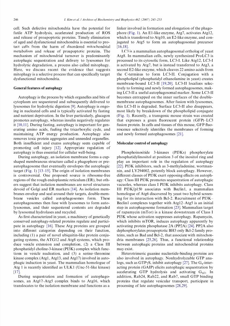
In one model, PT pores are composed of the voltagedependent anion channel (VDAC) in the outer membrane,the adenine nucleotide translocator (ANT) in the inner
membrane and CypD in the matrix space (Fig. 2a)
Fig. 1. Scheme of mitophagy. Atg12–Atg5–Atg16 and LC3 complexes localize to isolation membranes. In nutrient deprivation (starvation), isolationmembranes target individual mitochondria by unknown signals in a process inhibited by the PI3K inhibitors, 3-methyladenine (3MA) and wortmannin.Isolation membranes completely envelop individual mitochondria to form double-membrane vesicles (autophagosomes). After this sequestration,mitochondria depolarize in a CsA and NIM811 sensitive fashion, and Atg12–Atg5/Atg16 complexes are released from the autophagosomal surface.Autophagosomes then acidify and fuse with lysosomal vesicles to form autolysosomes. Lysosomal hydrolases digest the inner autophagosomal membraneand degrade LC3 trapped inside autophagosomes. Remaining LC3 on the surface of autophagosomes is released. After mitochondrial damage,mitochondria first depolarize and then are recognized and sequestered by isolation membranes recognizing unknown markers on the damagedmitochondria. 3MA and wortmannin do not inhibit this process but actually seem to augment it. In both pathways, sequestered mitochondria arecompletely digested and their molecular components recycled to the cytoplasm.
I. Kim et al. / Archives of Biochemistry and Biophysics 462 (2007) 245–253 247
7/28/2019 Selective degradation of mitochondria by mitophagy
http://slidepdf.com/reader/full/selective-degradation-of-mitochondria-by-mitophagy 4/9
[43–46]. Other proteins, such as creatine kinase (intermem-brane space), hexokinase (outer membrane), and Bax(outer membrane), are also proposed to contribute to thecomposition of PT pores. However, the MPT still occursin cells types like hepatocytes that lack creatine kinaseand hexokinase and in ANT-deficient mitochondria iso-lated from conditional double ANT knockout mice [47].Most recently, CypD knockout mice have been developed,and mitochondria from these mitochondria still display anMPT, but the MPT observed is insensitive to CsA andrequires higher concentrations of calcium for induction[48].
An alternative model of the MPT has been proposedthat accounts for these observations (Fig. 2b) [49]. Thismodel postulates that PT pores form as a consequenceof misfolding of integral membrane proteins caused byROS, reactive chemicals, and other stresses. Because mis-folding exposes hydrophilic surfaces to the hydrophobicmembrane bilayer, the proteins aggregate at these hydro-philic surfaces to enclose channels that conduct all aque-ous solutes smaller in size than the channel diameter.Since such permeabilizatoin would be catastrophic tomitochondrial function, chaperones have evolved, includ-ing cyclophilin D, that block conductance through thesenascent channels. Other chaperones remain to be identi-
fied, although indirect evidence suggests that the smallheat shock protein, Hsp25/27, and the Rieske iron sulfurprotein may be involved [50,51]. When matrix calciumrises to high levels, PT pores open to induce the MPT,an effect mediated by CypD and blocked by CsA. Whenformation of nascent PT pores from misfolded proteinaggregates exceeds the number of chaperones that canregulate and close these pores, an unregulated MPToccurs that is CsA-insensitive and calcium-independent.This change from a regulated to an unregulated PT poreoccurs as the time and strength of MPT inductionincreases. On a molar basis, ANT is the most abundant
inner membrane protein and thus is often a target of
stresses causing protein misfolding. However, other pro-teins can also misfold, which explains MPT onset inANT knockout mice. Since CypD participates in calciumsensing, the model also explains the greater requirementfor calcium for the MPT in CypD deficient mitochondria.Lastly, the model explains why completely exogenouspore-forming peptides like mastoparan and alamethicininduce a CsA-sensitive and calcium-dependent MPT atlow concentrations but CsA-insensitive and calcium-inde-pendent mitochondrial swelling at higher concentrations[49,52]. At low concentration, chaperones recognize thepore-forming peptides as misfolded protein aggregates
and block their conductance, but as the chaperone supplybecomes exhausted conductance can no longer beblocked, and mitochondrial swelling, depolarization, anduncoupling ensue.
Mitophagy induced by nutrient deprivation
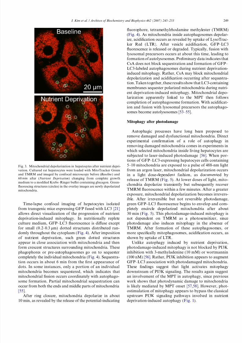
A role of the MPT in mitophagy is implicated in cul-tured hepatocytes during nutrient deprivation. Autophagicstimulation of rat hepatocytes by serum deprivation andglucagon (a hormone released to the liver during fasting)increases the rate of spontaneous depolarization of mito-chondria by 5-fold to about 1% of mitochondria per hour
(Fig. 3) [53]. These depolarized mitochondria move intoacidic vacuoles, which also increase in number after nutri-ent deprivation. The acidic structures containing mitochon-drial remnants are autophagosomes and autolysosomes,and serial imaging reveals an average mitochondrial diges-tion time of about 7 min after autophagic sequestration[54]. CsA, the MPT blocker, suppresses both mitochondrialdepolarization during nutrient deprivation and the prolifer-ation of autophagosomes and autolysosomes. Tacrolimus,an immunosuppressant that does not block the MPT, doesnot block autophagosomal proliferation, whereas NIM811,a CsA analog and MPT inhibitor that is not immunosup-
pressive, does block [53,54].
Fig. 2. Models of the permeability transition pore. In one model (a), the PT pore is composed of ANT from the inner membrane (IM), CypD from thematrix, VDAC from the outer membrane (OM) and other proteins, including hexokinase (HK), creatine kinase (CK) and Bax, a proapoptotic Bcl2 familymember. Ca2+, inorganic phosphate (Pi), ROS, and oxidized pyridine nucleotides NAD(P)+ and glutathione (GSSG) promote PT pore opening, whereasCsA, Mg2+ and pH less than 7 inhibit opening. In an alternative model (b), PT pores form from misfolding and aggregation of damaged mitochondrialmembrane proteins at hydrophilic surfaces facing the hydrophobic membrane bilayer. CypD and other chaperones bind to the nascent PT pores and blockconductance of solutes through the aqueous channels formed by the protein clusters. High Ca 2+ opens these regulated channels acting through CypD, aneffect blocked by CsA. As misfolded protein clusters exceeds the number of chaperones available to regulate them, constitutively open unregulatedchannels form that are not inhibited by CsA.
248 I. Kim et al. / Archives of Biochemistry and Biophysics 462 (2007) 245–253
7/28/2019 Selective degradation of mitochondria by mitophagy
http://slidepdf.com/reader/full/selective-degradation-of-mitochondria-by-mitophagy 5/9
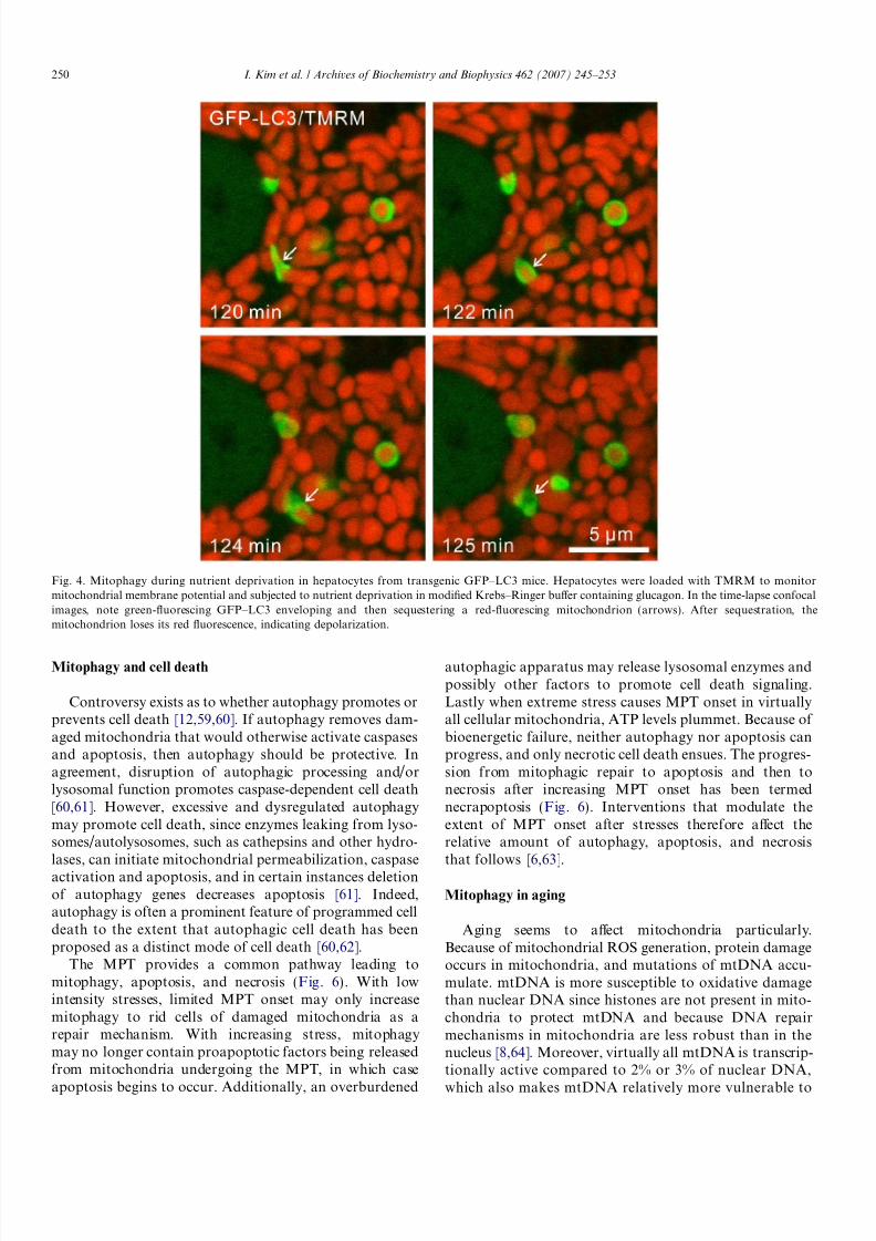
Time-lapse confocal imaging of hepatocytes isolatedfrom transgenic mice expressing GFP fused with LC3 [21]allows direct visualization of the progression of nutrientdeprivation-induced mitophagy. In nutritionally repleteculture medium, GFP–LC3 fluorescence is diffuse exceptfor small (0.2–0.3 lm) dotted structures distributed ran-domly throughout the cytoplasm (Fig. 4). After impositionof nutrient deprivation, such green dotted structuresappear in close association with mitochondria and thenform crescent structures surrounding mitochondria. Thesephagophores or pre-autophagosomes go on to sequestercompletely the individual mitochondria (Fig. 4). Sequestra-tion occurs in about 6 min from the first appearance of dots. In some instances, only a portion of an individualmitochondria becomes sequestered, which indicates thatmitochondrial fission occurs coordinately with autophago-some formation. Partial mitochondrial sequestration canoccur from both the ends and middle parts of mitochondria[55].
After ring closure, mitochondria depolarize in about
10 min, as revealed by the release of the potential-indicating
fluorophore, tetramethylrhodamine methylester (TMRM)(Fig. 4). As mitochondria inside autophagosomes depolar-ize, acidification occurs as revealed by uptake of LysoTrac-ker Red (LTR). After vesicle acidification, GFP–LC3fluorescence is released or degraded. Typically, fusion withlysosomal precursors occurs at about this time, leading to
formation of autolysosomes. Preliminary data indicates thatCsA does not block sequestration and formation of GFP– LC3-labeled autophagosomes during nutrient deprivation-induced mitophagy. Rather, CsA may block mitochondrialdepolarization and acidification occurring after sequestra-tion. Taken together, these results show that LC3-containingmembranes sequester polarized mitochondria during nutri-ent deprivation-induced mitophagy. Mitochondrial depo-larization apparently linked to the MPT then followscompletion of autophagosome formation. With acidificat-ion and fusion with lysosomal precursors the autophago-somes become autolysosomes [53–55].
Mitophagy after photodamage
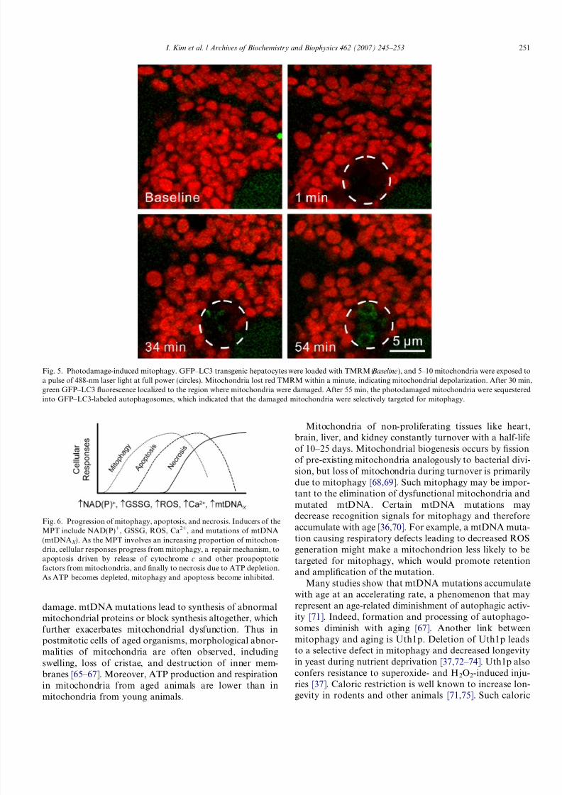
Autophagic processes have long been proposed toremove damaged and dysfunctional mitochondria. Directexperimental confirmation of a role of autophagy inremoving damaged mitochondria comes in experiments inwhich selected mitochondria inside living hepatocytes aresubjected to laser-induced photodamage [56]. When por-tions of GFP–LC3-expressing hepatocyes cells containing5–10 mitochondria are exposed to a pulse of 488-nm lightfrom an argon laser, mitochondrial depolarization occursin a light dose-dependent fashion, as documented by
release of TMRM (Fig. 5). At lower doses of light, mito-chondria depolarize transiently but subsequently recoverTMRM fluorescence within a few minutes. After a greaterexposure, mitochondrial depolarization becomes irrevers-ible. After irreversible but not reversible photodamage,green GFP–LC3 fluorescence begins to envelop and com-pletely encircle depolarized mitochondria after about30 min (Fig. 5). This photodamage-induced mitophagy isnot dependent on TMRM as a photosensitizer, sincephotodamage also induces mitophagy in the absence of TMRM. After formation of these autophagosomes, ormore specifically mitophagosomes, acidification occurs, asshown by uptake of LTR.
Unlike autophagy induced by nutrient deprivation,photodamage-induced mitophagy is not blocked by PI3Kinhibition with 3-methyladenine (10 mM) or wortmannin(100 nM) [56]. Rather, PI3K inhibition appears to augmentGFP–LC3 association with photodamaged mitochondria.These findings suggest that light activates mitophagydownstream of PI3K signaling. The results again suggestan involvement of the MPT in autophagy, since previouswork shows that photodynamic damage to mitochondriais likely mediated by MPT onset [57,58]. However, phot-ostimulation of mitophagy appears to bypass the classicalupstream PI3K signaling pathways involved in nutrient
deprivation-induced autophagy (Fig. 1).
Fig. 3. Mitochondrial depolarization in hepatocytes after nutrient depri-vation. Cultured rat hepatocytes were loaded with MitoTracker Greenand TMRM and imaged by confocal microscopy before (Baseline) and60 min after (Nutrient Deprivation) changing from complete growthmedium to a modified Krebs–Ringer buffer containing glucagon. Green-fluorescing structures (circles) in the overlay images are newly depolarizedmitochondria.
I. Kim et al. / Archives of Biochemistry and Biophysics 462 (2007) 245–253 249
7/28/2019 Selective degradation of mitochondria by mitophagy
http://slidepdf.com/reader/full/selective-degradation-of-mitochondria-by-mitophagy 6/9
Mitophagy and cell death
Controversy exists as to whether autophagy promotes orprevents cell death [12,59,60]. If autophagy removes dam-aged mitochondria that would otherwise activate caspasesand apoptosis, then autophagy should be protective. Inagreement, disruption of autophagic processing and/orlysosomal function promotes caspase-dependent cell death[60,61]. However, excessive and dysregulated autophagymay promote cell death, since enzymes leaking from lyso-somes/autolysosomes, such as cathepsins and other hydro-lases, can initiate mitochondrial permeabilization, caspaseactivation and apoptosis, and in certain instances deletionof autophagy genes decreases apoptosis [61]. Indeed,autophagy is often a prominent feature of programmed celldeath to the extent that autophagic cell death has beenproposed as a distinct mode of cell death [60,62].
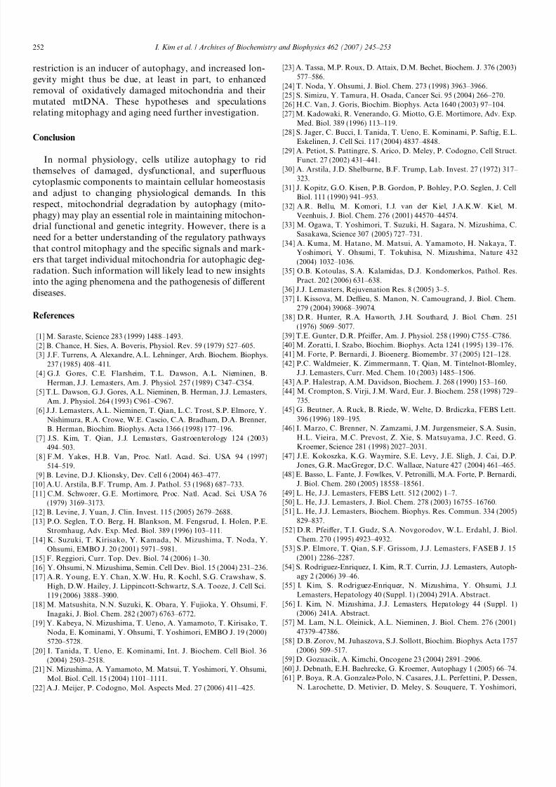
The MPT provides a common pathway leading tomitophagy, apoptosis, and necrosis (Fig. 6). With lowintensity stresses, limited MPT onset may only increasemitophagy to rid cells of damaged mitochondria as arepair mechanism. With increasing stress, mitophagymay no longer contain proapoptotic factors being releasedfrom mitochondria undergoing the MPT, in which case
apoptosis begins to occur. Additionally, an overburdened
autophagic apparatus may release lysosomal enzymes andpossibly other factors to promote cell death signaling.Lastly when extreme stress causes MPT onset in virtuallyall cellular mitochondria, ATP levels plummet. Because of bioenergetic failure, neither autophagy nor apoptosis canprogress, and only necrotic cell death ensues. The progres-sion from mitophagic repair to apoptosis and then tonecrosis after increasing MPT onset has been termednecrapoptosis (Fig. 6). Interventions that modulate theextent of MPT onset after stresses therefore affect therelative amount of autophagy, apoptosis, and necrosisthat follows [6,63].
Mitophagy in aging
Aging seems to affect mitochondria particularly.Because of mitochondrial ROS generation, protein damageoccurs in mitochondria, and mutations of mtDNA accu-mulate. mtDNA is more susceptible to oxidative damagethan nuclear DNA since histones are not present in mito-chondria to protect mtDNA and because DNA repairmechanisms in mitochondria are less robust than in thenucleus [8,64]. Moreover, virtually all mtDNA is transcrip-tionally active compared to 2% or 3% of nuclear DNA,
which also makes mtDNA relatively more vulnerable to
Fig. 4. Mitophagy during nutrient deprivation in hepatocytes from transgenic GFP–LC3 mice. Hepatocytes were loaded with TMRM to monitormitochondrial membrane potential and subjected to nutrient deprivation in modified Krebs–Ringer buffer containing glucagon. In the time-lapse confocalimages, note green-fluorescing GFP–LC3 enveloping and then sequestering a red-fluorescing mitochondrion (arrows). After sequestration, themitochondrion loses its red fluorescence, indicating depolarization.
250 I. Kim et al. / Archives of Biochemistry and Biophysics 462 (2007) 245–253
7/28/2019 Selective degradation of mitochondria by mitophagy
http://slidepdf.com/reader/full/selective-degradation-of-mitochondria-by-mitophagy 7/9
damage. mtDNA mutations lead to synthesis of abnormalmitochondrial proteins or block synthesis altogether, whichfurther exacerbates mitochondrial dysfunction. Thus inpostmitotic cells of aged organisms, morphological abnor-malities of mitochondria are often observed, includingswelling, loss of cristae, and destruction of inner mem-branes [65–67]. Moreover, ATP production and respirationin mitochondria from aged animals are lower than in
mitochondria from young animals.
Mitochondria of non-proliferating tissues like heart,brain, liver, and kidney constantly turnover with a half-lifeof 10–25 days. Mitochondrial biogenesis occurs by fissionof pre-existing mitochondria analogously to bacterial divi-sion, but loss of mitochondria during turnover is primarilydue to mitophagy [68,69]. Such mitophagy may be impor-tant to the elimination of dysfunctional mitochondria andmutated mtDNA. Certain mtDNA mutations maydecrease recognition signals for mitophagy and thereforeaccumulate with age [36,70]. For example, a mtDNA muta-tion causing respiratory defects leading to decreased ROSgeneration might make a mitochondrion less likely to be
targeted for mitophagy, which would promote retentionand amplification of the mutation.
Many studies show that mtDNA mutations accumulatewith age at an accelerating rate, a phenomenon that mayrepresent an age-related diminishment of autophagic activ-ity [71]. Indeed, formation and processing of autophago-somes diminish with aging [67]. Another link betweenmitophagy and aging is Uth1p. Deletion of Uth1p leadsto a selective defect in mitophagy and decreased longevityin yeast during nutrient deprivation [37,72–74]. Uth1p alsoconfers resistance to superoxide- and H2O2-induced inju-ries [37]. Caloric restriction is well known to increase lon-
gevity in rodents and other animals [71,75]. Such caloric
Fig. 5. Photodamage-induced mitophagy. GFP–LC3 transgenic hepatocytes were loaded with TMRM (Baseline), and 5–10 mitochondria were exposed toa pulse of 488-nm laser light at full power (circles). Mitochondria lost red TMRM within a minute, indicating mitochondrial depolarization. After 30 min,green GFP–LC3 fluorescence localized to the region where mitochondria were damaged. After 55 min, the photodamaged mitochondria were sequesteredinto GFP–LC3-labeled autophagosomes, which indicated that the damaged mitochondria were selectively targeted for mitophagy.
Fig. 6. Progression of mitophagy, apoptosis, and necrosis. Inducers of theMPT include NAD(P)+, GSSG, ROS, Ca2+, and mutations of mtDNA(mtDNAX ). As the MPT involves an increasing proportion of mitochon-dria, cellular responses progress from mitophagy, a repair mechanism, to
apoptosis driven by release of cytochrome c and other proapoptoticfactors from mitochondria, and finally to necrosis due to ATP depletion.As ATP becomes depleted, mitophagy and apoptosis become inhibited.
I. Kim et al. / Archives of Biochemistry and Biophysics 462 (2007) 245–253 251
7/28/2019 Selective degradation of mitochondria by mitophagy
http://slidepdf.com/reader/full/selective-degradation-of-mitochondria-by-mitophagy 8/9
7/28/2019 Selective degradation of mitochondria by mitophagy
http://slidepdf.com/reader/full/selective-degradation-of-mitochondria-by-mitophagy 9/9
G. Pierron, P. Codogno, G. Kroemer, Mol. Cell Biol. 25 (2005)1025–1040.
[62] Y. Tsujimoto, S. Shimizu, Cell Death Differ. 12 (Suppl. 2) (2005)1528–1534.
[63] H. Malhi, G.J. Gores, J.J. Lemasters, Hepatology 43 (2006)S31–S44.
[64] V.A. Bohr, Free Radic. Biol. Med. 32 (2002) 804–812.[65] M. Ermini, Gerontology 22 (1976) 301–316.[66] E. Beregi, O. Regius, T. Huttl, Z. Gobl, Z. Gerontol. 21 (1988) 83–86.[67] A. Terman, Gerontology 41 (Suppl. 2) (1995) 319–326.[68] R.A. Menzies, P.H. Gold, J. Biol. Chem. 246 (1971) 2425–2429.
[69] G. Attardi, G. Schatz, Annu. Rev. Cell Biol. 4 (289–333) (1988)289–333.
[70] A.D. de Grey, BioEssays 19 (1997) 161–166.[71] E. Bergamini, Mol. Aspects Med. 27 (2006) 403–410.[72] N. Camougrand, I. Kissova, G. Velours, S. Manon, FEMS Yeast
Res. 5 (2004) 133–140.[73] B.K. Kennedy, L. Guarente, Trends Genet. 12 (1996) 355–359.[74] B.K. Kennedy, N.R. Austriaco Jr., J. Zhang, L. Guarente, Cell 80
(1995) 485–496.[75] E. Bergamini, G. Cavallini, A. Donati, Z. Gori, Biomed. Pharmac-
other. 57 (2003) 203–208.
I. Kim et al. / Archives of Biochemistry and Biophysics 462 (2007) 245–253 253