seasonal changes in the histology of the ovaries of nile ...vetanat.com/v11-pdf/1.pdf · seasonal...
TRANSCRIPT

J. Vet. Anat. Vol 6 No 2, (2013) 1 - 211
Histology of the Nile tilapia ovaries El-Saba et al.
Seasonal changes in the histology of the ovaries of Nile tilapia (Oreochromis niloticus)
El-Saba A., Abd Rabou M.I., El-Sakhawy M.A., El-Shammaa. M. A. and Hussein S.M.* *Department of Cytology & Histology Faculty of Veterinary Medicine Cairo University With 20 figures Received March, accepted for publication May 2013
Abstract Basic Histology of Ovaries of Nile tilapia (Oreochromis niloticus) was studied. Sampling was initiated of a total 40 female sexually mature Nile tilapia fish were collected over the period from September 2009 to Au-gust 2010. Ovaries were processed by standard histological technique. Histological characteristics of ovari-an tissues and oocyte stages were studied by light microscopy. It re-vealed different histological struc-ture of each oocyte developmental stage: Oogonia stage; Chromatin nucleolar stage; Perinucleolar stage; Cortical alveoli formation stage; Vitellogenic (yolk) stage; Postvitellogenic (mature) stage. Yolk nucleus (Balbiani bodies) was noticed in the ooplasm of the peri-nucleolar stage .During breeding season the ovary was surrounded by thin tunica albuginea. The ovig-erous lamellae contained oocyte in active vitellogenesis, In addition;
atretic and post ovulatory follicles were increased. During winter, the tunica albuginea reached a max-imuem thickness and contained oo-cytes in previtellogenic stages. Keyword: tilapia, ovary, oocyte, Introduction Nile tilapia (Oreochromis niloticus) is a fish of economic importance in tropical and subtropical countries. Basic study on histology of O. nilot-icus is still limited especially in re-productive system. Some basic knowledge was mentioned that dip-loid ovaries from the fish of six to eight months of age contained oo-gonia and maturing previtellogenic and vitellogenic oocytes with irregu-lar nuclei and vacuolated cytoplasm associated with endogenous and exogenous yolk formation (Hussain et al., 1996). Reproductive devel-opment and reproductive histology in female are well understood by
J. Vet. Anat. Vol 6 No 2, (2013) 1 - 212
Histology of the Nile tilapia ovaries El-Saba et al.
histological techniques. Histology is the most accurate method to deter-mine the reproductive state of fe-male fish (West, 1990). The ovarian histological pattern of teleosts was described according to the division of ovarian tissues into seven or eight stages of maturity based upon the dominant gametogenic cell type present (Crim and Glebe, 1990). The study on histology of female reproductive organ of O. niloticus will provide a basic knowledge of reproductive system of the fish and will be useful for further applica-tions. Material and methods Specimens of Nile tilapia (Oreo-chromis niloticus) were collected monthly at the same time of the day from the River Nile at Giza. A total of 40 female sexually mature Nile tilapia fish were collected over the period from September 2009 to Au-gust 2010. Fish were transported alive to the central lab. of Cytology and Histology department, faculty of Veterinary Medicine Cairo Universi-ty. Fish were physically examined to ensure that they were free from any pathological changes. The males were distinguished from the females by the examination of the urogenital area. Each Nile tilapia was weighted (700-850 gms) to ensure that all fish sexually mature as mentioned by
Popma and Masser (1999). The ovaries were removed immediately after decapitation and sample were carefully separated of 1 cubic cm. Sections were taken quickly from anterior, middle and posterior parts, fixed in formol-sublimte and neutral buffered formalin for about 24hs., also Bouin′s fluid was used. The samples were dehydrated, embed-ded in paraplast and following that 5-6 μm. sections were cut in cross and longitudinal sections obtained for ovaries. In Oreochromis niloticus, the spring, summer and autumn were consid-ered breeding season (spawning season) while winter considered non breeding season (Dougbag et al., 1988c). Section stained with Harris haema-toxylin and eosin for general histo-logical examination; Gomori’s reticu-line method for demonstration of reticular fibers; Periodic acid Schiff and Periodic acid Schiff - Alcian blue (PH 2.5) combination for identi-fication and differentiation of both neutral and acid mucopolysaccha-rides (Drury and Wallington,1980). Results The histological appearance of the ovary during spring, summer and autumn seasons (from March to November)
The ovary of the Nile tilapia (Oreo-chromis niloticus) was surrounded by thin tunica albuginea which was consisted of a vascular collagenous connective tissue contained smooth muscle cells (Fig. 1). Moreover, a network of reticular fibers was pre-sent (Fig. 2). The tunica albuginea was covered externally by squa-mous epithelium which was the mesothelium of the visceral perito-neum. The tunica albuginea pro-jected inside the lumen of the ovary as numerous fine strands of con-nective tissue folds called ovigerous lamellae (Fig. 1) that contained oo-gonia and oocytes in follicles in var-ious stages of development without any arrangment (Fig. 3). During the breeding seasons the ovigerous lamellae were thick and completely occupied the ovarian cavity (ovocoel) and the ovary was in active vitellogenesis. Oocytes in all stages of development from peri-nucleolar to mature stages could be identified, but late stages of vitello-genesis were dominant (Fig. 3). The ovarian follicles of Nile tilapia passed six developmental stages according to the changes in size, nucleus, ooplasm and egg mem-branes of the developing ova. The stages are: oogonia stage, chroma-tin nucleolus stage, perinucleolar stage, cortical alveolar stage, yolk
globule stage and mature follicles. In addition, atretic and post ovulato-ry follicles were observed. 1- Oogonia stage: The oogonia, the smallest germ cells in the ovary, were found in groups or nests that embedded within the ovig-erous lamellae associated with the germinal epithelium (Fig. 4). They were small spherical cells with large basophilic nuclei with a single nucleo-lus. Their ooplasm was scanty, faintly basophilic thin rim surround the nu-cleus, and was periodic acid Schiff negative. The oogonia were found to be abundant during these seasons. They divided by mitosis to give prima-ry oocyte. 2- Chromatin nucleolus stage (Ear-ly oocytes) which were the smallest previtellogenic follicles. Each one consisted of an oocyte with a large nucleus and surrounded by a thin follicular layer. The nucleus was ec-centrically located and occupied large part of the oocyte. The chromatin of the nucleus appeared thread like. The nucleoli increased in number which distributed throughout the nu-cleus. The ooplasm was homoge-nous, deeply basophilic (Fig. 5) and they gave periodic acid Schiff nega-tive reaction. 3- Perinucleolar stage (Late oo-cytes) which is characterized by in-

J. Vet. Anat. Vol 6 No 2, (2013) 1 - 213
Histology of the Nile tilapia ovaries El-Saba et al.
histological techniques. Histology is the most accurate method to deter-mine the reproductive state of fe-male fish (West, 1990). The ovarian histological pattern of teleosts was described according to the division of ovarian tissues into seven or eight stages of maturity based upon the dominant gametogenic cell type present (Crim and Glebe, 1990). The study on histology of female reproductive organ of O. niloticus will provide a basic knowledge of reproductive system of the fish and will be useful for further applica-tions. Material and methods Specimens of Nile tilapia (Oreo-chromis niloticus) were collected monthly at the same time of the day from the River Nile at Giza. A total of 40 female sexually mature Nile tilapia fish were collected over the period from September 2009 to Au-gust 2010. Fish were transported alive to the central lab. of Cytology and Histology department, faculty of Veterinary Medicine Cairo Universi-ty. Fish were physically examined to ensure that they were free from any pathological changes. The males were distinguished from the females by the examination of the urogenital area. Each Nile tilapia was weighted (700-850 gms) to ensure that all fish sexually mature as mentioned by
Popma and Masser (1999). The ovaries were removed immediately after decapitation and sample were carefully separated of 1 cubic cm. Sections were taken quickly from anterior, middle and posterior parts, fixed in formol-sublimte and neutral buffered formalin for about 24hs., also Bouin′s fluid was used. The samples were dehydrated, embed-ded in paraplast and following that 5-6 μm. sections were cut in cross and longitudinal sections obtained for ovaries. In Oreochromis niloticus, the spring, summer and autumn were consid-ered breeding season (spawning season) while winter considered non breeding season (Dougbag et al., 1988c). Section stained with Harris haema-toxylin and eosin for general histo-logical examination; Gomori’s reticu-line method for demonstration of reticular fibers; Periodic acid Schiff and Periodic acid Schiff - Alcian blue (PH 2.5) combination for identi-fication and differentiation of both neutral and acid mucopolysaccha-rides (Drury and Wallington,1980). Results The histological appearance of the ovary during spring, summer and autumn seasons (from March to November)
The ovary of the Nile tilapia (Oreo-chromis niloticus) was surrounded by thin tunica albuginea which was consisted of a vascular collagenous connective tissue contained smooth muscle cells (Fig. 1). Moreover, a network of reticular fibers was pre-sent (Fig. 2). The tunica albuginea was covered externally by squa-mous epithelium which was the mesothelium of the visceral perito-neum. The tunica albuginea pro-jected inside the lumen of the ovary as numerous fine strands of con-nective tissue folds called ovigerous lamellae (Fig. 1) that contained oo-gonia and oocytes in follicles in var-ious stages of development without any arrangment (Fig. 3). During the breeding seasons the ovigerous lamellae were thick and completely occupied the ovarian cavity (ovocoel) and the ovary was in active vitellogenesis. Oocytes in all stages of development from peri-nucleolar to mature stages could be identified, but late stages of vitello-genesis were dominant (Fig. 3). The ovarian follicles of Nile tilapia passed six developmental stages according to the changes in size, nucleus, ooplasm and egg mem-branes of the developing ova. The stages are: oogonia stage, chroma-tin nucleolus stage, perinucleolar stage, cortical alveolar stage, yolk
globule stage and mature follicles. In addition, atretic and post ovulato-ry follicles were observed. 1- Oogonia stage: The oogonia, the smallest germ cells in the ovary, were found in groups or nests that embedded within the ovig-erous lamellae associated with the germinal epithelium (Fig. 4). They were small spherical cells with large basophilic nuclei with a single nucleo-lus. Their ooplasm was scanty, faintly basophilic thin rim surround the nu-cleus, and was periodic acid Schiff negative. The oogonia were found to be abundant during these seasons. They divided by mitosis to give prima-ry oocyte. 2- Chromatin nucleolus stage (Ear-ly oocytes) which were the smallest previtellogenic follicles. Each one consisted of an oocyte with a large nucleus and surrounded by a thin follicular layer. The nucleus was ec-centrically located and occupied large part of the oocyte. The chromatin of the nucleus appeared thread like. The nucleoli increased in number which distributed throughout the nu-cleus. The ooplasm was homoge-nous, deeply basophilic (Fig. 5) and they gave periodic acid Schiff nega-tive reaction. 3- Perinucleolar stage (Late oo-cytes) which is characterized by in-
J. Vet. Anat. Vol 6 No 2, (2013) 1 - 214
Histology of the Nile tilapia ovaries El-Saba et al.
creasing the size of the oocyte and its nucleus became slightly basophilic with increased number of nucleoli, arranged themselves in the peripheral part of the nucleus. The ooplasm was basophilic and surrounded by a sim-ple follicular epithelium which made up of flattened squamous cells (Fig. 6). In the larger oocytes, the ooplasm became frothy with the appearance of few unstained vacuoles. Ooplasm contained yolk nucleus (Balbiani bod-ies), which appeared as juxtanuclear basophilic round mass (Fig. 7). 4- Cortical alveolar stage (vacuo-lated follicles) which is characterized by marked increase in size of the oo-cytes and nuclei. The most character-istic feature of these follicles was the appearance of large number of un-stained vacuoles (yolk vesicles) in the periphery of the ooplasm. However, yolk granules appeared within the ooplasm in between the yolk vesicles (Fig. 5). Zona pellucida (oolemma) was a cel-lular thin hyaline acidophilic mem-brane (Fig. 8). The follicular layer was formed of cuboidal cells. The stroma was formed of flat thecal cells that surrounded the follicular layer (Fig. 9). 5- Vitellogenesis (yolk globule stage) which is characterized by in-
creasing of yolk vesicles number and size toward the center, and by yolk globules appeared toward the center of the oocyte. These yolk globules gave positive reaction with periodic acid Schiff (Figs. 10 and 11). The nu-clei became relatively smaller and contained fewer numbers of nucleoli and migrated toward periphery. The zona pellucida appeared thicker and the follicular epithelium made up of cuboidal cells. Theca folliculi was di-vided into outer vascular collagenous connective tissue thecal layer and inner cellular theca cells. Basal lami-na, periodic acid schiff positive was found between the follicular epitheli-um and thecal layer. 6- Mature follicle (Postvitellogenic stage), these follicles showed a marked increase in size and reached the final growth stage (Fig. 12). The most characteristic feature of these follicles was the migration of their nu-clei from the center to eccentric posi-tion (Fig. 13). Their ooplasm is char-acterized by being full of large yolk globules. These globules were peri-odic acid Schiff positive (Fig. 14) and alcian blue negative. The follicular epithelium appeared as high cuboidal cells which had vacuolated, faintly stained cytoplasm and with dark stained nuclei (Fig. 15). Zona pelluci-da was thick and periodic acid Schiff positive only (Fig. 14). The theca fol-liculi were composed of squamous
cells with flat nuclei. The mature (postvitellogenic) follicles were com-mon and abundant during these sea-sons of the year. Post-ovulatory follicles: They were formed from the collapse of the follicle after ovulation. They showed a central lumen and a wall formed of follicular cell layer and the theca (Fig. 16). Atretic follicles: Two types of atresia were recog-nized, hypertrophic atresia and cyst-ic atresia. The hypertrophic atresia was the most numerous type. The follicular cells were increased both in size and number invading the ooplasm. Blood cells were seen between the follicular cells and yolk. Later on, the follicle collapse and finally replaced by stromal tissue (Fig. 17). The cystic atresia showed degener-ation of the oocyte. The follicular epithelium thickened and was sepa-rated from the oocyte. Later, there was a rupture of the oocyte. The follicle became a small mass of cells or a cyst-like structure. The atretic follicles were found throughout the year especially during these sea-sons. The histological appearance of
the ovary during winter season (from December to February) The tunica albuginea surrounding the ovary reached a maximum thickness. Also, the stromal connec-tive tissue was increased and con-tained much amount of smooth muscle fibers (Fig. 18). The oviger-ous lamellae were filled with pre-vitellogenic oocytes in oogonium stage, chromatin nucleolus stages and perinucleolar stage (Fig. 19). The lamellae were disrupted and disorganized with several empty spaces and extensive vasculariza-tion. Remnants of atretic follicles were present throughout the ovary (Fig. 20). The previtellogenic stages were abundant while mature ovarian follicles were few. Discussion The present work was carried out on 40 female specimens of Nile ti-lapia (Oreochromis niloticus) throu-ghout the year, in order to observe the morphological and the histologi-cal changes in the ovaries during the different seasons of the year. The results showed that the breed-ing season for reproduction was from March to November, while non-breeding season was from Decem-ber to February. These current find-ings not simulate those of Caputo et al.(2003) in Crystallogobius linearis;

J. Vet. Anat. Vol 6 No 2, (2013) 1 - 215
Histology of the Nile tilapia ovaries El-Saba et al.
creasing the size of the oocyte and its nucleus became slightly basophilic with increased number of nucleoli, arranged themselves in the peripheral part of the nucleus. The ooplasm was basophilic and surrounded by a sim-ple follicular epithelium which made up of flattened squamous cells (Fig. 6). In the larger oocytes, the ooplasm became frothy with the appearance of few unstained vacuoles. Ooplasm contained yolk nucleus (Balbiani bod-ies), which appeared as juxtanuclear basophilic round mass (Fig. 7). 4- Cortical alveolar stage (vacuo-lated follicles) which is characterized by marked increase in size of the oo-cytes and nuclei. The most character-istic feature of these follicles was the appearance of large number of un-stained vacuoles (yolk vesicles) in the periphery of the ooplasm. However, yolk granules appeared within the ooplasm in between the yolk vesicles (Fig. 5). Zona pellucida (oolemma) was a cel-lular thin hyaline acidophilic mem-brane (Fig. 8). The follicular layer was formed of cuboidal cells. The stroma was formed of flat thecal cells that surrounded the follicular layer (Fig. 9). 5- Vitellogenesis (yolk globule stage) which is characterized by in-
creasing of yolk vesicles number and size toward the center, and by yolk globules appeared toward the center of the oocyte. These yolk globules gave positive reaction with periodic acid Schiff (Figs. 10 and 11). The nu-clei became relatively smaller and contained fewer numbers of nucleoli and migrated toward periphery. The zona pellucida appeared thicker and the follicular epithelium made up of cuboidal cells. Theca folliculi was di-vided into outer vascular collagenous connective tissue thecal layer and inner cellular theca cells. Basal lami-na, periodic acid schiff positive was found between the follicular epitheli-um and thecal layer. 6- Mature follicle (Postvitellogenic stage), these follicles showed a marked increase in size and reached the final growth stage (Fig. 12). The most characteristic feature of these follicles was the migration of their nu-clei from the center to eccentric posi-tion (Fig. 13). Their ooplasm is char-acterized by being full of large yolk globules. These globules were peri-odic acid Schiff positive (Fig. 14) and alcian blue negative. The follicular epithelium appeared as high cuboidal cells which had vacuolated, faintly stained cytoplasm and with dark stained nuclei (Fig. 15). Zona pelluci-da was thick and periodic acid Schiff positive only (Fig. 14). The theca fol-liculi were composed of squamous
cells with flat nuclei. The mature (postvitellogenic) follicles were com-mon and abundant during these sea-sons of the year. Post-ovulatory follicles: They were formed from the collapse of the follicle after ovulation. They showed a central lumen and a wall formed of follicular cell layer and the theca (Fig. 16). Atretic follicles: Two types of atresia were recog-nized, hypertrophic atresia and cyst-ic atresia. The hypertrophic atresia was the most numerous type. The follicular cells were increased both in size and number invading the ooplasm. Blood cells were seen between the follicular cells and yolk. Later on, the follicle collapse and finally replaced by stromal tissue (Fig. 17). The cystic atresia showed degener-ation of the oocyte. The follicular epithelium thickened and was sepa-rated from the oocyte. Later, there was a rupture of the oocyte. The follicle became a small mass of cells or a cyst-like structure. The atretic follicles were found throughout the year especially during these sea-sons. The histological appearance of
the ovary during winter season (from December to February) The tunica albuginea surrounding the ovary reached a maximum thickness. Also, the stromal connec-tive tissue was increased and con-tained much amount of smooth muscle fibers (Fig. 18). The oviger-ous lamellae were filled with pre-vitellogenic oocytes in oogonium stage, chromatin nucleolus stages and perinucleolar stage (Fig. 19). The lamellae were disrupted and disorganized with several empty spaces and extensive vasculariza-tion. Remnants of atretic follicles were present throughout the ovary (Fig. 20). The previtellogenic stages were abundant while mature ovarian follicles were few. Discussion The present work was carried out on 40 female specimens of Nile ti-lapia (Oreochromis niloticus) throu-ghout the year, in order to observe the morphological and the histologi-cal changes in the ovaries during the different seasons of the year. The results showed that the breed-ing season for reproduction was from March to November, while non-breeding season was from Decem-ber to February. These current find-ings not simulate those of Caputo et al.(2003) in Crystallogobius linearis;

J. Vet. Anat. Vol 6 No 2, (2013) 1 - 216
Histology of the Nile tilapia ovaries El-Saba et al.
Cinquetti and Dramis (2003) in Pa-dogobius martensi and El-Hafez et al. (2009) in Oreochromis niloticus who mentioned that, the breeding season is between April and Sep-tember, while the non-breeding season between October and march.
El-Zarka et al. (1970) observed that the spawning was more protracted- from April to August in delta Nile tilapia (Oreochromis niloticus). While, Trewevas (1983) mentioned that spawning occurs between April to May in Nile tilapia (Oreochromis niloticus).
The present study revealed that the ovary of the Nile tilapia (Oreo-chromis niloticus) was covered by tunica albuginea which was consist-ed of dense collagenous connective tissue, elastic fibers and network of reticular fibers. The ovarian wall was supported with smooth muscle cells and this agreed with the result of Rizkalla (1970) in Clarias lazera; Yoakim (1971) in Synodontus schall; Khallaf et al. (1991) in Bagrus bayad; Gaber (2000) in Bagrus docmac and Bagrus bayad and El Hafez et al. (2009) in Oreo-chromis niloticus and in other parts of the wall, there were longitudinal smooth muscle cells only as those obtained by Gaber (2000) in Bagrus docmac and Bagrus bayad.
This tunica albuginea had no uni-form thickness around the year that was similar to those obtained by Gaber (2000) in Bagrus docmac and Bagrus bayad and El Hafez et al. (2009) in Oreochromis niloticus. As the tunica albuginea reached a maximum thickness during winter (resting season) and decreased dur-ing the following breeding seasons (spring, summer and autumn). Numerous ovigerous lamellae pro-jected from the tunica albuginea into the interior of the ovary. Each lamel-la acted as a unit and contained oo-gonia and various developmental stages of the follicles. These find-ings were similar to Van den Hurk and Peute (1979) in rainbow trout; Dougbag et al. (1988c) in Oreo-chromis niloticus; Ismail (1992) in Clarias lazera; Mousa (1998) and El-Gohary (2001) in Oreochromis niloticus; Dutta and Maxwell (2003) in Lepomis macrochirus and El-Hafez et al. (2009) in Oreochromis niloticus.
The early germ cells were first found on the edge of the lamellae, and as the oocytes matured and increased in size, they were pushed deeper into the stroma and when matured they were ovulated into the lumen. According to the classification of Wallace and Selman (1981), the ovary of the Oreochromis niloticus showed an asynchronous mode,
since not all the oocytes were at the same stage of development at any time so; the Oreochromis niloticus had a prolonged spawning period. This observation agreed with Mous-tafa (1984) in Clarias lazera; Zaki et al. (1986a) in Clarias gariepinus and El-Zoghby et al. (2009) in Clarias lazera.
Ovaries of tilapia were of cystovari-an type, as the ovarian cavity is connected directly with the oviduct (Alka′abi, 1996), subsequently the Oreochromis niloticus don′t release their mature ova into coelemic cavi-ty. The overall pattern of oocytes development in Oreochromis nilot-icus was the same as in other tele-ost species (Coward and Bromage, 1998).
Van den Hurk and Peute (1979) in rainbow trout (Salmo gairdneri) di-vided into five stages: 1- previtello-genic, 2- vitellogenic, 3- mature, 4- postovulatory and 5-atretic follicles. On the other hand, Dougbag et al. (1988c) observed eight stages of oocyes development in Oreochro-mis niloticus.
In agreement with Alka′abi (1996) and El-Hafez et al. (2009), the pro-cess of oogenesis in Oreochromis niloticus was classified according to the changes in size, nucleus, cyto-plasm and egg membranes of the developing ova into six stages be-ginning with oogonia. These stages
were oogonia, chromatin nucleolus stages, perinucleolar stage, yolk vesicle stage, yolk globule stage and mature stage. Coward and Bromage (1998) recorded nine stages for oogenesis process in Ti-lapia zilli. However, Essa (2011) in Oreochromis niloticus stated that the developmental stages of oo-cytes were classified into five stages according to West (1990), chromatin nucleolus stages, perinucleolar stage, cortical alveoli formation stage, vitellogenic stage and mature stage.
A characteristic Balbiani bodies sim-ilar to that seen by Yoakim (1971) in schilbe mystus; Van den Hurk and Peute (1979) in the rainbow trout (Salmo gairdneri); Alka′abi (1996) in Oreochromis niloticus; Hamdoon and Zayed (1998) in Oreochromis niloticus; Bardakci et al. (2000) in teleost and El-Hafez et al. (2009) in Oreochromis niloticus was present in our investigation. Norrevang (1968) and Guraya (1979) revealed that most teleost oocytes accumu-lated a small juxtanuclear basophilic mass, which was termed yolk nu-cleus or Balbiani bodies. They were composed of various cellular orga-nelles such as mitochondria, Golgi bodies, smooth endoplasmic reticu-lum, multivesivular bodies and lipid granules in Salmo gairdneri (Beams and Kessel, 1973) and in teleosts (Wallace and Selman, 1981).

J. Vet. Anat. Vol 6 No 2, (2013) 1 - 217
Histology of the Nile tilapia ovaries El-Saba et al.
Cinquetti and Dramis (2003) in Pa-dogobius martensi and El-Hafez et al. (2009) in Oreochromis niloticus who mentioned that, the breeding season is between April and Sep-tember, while the non-breeding season between October and march.
El-Zarka et al. (1970) observed that the spawning was more protracted- from April to August in delta Nile tilapia (Oreochromis niloticus). While, Trewevas (1983) mentioned that spawning occurs between April to May in Nile tilapia (Oreochromis niloticus).
The present study revealed that the ovary of the Nile tilapia (Oreo-chromis niloticus) was covered by tunica albuginea which was consist-ed of dense collagenous connective tissue, elastic fibers and network of reticular fibers. The ovarian wall was supported with smooth muscle cells and this agreed with the result of Rizkalla (1970) in Clarias lazera; Yoakim (1971) in Synodontus schall; Khallaf et al. (1991) in Bagrus bayad; Gaber (2000) in Bagrus docmac and Bagrus bayad and El Hafez et al. (2009) in Oreo-chromis niloticus and in other parts of the wall, there were longitudinal smooth muscle cells only as those obtained by Gaber (2000) in Bagrus docmac and Bagrus bayad.
This tunica albuginea had no uni-form thickness around the year that was similar to those obtained by Gaber (2000) in Bagrus docmac and Bagrus bayad and El Hafez et al. (2009) in Oreochromis niloticus. As the tunica albuginea reached a maximum thickness during winter (resting season) and decreased dur-ing the following breeding seasons (spring, summer and autumn). Numerous ovigerous lamellae pro-jected from the tunica albuginea into the interior of the ovary. Each lamel-la acted as a unit and contained oo-gonia and various developmental stages of the follicles. These find-ings were similar to Van den Hurk and Peute (1979) in rainbow trout; Dougbag et al. (1988c) in Oreo-chromis niloticus; Ismail (1992) in Clarias lazera; Mousa (1998) and El-Gohary (2001) in Oreochromis niloticus; Dutta and Maxwell (2003) in Lepomis macrochirus and El-Hafez et al. (2009) in Oreochromis niloticus.
The early germ cells were first found on the edge of the lamellae, and as the oocytes matured and increased in size, they were pushed deeper into the stroma and when matured they were ovulated into the lumen. According to the classification of Wallace and Selman (1981), the ovary of the Oreochromis niloticus showed an asynchronous mode,
since not all the oocytes were at the same stage of development at any time so; the Oreochromis niloticus had a prolonged spawning period. This observation agreed with Mous-tafa (1984) in Clarias lazera; Zaki et al. (1986a) in Clarias gariepinus and El-Zoghby et al. (2009) in Clarias lazera.
Ovaries of tilapia were of cystovari-an type, as the ovarian cavity is connected directly with the oviduct (Alka′abi, 1996), subsequently the Oreochromis niloticus don′t release their mature ova into coelemic cavi-ty. The overall pattern of oocytes development in Oreochromis nilot-icus was the same as in other tele-ost species (Coward and Bromage, 1998).
Van den Hurk and Peute (1979) in rainbow trout (Salmo gairdneri) di-vided into five stages: 1- previtello-genic, 2- vitellogenic, 3- mature, 4- postovulatory and 5-atretic follicles. On the other hand, Dougbag et al. (1988c) observed eight stages of oocyes development in Oreochro-mis niloticus.
In agreement with Alka′abi (1996) and El-Hafez et al. (2009), the pro-cess of oogenesis in Oreochromis niloticus was classified according to the changes in size, nucleus, cyto-plasm and egg membranes of the developing ova into six stages be-ginning with oogonia. These stages
were oogonia, chromatin nucleolus stages, perinucleolar stage, yolk vesicle stage, yolk globule stage and mature stage. Coward and Bromage (1998) recorded nine stages for oogenesis process in Ti-lapia zilli. However, Essa (2011) in Oreochromis niloticus stated that the developmental stages of oo-cytes were classified into five stages according to West (1990), chromatin nucleolus stages, perinucleolar stage, cortical alveoli formation stage, vitellogenic stage and mature stage.
A characteristic Balbiani bodies sim-ilar to that seen by Yoakim (1971) in schilbe mystus; Van den Hurk and Peute (1979) in the rainbow trout (Salmo gairdneri); Alka′abi (1996) in Oreochromis niloticus; Hamdoon and Zayed (1998) in Oreochromis niloticus; Bardakci et al. (2000) in teleost and El-Hafez et al. (2009) in Oreochromis niloticus was present in our investigation. Norrevang (1968) and Guraya (1979) revealed that most teleost oocytes accumu-lated a small juxtanuclear basophilic mass, which was termed yolk nu-cleus or Balbiani bodies. They were composed of various cellular orga-nelles such as mitochondria, Golgi bodies, smooth endoplasmic reticu-lum, multivesivular bodies and lipid granules in Salmo gairdneri (Beams and Kessel, 1973) and in teleosts (Wallace and Selman, 1981).

J. Vet. Anat. Vol 6 No 2, (2013) 1 - 218
Histology of the Nile tilapia ovaries El-Saba et al.
Guraya (1979) stated that, although the role of Balbiani bodies was yet not clear, it had been considered that the yolk nucleus function as a center for the formation of organells within the oocytes.
The oogonia, small spherical cells with large nuclei, were arranged in nests. This finding was similar to Yoakim (1971) in Synodontus schall ; Dougbag et al. (1988c) in Oreo-chromis niloticus; Gaber (2000) Bagrus docmac and Bagrus bayad and El-Ghohary (2001) in Oreo-chromis niloticus. The oocytes in the previtellogenic stages had baso-philic cytoplasm and did not have yet yolk granules. They were classi-fied into two phases; a) early oo-cytes which were small oocytes with homogenous deeply basophilic oo-plasm and b) late oocytes which were larger and had less basophilic and frothy ooplasm. They were found throughout the year, but were common in the non-breeding sea-son (winter) and less abundant dur-ing spring, summer and autumn (spawning seasons). This result come in agreement with that re-vealed by Yoakim (1971) in Syn-odontus schall; Dougbag et al. (1988c) in Oreochromis niloticus; Salem (1991) in Lethrinus bungus; Ismail (1992) in Clarias lazera and El Hafez et al. (2009) in Oreo-chromis niloticus.
The vitellogenic follicles cytoplasm became acidophilic due to the dep-osition of vitellogenin into the oo-cytes (vitellogenesis). They were characterized by the appearance of unstained yolk vesicles in the pe-riphery of the ooplasm and later the appearance of the extravesicular small eosinophilic yolk granules. These granules appeared firstly at the periphery of the ooplasm then aggregated toward the center of the oocytes. This finding was similar to results of Yoakim (1971) in Syn-odontus schall and Gaber (2000) in Bagrus docmac and Bagrus bayad. These vitellogenic follicles were de-creased during winter as it was rest-ing season but, abundant during spring, summer and autumn (spaw-ning American plaice contained oo-cytes undergoing vitellogenesis in-dicate spawning activity. Yolk globule stage was the most important phase of oocyte develop-ment; since it was during this phase, vitellogenesis occured, resulting in an extensive oocyte growth. Coward and Bromage (1998) in Tilapia zillii and Chmilevskii and Kameneva (2003) in Tilapia mossambica stated that oocytes were enlarged chiefly by rapid incorporation of large amounts of exogenous hepatically derived vitellogenin. While Patino (1997); Arockiaraj et al. (2004) re-ported that growing ovarian follicles
produced steroid hormones. This steroid left the follicle via blood ves-sels supplying the theca cell layer and was transported to the liver where it induced the production of vitellogenin. Vitellogenin was trans-ferred to the ovary via circulation, where it taken up by the oocyte and was deposited as yolk protein which serves as building and energy mate-rial after fertilization. The mature or post-vitellogenic folli-cles characterized by migration of their nuclei toward the animal pole, with presence of large yolk globules. This finding was similar to the re-sults of Yoakim (1971) in Synodon-tus schall; Van den Hurk and Peute (1979) in the rainbow trout (Salmo gairdneri) and Gaber (2000) in Bag-rus docmac and Bagrus bayad. The postvitellogenic follicles were com-mon and abundant during spring as they were in the beginning of the spawning and ready to spawn and ovulate, while less abundant during winter season, as most of them were already spawned. The egg wall was consisted of zona pellucida, follicular epithelium and theca folliculi. Where zona pellucida was considered as secretory prod-uct from the follicular cells, its ap-pearance for first time could vary among fish species. In this study, it was firstly seen in larger late oo-
cytes. This observation was similar to those of Ismail (1992) in Clarias lazera, Latif and Saady (1973) in Oreochromis niloticus and Davis (1977) in Tilapia tandanus. On the other hand, it appeared at the end of the yolk vesicle stage by Fahmy (1997) in Clarias ruepplli but ap-peared at early vacuolated oocytes as reported by Gaber (2000) in Bagrus docmac and Bagrus bayad. The thickness of zona pellucida was not the same throughout the differ-ent stages, but it began in larger late oocytes as very thin acidophilic membrane, then increased at both early and late vacuolated oocytes and reached its maximum thickness at mature follicles. In our study, the zona pollicida had a strongly period-ic acid schiff (PAS) positive reaction that indicated presence of mucpoly-saccharides. This was similar to those results of Yoakim (1971) in Synodontus schall; Alves et al. (1983) in Oreochromis niloticus; Van den Hurk and Peute (1985) in Clarias gariepinus; Emel (1992) in the channel catfish and Gaber (2000) in Bagrus docmac and Bag-rus bayad.
The follicular epithelium differed among the different stages where it appeared in the previtellogenic folli-cles as flattened squamous cells and reached its high columnar cells in the mature follicles which ap-

J. Vet. Anat. Vol 6 No 2, (2013) 1 - 219
Histology of the Nile tilapia ovaries El-Saba et al.
Guraya (1979) stated that, although the role of Balbiani bodies was yet not clear, it had been considered that the yolk nucleus function as a center for the formation of organells within the oocytes.
The oogonia, small spherical cells with large nuclei, were arranged in nests. This finding was similar to Yoakim (1971) in Synodontus schall ; Dougbag et al. (1988c) in Oreo-chromis niloticus; Gaber (2000) Bagrus docmac and Bagrus bayad and El-Ghohary (2001) in Oreo-chromis niloticus. The oocytes in the previtellogenic stages had baso-philic cytoplasm and did not have yet yolk granules. They were classi-fied into two phases; a) early oo-cytes which were small oocytes with homogenous deeply basophilic oo-plasm and b) late oocytes which were larger and had less basophilic and frothy ooplasm. They were found throughout the year, but were common in the non-breeding sea-son (winter) and less abundant dur-ing spring, summer and autumn (spawning seasons). This result come in agreement with that re-vealed by Yoakim (1971) in Syn-odontus schall; Dougbag et al. (1988c) in Oreochromis niloticus; Salem (1991) in Lethrinus bungus; Ismail (1992) in Clarias lazera and El Hafez et al. (2009) in Oreo-chromis niloticus.
The vitellogenic follicles cytoplasm became acidophilic due to the dep-osition of vitellogenin into the oo-cytes (vitellogenesis). They were characterized by the appearance of unstained yolk vesicles in the pe-riphery of the ooplasm and later the appearance of the extravesicular small eosinophilic yolk granules. These granules appeared firstly at the periphery of the ooplasm then aggregated toward the center of the oocytes. This finding was similar to results of Yoakim (1971) in Syn-odontus schall and Gaber (2000) in Bagrus docmac and Bagrus bayad. These vitellogenic follicles were de-creased during winter as it was rest-ing season but, abundant during spring, summer and autumn (spaw-ning American plaice contained oo-cytes undergoing vitellogenesis in-dicate spawning activity. Yolk globule stage was the most important phase of oocyte develop-ment; since it was during this phase, vitellogenesis occured, resulting in an extensive oocyte growth. Coward and Bromage (1998) in Tilapia zillii and Chmilevskii and Kameneva (2003) in Tilapia mossambica stated that oocytes were enlarged chiefly by rapid incorporation of large amounts of exogenous hepatically derived vitellogenin. While Patino (1997); Arockiaraj et al. (2004) re-ported that growing ovarian follicles
produced steroid hormones. This steroid left the follicle via blood ves-sels supplying the theca cell layer and was transported to the liver where it induced the production of vitellogenin. Vitellogenin was trans-ferred to the ovary via circulation, where it taken up by the oocyte and was deposited as yolk protein which serves as building and energy mate-rial after fertilization. The mature or post-vitellogenic folli-cles characterized by migration of their nuclei toward the animal pole, with presence of large yolk globules. This finding was similar to the re-sults of Yoakim (1971) in Synodon-tus schall; Van den Hurk and Peute (1979) in the rainbow trout (Salmo gairdneri) and Gaber (2000) in Bag-rus docmac and Bagrus bayad. The postvitellogenic follicles were com-mon and abundant during spring as they were in the beginning of the spawning and ready to spawn and ovulate, while less abundant during winter season, as most of them were already spawned. The egg wall was consisted of zona pellucida, follicular epithelium and theca folliculi. Where zona pellucida was considered as secretory prod-uct from the follicular cells, its ap-pearance for first time could vary among fish species. In this study, it was firstly seen in larger late oo-
cytes. This observation was similar to those of Ismail (1992) in Clarias lazera, Latif and Saady (1973) in Oreochromis niloticus and Davis (1977) in Tilapia tandanus. On the other hand, it appeared at the end of the yolk vesicle stage by Fahmy (1997) in Clarias ruepplli but ap-peared at early vacuolated oocytes as reported by Gaber (2000) in Bagrus docmac and Bagrus bayad. The thickness of zona pellucida was not the same throughout the differ-ent stages, but it began in larger late oocytes as very thin acidophilic membrane, then increased at both early and late vacuolated oocytes and reached its maximum thickness at mature follicles. In our study, the zona pollicida had a strongly period-ic acid schiff (PAS) positive reaction that indicated presence of mucpoly-saccharides. This was similar to those results of Yoakim (1971) in Synodontus schall; Alves et al. (1983) in Oreochromis niloticus; Van den Hurk and Peute (1985) in Clarias gariepinus; Emel (1992) in the channel catfish and Gaber (2000) in Bagrus docmac and Bag-rus bayad.
The follicular epithelium differed among the different stages where it appeared in the previtellogenic folli-cles as flattened squamous cells and reached its high columnar cells in the mature follicles which ap-

J. Vet. Anat. Vol 6 No 2, (2013) 1 - 2110
Histology of the Nile tilapia ovaries El-Saba et al.
peared to have both neutral and acidic mucopolysaccharides as it gave positive periodic acid Schiff (PAS) and alcian blue reaction, a result that was supported by Alves et al. (1983) in Oreochromis nilot-icus. The follicular epithelium was covered by thin theca folliculi. This similar to those recorded by Davis (1977) in Tandanus tandanus; Hus-sein and Abbas (1985) in two spe-cies of genus Morone; Van den Hurk and Peute (1985) in Clarias gariepinus; Khallaf et al. (1991) in Bagrus bayad and Gaber (2000) in Bagrus docmac and Bagrus bayad.
In agreement with Braekevelt and Memillan (1967) in Eucolia incon-stans; Dougbag et al. (1988c) and Essa (2011) in Oreochromis nilot-icus two types of atresia, hyper-trophic and cystic were present in our study. El-Hafez et al. (2009) in Oreochromis niloticus stated that the presence of follicular atresia seem to be a very common phe-nomenon of the teleost ovary. The atretic follicles were usually seen at any stage. The atretic follicles were present throughout the year but, they were abundant during winter (resting ovaries). These atretic folli-cles indicated the spawned individ-uals. In our investigation it was apparent that during breeding seasons (spring, summer and autumn) the
ovaries were filled with vitellogenic and mature follicles, while in non- breeding seasons (winter), the ova-ries revealed predominance of pre- vitellogenic stages. References Alka′abi, N.A.O. (1996): Histologi cal and histochemical comparative studies on gonads of Grouper fish in Arabian Gulf and Tilapia fish in aq-uacultures. Ph.D. Department of Zoology, Collage of Science for Girls. Dammam- KSA. Alves, M.M., Leme Dos Santos, H.S., Lopes, R.A., Petenusci, S.O. and Haiyashi, C. (1983): Rhythm of development in the oocyte of the tilapia Oreochromis niloticus L. (Pi-sces: Cichlidae); a morphometric and histochemical study. Gegen-baurs Morphol Jahrb., 129: 575 - 592. Arockiaraj, A.J.; Haniffa, M.A.; Seetharaman, S. and Singh, S. (2004): Cyclic changes in gonadal maturation and histological observa-tions of threatened fresh water cat-fish "Narikeliru" (Jerdon, 1849). J. Acta Icth. Et Pisc. (34) 2: 253 – 266. Bardakci, F.; Ozansoy, U and Koptagel, E. (2000): A comparison of oogenesis under constant and fluctuating temperatures in Doctor
fish, Garra Rufa Heckel, 1843 (Tel-eostei: Cyprinidae).
Beams, H. W. and Kessel, R. G. (1973): Oocyte structure and early vitellogenesis in the trout, Salmo gairdneri. Am.J.Anat. 136: 105-122.
Braekevelt, C.R. and Memillan, D.B. (1967): Cyclic changes in the ovary of brook stickleback, Eucolia inconstans. J.Morph.,123:369 - 373.
Caputo, V.; Mesa, M. L.; Candi, G. and Cerioni, P. N. (2003): The re-productive biology of the crystal go-by with a comparison to that of the transparent goby. Journal of Fish Biology (6) 2: 375 – 385. Chmilevskii, D.A. and Kameneva, T.O. (2003): Oogenesis of Tilapia mossambica. IV. Yolk formation. J. Tsitologiia (45) 1: 5 – 13. Cinquetti, R. and Dramis, L. (2003): Histological, histochemichal, enzyme histochemical and ultrasa-tructural investigations of the testis of Padogobius martensi between annual breeding seasons. J. Fish Biol., 63: 1402 - 1428. Coward, K. and Bromage, N.R. (1998): Histological classification of oocyte growth and the dynamics of ovarian recrudescence in Tilapia zilli. Journal of Fish Biology 53: 285–302.
Crim, L. W. and B. D. Glebe.( 1990): Reproduction. In: C. B. Schreck and P. B. Moyle (eds.), Methods for Fish Biology, pp. 529-553. American Fisheries Socie-ty,Bethesda. Davis, T.L.O. (1977): Reproductive biology of the fresh water cartfsh, Tandanus tandanus, in the Gwydir River, Autralia. I. Structure of the gonad. Aust. J. Mar. Fresh water Res., 28: 139 - 158. Dougbag, A.; El-Gazzawy, E.; Kassem, A.; El-Shewemi, S.; Abd El-Aziz, M. and Amin, M. (1988c): Histological and histochemical stud-ies on the ovary of Tilapia niloticus. I. Basic structures. Alex. J. Vet. Sci., 4: 26 - 38.
Drury, R. A. B. and Wallington, E. A. (1980): Carleton’s histological technique. Fourth Edition Oxford University Press, New York, Toron-to. Dutta, H. M. and Maxwell, L. (2003): Histological examination of sublethal effects of Diazinon on ova-ry of bluegill, Lepomis macrochirus. Environ. Poll. 121, 95–102. El-Gohary, N.M.A. (2001): The ef-fect of water quality on the repro-ductive biology of the Nile tilapia, Oreochromis niloticus in Lake Man-

J. Vet. Anat. Vol 6 No 2, (2013) 1 - 2111
Histology of the Nile tilapia ovaries El-Saba et al.
peared to have both neutral and acidic mucopolysaccharides as it gave positive periodic acid Schiff (PAS) and alcian blue reaction, a result that was supported by Alves et al. (1983) in Oreochromis nilot-icus. The follicular epithelium was covered by thin theca folliculi. This similar to those recorded by Davis (1977) in Tandanus tandanus; Hus-sein and Abbas (1985) in two spe-cies of genus Morone; Van den Hurk and Peute (1985) in Clarias gariepinus; Khallaf et al. (1991) in Bagrus bayad and Gaber (2000) in Bagrus docmac and Bagrus bayad.
In agreement with Braekevelt and Memillan (1967) in Eucolia incon-stans; Dougbag et al. (1988c) and Essa (2011) in Oreochromis nilot-icus two types of atresia, hyper-trophic and cystic were present in our study. El-Hafez et al. (2009) in Oreochromis niloticus stated that the presence of follicular atresia seem to be a very common phe-nomenon of the teleost ovary. The atretic follicles were usually seen at any stage. The atretic follicles were present throughout the year but, they were abundant during winter (resting ovaries). These atretic folli-cles indicated the spawned individ-uals. In our investigation it was apparent that during breeding seasons (spring, summer and autumn) the
ovaries were filled with vitellogenic and mature follicles, while in non- breeding seasons (winter), the ova-ries revealed predominance of pre- vitellogenic stages. References Alka′abi, N.A.O. (1996): Histologi cal and histochemical comparative studies on gonads of Grouper fish in Arabian Gulf and Tilapia fish in aq-uacultures. Ph.D. Department of Zoology, Collage of Science for Girls. Dammam- KSA. Alves, M.M., Leme Dos Santos, H.S., Lopes, R.A., Petenusci, S.O. and Haiyashi, C. (1983): Rhythm of development in the oocyte of the tilapia Oreochromis niloticus L. (Pi-sces: Cichlidae); a morphometric and histochemical study. Gegen-baurs Morphol Jahrb., 129: 575 - 592. Arockiaraj, A.J.; Haniffa, M.A.; Seetharaman, S. and Singh, S. (2004): Cyclic changes in gonadal maturation and histological observa-tions of threatened fresh water cat-fish "Narikeliru" (Jerdon, 1849). J. Acta Icth. Et Pisc. (34) 2: 253 – 266. Bardakci, F.; Ozansoy, U and Koptagel, E. (2000): A comparison of oogenesis under constant and fluctuating temperatures in Doctor
fish, Garra Rufa Heckel, 1843 (Tel-eostei: Cyprinidae).
Beams, H. W. and Kessel, R. G. (1973): Oocyte structure and early vitellogenesis in the trout, Salmo gairdneri. Am.J.Anat. 136: 105-122.
Braekevelt, C.R. and Memillan, D.B. (1967): Cyclic changes in the ovary of brook stickleback, Eucolia inconstans. J.Morph.,123:369 - 373.
Caputo, V.; Mesa, M. L.; Candi, G. and Cerioni, P. N. (2003): The re-productive biology of the crystal go-by with a comparison to that of the transparent goby. Journal of Fish Biology (6) 2: 375 – 385. Chmilevskii, D.A. and Kameneva, T.O. (2003): Oogenesis of Tilapia mossambica. IV. Yolk formation. J. Tsitologiia (45) 1: 5 – 13. Cinquetti, R. and Dramis, L. (2003): Histological, histochemichal, enzyme histochemical and ultrasa-tructural investigations of the testis of Padogobius martensi between annual breeding seasons. J. Fish Biol., 63: 1402 - 1428. Coward, K. and Bromage, N.R. (1998): Histological classification of oocyte growth and the dynamics of ovarian recrudescence in Tilapia zilli. Journal of Fish Biology 53: 285–302.
Crim, L. W. and B. D. Glebe.( 1990): Reproduction. In: C. B. Schreck and P. B. Moyle (eds.), Methods for Fish Biology, pp. 529-553. American Fisheries Socie-ty,Bethesda. Davis, T.L.O. (1977): Reproductive biology of the fresh water cartfsh, Tandanus tandanus, in the Gwydir River, Autralia. I. Structure of the gonad. Aust. J. Mar. Fresh water Res., 28: 139 - 158. Dougbag, A.; El-Gazzawy, E.; Kassem, A.; El-Shewemi, S.; Abd El-Aziz, M. and Amin, M. (1988c): Histological and histochemical stud-ies on the ovary of Tilapia niloticus. I. Basic structures. Alex. J. Vet. Sci., 4: 26 - 38.
Drury, R. A. B. and Wallington, E. A. (1980): Carleton’s histological technique. Fourth Edition Oxford University Press, New York, Toron-to. Dutta, H. M. and Maxwell, L. (2003): Histological examination of sublethal effects of Diazinon on ova-ry of bluegill, Lepomis macrochirus. Environ. Poll. 121, 95–102. El-Gohary, N.M.A. (2001): The ef-fect of water quality on the repro-ductive biology of the Nile tilapia, Oreochromis niloticus in Lake Man-

J. Vet. Anat. Vol 6 No 2, (2013) 1 - 2112
Histology of the Nile tilapia ovaries El-Saba et al.
zalah. Ph.D. Thesis. Faculty of Sci-ence. Ain Shams University. El-Hafez, E.A.A.; Mahmoud, D.M. M. ; Ahmed, SH.M. and Hassan, A.H.S (2009): Histomorphological changes in the ovaries of Oreo-chromis niloticus during breeding and non-breeding seasons. Assiut Vet. Med. J. 55: 227 - 253. EL-Zarka, S.; Shaheen, A.H. and EL-Aleem, A.A. (1970): Reproduc-tion of Tilapia nilotica. Bulletin of the institute of Oceanography and fish-eries, Cairo 1: 182 – 192. El-Zoghby, I. M. A.; Bakry, H. H.; Ghallab, A. M. and Emam, M. A. (2009): Histological studies on the gonads of the catfish during differ-ent seasons. Lucrari Stiintifice USAMV Lasi, Medicina Veterinara, vol 52(11): 352 – 362. Essa, O.M. (2011): Histology of gonads of tilapia fish (tilapia nilot-icus). Ph.D. Faculty of Veterinary Medicin. Cairo University.
Fahmy, A.F.A. (1997): Some as-pects of the reproductive biology of Chrysichthys rueppelli. M.Sc.Thesis. Faculty of Science. Zagazig Univer-isty. Gaber, S.A.O. (2000): Biological, histological and histochemical stud-
ies on the reproductive organs and pituitary gland of Bagrus docmac and Bagrus bayad in the Nile water, with special reference to the Ultra-structure of supporting tissues. Ph. D.Thesis. Faculty of Science, Zaga-zig Univeristy. Guraya, S.S. (1979): Recent ad-vances in the morphology, cyto-chemistry and function of Balbiani’s vitelline body in animal oocytes. Znt. Reu. Cytol. 59: 249-321. Hamdoon, N.T. and Zayed, A.E. (1998): Feminization of Nile tilapia Oreochromis niloticus by oral ad-ministration of sex reversal hormone “Diethylstilbsterol". J. Assuit Vet. Med. (39) 78: 117 – 129. Hussein, Kh. and Abbas, A. (1985): The state of gonads of two species of genus Morone in Lake Manzalah. Egypt. J. Histol., 8: 33 - 36. Hussain, M. G., D. J. Penman and B. J. McAndrew.( 1996): Effects of triploidy on sexual maturation and reproduction in Nile tilapia, Oreo-chromis niloticus L. Proceedings of the Third International Symposium on Tilapia in Aquaculture 3: 320-325. Ismail, R.S. (1992): Physiological study on spawning in some fishes
(Clarias lazera). M.V.Sc. Thesis. Animal physiology. Zagazig Univer-sity (Benha branch). Khallaf, E.A; El-Saadany, M.M. and Authman, M. (1991): Oogene-sis of Bagrus bayad (Forsk.). J. Egypt. Ger. Soc. Zool., 4: 1 - 4. Latif, A.F.A. and Rashid, M.M. (1972): [Cited in Trewavas (1983); not available for checking]. Studies on Tilapia nilotica from Lake Nas-ser. I.Macroscopic characters of go-nads.Bulletin of Institute of Oceano-graphy and Fisheries. Cairo: pp 215-238. Latif, A.F.A. and Saady, B.E. (1973): Oogenesis in the Nile bolti, Tilapia nilotica L. Bull. Inst. Ocean. Fish, 13:183 - Ovarian development and related condition changes in American plaice. J. Fish Biol., 53: 928 - 944. Mousa, A. M. (1998): Immunocyto-chemical and Histological studies on the reproductive endocrine glands of the Nile tilapia, Oreochromis nilot-icus (Teleostei, cichlidae). J. Egypt.Ger.soc.zool., 27: 109 - 134. Moustafa, Z.A. (1984): Biological studies of the catfish, Clarias lazera, with special references to the histo-logical changes in the ovarian cycle and the peculiarities of the fecundi-
ty. M.Sc. Thesis. Faculty of Science. Zagazig Univeristy. Norrevang, A. (1968): Electron mi-croscopic morphology of oogenesis. Znt. Reo. Cytol. 23: 113-186. Patino, R. (1997): Manipulation of the reproductive system of fishes by means of exogenous chemicals. The Progressive Fish Culturist 59: 118 – 128. Popma, T. and Masser, M. (1999): Tilapia life history and biology. SRAC Publication No. 283 Rizkalla, W. (1970): Studies on the gonads of the teleost Nile catfish, Claria lazera (C. and V.), with spe-cial reference to their endocrine tis-sue. Acta Veterinaria, 20:1- 12. Salem, S.A. (1991): On the sexual cycle of Lethrinus bungus (Ehrenb.) in the Egyptian Red Sea coast. I. Microscopic Peculiarities of the ova-ry. Egypt. J. Histol., 14: 55 -62. Trewevas, E. (1983): Tilapia Fishes of the Genera Sarotherodon, Oreo-chromis and Danakilia. British Mu-seum of Natural History, Publ. Num. 878.Comstock. Comstock Publish-ing Associates. Ithica, New York, pp: 583.

J. Vet. Anat. Vol 6 No 2, (2013) 1 - 2113
Histology of the Nile tilapia ovaries El-Saba et al.
zalah. Ph.D. Thesis. Faculty of Sci-ence. Ain Shams University. El-Hafez, E.A.A.; Mahmoud, D.M. M. ; Ahmed, SH.M. and Hassan, A.H.S (2009): Histomorphological changes in the ovaries of Oreo-chromis niloticus during breeding and non-breeding seasons. Assiut Vet. Med. J. 55: 227 - 253. EL-Zarka, S.; Shaheen, A.H. and EL-Aleem, A.A. (1970): Reproduc-tion of Tilapia nilotica. Bulletin of the institute of Oceanography and fish-eries, Cairo 1: 182 – 192. El-Zoghby, I. M. A.; Bakry, H. H.; Ghallab, A. M. and Emam, M. A. (2009): Histological studies on the gonads of the catfish during differ-ent seasons. Lucrari Stiintifice USAMV Lasi, Medicina Veterinara, vol 52(11): 352 – 362. Essa, O.M. (2011): Histology of gonads of tilapia fish (tilapia nilot-icus). Ph.D. Faculty of Veterinary Medicin. Cairo University.
Fahmy, A.F.A. (1997): Some as-pects of the reproductive biology of Chrysichthys rueppelli. M.Sc.Thesis. Faculty of Science. Zagazig Univer-isty. Gaber, S.A.O. (2000): Biological, histological and histochemical stud-
ies on the reproductive organs and pituitary gland of Bagrus docmac and Bagrus bayad in the Nile water, with special reference to the Ultra-structure of supporting tissues. Ph. D.Thesis. Faculty of Science, Zaga-zig Univeristy. Guraya, S.S. (1979): Recent ad-vances in the morphology, cyto-chemistry and function of Balbiani’s vitelline body in animal oocytes. Znt. Reu. Cytol. 59: 249-321. Hamdoon, N.T. and Zayed, A.E. (1998): Feminization of Nile tilapia Oreochromis niloticus by oral ad-ministration of sex reversal hormone “Diethylstilbsterol". J. Assuit Vet. Med. (39) 78: 117 – 129. Hussein, Kh. and Abbas, A. (1985): The state of gonads of two species of genus Morone in Lake Manzalah. Egypt. J. Histol., 8: 33 - 36. Hussain, M. G., D. J. Penman and B. J. McAndrew.( 1996): Effects of triploidy on sexual maturation and reproduction in Nile tilapia, Oreo-chromis niloticus L. Proceedings of the Third International Symposium on Tilapia in Aquaculture 3: 320-325. Ismail, R.S. (1992): Physiological study on spawning in some fishes
(Clarias lazera). M.V.Sc. Thesis. Animal physiology. Zagazig Univer-sity (Benha branch). Khallaf, E.A; El-Saadany, M.M. and Authman, M. (1991): Oogene-sis of Bagrus bayad (Forsk.). J. Egypt. Ger. Soc. Zool., 4: 1 - 4. Latif, A.F.A. and Rashid, M.M. (1972): [Cited in Trewavas (1983); not available for checking]. Studies on Tilapia nilotica from Lake Nas-ser. I.Macroscopic characters of go-nads.Bulletin of Institute of Oceano-graphy and Fisheries. Cairo: pp 215-238. Latif, A.F.A. and Saady, B.E. (1973): Oogenesis in the Nile bolti, Tilapia nilotica L. Bull. Inst. Ocean. Fish, 13:183 - Ovarian development and related condition changes in American plaice. J. Fish Biol., 53: 928 - 944. Mousa, A. M. (1998): Immunocyto-chemical and Histological studies on the reproductive endocrine glands of the Nile tilapia, Oreochromis nilot-icus (Teleostei, cichlidae). J. Egypt.Ger.soc.zool., 27: 109 - 134. Moustafa, Z.A. (1984): Biological studies of the catfish, Clarias lazera, with special references to the histo-logical changes in the ovarian cycle and the peculiarities of the fecundi-
ty. M.Sc. Thesis. Faculty of Science. Zagazig Univeristy. Norrevang, A. (1968): Electron mi-croscopic morphology of oogenesis. Znt. Reo. Cytol. 23: 113-186. Patino, R. (1997): Manipulation of the reproductive system of fishes by means of exogenous chemicals. The Progressive Fish Culturist 59: 118 – 128. Popma, T. and Masser, M. (1999): Tilapia life history and biology. SRAC Publication No. 283 Rizkalla, W. (1970): Studies on the gonads of the teleost Nile catfish, Claria lazera (C. and V.), with spe-cial reference to their endocrine tis-sue. Acta Veterinaria, 20:1- 12. Salem, S.A. (1991): On the sexual cycle of Lethrinus bungus (Ehrenb.) in the Egyptian Red Sea coast. I. Microscopic Peculiarities of the ova-ry. Egypt. J. Histol., 14: 55 -62. Trewevas, E. (1983): Tilapia Fishes of the Genera Sarotherodon, Oreo-chromis and Danakilia. British Mu-seum of Natural History, Publ. Num. 878.Comstock. Comstock Publish-ing Associates. Ithica, New York, pp: 583.

J. Vet. Anat. Vol 6 No 2, (2013) 1 - 2114
Histology of the Nile tilapia ovaries El-Saba et al.
Van den Hurk. R. and Peute, J. (1979): Cyclic changes in the ovary of the rainbow trout, Salmo gaird-neri, with special reference to sites of steroidogenesis. Cell Tissue Res. 199: 289 – 306. Van den Hurk, R. and Peute, J. (1985): Functional aspects of the post-ovulatory follicle in the ovary of the African catfish, Clarias gariepi-nus after induced ovulation. An ul-trastructural and enzyme histo-chemical study. Cell. Tiss. Res., 240 : 199 - 208. Wallace, R.A. and Selman, K. (1981): Cellular and dynamic aspect of oocyte growth in teleosts. Amer. Zool., 21: 325 - 343.
West, G. (1990): Methods of as-sessing ovarian development in fishes: a review. Aust. J. Mar.Fresh-water Res. 41: 199-222. Yoakim, E.G. (1971): Seasonal var-iations in the pituitary gland and gonads of the Nile catfish (Synodon-tus schall) in relation to its reproduc-tive cycle. PhD. Thesis. Faculty of Science. Ain Shams University. Zaki, M.I.; Dowidar, M.N. and abdala, A. (1986a): Reproductive biology of Clarias gariepinus (Syn, lazera) Burchell (clariidae) in lake Manzalah, Egypt. I. Structure of the ovaries. Folia Morphologica, 34: 301 - 306.
Fig (1): Section of ovary of Nile tilapia during spring showing: Capsule (Ca) and Ovigerous lamellae (arrow). H&E, X 40 1
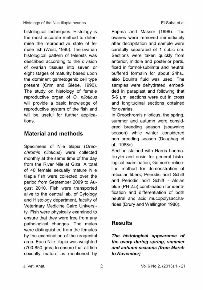
Fig (2): Section of ovary of Nile tilapia during summer showing: Cap-sule (Ca) and Blood ves-sels (B.V). Gomeri′s retic-uline stain, X 400
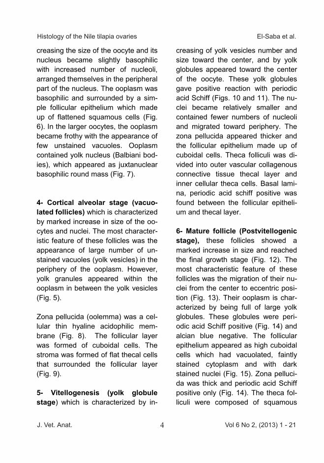
Fig (3): Section of ovary of Nile tilapia during spring showing: - Capsule (Ca); Stroma (St); Mature follicle (M) and Cortical al-veolar stage (C). H&E, X 40
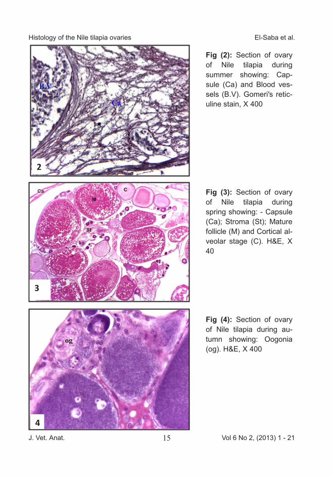
Fig (4): Section of ovary of Nile tilapia during au-tumn showing: Oogonia (og). H&E, X 400
2
3
4
J. Vet. Anat. Vol 6 No 2, (2013) 1 - 2115
Histology of the Nile tilapia ovaries El-Saba et al.
Van den Hurk. R. and Peute, J. (1979): Cyclic changes in the ovary of the rainbow trout, Salmo gaird-neri, with special reference to sites of steroidogenesis. Cell Tissue Res. 199: 289 – 306. Van den Hurk, R. and Peute, J. (1985): Functional aspects of the post-ovulatory follicle in the ovary of the African catfish, Clarias gariepi-nus after induced ovulation. An ul-trastructural and enzyme histo-chemical study. Cell. Tiss. Res., 240 : 199 - 208. Wallace, R.A. and Selman, K. (1981): Cellular and dynamic aspect of oocyte growth in teleosts. Amer. Zool., 21: 325 - 343.
West, G. (1990): Methods of as-sessing ovarian development in fishes: a review. Aust. J. Mar.Fresh-water Res. 41: 199-222. Yoakim, E.G. (1971): Seasonal var-iations in the pituitary gland and gonads of the Nile catfish (Synodon-tus schall) in relation to its reproduc-tive cycle. PhD. Thesis. Faculty of Science. Ain Shams University. Zaki, M.I.; Dowidar, M.N. and abdala, A. (1986a): Reproductive biology of Clarias gariepinus (Syn, lazera) Burchell (clariidae) in lake Manzalah, Egypt. I. Structure of the ovaries. Folia Morphologica, 34: 301 - 306.
Fig (1): Section of ovary of Nile tilapia during spring showing: Capsule (Ca) and Ovigerous lamellae (arrow). H&E, X 40 1
Fig (2): Section of ovary of Nile tilapia during summer showing: Cap-sule (Ca) and Blood ves-sels (B.V). Gomeri′s retic-uline stain, X 400
Fig (3): Section of ovary of Nile tilapia during spring showing: - Capsule (Ca); Stroma (St); Mature follicle (M) and Cortical al-veolar stage (C). H&E, X 40
Fig (4): Section of ovary of Nile tilapia during au-tumn showing: Oogonia (og). H&E, X 400
2
3
4

J. Vet. Anat. Vol 6 No 2, (2013) 1 - 2116
Histology of the Nile tilapia ovaries El-Saba et al. Fig (5): Section of ovary of Nile tilapia during summer showing: Cortical alveolar stage (C); Follicu-lar cells (F) and Chroma-tin nucleolus stage (Cn). H&E, X 400 Fig (6): Section of ovary of Nile tilapia during spring showing: Atretic fol-licles (A); Chromatin nu-cleolus stage (Cn); Peri-nucleolar stage (Pe) and Follicular cells (F). H&E, X 100
Fig (7): Section of ovary of Nile tilapia during summer showing: Follic-ular cells (F); Peri-nucleolar stage (Pe) and Balbiani body (Arrow). H&E, X 400
5
6
7
Fig (8): Section of ovary of Nile tilapia during summer showing: Follicular cells (F); Yolk granules (g); Cortical alveolar stage follicle (C) and Zona pellucida (Z). H&E, X 400 Fig (9): Section of ovary of Nile tilapia during spring showing: Cortical alveoli (C); Follicular cells (F); Thecal layer (Th); Zona pellucida (Z) and Yolk globules in mature follicle (gl). H&E, X 1000 Fig (10): Section of ovary of Nile tilapia during spring showing yolk globule stage. Note: Follicular cells (F); Zo-na pellucida (Z) and Base-ment membrane periodic ac-id Schiff positive (arrow), pe-riodic acid Schiff (PAS) and Alcian blue (AB) (PH 2.5), X400
8
9
10

J. Vet. Anat. Vol 6 No 2, (2013) 1 - 2117
Histology of the Nile tilapia ovaries El-Saba et al. Fig (5): Section of ovary of Nile tilapia during summer showing: Cortical alveolar stage (C); Follicu-lar cells (F) and Chroma-tin nucleolus stage (Cn). H&E, X 400 Fig (6): Section of ovary of Nile tilapia during spring showing: Atretic fol-licles (A); Chromatin nu-cleolus stage (Cn); Peri-nucleolar stage (Pe) and Follicular cells (F). H&E, X 100
Fig (7): Section of ovary of Nile tilapia during summer showing: Follic-ular cells (F); Peri-nucleolar stage (Pe) and Balbiani body (Arrow). H&E, X 400
5
6
7
Fig (8): Section of ovary of Nile tilapia during summer showing: Follicular cells (F); Yolk granules (g); Cortical alveolar stage follicle (C) and Zona pellucida (Z). H&E, X 400 Fig (9): Section of ovary of Nile tilapia during spring showing: Cortical alveoli (C); Follicular cells (F); Thecal layer (Th); Zona pellucida (Z) and Yolk globules in mature follicle (gl). H&E, X 1000 Fig (10): Section of ovary of Nile tilapia during spring showing yolk globule stage. Note: Follicular cells (F); Zo-na pellucida (Z) and Base-ment membrane periodic ac-id Schiff positive (arrow), pe-riodic acid Schiff (PAS) and Alcian blue (AB) (PH 2.5), X400
8
9
10

J. Vet. Anat. Vol 6 No 2, (2013) 1 - 2118
Histology of the Nile tilapia ovaries El-Saba et al. Fig (11): Section of ovary of Nile tilapia during spring showing yolk globule stage. Note: Follicular cells (F); Zona pellucida (Z); Yolk globules (gl); Thecal layer (Th); Basement membrane periodic acid Schiff positive (arrow). peri-odic acid Schiff (PAS) and alcian blue (AB) (PH 2.5), X1000 Fig (12): Section of ovary of Nile tilapia during summer showing: Mature follicles (M); Cortical al-veolar stage (C); Stroma (St). H&E, X 100
Fig (13): Section of ovary of Nile tilapia during summer showing mature follicle: Migra-tory nucleus in the mature folli-cles (N). H&E, X 100
11
12
13
12
Fig (14): Section of ovary of Nile tilapia during summer showing mature follicle. Note: Follicular cells (F); Zona pellu-cida (Z) and Yolk globules (gl). Periodic acid Schiff (PAS), X400
Fig (15): Section of ovary of Nile tilapia during spring show-ing: Follicular cells (F); Zona pellucida (Z); Basement mem-brane (arrow) and Thecal layer (Th). H&E, X1000
Fig (16): Section of ovary of Nile tilapia during spring show-ing: Post ovulatory follicle (ar-row). H & E, X 100
14
15
16

J. Vet. Anat. Vol 6 No 2, (2013) 1 - 2119
Histology of the Nile tilapia ovaries El-Saba et al. Fig (11): Section of ovary of Nile tilapia during spring showing yolk globule stage. Note: Follicular cells (F); Zona pellucida (Z); Yolk globules (gl); Thecal layer (Th); Basement membrane periodic acid Schiff positive (arrow). peri-odic acid Schiff (PAS) and alcian blue (AB) (PH 2.5), X1000 Fig (12): Section of ovary of Nile tilapia during summer showing: Mature follicles (M); Cortical al-veolar stage (C); Stroma (St). H&E, X 100
Fig (13): Section of ovary of Nile tilapia during summer showing mature follicle: Migra-tory nucleus in the mature folli-cles (N). H&E, X 100
11
12
13
12
Fig (14): Section of ovary of Nile tilapia during summer showing mature follicle. Note: Follicular cells (F); Zona pellu-cida (Z) and Yolk globules (gl). Periodic acid Schiff (PAS), X400
Fig (15): Section of ovary of Nile tilapia during spring show-ing: Follicular cells (F); Zona pellucida (Z); Basement mem-brane (arrow) and Thecal layer (Th). H&E, X1000
Fig (16): Section of ovary of Nile tilapia during spring show-ing: Post ovulatory follicle (ar-row). H & E, X 100
14
15
16

J. Vet. Anat. Vol 6 No 2, (2013) 1 - 2120
Histology of the Nile tilapia ovaries El-Saba et al.
Fig (17): Section of ovary of Nile tilapia during autumn show-ing: Atretic follicles (A). H&E, X400
Fig (18): Section of ovary of Nile tilapia during winter show-ing: Capsule (Ca) and Circular muscle layer (arrow). H&E, X 400
Fig (19): Section of ovary of Nile tilapia during winter show-ing: Chromatin nucleolus stage (Cn); Perinucleolar stage (Pe); Cortical alveolar stage (C) and Thick stroma (St). H&E, X40
18
17
19
Fig (20): Section of ovary of Nile tilapia during winter show-ing: Chromatin nucleolus stage (Cn); Perinucleolar stage (Pe); Cortical alveolar stage (C); Atretic follicles (A) and Thick stroma (St). Stain: H&E, X40
20

J. Vet. Anat. Vol 6 No 2, (2013) 1 - 2121
Histology of the Nile tilapia ovaries El-Saba et al. Fig (17): Section of ovary of Nile tilapia during autumn show-ing: Atretic follicles (A). H&E, X400
Fig (18): Section of ovary of Nile tilapia during winter show-ing: Capsule (Ca) and Circular muscle layer (arrow). H&E, X 400
Fig (19): Section of ovary of Nile tilapia during winter show-ing: Chromatin nucleolus stage (Cn); Perinucleolar stage (Pe); Cortical alveolar stage (C) and Thick stroma (St). H&E, X40
18
17
19
Fig (20): Section of ovary of Nile tilapia during winter show-ing: Chromatin nucleolus stage (Cn); Perinucleolar stage (Pe); Cortical alveolar stage (C); Atretic follicles (A) and Thick stroma (St). Stain: H&E, X40
20