schistosoma mansoni antigens alter activation markers and ... tvs... · schistosoma mansoni...
TRANSCRIPT
Sp
TLMa
b
c
d
e
f
g
a
ARR2AA
KASLSS
1
a
ss
h0
Acta Tropica 166 (2017) 268–279
Contents lists available at ScienceDirect
Acta Tropica
jo u r n al homep age: www.elsev ier .com/ locate /ac ta t ropica
chistosoma mansoni antigens alter activation markers and cytokinerofile in lymphocytes of patients with asthma
arcísio Vila Verde Santana de Almeidaa, Jamille Souza Fernandesa, Diego Mota Lopesa,orena Santana Andradea, Sérgio Costa Oliveirab,c, Edgar M. Carvalhoa,b,d,aria Ilma Araujoa,b,e, Álvaro A. Cruza,f, Luciana Santos Cardosoa,b,g,∗
Servic o de Imunologia, Hospital Universitário Professor Edgard Santos, Universidade Federal da Bahia, Salvador, Bahia, BrazilInstituto Nacional de Ciência e Tecnologia de Doenc as Tropicais (INCT-DT/CNPq), Salvador, Bahia, BrazilDepartamento de Bioquímica e Imunologia, Universidade Federal de Minas Gerias, BrazilCentro de Pesquisas Gonc alo Moniz, FIOCRUZ, Salvador, Bahia, BrazilEscola Baiana de Medicina e Saúde Pública, Salvador, Bahia, BrazilProAR—Núcleo de Excelência em Asma, UFBA, Salvador, Bahia, BrazilDepartamento de Análises Clínicas e Toxicológicas, Faculdade de Farmácia, UFBA, Brazil
r t i c l e i n f o
rticle history:eceived 25 January 2016eceived in revised form2 November 2016ccepted 1 December 2016vailable online 5 December 2016
eywords:sthmachistosoma mansoni antigensymphocytesm29m29TSP-2
a b s t r a c t
Asthma is a chronic disease characterized by airway inflammation, obstruction and hyperresponsiveness.Severe asthma affects a small proportion of subjects but results in most of the morbidity, costs andmortality associated with the disease. Studies have suggested that Schistosoma mansoni infection reducesthe severity of asthma and prevent atopy.Objective: We evaluated the ability of S. mansoni antigens, Sm29 and Sm29TSP-2 to modulate lymphocyteactivation status in response to the allergen of the mite Dermatophagoides pteronyssinus (Der p1) in cellcultures of individuals with asthma.Methods: Thirty four patients were enrolled in this study: seventeen patients with severe asthma (SAgroup), seventeen patients with mild asthma (MA group) and six controls with no asthma. Peripheralblood mononuclear cells (PBMC) were obtained and stimulated with Sm29 and Sm29TSP-2 in the pres-ence or absence of Der p1. The expression of surface markers and cytokines on lymphocytes was evaluatedby flow cytometry and the levels of IL-10 in the culture supernatant were determined by ELISA.Results: The addition of Sm29 and Sm29TSP-2 antigens to PBMC cultures from both groups of subjectswith asthma stimulated with Der p1 reduced the frequency of CD4+CD25low cells whereas and increasedfrequency of CD4+CD25high population was observed compared to unstimulated cultures. Moreover, cul-tures stimulated with Sm29TSP-2 showed a reduction in the frequency of T cells expressing CD69, IFN-�,TNF and TGF-� in the MA group and an increase in the frequency of CD4+TSLPR+ T cells in the SA group.The addition of Sm29 to the cultures reduced the frequency of CD4+CD69+ and CD4+IL-5+ T cells in all asth-
+
matic groups, and reduced the frequency of CD4 T cells expressing IL-13 in the MA group. The culturesstimulated with Sm29 and Sm29TSP-2 showed an increase in the level of IL-10 in the supernatants.Conclusion: These results suggest that the addition of Sm29 and Sm29TSP-2 to the cells cultures fromsubjects with asthma reduced cell activation markers and altered the cytokine production pattern in aontro
way that can potentialy c. Introduction
Severe asthma affects approximately 5% to 10% of patients withsthma worldwide and is characterized by the persistence of symp-
∗ Corresponding author at: Servic o de Imunologia, Complexo Hospitalar Univer-itário Professor Edgard Santos, Universidade Federal da Bahia, Rua João das Botas/n, Canela, 40110-160 Salvador, Bahia, Brazil.
E-mail addresses: [email protected], [email protected] (L.S. Cardoso).
ttp://dx.doi.org/10.1016/j.actatropica.2016.12.002001-706X/© 2016 Elsevier B.V. All rights reserved.
l the inflammatory response associated with asthma.© 2016 Elsevier B.V. All rights reserved.
toms, frequent exacerbations, reduced lung function and a needfor high doses of inhaled corticosteroids (Antonicelli et al., 2004;Bousquet et al., 2010; Hekking et al., 2014; Moore et al., 2007;Von Bulow et al., 2014). The treatment of severe asthma is difficult,costly and bring the risk of adverse events. Only a small proportionof subjects with severe asthma reaches total control of symptoms
and exacerbations, which affect significantly their quality of lifeand results in higher expenditures with the disease, often related toemergency room visits, hospitalizations and the use of other sort of
T.V.V.S. de Almeida et al. / Acta Tropica 166 (2017) 268–279 269
Table 1Characteristics of the study population.
Mild Asthma (n = 17) Severe Asthma (n = 17) Healthy Controls (n = 6) P
Age (years)a (mean ± DP) 38,8 ± 14,3 46,5 ± 11,4 43,5 ± 13,3 >0,05Female gender n(%)b 13 (76,4) 11 (64,7) 5 (83,3) >0,05Positivity to the skin prick test to Der p1 n (%)b 4 (23,5) 6 (35,2) 0 (0)c <0,0001SWAP-specific IgE (mean ± SD) 0.12 ± 0.03 0.05 ± 0.04 0.14 ± 0.07 >0.05
Cutoff IgE: 0.36.
he
bbaie1VAlMwsMirmtO
smTeac2aacP(P
iihsr2eTnwwSo2se1t
a ANOVA.b Chi-square.c HC group vs MA group and HC group vs SA group.
eath resources (Antonicelli et al., 2004; Franco et al., 2009; O’Neillt al., 2015; Santos et al., 2007).
In recent years, many studies have suggested that infectiony helminths may modulate the allergic response in asthma,eing associated to lower frequency of positive skin prick tests toeroallergens, and a lower prevalence of atopy in general amongndividuals living in helminth-endemic areas (Alcantara-Nevest al., 2014; Araujo et al., 2000; Cooper et al., 2003; Hagel et al.,993; Lynch et al., 1993; Lynch et al., 1987; Medeiros et al., 2004;an den Biggelaar et al., 2001; Van den Biggelaar et al., 2000).sthmatics individuals infected with Schistosoma mansoni have
ess severe asthma symptoms as compared to uninfected patients.oreover, the anthelmintic treatment against S. mansoni led to aorsening of asthma symptoms and to an increase in allergen-
pecific serum IgE (Almeida et al., 2012; Campolina et al., 2013;edeiros et al., 2003; Van den Biggelaar et al., 2004). Furthermore,
nfection with S. mansoni has been associated with a reduced Th2esponse in vitro and in murine model with an increase in regulatoryechanisms that may be associated with the control of inflamma-
ion and improvement in asthma symptoms (Araujo et al., 2004;liveira et al., 2009; Smits et al., 2007).
Regulatory T cell activation have been one of the main hypothe-es to explain the inverse relationship between allergy and S.ansoni infection (Layland et al., 2013; Van der Vlugt et al., 2012).
he literature has described CD4+ T lymphocytes that do notxpress CD25 (CD25neg) as young effector cells that are not yetctivated, while those with a low expression of CD25 (CD25low) areonsidered activated responder CD4+T cells (Baecher-Allan et al.,001). CD4+ T cells with a high expression of CD25 (CD25high) aressociated with the suppression of the immune response and are,ble to control the activation and proliferation of activated cells byell-cell contact via costimulatory molecules such as CTLA- 4 andD-1, or by the production of regulatory cytokines, such as IL-10Baecher-Allan et al., 2001; Gangi et al., 2005; Okita et al., 2009;ontoux et al., 2002; Sojka et al., 2009; Uhlig et al., 2006).
The studies mentioned above provided support to furthernvestigating the use of parasite antigens to down-modulate thenflammatory response observed in subjects with asthma. Studiesave shown that chronic helminth infections, especially Schisto-oma mansoni, possesses the ability to modulate the inflammatoryesponse associated to both, Th1 (Bafica et al., 2011; Lima et al.,013) and Th2 (Cardoso et al., 2012; Cardoso et al., 2010; Cardosot al., 2006a,b; Pacifico et al., 2009) immune-mediated diseases.hese findings have provided the rationale for the use of recombi-ant S. mansoni proteins in in vitro studies with cells from patientsith asthma in an attempt to modulate the response associatedith inflammatory process. Studies have shown that Sm29 and
mTSP-2 antigens are secreted by the membrane and/or tegumentf the S. mansoni adult worm (Cardoso et al., 2006a,b; Tran et al.,006). Proteins secreted or localized on the surface of Schistosoma
pp., which are in intimate contact with host tissues, might be moreffective in triggering immunoregulatory processes (Simpson et al.,990). The Sm29 is a membrane-bound glycoprotein located on theegument of the adult worm and lung stage schistosomula (Cardosoet al., 2006a,b). SmTSP-2 is a recombinant protein (tetraspanin)from S. mansoni tegument (Tran et al., 2006). These antigens havebeen evaluated by our group regarding their potential to down-modulate inflammatory cytokines and to induces IL-10 productionin vitro in PBMC from individuals with cutaneous leishmaniasis,HTLV-1 infection and asthma (Bafica et al., 2011; Cardoso et al.,2010; Lima et al., 2013). Thus, the identification of parasite anti-gens with the potential to prevent or attenuate the inflammatoryresponse associated with asthma represents a promising strategyfor an alternative intervention to control this chronic illness.
2. Materials and methods
2.1. Features of the studied subjects
In this study, we recruited 17 consecutive patients with severeasthma followed up for over one year in the Program for the Controlof Asthma of Bahia (ProAR), a reference center for severe asthma inSalvador, Bahia, Brazil, 17 patients with mild asthma and 6 healthcontrols (HC), recruited consecutively from the same communitiespatients with severe asthma live, invited to volunteer by publicadvertisement in health facilities and public transportation. Sub-jects were recruited from January 2013 until July 2015 and theblood samples processed immediately upon collection. Subjectswere not included if they had an exacerbation in the last monthregardless of using oral corticosteroids or not. At the time of bloodsamples collection all subjects with severe asthma were treatedwith a combination of medium to high dose of inhaled corticos-teroids (800mcg to 1600 of budesonide or equivalent) and longacting beta 2 agonist. Those with mild asthma were not receivinginhaled corticosteroids. Individuals with severe asthma were iden-tified as having untreated severe asthma at enrollment in ProAR(from 2003), according to the NIH Guidelines for Asthma (NIH-NHBLI. Guidelines for the Diagnosis and Management of Asthma,1997) and a WHO consultation on severe asthma (Bousquet et al.,2010). In brief, they had any one of the following: (i) symp-toms daily, continuous; (ii) activities limited daily (symptoms withminor efforts); (iii) nocturnal symptoms over 2 times a week; (iv)use of bronchodilators ≥2 times a day; (v) Peak Expiratory Flow(PEF) or Forced Expiratory Volume in one second (FEV1): <60% ofpredict. Subjects with mild asthma were recruited on the basis oftheir history of asthma, having their diagnosis validated by a doc-tor in the research facility. We did not include in this study currentsmokers individuals and those with a positive serology for Cha-gas disease, HIV, HTLV-1, or hepatitis virus types B and C, all ofwhich are conditions that could interfere with the immunologi-cal response. All participants were submitted to skin prick test toDermatophagoides pteronyssinus antigen 1 (Der p1) and a panel ofthe most common aeroallergens including other house dust mites,
coacoroach, molds, cat, dog and grass. To rule out the effect ofS. mansoni previous exposure in immunological assays, we alsoexcluded individuals who had a positive S. mansoni infection orexposure to this parasite any time in his/her life. In the urban area
2 cta Tr
ot
ma1aArHa(
2
etaamt
2
sd
2
FthpaGwtacf(a
Scoaee1a(ri
cIse(T�
70 T.V.V.S. de Almeida et al. / A
f Salvador da Bahia there is no report of Schistosoma infection inhe last decades.
Furthermore, we measured the levels of serum-specific IgE to S.ansoni soluble adult worm antigen (SWAP) and performed par-
sitological assays by Hoffman et al. technique (Hoffman et al.,934). There was no significant difference in the mean age, gendernd levels of serum-specific IgE to SWAP among groups evaluated.dditionally, there were no significant differences regarding theesponse to skin prick test (SPT) to Der p1 between asthmatics. TheC group was negative to the SPT to Der p1 (Table 1). All individu-ls were negative for S. mansoni infection by parasitological examsnot shown).
.2. Ethical statement
The Ethics Committee of Maternidade Climério de Oliveira, Fed-ral University of Bahia (License Number: 095/2009) approvedhe present study. Written informed consent was obtained fromll patients and controls. All participants who had complaints ofsthma and allergies were properly oriented regarding environ-ental control, prescribed the medication required and referred to
he most convenient health facility.
.3. SWAP-specific IgE measurements in human serum
Levels of SWAP-specific IgE were measured in serum from alltudied individuals using an indirect ELISA technique, as previouslyescribed (Figueiredo et al., 2012; Souza-Atta et al., 1999).
.4. Cell culture and flow cytometry assays
Peripheral blood mononuclear cells (PBMCs) were isolated usingicoll-Hypaque gradient sedimentation and adjusted to a concen-ration of 3 × 10⁵/mL in RPMI 1640 medium containing 10% normaluman serum (AB positive and heat inactivated), 100 U/mL ofenicillin, 100 mg/mL of streptomycin, 2 mmol/L of l-glutamine,nd 30 mmol/L of HEPES (all from Life Technologies GIBCO, BRL,aithersburg, MS). Cells were cultured in vitro either stimulatedith 10 �g/mL of the S. mansoni antigens Sm29 and Sm29TSP-2 in
he presence or absence of Der p1 (Cosmo Bio, LTD.; Tokyo, Japan)t the concentration of 5 �g/mL for 48 h at 37 ◦C in an atmosphereontaining 5% CO2. After incubation, supernatants were harvestedor IL-10 measurement by ELISA using commercially available kitsR&D Systems, Inc.), and the cells were stained for flow cytometrys described below.
During the last 4 h of culture, Brefeldin A (10 �g/mL; Sigma,t. Louis, MO), which impairs protein secretion by the Golgiomplex, was added to the cultures. Cells were stained with flu-rescently conjugated mouse anti-human monoclonal antibodiesgainst human CD3 (clone OKT3, eBioscience), CD4 (clone OKT4,Bioscience), CD25 (clone BC96, eBioscience), CD28 (clone CD28.2,Bioscience), CD69 (clone L78, Becton Dickinson), CTLA-4 (clone4D3, eBioscience) and TSLPR (clone 1A6, eBioscience), and thennalyzed for 100000 events per sample using a flow cytometerFACSCanto, Becton Dickinson, San Jose, CA). Limits for the quad-ant markers were set based on negative populations and controlssotype (data not shown).
Intracellular staining was performed with a PE-labeled mono-lonal antibody against human FoxP3 (clone 236A/E7, eBioscience),L-10 (clone JES3-19F1, eBioscience), TGF-� (clone TW4-2F8, eBio-cience), IFN-� (clone GZ-4, eBioscience), TNF (clone MAb11,
Bioscience), IL-17A (clone eBio64DEC17, eBioscience), IL-13PVM13-1, eBioscience) and IL-5 (clone JES1-39D10, eBioscience).he Clone TW4-2F8 (eBioscience) evaluated detects LAP/pro-TGF-1.opica 166 (2017) 268–279
The frequency of positive cells was analyzed using the programFlowJoTM (Tree Star, USA). The lymphocyte region was defined bynonspecific fluorescence with forward scatter (FSC) and side scat-ter (SSC) used to indicate cell size and granularity, respectively.The cells were also gated based on their expression of CD3 and CD4(Fig. 1A). Fig. 1B and C shows a representative gate strategy to anal-yse the different CD4+CD25+ and CD4+CD69+ T cell populations,respectively.
2.5. Antigen stimulation
The Schistosoma mansoni tegument antigens Sm29 andSm29TSP-2, used in this study were provided by Dr. Sérgio C.Oliveira from the Institute of Biological Science, Department ofBiochemistry and Immunology, UFMG, Brazil. The recombinantproteins were cloned in E. coli (Cardoso et al., 2006a,b; Pinheiroet al., 2014) and were tested for the presence of lipopolysaccharide(LPS) using a commercially available LAL Chromogenic Kit (CAM-BREX).The level of LPS was below the detection limit (data notshown).
2.6. Statistical analysis and sample size
Statistical analysis and graphical representation were per-formed using Graphpad PRISM 5.0 software (La Jolla, CA, USA).Comparisons among age of groups were performed using ANOVAand for immunological assays we used Kruskal Wallis test withDunns pos-test. Comparisons among gender and positivity skinprick test and SWAP-specific IgE were performed using chi-squaretest. All statistical tests were two-tailed and statistical signif-icance was established at the 95 percent confidence interval.P-value < 0.05 were considered significant. The sample size calcula-tion was performed based on the frequency of IL-10 expression inCD4+CD25+ T cells from healthy subjects with mild asthma stimu-lated with Der p1 plus Sm29 (Cardoso et al., 2011). A minimum sizeof 16 patients per group would be sufficient to detect significantdifferences.
3. Results
3.1. Effect of S. mansoni antigens on the expression of CD25molecules on CD4+ T lymphocytes
Different degrees of CD25 expression on CD4+ T lymphocyteshave been associated with either activation or regulatory profile inthese cells. Therefore, we decided to analyze the expression of thismolecule in T cell populations from subjects with mild asthma (MA)and severe asthma (SA) after the addition of S. mansoni antigens tothe cultures in the presence of Der p1 antigen.
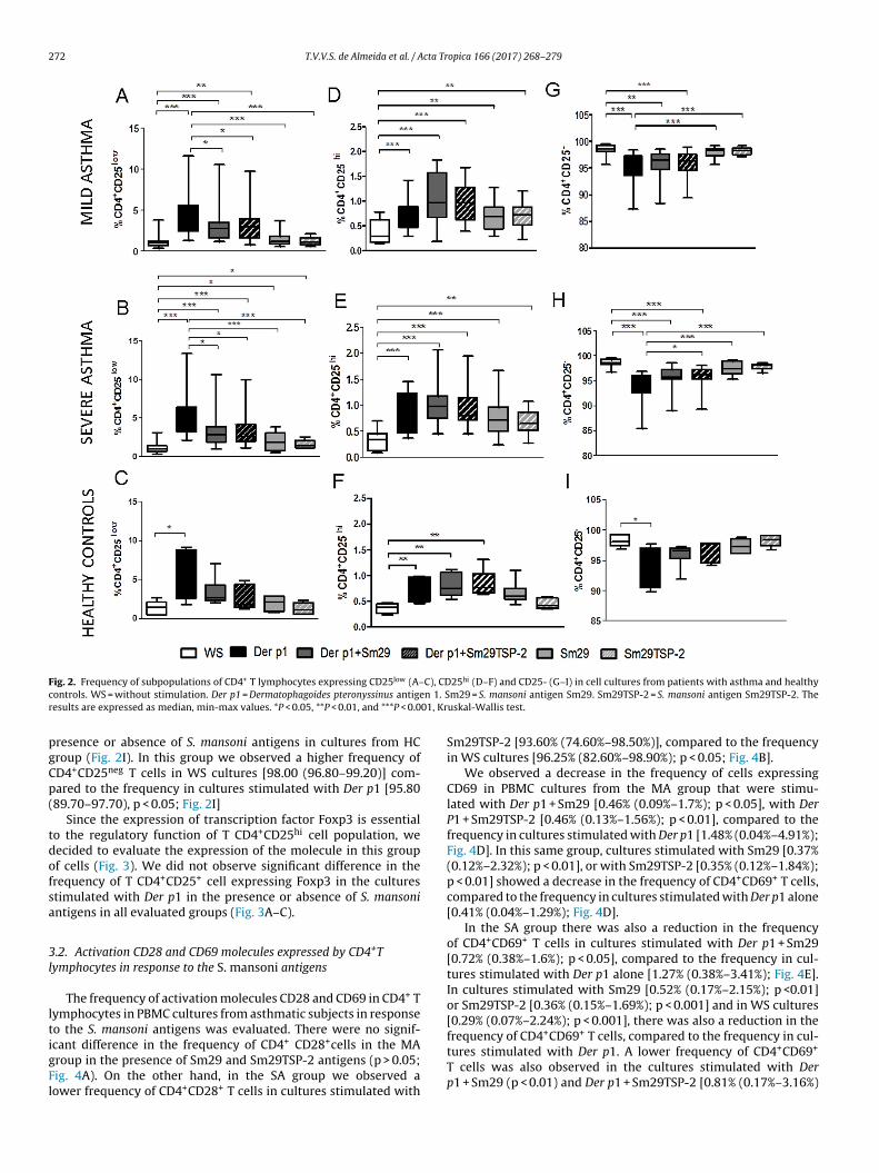
In the MA group, there was a reduction in the fre-quency of CD4+CD25low T cells in cultures stimulated with Derp1 + Sm29 [median = 2.68% (min–max = 1.16%–10.50%); p < 0.05],Der p1 + Sm29TSP-2 [2.89% (0.76%–9.73%); p <0.05], Sm29 [1.12%(0.49%–3.64%) p < 0.001] or Sm29TSP-2 [1.08% (0.53%–2.09%);p < 0.001] compared to the frequency in cultures stimulated withDer p1 alone [3.93% (1.30%–11.60%); Fig. 2A]. Additionally, weobserved a lower frequency of CD4+CD25low T cells in cultures with-out stimulation (WS cultures) [0.97% (0.34%–3.82%)] than in thosestimulated with Der p1 (p < 0.001), Der p1 + Sm29 (p < 0.001), Derp1 + Sm29TSP-2 (p < 0.001), Sm29 (p < 0.05) or Sm29TSP-2 (p < 0.05;Fig. 2A).
In the SA group we found similar results with a reduction in
the frequency of CD4+CD25low T cells in cultures stimulated withDer p1 + Sm29 [2.77% (0.90%–10.60%); p < 0.05], Der p1 + Sm29TSP-2 [2.63% (1.07%–9.92%); p < 0.05], Sm29 [1.78% (0.44%–3.78%)p < 0.001] or Sm29TSP-2 [1.36% (0.97%–2.46%); p < 0.001] compared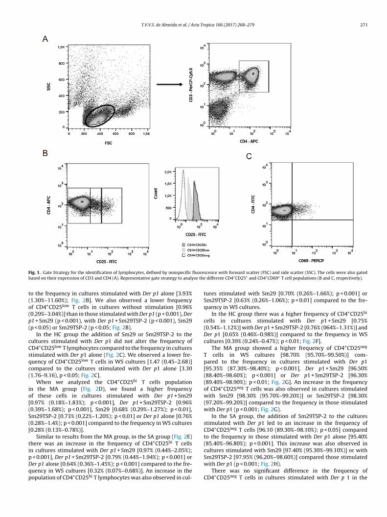
T.V.V.S. de Almeida et al. / Acta Tropica 166 (2017) 268–279 271
F fluoreb se the
t(o(p(
cCsqc(
io[(S([
tipDqp
ig. 1. Gate Strategy for the identification of lymphocytes, defined by nonspecific
ased on their expression of CD3 and CD4 (A). Representative gate strategy to analy
o the frequency in cultures stimulated with Der p1 alone [3.93%1.30%–11.60%); Fig. 2B]. We also observed a lower frequencyf CD4+CD25low T cells in cultures without stimulation [0.96%0.29%–3.04%)] than in those stimulated with Der p1 (p < 0.001), Der1 + Sm29 (p < 0.001), with Der p1 + Sm29TSP-2 (p < 0.001), Sm29p < 0.05) or Sm29TSP-2 (p < 0.05; Fig. 2B).
In the HC group the addition of Sm29 or Sm29TSP-2 to theultures stimulated with Der p1 did not alter the frequency ofD4+CD25low T lymphocytes compared to the frequency in culturestimulated with Der p1 alone (Fig. 2C). We observed a lower fre-uency of CD4+CD25low T cells in WS cultures [1.47 (0.45–2.68)]ompared to the cultures stimulated with Der p1 alone [3.301.76–9.16), p < 0.05; Fig. 2C].
When we analyzed the CD4+CD25hi T cells populationn the MA group (Fig. 2D), we found a higher frequencyf these cells in cultures stimulated with Der p1 + Sm290.97% (0.18%–1.83%); p < 0.001], Der p1 + Sm29TSP-2 [0.96%0.39%–1.68%); p < 0.001], Sm29 [0.68% (0.29%–1.27%); p < 0.01],m29TSP-2 [0.73% (0.22%–1.20%); p < 0.01] or Der p1 alone [0.76%0.28%–1.4%); p < 0.001] compared to the frequency in WS cultures0.28% (0.13%–0.78%)].
Similar to results from the MA group, in the SA group (Fig. 2E)here was an increase in the frequency of CD4+CD25hi T cellsn cultures stimulated with Der p1 + Sm29 [0.97% (0.44%–2.05%);
< 0.001], Der p1 + Sm29TSP-2 [0.79% (0.44%–1.94%); p < 0.001] orer p1 alone [0.64% (0.36%–1.45%); p < 0.001] compared to the fre-uency in WS cultures [0.32% (0.07%–0.68%)]. An increase in theopulation of CD4+CD25hi T lymphocytes was also observed in cul-
scence with forward scatter (FSC) and side scatter (SSC). The cells were also gated different CD4+CD25+ and CD4+CD69+ T cell populations (B and C, respectively).
tures stimulated with Sm29 [0.70% (0.26%–1.66%); p < 0.001] orSm29TSP-2 [0.63% (0.26%–1.06%); p < 0.01] compared to the fre-quency in WS cultures.
In the HC group there was a higher frequency of CD4+CD25hi
cells in cultures stimulated with Der p1 + Sm29 [0.75%(0.54%–1.12%)] with Der p1 + Sm29TSP-2 [0.76% (064%–1.31%)] andDer p1 [0.65% (0.46%–0.98%)] compared to the frequency in WScultures [0.39% (0.24%–0.47%); p < 0.01; Fig. 2F].
The MA group showed a higher frequency of CD4+CD25neg
T cells in WS cultures [98.70% (95.70%–99.50%)] com-pared to the frequency in cultures stimulated with Der p1[95.35% (87.30%–98.40%); p < 0.001], Der p1 + Sm29 [96.50%(88.40%–98.60%); p < 0.001] or Der p1 + Sm29TSP-2 [96.30%(89.40%–98.90%); p < 0.01; Fig. 2G]. An increase in the frequencyof CD4+CD25neg T cells was also observed in cultures stimulatedwith Sm29 [98.30% (95.70%–99.20%)] or Sm29TSP-2 [98.30%(97.20%–99.20%)] compared to the frequency in those stimulatedwith Der p1 (p < 0.001; Fig. 2G).
In the SA group, the addition of Sm29TSP-2 to the culturesstimulated with Der p1 led to an increase in the frequency ofCD4+CD25neg T cells [96.10 (89.30%–98.10%); p < 0.05] comparedto the frequency in those stimulated with Der p1 alone [95.40%(85.40%–96.80%); p < 0.001]. This increase was also observed incultures stimulated with Sm29 [97.40% (95.30%–99.10%)] or with
Sm29TSP-2 [97.95% (96.20%–98.60%)] compared those stimulatedwith Der p1 (p < 0.001; Fig. 2H).There was no significant difference in the frequency ofCD4+CD25neg T cells in cultures stimulated with Der p 1 in the
272 T.V.V.S. de Almeida et al. / Acta Tropica 166 (2017) 268–279
F C), CDc n 1. Sr 01, Kr
pgCp(
tdofsa
3l
ltigFl
ig. 2. Frequency of subpopulations of CD4+ T lymphocytes expressing CD25low (A–ontrols. WS = without stimulation. Der p1 = Dermatophagoides pteronyssinus antigeesults are expressed as median, min-max values. *P < 0.05, **P < 0.01, and ***P < 0.0
resence or absence of S. mansoni antigens in cultures from HCroup (Fig. 2I). In this group we observed a higher frequency ofD4+CD25neg T cells in WS cultures [98.00 (96.80–99.20)] com-ared to the frequency in cultures stimulated with Der p1 [95.8089.70–97.70), p < 0.05; Fig. 2I]
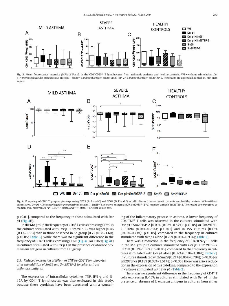
Since the expression of transcription factor Foxp3 is essentialo the regulatory function of T CD4+CD25hi cell population, weecided to evaluate the expression of the molecule in this groupf cells (Fig. 3). We did not observe significant difference in therequency of T CD4+CD25+ cell expressing Foxp3 in the culturestimulated with Der p1 in the presence or absence of S. mansonintigens in all evaluated groups (Fig. 3A–C).
.2. Activation CD28 and CD69 molecules expressed by CD4+Tymphocytes in response to the S. mansoni antigens
The frequency of activation molecules CD28 and CD69 in CD4+ Tymphocytes in PBMC cultures from asthmatic subjects in responseo the S. mansoni antigens was evaluated. There were no signif-
cant difference in the frequency of CD4+ CD28+cells in the MAroup in the presence of Sm29 and Sm29TSP-2 antigens (p > 0.05;ig. 4A). On the other hand, in the SA group we observed aower frequency of CD4+CD28+ T cells in cultures stimulated with25hi (D–F) and CD25- (G–I) in cell cultures from patients with asthma and healthym29 = S. mansoni antigen Sm29. Sm29TSP-2 = S. mansoni antigen Sm29TSP-2. Theuskal-Wallis test.
Sm29TSP-2 [93.60% (74.60%–98.50%)], compared to the frequencyin WS cultures [96.25% (82.60%–98.90%); p < 0.05; Fig. 4B].
We observed a decrease in the frequency of cells expressingCD69 in PBMC cultures from the MA group that were stimu-lated with Der p1 + Sm29 [0.46% (0.09%–1.7%); p < 0.05], with DerP1 + Sm29TSP-2 [0.46% (0.13%–1.56%); p < 0.01], compared to thefrequency in cultures stimulated with Der p1 [1.48% (0.04%–4.91%);Fig. 4D]. In this same group, cultures stimulated with Sm29 [0.37%(0.12%–2.32%); p < 0.01], or with Sm29TSP-2 [0.35% (0.12%–1.84%);p < 0.01] showed a decrease in the frequency of CD4+CD69+ T cells,compared to the frequency in cultures stimulated with Der p1 alone[0.41% (0.04%–1.29%); Fig. 4D].
In the SA group there was also a reduction in the frequencyof CD4+CD69+ T cells in cultures stimulated with Der p1 + Sm29[0.72% (0.38%–1.6%); p < 0.05], compared to the frequency in cul-tures stimulated with Der p1 alone [1.27% (0.38%–3.41%); Fig. 4E].In cultures stimulated with Sm29 [0.52% (0.17%–2.15%); p <0.01]or Sm29TSP-2 [0.36% (0.15%–1.69%); p < 0.001] and in WS cultures[0.29% (0.07%–2.24%); p < 0.001], there was also a reduction in the
+ +
frequency of CD4 CD69 T cells, compared to the frequency in cul-tures stimulated with Der p1. A lower frequency of CD4+CD69+T cells was also observed in the cultures stimulated with Derp1 + Sm29 (p < 0.01) and Der p1 + Sm29TSP-2 [0.81% (0.17%–3.16%)
T.V.V.S. de Almeida et al. / Acta Tropica 166 (2017) 268–279 273
Fig. 3. Mean fluorescence intensity (MFI) of Foxp3 in the CD4+CD25hi T lymphocytes from asthmatic patients and healthy controls. WS = without stimulation. Derp1 = Dermatophagoides pteronyssinus antigen 1. Sm29 = S. mansoni antigen Sm29. Sm29TSP–2 = S. mansoni antigen Sm29TSP-2. The results are expressed as median, min-maxvalues.
F (D, E as antigem
pp
t(pfim
3aa
1b
ig. 4. Frequency of CD4+ T lymphocytes expressing CD28 (A, B and C) and CD69
timulation. Der p1 = Dermatophagoides pteronyssinus antigen 1. Sm29 = S. mansoni
edian, min-max values. *P < 0.05,**P < 0.01, and ***P < 0.001, Kruskal-Wallis test.
< 0.01], compared to the frequency in those stimulated with Der1 (Fig. 4E).
In the MA group the frequency of CD4+ T cells expressing CD69 inhe cultures stimulated with Der p1 + Sm29TSP-2 was higher [0.460.13–1.56)] than in those observed in SA group [0.72 (0.38–1.60),
< 0.05; Table 3], while there was no significant difference in therequency of CD4+ T cells expressing CD28 (Fig. 4C) or CD69 (Fig. 4F)n cultures stimulated with Der p 1 in the presence or absence of S.
ansoni antigens in cultures from HC group.
.3. Reduced expression of IFN-� or TNF by CD4+T lymphocytesfter the addition of Sm29 and Sm29TSP-2 to cultures fromsthmatic patients
The expression of intracellular cytokines TNF, IFN-� and IL-7A by CD4+ T lymphocytes was also evaluated in this study,ecause these cytokines have been associated with a worsen-
nd F) in cell cultures from asthmatic patients and healthy controls. WS = withoutn Sm29. Sm29TSP–2 = S. mansoni antigen Sm29TSP-2. The results are expressed as
ing of the inflammatory process in asthma. A lower frequency ofCD4+TNF+ T cells was observed in the cultures stimulated withDer p1 + Sm29TSP-2 [0.09% (0.02%–0.87%); p < 0.05] or Sm29TSP-2 [0.09% (0.04%–0.73%); p < 0.01] and in WS cultures [0.13%(0.01%–0.73%); p < 0.05], compared to the frequency in culturesstimulated with Der p1 alone [0.20% (0.05%–0.93%); Table 2].
There was a reduction in the frequency of CD4+IFN-�+ T cellsin the MA group in cultures stimulated with Der p1 + Sm29TSP-2[0.21% (0.03%–1.38%); p < 0.05], compared to the frequency in cul-tures stimulated with Der p1 alone [0.32% (0.10%–1.38%); Table 2].In cultures stimulated with Sm29 [0.21% (0.06%–0.70%); p < 0.05] orSm29TSP-2 [0.18% (0.08%–1.51%); p < 0.05], there was also a reduc-tion in the expression of this cytokine, compared to the expressionin cultures stimulated with Der p1 (Table 2).
There was no significant difference in the frequency of CD4+ Tcells expressing IL-17A in cultures stimulated with Der p1 in thepresence or absence of S. mansoni antigens in cultures from either

274
T.V.V
.S. de
Alm
eida et
al. /
Acta
Tropica 166
(2017) 268–279
Table 2Frequency of CD4+ T lymphocytes expressing TNF, IFN-�, IL-17A, IL-5, IL-13, TSLPR, CTLA-4, IL-10 and TGF-� in cell cultures from asthmatics patients.
MILD Asthma SEVERE Asthma
WS Der p1 Der p1 + Sm29 Der p1 + Sm29TSP-2 Sm29 Sm29TSP-2 WS Der p1 Der p1 + Sm29 Der p1 + Sm29TSP-2 Sm29 Sm29TSP-2
CD4+TNF+ (%) 0.13 (0.02–0.73) 0.20 (0.06–0.93) † 0.15 (0.04–0.89) 0.10 (0.02–0.87) * 0.12 (0.05–0.48) 0.09 (0.04–0.73) ** 0.14 (0.06–1.97) 0.16 (0.05–2.96) 0.19 (0.04–0.36) 0.16 (0.07–0.28) 0.12 (0.02–0.85) 0.16 (0.07–0.74)CD4+IFN-�+ (%) 0.37 (0.03–1.02) 0.32 (0.10–1.38) 0.30 (0.05–1.77) 0.21 (0.03–1.38) * 0.21 (0.07–0.70) * 0.18 (0.08–1.51) * 0.12 (0.05–2.06) 0.23 (0.03–1.24) 0.21 (0.07–1.03) 0.20 (0.06–0.99) 0.12 (0.04–1.00) 0.18 (0.08–0.74)CD4+IL-17A+ (%) 0.16 (0.01–1.37) 0.29 (0.02–1.49) 0.14 (0.01–2.44) 0.17 (0.02–2.03) 0.16 (0.01–1.61) 0.11 (0.05–1.52) 0.31 (0.02–1.28) 0.23 (0.02–2.28) 0.21 (0.02–1.69) 0.31 (0.02–4.23) 0.19 (0.01–1.46) 0.20 (0.01–1.65)CD4+IL-5+ (%) 0.26 (0.02–1.79) 0.42 (0.07–2.35) 0.55 (0.14–1.36) 0.14 (0.02–1.64) 0.19 (0.02–1.21) * 0.22 (0.03–0.84) 0.28 (0.07–1.47) 0.25 (0.12–1.7) 0.40 (0.05–1.52) 0.32 (0.05–1.73) 0.11 (0.04–0.53)* 0.16 (0.01–0.35)CD4+IL-13+ (%) 0.93 (0.07–2,65) 0.78 (0.06–2.82) 0.78 (0.13–1.86) 1.07 (0.02–2.07) 0.69 (0.01–1.72) 0.42 (0.01–0.90) 0.31 (0.03–2.77) 0.45 (0.05–2.19) 0.43 (0.08–2.86) 0.66 (0.09–3.00) 0.19 (0.05–2.11)* 0.37 (0.05–2.46)CD4+TSLPR+ (%) 0.49 (0.09–0.78) 0.36 (0.04–4.31) 0.51 (0.15–2.40) 0.46 (0.14–1.23) 0.29 (0.08–1.02) 0.25 (0.11–0.45) 0.29 (0.08–0.95) 0.25 (0.10–0.91) 0.43 (0.12–1.21) 0.62 (0.12–1.52)*† 0.23 (0.13–1.84) 0.18 (0.07–0.92)CD4+CTLA-4+ (%) 1.35 (0.07–4.85) 1.73 (0.19–6.13) 2.03 (0.37–7.75) 2.49 (0.03–7.83) 1.6 (0.16–4.40) 1.5 (0.18–6.20) 0.72 (0.18–7.71) 0.50 (0.11–2.83) 1.45 (0.15–9.09) 1.30 (0.10–6.37) 0.97 (0.14–5.16) 1.36 (0.20–4.62)CD4+IL-10+ (%) 0.43 (0.04–1.78) 0.47 (0.04–2.22) 0.58 (0.04–3.09) 0.54 (0.05–2.07) 0.41 (0.05–2.03) 0.28 (0.03–1.89) 0.30 (0.07–2.60) 0.59 (0.07–1.93) 0.56 (0.05–2,27) 0.56 (0.04–2.44) 0.39 (0.07–2.26) 0.38 (0.05–1.49)CD4+TGF-�+ (%) 0.32 (0.09–1.79) 0.71a (0.07–3.70) 0.41 (0.07–1.46) 0.27 (0.07–1.36) * 0.36 (0.01–1.42) * 0.38 (0.09–1.68) * 0.27 (0.04–1.48) 0.24 (0.05–1.78) 0.37 (0.03–0.77) 0.28 (0.02–0.93) 0.24 (0.02–1.29) 0.26 (0.04–1.08)
WS = without stimulation. Der p1 = Dermatophagoides pteronyssinus antigen 1. Sm29 = S. mansoni antigen Sm29. Sm29TSP–2 = S. mansoni antigen Sm29TSP-2. The results are expressed as median, min-max values. *P < 0.05,**P < 0.01compared to Der p1 stimulated cultures. †P < 0,05 compared to WS cultures, Kruskal-Wallis test.ap < 0.05 Der p1 (MA group) vs Der p1 (SA group)
Table 3Expression of CD25, Foxp3, CD69 and CD28 by CD4+ T lymphocytes stimulated with S. mansoni antigens and the levels of IL-10 in supernatants of cell cultures from asthmatic patients.
MILD Asthma SEVERE Asthma
WS Der p1 Der p1 + Sm29 Der p1 + Sm29TSP-2 Sm29 Sm29TSP-2 WS Der p1 Der p1 + Sm29 Der p1 + Sm29TSP-2 Sm29 Sm29TSP-2
CD4+CD25low (%) 0.97 (0.34–3.8) 3.93 (1.30–11.6) 2.68 (1.16–10.5) 2.89 (0.76–9.73) 1.12 (0.49–3.64) 1.08 (0.53–2.09) 0.96 (0.29–3.04) 3.85 (2.06–13.30) 2.77 (0.90–10.60) 2.63 (1.07–9.92) 1.78 (0.44–3.78) 1.36 (0.97–2.46)
CD4+CD25hi (%) 0.28 (0.13–0.8) 0.76 (0.28–1.4) 0.97 (0.18–1.8) 0.96 (0.39–1.68) 0.68 (0.29–1.27) 0.73 (0.22–1.2) 0.32 (0.07–0.68) 0.64 (0.36–1.45) 0.97 (0.44–2.05) 0.79 (0.44–1.94) 0.70 (0.22–1.66) 0.63 (0.26–1.06)
CD4+CD25hi Foxp3(MIF) 56.30 (9.65–154.0) 51.25 (7.67–130.0) 41.81 (10.90–94.90 45.20 (5.00–161.0) 36.80 (14.5–132.0) 40.70 (3.49–118.0) 45.10 (6.98–147.0) 25.20 (6.99–168.0) 22.60 (7.74–97.30) 27.60 (7.19–93.60) 19.90 (6.68–109.0) 18.80 (8.24–109.0)CD4+CD25− (%) 98.70 (95.70–99.5) 95.35 (87.30–98.7) 96.50 (88.4–98.6) 96.30 (89.40–98.90) 98.30 (95.7–99.2) 98.30 (97.2–99.2) 98.60 (96.7–99.6) 95.40 (85.40–96.8) 95.70 (89.0−98.5) 96.10 (89,30–98.1) 97.40 (95.3–99.1) 97.95 (96.40–98.60)
CD4+CD69+ (%) 0.41 (0.04–1.2) 1.48 (0.04–4.9) 0.56 (0.09–1.76) 0.46 (0.13–1.56)* 0.37 (0.12–2.3) 0.35 (0.12–1.84) 0.29 (0.07–2.24) 1.27 (0.38–3.4) 0.72 (0.38–1.60) 0.81 (0.17–3.16) 0.52 (0.17–2.15) 0.36 (0.15–1.69)CD4+CD28+ (%) 95.10 (88.60–99.4) 95.15 (84.8–98.8) 95.85 (81.50–98.8) 94.65 (74.50–99.50) 94.95 (53.5–99.2) 94.65 (83.90–99.60) 96.25 (82.60–98.90) 94.35 (60.80–98.90) 95.50 (76.90–8.20) 94.10 (69.60–98.30) 95.50 (73.10–8.70) 93.60 (74.60–98.50)IL-10 (pg/ml) 15.62 (15.6–15.6) 15.62 (15.6–291.9) 780.30 (15.62–1000) 392.10 (15.62–1000) 704.40 (15.6–1000) 356.00 (15.6–1000) † 15.62 (15.6–17.10) 15.62 (15.6–39.40) 431.60 (15.6–1000) 233.30 (15.6–81.90) 407.90 (15.6–1000) 35.72 (15.6–793.20)
WS = without stimulation. Der p1 = Dermatophagoides pteronyssinus antigen 1. Sm29 = S. mansoni antigen Sm29. Sm29TSP–2 = S. mansoni antigen Sm29TSP-2. The results are expressed as median, min-max values. *p < 0.05 Derp1 + Sm29TSP-2 (MA group) vs Der p1 + Sm29TSP-2 (SA group). †p < 0.05 Sm29TSP-2 (MA group) vs Sm29TSP-2 (SA group).

cta Tr
gsfo
Ccm
3t
IclliT5(w
aMf(D
ttitc[
Ccm
3P
ic
do(tliwu[qitcS
i[(
T.V.V.S. de Almeida et al. / A
roup of asthmatic subjects (Table 2). In the SA group there was noignificant difference in the frequency of cells expressing the dif-erent cytokines in cultures stimulated with Der p1 in the presencer absence of S. mansoni antigens (Table 2).
We did not observe significant difference in the frequency ofD4+ T cells expressing the cytokines TNF, IFN-� and IL-17A inultures stimulated with Der p1 in the presence or absence of S.ansoni antigens in the HC group (data not shown).
.4. Expression of molecules associated with the Th2-profile afterhe addition of S. mansoni antigens to the cultures
The frequency of cells expressing the Th2-cytokines IL-5 andL-13 and the surface receptor TSLPR, was evaluated in PBMCultures from asthmatic patients. In the MA group there was aower frequency of T CD4+ cells expressing IL-5 in cultures stimu-ated with Sm29 [0.19% (0.01%–1.2%)], compared to the frequencyn those stimulated with Der p1 [0.42% (0.06%–3.35%); p < 0.05,able 2]. Similarly, in the SA group, a lower frequency of CD4+IL-+ T cells was observed in cultures stimulated with Sm29 [0.11%0.04%–0.53%)], compared to the frequency in cultures stimulatedith Der p1 [0.25% (0.12%–1.70%); p < 0.05; Table 2]
There was no difference in the frequency of CD4+IL-13+ T cellsfter the addition of S. mansoni antigens to the cultures from theA group (Table 2). In the SA group, however, there was a lower
requency of these cells in cultures stimulated with Sm29 [0.19%0.05%–2.11)], compared to the frequency in those stimulated wither p1 [0.45% (0.05%–2.29%); p < 0.05; Table 2].
The addition of Sm29 or Sm29TSP-2 antigens did not changehe expression of TSLPR in cultures stimulated with Der p1 inhe MA group (Table 2). However in the SA group there was anncreased frequency of TSLPR expression by CD4+ T cells in cul-ures stimulated with Der p1 + Sm29-TSP2 [0.62% (0.12%–1.5%)],ompared with the frequency in cultures stimulated with Der p10.25% (0.09%–0.91%); p < 0.05]; p < 0.05; Table 2].
We did not observe significant difference in the frequency ofD4+ T cells expressing the cytokines IL-5, IL-13 and TSLPR inultures stimulated with Der p1 in the presence or absence of S.ansoni antigens in the HC group (data not shown).
.5. Regulatory molecules expressed by CD4+ T lymphocytes inBMC cultures of asthmatic patients
In this study, we assessed the frequency of CD4+T cells express-ng the regulatory markers CTLA-4, IL-10 and TGF-� in PBMCultures from the patients in response to S. mansoni antigens.
The frequency of CD4+ cells expressing CTLA-4 and IL-10 did notiffer in cultures stimulated with Der p1 in the presence or absencef the S. mansoni antigens in either group of asthmatic patientsp > 0.05; Table 2). On the other hand, the addition of Sm29TSP-2 tohe cultures stimulated with Der p1 [0.27% (0.07%–1.36%); p < 0.05]ed to a reduction in the frequency of TCD4+ cells expressing TGF-�n the MA group, compared with the frequency in those stimulated
ith Der p1 alone [0.71% (0.06%–3.7%); Table 2]. Cultures stim-lated with Sm29 [0.36% (0.01%–1.42%); p < 0.05] or Sm29TSP-20.38% (0.08%–1.68%); p < 0.05] showed a reduction in the fre-uency of cells expressing TGF-� when compared to the frequency
n cultures stimulated with Der p1 alone (Table 2). In the SA grouphere was no significant difference in the frequency of these cells inultures stimulated with Der p1 in the presence or absence of the. mansoni antigens (Table 2).
Regarding the frequency of CD4+ T cells expressing TGF-�,t was higher in the group of individuals with mild asthma0.71 (0.07–3.70)] than in to the group with severe asthma [0.240.05–1.78), p < 0.05; Table 2].
opica 166 (2017) 268–279 275
In the HC group the mean frequency of CD4+ T cells expressingCTLA-4, IL-10 and TGF-� did not differs in cultures stimulated withDer p1 in the presence or absence of S. mansoni antigens (data notshown).
3.6. IL-10 levels in supernatant of PBMC cultures after theaddition of S. mansoni antigens
Since there was no difference in the IL-10 expression by CD4+
T lymphocytes, we evaluated the levels of this cytokine in PBMCsupernatants from the two different groups of asthmatic patients.
In the MA group, the addition of Sm29 [780.3 pg/mL(15.6–1000)] or Sm29TSP-2 antigens [392 pg/mL (15.6–1000)] tothe cultures stimulated with Der p1 resulted in increased lev-els of IL-10, compared to the levels in cultures stimulated withDer p1 alone [15.6 pg/mL (15.6–291); p < 0.001; Fig. 5A]. The lev-els of IL-10 were also greater in cultures stimulated with Sm29[704 pg/mL (15.6–1000)] or Sm29TSP-2 [356 pg/mL (15.6–1000)],when compared to the levels of those stimulated with Der p1(p < 0.001). Additionally, all cultures stimulated with S. mansoniantigens, independent of the presence of Der p1, showed an increasein IL-10 levels, compared to the levels in WS cultures [15.6 pg/mL(15.6–15.6) p < 0.01, Fig. 5A].
In SA group, it was observed that in the cultures stim-ulated with Der p1 + Sm29 [432 pg/mL (15.6–1000)] or Derp1 + Sm29TSP-2 [233 pg/mL (15.6–982)] showed higher levels ofIL-10 compared to the levels in cultures stimulated with Derp1 [15.6 pg/mL (15.6–139); p < 0.001; Fig. 4B]. There was also ahigher production of IL-10 in cultures stimulated with Sm29 alone[408 pg/mL (15.6–1000); p < 0.001] or Sm29TSP-2 alone [36 pg/mL(15.6–793.2); p < 0.01], compared to the levels in those stimulatedwith Der p1. Additionally, in cultures stimulated with S. mansoniantigens independently of the presence of Der p1, there was ahigher levels of IL-10 when compared to the levels in WS cultures[15.6 pg/mL (15.6–317); p < 0.001; Fig. 5B].
In the HC group there was an increase in IL-10 levels insupernatants from cultures stimulated Der p1 + Sm29 [703 pg/mL(15.6–1000)], and Sm29 [657 pg/mL (15.6–1000)] compared tothe levels in cultures stimulated with Der p1 alone [15.6 pg/mL(15.6–55); p < 0.05; Fig. 5C].
The levels of IL-10 in supernatants of PBMC cultures stimu-lated with Sm29TSP-2 was higher in individuals with mild asthma[356.00 (15.6–1000.00) than in those with severe asthma [35.72(15.6–793.20), p < 0.05; Table 3].
4. Discussion
This study aimed to evaluate the in vitro potential of S. man-soni antigens to modulate the inflammatory response of bloodmononuclear cells from subjects with asthma. Several studies haveassociated the expression of the CD25 molecule, which is the �chain of the IL-2 receptor, with the phenotype of natural regula-tory T cells derived from the thymus. These cells are responsiblefor important mechanisms of immune self-tolerance and con-trol of the immune response. Therefore, a deficiency in thesecells is associated with the development of autoimmune disorders(Baecher-Allan et al., 2001; Crispin et al., 2003; Ehrenstein et al.,2004; Kim et al., 2007; Sakaguchi et al., 1995)
The present study found that the addition of Sm29TSP-2 andSm29 antigens to PBMC cultures from patients with mild or severeasthma increased the frequency of CD4+CD25hi T lymphocytes,
reduced the frequency of CD4+CD25low T cells and increased thefrequency of CD4+CD25neg T cells compared to unstimulated cul-tures. The cultures stimulated with Der p1 alone also increasedfrequency of CD4+CD25hi T lymphocytes, however this increase
276 T.V.V.S. de Almeida et al. / Acta Tropica 166 (2017) 268–279
F A andp nsoni
*
wttTwtdtiT
rp2FhcfMrm
Catodm
ooiTSattfp
si(a
ig. 5. Levels of IL-10 in supernatant of cell cultures from patients with asthma (teronyssinus antigen 1. Sm29 = S. mansoni antigen Sm29. Sm29TSP–2 = S. maP < 0.05,**P < 0.01, and ***P < 0.001, Kruskal-Wallis test.
as also accompanied by an increase in CD4+CD25low. Important,herefore, was the fact that the addition of Sm29 or Sm29TSP-2 tohe cultures increased the frequency of CD25high cells, a marker of
regulatory cell, and different to the observed when Der p1 aloneas added, reduced the frequency of CD25low cells. It suggests that
he increase of CD25high cells in response to Der p1 does not result inown modulation of activated T cells. Indeed, one of the hypothesiso explain the existing inflammatory response in asthma and othermmune-based diseases is the absence or dysfunction of regulatory
cells.Studies have shown that individuals with asthma and allergic
hinitis have a deficiency in regulatory CD4+CD25hi T cells in theireripheral blood compared to healthy subjects (Pietruczuk et al.,012; Rojas-Ramos et al., 2015; Stelmaszczyk-Emmel et al., 2013).urther, the bronchoalveolar lavage fluid from asthmatic childrenas been shown to have a reduced frequency of CD4+CD25hi T cellsompared to the frequency in children without asthma, and thisrequency is even lower in untreated children (Hartl et al., 2007).
oreover, CD4+CD25hi T cells are able to suppress the allergicesponse of airway inflammation in an ovalbumin-induced mouseodel of asthma (Xia et al., 2006).There was no significant difference in the frequency of
D4+CD25hi T cells expressing Foxp3 after addition of the S. mansonintigens. In an experimental model of ovalbumin-induced asthma,he regulation of the exacerbated inflammatory immune responsebserved by immunization with the Sm29 antigen was not depen-ent on Foxp3 (Cardoso et al., 2010). It suggests that there are otherechanisms associated to the regulatory property of Sm29antigen.In the present study the effect of Sm29 and Sm29TSP-2 antigens
n lymphocyte activation status was also evaluated. The additionf Sm29 was shown to reduce the frequency of CD4+CD69+ T cellsn cultures stimulated with Der p1 in subjects with severe asthma.his reduction was also observed upon the addition of Sm29 orm29TSP-2 to cultures from the group of individuals with mildsthma, suggesting that these antigens have the ability to reducehe activation of CD4+ T cells in response to an allergen. In the cul-ures stimulated with Der p1 alone, there was an increase in therequency of CD4+ T lymphocytes expressing CD69 in cultures fromatients with severe or mild asthma.
Studies have shown that the development of airway hyperre-
ponsiveness induced by ovalbumin in mice is associated with anncrease in the number of CD4+CD69+ T lymphocytes in the airwaysZosky et al., 2009). CD69-deficient mice have been shown to havesignificant reduction in their Th2 response and in the migration
B) and healthy controls (C). WS = without stimulation. Der p1 = Dermatophagoidesantigen Sm29TSP-2. The results are expressed as median, min-max values.
of lymphocytes into the lung (Miki-Hosokawa et al., 2009). Fur-thermore, both the peripheral blood and sputum of patients withasthma have a high number of CD69+ lymphocytes and this numberis increased after stimulation by an allergen (Lourenco et al., 2009;Pelikan 2014).
In the group of individuals with mild asthma, we observed thatthe addition of Sm29TSP-2 to cultures stimulated with Der p1 ledto a reduction in the proportion of CD4+ T cells expressing inflam-matory cytokines IFN-� and TNF.
Studies on the role of IFN-� in asthma have shown an increasein this cytokine in the sputum of patients with asthma compared tothat in non-asthmatics. The association of IFN-� with disease sever-ity has also been shown (Cho et al., 2005). The role of this cytokine inthe immunopathogenesis of asthma in the murine model is highlycontroversial. Mice deficient in the IFN-� receptor show a per-petuation of the inflammatory Th2 response along with persistenteosinophilic inflammation (Coyle et al., 1996).
TNF is another cytokine widely involved in the inflammatoryprocess observed in asthma patients, and studies have associatedthis cytokine with severe and refractory asthma (Berry et al., 2006;Thomas and Heywood, 2002; Thomas et al., 1995). Treatment ofasthmatic individuals with a TNF antagonist prevents exacerba-tions of the disease (Berry et al., 2006; Erin et al., 2006; Morjariaet al., 2008).
Previous studies have demonstrated the ability of S. mansoniinfection or its antigens (including Sm29), to reduce the produc-tion of Th2 cytokines in a murine model of airway inflammationand in PBMC cultures from asthmatic patients (Araujo et al., 2004;Cardoso et al., 2010; Cardoso et al., 2011). In the present study, weobserved a lower frequency of CD4+ T cells expressing IL-5 and IL-13 in cultures from subjects with severe asthma stimulated withSm29, compared to the frequency in cultures stimulated with onlyDer p1. This reduction was not observed after the addition of Sm29to cultures stimulated with Der p1. In the mild asthma group, therewas also a reduction in the frequency of CD4+IL-5+ T cells in cul-tures stimulated with Sm29, compared to cultures stimulated withDer p1 alone.
These findings agree with previous studies, which have demon-strated the ability of Sm29 and other antigens of S. mansoni toreduce the production of IL-5 in PBMC cultures from asthmatics
individuals (Cardoso et al., 2011). Additionally, this antigen hasbeen shown to down-modulate the allergic immune response inan experimental model of asthma, decreasing the production ofthe Th2 cytokines IL-4 and IL-5, the levels of specific serum IgE and
cta Tr
oe
IKsDirgse
aattoassp(2sa2
orn2hlhive
aDeTphppegPCpH
wralwtmg
tnl
T.V.V.S. de Almeida et al. / A
f eosinophil peroxidase in bronchoalveolar lavage fluid (Cardosot al., 2010).
The literature has described cytokines, such as IL-33, TSLP andL-25, that act to maintain the Th2 response (Ballantyne et al., 2007;ondo et al., 2008; Ying et al., 2008; Ying et al., 2005). In the presenttudy, the addition of Sm29TSP-2 to the cultures stimulated wither p1 from the group of patients with severe asthma led to an
ncrease in the frequency of CD4+ T lymphocytes expressing TSLPeceptors. TSLP has been associated with the initiation and pro-ression of the allergic inflammation observed in asthma, acting inynergy with IL-33 and IL-25 to support the Th2 response (Saenzt al., 2008; Ying et al., 2008; Ying et al., 2005).
The effect of S. mansoni antigens on the expression of moleculesssociated with the regulation of immune response, was alsossessed in this study. In the group of patients with mild asthma,he addition of Sm29TSP-2 to cultures stimulated with Der p1 ledo a reduction in the frequency of CD4+TGF-�+, which was alsobserved in cultures stimulated with Sm29 alone or Sm29TSP-2lone. Although considered to be a regulatory cytokine, severaltudies have consistently associated TGF-� with the pathogene-is of asthma, because it is associated with the fibrotic remodelingrocess of airway epithelium that results in loss of lung functionAl-Alawi et al., 2014; Fichtner-Feigl et al., 2006; Itoigawa et al.,015; Lee et al., 2001; Xu et al., 2003). Furthermore, there are datahowing high levels of TGF-� in the bronchoalveolar lavage fluidnd bronchial biopsies from individuals with asthma (Kokturk et al.,003; Redington et al., 1997).
IL-10 has been described as a key cytokine in the inhibitionf inflammatory response in asthma. It suppresses airway hyper-esponsiveness, epithelial hyperplasia, eosinophilia and airwayeutrophilia in an experimental model of asthma (Akbari et al.,001; Nabe et al., 2012; Oh et al., 2002; Tournoy et al., 2000). Inumans it has been reported that subjects with asthma have low
evels of IL-10 in both serum and bronchoalveolar lavage fluid. Theyave also been show to have a decreased production of this cytokine
n PBMC cultures when compared to production in healthy indi-iduals (Borish et al., 1996; Gupta et al., 2014; Raeiszadeh Jahromit al., 2014).
In the present study, the addition of Sm29 or Sm29TSP-2 led ton increase in the levels of IL-10 in PBMC cultures stimulated wither p1 in the group from individuals with severe asthma. The sameffect was observed in cultures from the group with mild asthma.hese findings agree with our previous studies demonstrating thatatients with asthma infected with S. mansoni and other helminthsave an increased in vitro production of IL-10 and a decreasedroduction of Th2 cytokines, in response to Der p1, when com-ared to the production in uninfected asthmatic patients (Araujot al., 2004). Moreover, in our previous studies, S. mansoni anti-ens, including Sm29, they were able to induce IL-10 production byBMCs from uninfected asthmatic patients (Cardoso et al., 2006a,b;ardoso et al., 2011). These antigens were also able to induce IL-10roduction in vitro in other disease models, such leishmaniasis andTLV-1 infection (Lima et al., 2013; Lopes et al., 2014).
When we compared the two groups of asthmatic individualse did not observe significant differences in the levels of IL-10 in
esponse to S. mansoni antigens in cultures stimulated with thellergen Der p1. The find of higher levels of IL-10 in cultures stimu-ated with Sm29TSP-2 alone in the MA group compared to SA group
as an isolated finding and does not appear to be sufficient to statehat this antigen benefits one or other group. Indeed, the most cell
arkers evaluated in this study showed a similar pattern betweenroups.
The increase in the levels of IL-10 in the supernatant of cul-ures stimulated with Der p1 + Sm29 and Der p1 + Sm29TSP-2 wasot accompanied by the increased expression of this cytokine in
ymphocytes, suggesting the existence of other cell sources of IL-
opica 166 (2017) 268–279 277
10. Previously, Cardoso et al. (2011) showed that monocytes and Blymphocytes of patients with mild asthma produced high levels ofIL-10 after PBMC stimulation with Sm29.
Subjects with severe asthma were receiving inhaled corticos-teroids at medium to high doses. Although such medications aredirectly active in the airway mucosa, there may be some small sys-temic bioavailability and effects. Therefore we cannot rule out thepossibility some observations on the ex-vivo behavior of their PBMCmight bear some influence of these medications. However, it wouldbe unethical to withdraw treatment of patients with severe asthmafor the purpose of the study.
Our results demonstrate that S. mansoni antigens, Sm29 andSm29TSP-2, were able to down-modulate the in vitro inflam-matory asthma response by reducing the levels of activationmarkers in T lymphocytes, reducing the expression of Th1 andTh2-type cytokines and inducing IL-10 production. Thus, theseresults suggest that the use of parasite antigens is promising in thedevelopment of strategies for controlling the dysregulated immuneresponse that occurs with asthma.
Conflict of interests
There is no conflict of interests.
Funding
Alvaro A. Cruz was awarded a grant to contitute Nucleode Excelência em Asma, Universidade Federal da Bahia fromCNPq/FAPESB (Edital 020/2009 – PRONEX – 6353 – PNX0018/2009).An additional grant was obtained by an investigator initiatedproposal supported by Trust in Science, a Glaxo SmithKline’s pro-gramme.
Acknowledgments
The authors would like to thanks to Dr. Ila Muniz for the assis-tance, to Juliana Viana, Paula Almeida and Aline Lima for theirsupport, A. Cruz, E. Carvalho, L. Cardoso, S. C. Oliveira and M.I.Araujo are investigators supported by The Conselho Nacional deDesenvolvimento Científico e Tecnológico (CNPq).
References
Akbari, O., DeKruyff, R.H., Umetsu, D.T., 2001. Pulmonary dendritic cells producingIL-10 mediate tolerance induced by respiratory exposure to antigen. Nat.Immunol. 2, 725–731.
Al-Alawi, M., Hassan, T., Chotirmall, S.H., 2014. Transforming growth factor betaand severe asthma: a perfect storm. Respir. Med. 108, 1409–1423.
Alcantara-Neves, N.M.S.G.B.G., Veiga de, R.V., Figueiredo, C.A., Fiaccone, R.L.,Conceicao, J.S., Cruz, A.A., Rodrigues, L.C., Cooper, P.J., Pontes-de-Carvalho, L.C.,Barreto, M.L., 2014. Effects of helminth co-infections on atopy, asthma andcytokine production in children living in a poor urban area in Latin America.BMC Res. Notes 7, 817.
Almeida, M.C., Lima, G.S., Cardoso, L.S., Souza, R.P., Campos, R.A., Cruz, A.A.,Figueiredo, J.P., Oliveira, R.R., Carvalho, E.M., Araujo, M.I., 2012. The effect ofantihelminthic treatment on subjects with asthma from an endemic area ofschistosomiasis: a randomized, double-blinded, and placebo-controlled trial. J.Parasitol. Res. 2012, 296856.
Antonicelli, L., Bucca, C., Neri, M., Benedetto, F., Sabbatani, P., Bonifazi, F., Eichler,H.G., Zhang, Q., Yin, D.D., 2004. Asthma severity and medical resourceutilisation. Eur. Respir. J. 23, 723–729.
Araujo, M.I., Lopes, A.A., Medeiros, M., Cruz, A.A., Sousa-Atta, L., Sole, D., Carvalho,E.M., 2000. Inverse association between skin response to aeroallergens andSchistosoma mansoni infection. Int. Arch. Allergy Immunol. 123, 145–148.
Araujo, M.I., Hoppe, B., Medeiros, M., Alcantara Jr., L., Almeida, M.C., Schriefer, A.,Oliveira, R.R., Kruschewsky, R., Figueiredo, J.P., Cruz, A.A., Carvalho, E.M., 2004.Impaired T helper 2 response to aeroallergen in helminth-infected patients
with asthma. J. Infect. Dis. 190, 1797–1803.Bafica, A.M., Cardoso, L.S., Oliveira, S.C., Loukas, A., Varela, G.T., Oliveira, R.R.,Bacellar, O., Carvalho, E.M., Araujo, M.I., 2011. Schistosoma mansoni antigensalter the cytokine response in vitro during cutaneous leishmaniasis. Mem. Inst.Oswaldo Cruz 106, 856–863.

2 cta Tr
B
B
B
B
B
C
C
C
C
C
C
C
C
C
E
E
F
F
F
G
G
H
H
78 T.V.V.S. de Almeida et al. / A
aecher-Allan, C.J., Brown, A., Freeman, G.J., Hafler, D.A., 2001. CD4 CD25highregulatory cells in human peripheral blood. J. Immunol. 167, 1245–1253.
allantyne, S.J., Barlow, J.L., Jolin, H.E., Nath, P., Williams, A.S., Chung, K.F., Sturton,G., Wong, S.H., McKenzie, A.N., 2007. Blocking IL-25 prevents airwayhyperresponsiveness in allergic asthma. J. Allergy Clin. Immunol. 120,1324–1331.
erry, M.A., Hargadon, B., Shelley, M., Parker, D., Shaw, D.E., Green, R.H., Bradding,P., Brightling, C.E., Wardlaw, A.J., Pavord, I.D., 2006. Evidence of a role of tumornecrosis factor alpha in refractory asthma. N. Engl. J. Med. 354, 697–708.
orish, L., Aarons, A., Rumbyrt, J., Cvietusa, P., Negri, J., Wenzel, S., 1996.Interleukin-10 regulation in normal subjects and patients with asthma. J.Allergy Clin. Immunol. 97, 1288–1296.
ousquet, J., Mantzouranis, E., Cruz, A.A., Ait-Khaled, N., Baena-Cagnani, C.E.,Bleecker, E.R., Brightling, C.E., Burney, P., Bush, A., Busse, W.W., Casale, T.B.,Chan-Yeung, M., Chen, R., Chowdhury, B., Chung, K.F., Dahl, R., Drazen, J.M.,Fabbri, L.M., Holgate, S.T., Kauffmann, F., Haahtela, T., Khaltaev, N., Kiley, J.P.,Masjedi, M.R., Mohammad, Y., O’Byrne, P., Partridge, M.R., Rabe, K.F., Togias, A.,van Weel, C., Wenzel, S., Zhong, N., Zuberbier, T., 2010. Uniform definition ofasthma severity, control, and exacerbations: document presented for theWorld Health Organization Consultation on Severe Asthma. J. Allergy Clin.Immunol. 126, 926–938.
ampolina, S.S., Araujo, M.S., Rezende, T.M., Matoso, L., Quites, H.F.,Teixeira-Carvalho, A., Martins-Filho, O.A., Gazzinelli, A., Correa-Oliveira, R.,2013. Effective anthelmintic therapy of residents living in endemic area of highprevalence for Hookworm and Schistosoma mansoni infections enhances thelevels of allergy risk factor anti-Der p1 IgE. Results Immunol. 5, 6–12.
ardoso, F.C., Pacifico, R.N., Mortara, R.A., Oliveira, S.C., 2006a. Human antibodyresponses of patients living in endemic areas for schistosomiasis to thetegumental protein Sm29 identified through genomic studies. Clin. Exp.Immunol. 144, 382–391.
ardoso, L.S., Oliveira, S.C., Pacifico, L.G., Goes, A.M., Oliveira, R.R., Fonseca, C.T.,Carvalho, E.M., Araujo, M.I., 2006b. Schistosoma mansoni antigen-driveninterleukin-10 production in infected asthmatic individuals. Mem. Inst.Oswaldo Cruz 101 (Suppl. 1), 339–343.
ardoso, L.S., Oliveira, S.C., Goes, A.M., Oliveira, R.R., Pacifico, L.G., Marinho, F.V.,Fonseca, C.T., Cardoso, F.C., Carvalho, E.M., Araujo, M.I., 2010. Schistosomamansoni antigens modulate the allergic response in a murine model ofovalbumin-induced airway inflammation. Clin. Exp. Immunol.
ardoso, L.S., Oliveira, S.C., Souza, R.P., Góes, A.M., Oliveira, R.R., Alcântara, L.M.,Almeida, M.C., Cravalho, E.M., Araújo, M.I., 2011. Schistosoma mansoni antigensmodulate allergic response In vitro in cells of asthmatic individuals. Drug Dev.Res. 72, 538–548.
ho, S.H., Stanciu, L.A., Holgate, S.T., Johnston, S.L., 2005. Increased interleukin-4,interleukin-5, and interferon-gamma in airway CD4+ and CD8+ T cells in atopicasthma. Am. J. Respir. Crit. Care Med. 171, 224–230.
ooper, P.J., Chico, M.E., Rodrigues, L.C., Ordonez, M., Strachan, D., Griffin, G.E.,Nutman, T.B., 2003. Reduced risk of atopy among school-age children infectedwith geohelminth parasites in a rural area of the tropics. J. Allergy Clin.Immunol. 111, 995–1000.
oyle, A.J., Tsuyuki, S., Bertrand, C., Huang, S., Aguet, M., Alkan, S.S., Anderson, G.P.,1996. Mice lacking the IFN-gamma receptor have impaired ability to resolve alung eosinophilic inflammatory response associated with a prolonged capacityof T cells to exhibit a Th2 cytokine profile. J. Immunol. 156, 2680–2685.
rispin, J.C., Martinez, A., Alcocer-Varela, J., 2003. Quantification of regulatory Tcells in patients with systemic lupus erythematosus. J. Autoimmun. 21,273–276.
hrenstein, M.R., Evans, J.G., Singh, A., Moore, S., Warnes, G., Isenberg, D.A., Mauri,C., 2004. Compromised function of regulatory T cells in rheumatoid arthritisand reversal by anti-TNFalpha therapy. J. Exp. Med. 200, 277–285.
rin, E.M., Leaker, B.R., Nicholson, G.C., Tan, A.J., Green, L.M., Neighbour, H.,Zacharasiewicz, A.S., Turner, J., Barnathan, E.S., Kon, O.M., Barnes, P.J., Hansel,T.T., 2006. The effects of a monoclonal antibody directed against tumornecrosis factor-alpha in asthma. Am. J. Respir. Crit. Care Med. 174, 753–762.
ichtner-Feigl, S., Strober, W., Kawakami, K., Puri, R.K., Kitani, A., 2006. IL-13signaling through the IL-13alpha2 receptor is involved in induction ofTGF-beta1 production and fibrosis. Nat. Med. 12, 99–106.
igueiredo, J.P., Oliveira, R.R., Cardoso, L.S., Barnes, K.C., Grant, A.V., Carvalho, E.M.,Araujo, M.I., 2012. Adult worm-specific IgE/IgG4 balance is associated with lowinfection levels of Schistosoma mansoni in an endemic area. Parasite Immunol.34, 604–610.
ranco, R., Nascimento, H.F., Cruz, A.A., Santos, A.C., Souza-Machado, C., Ponte, E.V.,Souza-Machado, A., Rodrigues, L.C., Barreto, M.L., 2009. The economic impactof severe asthma to low-income families. Allergy 64, 478–483.
angi, E., Vasu, C., Cheatem, D., Prabhakar, B.S., 2005. IL-10-producing CD4 +CD25+ regulatory T cells play a critical role in granulocyte-macrophagecolony-stimulating factor-induced suppression of experimental autoimmunethyroiditis. J. Immunol. 174, 7006–7013.
upta, A., Dimeloe, S., Richards, D.F., Chambers, E.S., Black, C., Urry, Z., Ryanna, K.,Xystrakis, E., Bush, A., Saglani, S., Hawrylowicz, C.M., 2014. Defective IL-10expression and in vitro steroid-induced IL-17A in paediatric severetherapy-resistant asthma. Thorax 69, 508–515.
agel, I., Lynch, N.R., DiPrisco, M.C., Lopez, R.I., Garcia, N.M., 1993. Allergicreactivity of children of different socioeconomic levels in tropical populations.Int. Arch. Allergy Immunol. 101, 209–214.
artl, D., Koller, B., Mehlhorn, A.T., Reinhardt, D., Nicolai, T., Schendel, D.J., Griese,M., Krauss-Etschmann, S., 2007. Quantitative and functional impairment of
opica 166 (2017) 268–279
pulmonary CD4 + CD25hi regulatory T cells in pediatric asthma. J. Allergy Clin.Immunol. 119, 1258–1266.
Hekking, P.P., Wener, R.R., Amelink, M., Zwinderman, A.H., Bouvy, M.L., Bel, E.H.,2014. The prevalence of severe refractory asthma. J. Allergy Clin. Immunol.135, 896–902.
Hoffman, W.A., Pons, J.A., Janer, J.L., 1934. Sedimentation concentration method inSchistosomiasis mansoni. Publ. Health Trop. Med. 9, 283–298.
Itoigawa, Y., Harada, N., Harada, S., Katsura, Y., Makino, F., Ito, J., Nurwidya, F., Kato,M., Takahashi, F., Atsuta, R., Takahashi, K., 2015. TWEAK enhancesTGF-beta-induced epithelial-mesenchymal transition in human bronchialepithelial cells. Respir. Res. 16, 48.
Kim, J.M., Rasmussen, J.P., Rudensky, A.Y., 2007. Regulatory T cells preventcatastrophic autoimmunity throughout the lifespan of mice. Nat. Immunol. 8,191–197.
Kokturk, N., Tatlicioglu, T., Memis, L., Akyurek, N., Akyol, G., 2003. Expression oftransforming growth factor beta1 in bronchial biopsies in asthma and COPD. J.Asthma 40, 887–893.
Kondo, Y., Yoshimoto, T., Yasuda, K., Futatsugi-Yumikura, S., Morimoto, M.,Hayashi, N., Hoshino, T., Fujimoto, J., Nakanishi, K., 2008. Administration ofIL-33 induces airway hyperresponsiveness and goblet cell hyperplasia in thelungs in the absence of adaptive immune system. Int. Immunol. 20, 791–800.
Layland, L.E., Straubinger, K., Ritter, M., Loffredo-Verde, E., Garn, H., Sparwasser, T.,Prazeres da Costa, C., 2013. Schistosoma mansoni-mediated suppression ofallergic airway inflammation requires patency and Foxp3+ Treg cells. PLoSNegl. Trop. Dis. 7, e2379.
Lee, C.G., Homer, R.J., Zhu, Z., Lanone, S., Wang, X., Koteliansky, V., Shipley, J.M.,Gotwals, P., Noble, P., Chen, Q., Senior, R.M., Elias, J.A., 2001. Interleukin-13induces tissue fibrosis by selectively stimulating and activating transforminggrowth factor beta(1). J. Exp. Med. 194, 809–821.
Lima, L.M., Santos, S.B., Oliveira, R.R., Cardoso, L.S., Oliveira, S.C., Goes, A.M., Loukas,A., Carvalho, E.M., Araujo, M.I., 2013. Schistosoma antigens downmodulate thein vitro inflammatory response in individuals infected with human T celllymphotropic virus type 1. Neuroimmunomodulation 20, 233–238.
Lopes, D.M., Fernandes, J.S., Cardoso, T.M., Bafica, A.M., Oliveira, S.C., Carvalho, E.M.,Araujo, M.I., Cardoso, L.S., 2014. Dendritic cell profile induced by Schistosomamansoni antigen in cutaneous leishmaniasis patients. BioMed Res. Int., 743069.
Lourenco, O., Mafalda Fonseca, A., Taborda-Barata, L., 2009. T cells in sputum ofasthmatic patients are activated independently of disease severity or control.Allergol. Immunopathol. (Madr.) 37, 285–292.
Lynch, N.R., Lopez, R.I., Di Prisco-Fuenmayor, M.C., Hagel, I., Medouze, I., Viana, G.,Ortega, C., Prato, G., 1987. Allergic reactivity and socio-economic level in atropical environment. Clin. Allergy 17, 199–207.
Lynch, N.R., Hagel, I., Perez, M., Di Prisco, M.C., Lopez, R., Alvarez, N., 1993. Effect ofanthelmintic treatment on the allergic reactivity of children in a tropical slum.J. Allergy Clin. Immunol. 92, 404–411.
Medeiros Jr., M., Figueiredo, J.P., Almeida, M.C., Matos, M.A., Araujo, M.I., Cruz, A.A.,Atta, A.M., Rego, M.A., de Jesus, A.R., Taketomi, E.A., Carvalho, E.M., 2003.Schistosoma mansoni infection is associated with a reduced course of asthma. J.Allergy Clin. Immunol. 111, 947–951.
Medeiros, M., Almeida Jr., M.C., Figueiredo, J.P., Atta, A.M., Mendes, C.M., Araujo,M.I., Taketomi, E.A., Terra, S.A., Silva, D.A., Carvalho, E.M., 2004. Low frequencyof positive skin tests in asthmatic patients infected with Schistosoma mansoniexposed to high levels of mite allergens. Pediatr. Allergy Immunol. 15,142–147.
Miki-Hosokawa, T., Hasegawa, A., Iwamura, C., Shinoda, K., Tofukuji, S., Watanabe,Y., Hosokawa, H., Motohashi, S., Hashimoto, K., Shirai, M., Yamashita, M.,Nakayama, T., 2009. CD69 controls the pathogenesis of allergic airwayinflammation. J. Immunol. 183, 8203–8215.
Moore, W.C., Bleecker, E.R., Curran-Everett, D., Erzurum, S.C., Ameredes, B.T.,Bacharier, L., Calhoun, W.J., Castro, M., Chung, K.F., Clark, M.P., Dweik, R.A.,Fitzpatrick, A.M., Gaston, B., Hew, M., Hussain, I., Jarjour, N.N., Israel, E., Levy,B.D., Murphy, J.R., Peters, S.P., Teague, W.G., Meyers, D.A., Busse, W.W., Wenzel,S.E., 2007. Characterization of the severe asthma phenotype by the nationalheart, lung, and blood institute’s severe asthma research program. J. AllergyClin. Immunol. 119, 405–413.
Morjaria, J.B., Chauhan, A.J., Babu, K.S., Polosa, R., Davies, D.E., Holgate, S.T., 2008.The role of a soluble TNFalpha receptor fusion protein (etanercept) incorticosteroid refractory asthma: a double blind, randomised, placebocontrolled trial. Thorax 63, 584–591.
Nabe, T., Ikedo, A., Hosokawa, F., Kishima, M., Fujii, M., Mizutani, N., Yoshino, S.,Ishihara, K., Akiba, S., Chaplin, D.D., 2012. Regulatory role of antigen-inducedinterleukin-10, produced by CD4(+) T cells, in airway neutrophilia in a murinemodel for asthma. Eur. J. Pharmacol. 677, 154–162.
O’Neill, S., Sweeney, J., Patterson, C.C., Menzies-Gow, A., Niven, R., Mansur, A.H.,Bucknall, C., Chaudhuri, R., Thomson, N.C., Brightling, C.E., O’Neill, C.E., Heaney,L.G., 2015. The cost of treating severe refractory asthma in the UK: aneconomic analysis from the British Thoracic Society Difficult Asthma Registry.Thorax 70, 376–378.
Oh, J.W., Seroogy, C.M., Meyer, E.H., Akbari, O., Berry, G., Fathman, C.G., Dekruyff,R.H., Umetsu, D.T., 2002. CD4 T-helper cells engineered to produce IL-10prevent allergen-induced airway hyperreactivity and inflammation. J. Allergy
Clin. Immunol. 110, 460–468.Okita, R., Yamaguchi, Y., Ohara, M., Hironaka, K., Okawaki, M., Nagamine, I., Ikeda,T., Emi, A., Hihara, J., Okada, M., 2009. Targeting of CD4 + CD25high cells whilepreserving CD4 + CD25low cells with low-dose chimeric anti-CD25 antibody inadoptive immunotherapy of cancer. Int. J. Oncol. 34, 563–572.

cta Tr
O
P
P
P
P
P
R
R
R
S
S
S
S
S
S
S
S
Immunol. 181, 2790–2798.Zosky, G.R., Larcombe, A.N., White, O.J., Burchell, J.T., Von Garnier, C., Holt, P.G.,
T.V.V.S. de Almeida et al. / A
liveira, R.R., Gollob, K.J., Figueiredo, J.P., Alcantara, L.M., Cardoso, L.S., Aquino, C.S.,Campos, R.A., Almeida, M.C., Carvalho, E.M., Araujo, M.I., 2009. Schistosomamansoni infection alters co-stimulatory molecule expression and cellactivation in asthma. Microbes Infect. 11, 223–229.
acifico, L.G., Marinho, F.A., Fonseca, C.T., Barsante, M.M., Pinho, V., Sales Junior,P.A., Cardoso, L.S., Araujo, M.I., Carvalho, E.M., Cassali, G.D., Teixeira, M.M.,Oliveira, S.C., 2009. Schistosoma mansoni antigens modulate experimentalallergic asthma in a murine model: a major role for CD4+ CD25+ Foxp3+ T cellsindependent of interleukin-10. Infect Immun. 77, 98–107.
elikan, Z., 2014. Expression of surface markers on the blood cells during thedelayed asthmatic response to allergen challenge. Allergy Rhinol. (Providence)5, 96–109.
ietruczuk, M., Eusebio, M., Kraszula, L., Kupczyk, M., Kuna, P., 2012. Phenotypiccharacterization of ex vivo CD4 + CD25highCD127low immune regulatory Tcells in allergic asthma: pathogenesis relevance of their FoxP3, GITR, CTLA-4and FAS expressions. J. Biol. Regul. Homeost. Agents 26, 627–639.
inheiro, C.S., Ribeiro, A.P.D., Cardoso, F.C., Martins, V.P., Figueiredo, B.C.P., Assis,N.R.G., Morais, S.B., Caliari, M.V., Loukas, A., Oliveira, S.C., 2014. A multivalentchimeric vaccine composed of Schistosoma mansoni SmTSP-2 and Sm29 wasable to induce protection against infection in mice. Parasite Immunol. 36,303–312.
ontoux, C., Banz, A., Papiernik, M., 2002. Natural CD4 CD25(+) regulatory T cellscontrol the burst of superantigen-induced cytokine production: the role ofIL-10. Int. Immunol. 14, 233–239.
aeiszadeh Jahromi, S., Mahesh, P.A., Jayaraj, B.S., Madhunapantula, S.R., Holla, A.D.,Vishweswaraiah, S., Ramachandra, N.B., 2014. Serum levels of IL-10, IL-17F andIL-33 in patients with asthma: a case-control study. J. Asthma 51, 1004–1013.
edington, A.E., Madden, J., Frew, A.J., Djukanovic, R., Roche, W.R., Holgate, S.T.,Howarth, P.H., 1997. Transforming growth factor-beta 1 in asthma.Measurement in bronchoalveolar lavage fluid. Am. J. Respir. Crit. Care Med.156, 642–647.
ojas-Ramos, E., Martinez-Jimenez, N.E., Verdejo-Hernandez, B., Vazquez, G., 2015.Expression of CD152 and CD137 on T regulatory cells in rhinitis and bronchialasthma patients. Rev. Alerg. Mex. 62, 118–124.
aenz, S.A., Taylor, B.C., Artis, D., 2008. Welcome to the neighborhood: epithelialcell-derived cytokines license innate and adaptive immune responses atmucosal sites. Immunol. Rev. 226, 172–190.
akaguchi, S., Sakaguchi, N., Asano, M., Itoh, M., Toda, M., 1995. Immunologicself-tolerance maintained by activated T cells expressing IL-2 receptoralpha-chains (CD25). Breakdown of a single mechanism of self-tolerancecauses various autoimmune diseases. J. Immunol. 155, 1151–1164.
antos, L.A., Oliveira, M.A., Faresin, S.M., Santoro, I.L., Fernandes, A.L., 2007. Directcosts of asthma in Brazil: a comparison between controlled and uncontrolledasthmatic patients. Braz. J. Med. Biol. Res. 40, 943–948.
impson, A.J., Hagan, P., Hackett, F., Omer Ali, P., Smithers, S.R., 1990. Epitopesexpressed on very low Mr Schistosoma mansoni adult tegumental antigensconform to a general pattern of life-cycle cross-reactivity. Parasitology 100 (Pt.1), 73–81.
mits, H.H., Hammad, H., van Nimwegen, M., Soullie, T., Willart, M.A., Lievers, E.,Kadouch, J., Kool, M., Kos-van Oosterhoud, J., Deelder, A.M., Lambrecht, B.N.,Yazdanbakhsh, M., 2007. Protective effect of Schistosoma mansoni infection onallergic airway inflammation depends on the intensity and chronicity ofinfection. J. Allergy Clin. Immunol. 120, 932–940.
ojka, D.K., Hughson, A., Fowell, D.J., 2009. CTLA-4 is required by CD4 + CD25+ Tregto control CD4+ T-cell lymphopenia-induced proliferation. Eur. J. Immunol. 39,1544–1551.
ouza-Atta, M.L., Araujo, M.I., D’Oliveira Junior, A., Ribeiro-de-Jesus, A., Almeida,R.P., Atta, A.M., Carvalho, E.M., 1999. Detection of specific IgE antibodies inparasite diseases. Braz. J. Med. Biol. Res. 32, 1101–1105.
telmaszczyk-Emmel, A., Zawadzka-Krajewska, A., Szypowska, A., Kulus, M.,Demkow, U., 2013. Frequency and activation of CD4 + CD25 FoxP3+ regulatory
opica 166 (2017) 268–279 279
T cells in peripheral blood from children with atopic allergy. Int. Arch. AllergyImmunol. 162, 16–24.
Thomas, P.S., Heywood, G., 2002. Effects of inhaled tumour necrosis factor alpha insubjects with mild asthma. Thorax 57, 774–778.
Thomas, P.S., Yates, D.H., Barnes, P.J., 1995. Tumor necrosis factor-alpha increasesairway responsiveness and sputum neutrophilia in normal human subjects.Am. J. Respir. Crit. Care Med. 152, 76–80.
Tournoy, K.G., Kips, J.C., Pauwels, R.A., 2000. Endogenous interleukin-10suppresses allergen-induced airway inflammation and nonspecific airwayresponsiveness. Clin. Exp. Allergy 30, 775–783.
Tran, M.H., Pearson, M.S., Bethony, J.M., Smyth, D.J., Jones, M.K., Duke, M., Don, T.A.,McManus, D.P., Correa-Oliveira, R., Loukas, A., 2006. Tetraspanins on thesurface of Schistosoma mansoni are protective antigens against schistosomiasis.Nat. Med. 12, 835–840.
Uhlig, H.H., Coombes, J., Mottet, C., Izcue, A., Thompson, C., Fanger, A., Tannapfel, A.,Fontenot, J.D., Ramsdell, F., Powrie, F., 2006. Characterization ofFoxp3 + CD4 + CD25+ and IL-10-secreting CD4 + CD25+ T cells during cure ofcolitis. J. Immunol. 177, 5852–5860.
Van den Biggelaar, A.H., Van Ree, R., Rodrigues, L.C., Lell, B., Deelder, A.M.,Kremsner, P.G., Yazdanbakhsh, M., 2000. Decreased atopy in children infectedwith Schistosoma haematobium: a role for parasite-induced interleukin-10.Lancet 356, 1723–1727.
Van den Biggelaar, A.H., Lopuhaa, C., van Ree, R., van der Zee, J.S., Jans, J., Hoek, A.,Migombet, B., Borrmann, S., Luckner, D., Kremsner, P.G., Yazdanbakhsh, M.,2001. The prevalence of parasite infestation and house dust mite sensitizationin Gabonese schoolchildren. Int. Arch. Allergy Immunol. 126, 231–238.
Van den Biggelaar, A.H., Rodrigues, L.C., Van Ree, R., Van der Zee, J.S.,Hoeksma-Kruize, Y.C., Souverijn, J.H., Missinou, M.A., Borrmann, S., Kremsner,P.G., Yazdanbakhsh, M., 2004. Long-term treatment of intestinal helminthsincreases mite skin-test reactivity in Gabonese schoolchildren. J. Infect. Dis.189, 892–900.
Van der Vlugt, L.E., Labuda, L.A., Ozir-Fazalalikhan, A., Lievers, E., Gloudemans, A.K.,Liu, K.Y., Barr, T.A., Sparwasser, T., Boon, L., Ngoa, U.A., Feugap, E.N., Adegnika,A.A., Kremsner, P.G., Gray, D., Yazdanbakhsh, M., Smits, H.H., 2012.Schistosomes induce regulatory features in human and mouse CD1d(hi) Bcells: inhibition of allergic inflammation by IL-10 and regulatory T cells. PLoSOne 7, e30883.
Von Bulow, A., Kriegbaum, M., Backer, V., Porsbjerg, C., 2014. The prevalence ofsevere asthma and low asthma control among Danish adults. J. Allergy Clin.Immunol. Pract. 2, 759–767.
Xia, Z.W., Zhong, W.W., Xu, L.Q., Sun, J.L., Shen, Q.X., Wang, J.G., Shao, J., Li, Y.Z., Yu,S.C., 2006. Heme oxygenase-1-mediated CD4 + CD25high regulatory T cellssuppress allergic airway inflammation. J. Immunol. 177, 5936–5945.
Xu, Y.D., Hua, J., Mui, A., O’Connor, R., Grotendorst, G., Khalil, N., 2003. Release ofbiologically active TGF-beta1 by alveolar epithelial cells results in pulmonaryfibrosis. Am. J. Physiol. Lung Cell. Mol. Physiol. 285, L527–539.
Ying, S., O’Connor, B., Ratoff, J., Meng, Q., Mallett, K., Cousins, D., Robinson, D.,Zhang, G., Zhao, J., Lee, T.H., Corrigan, C., 2005. Thymic stromal lymphopoietinexpression is increased in asthmatic airways and correlates with expression ofTh2-attracting chemokines and disease severity. J. Immunol. 174, 8183–8190.
Ying, S., O’Connor, B., Ratoff, J., Meng, Q., Fang, C., Cousins, D., Zhang, G., Gu, S., Gao,Z., Shamji, B., Edwards, M.J., Lee, T.H., Corrigan, C.J., 2008. Expression andcellular provenance of thymic stromal lymphopoietin and chemokines inpatients with severe asthma and chronic obstructive pulmonary disease. J.
Turner, D.J., Wikstrom, M.E., Sly, P.D., Stumbles, P.A., 2009. Airwayhyperresponsiveness is associated with activated CD4+ T cells in the airways.Am. J. Physiol. Lung Cell. Mol. Physiol. 297, L373–379.