role of telomeres and telomerase in aging and...
TRANSCRIPT
584 | CANCER DISCOVERY�JUNE 2016 www.aacrjournals.org
REVIEW
Role of Telomeres and Telomerase in Aging and Cancer Jerry W. Shay 1 , 2
1 Department of Cell Biology, The University of Texas Southwestern Med-ical Center, Dallas, Texas. 2 Center of Excellence in Genomic Medicine Research, King Abdulaziz University, Jeddah, Saudi Arabia. Corresponding Author: Jerry W. Shay, The University of Texas Southwest-ern Medical Center, Department of Cell Biology, 5323 Harry Hines Boule-vard, Dallas, TX 75930-9039. Phone: 214-648-4201; Fax: 214-648-5814; E-mail: [email protected] doi: 10.1158/2159-8290.CD-16-0062©2016 American Association for Cancer Research.
ABSTRACT Telomeres progressively shorten throughout life. A hallmark of advanced malignan-cies is the ability for continuous cell divisions that almost universally correlates
with the stabilization of telomere length by the reactivation of telomerase. The repression of telomer-ase and shorter telomeres in humans may have evolved, in part, as an anticancer protection mechanism. Although there is still much we do not understand about the regulation of telomerase, it remains a very attractive and novel target for cancer therapeutics. This review focuses on the current state of advances in the telomerase area, identifi es outstanding questions, and addresses areas and methods that need refi nement.
Signifi cance: Despite many recent advances, telomerase remains a challenging target for cancer therapy. There are few telomerase-directed therapies, and many of the assays used to measure telomeres and telomerase have serious limitations. This review provides an overview of the current state of the fi eld and how recent advances could affect future research and treatment approaches. Cancer Discov; 6(6); 584–93. ©2016 AACR.
INTRODUCTION
Historical Background Telomere terminal transferase (telomerase) enzyme activity
(not the identifi cation of the genes encoding the components of telomerase) was discovered in 1985 in the single-cell organ-ism Tetrahymena ( 1 ). Almost a decade later, telomerase was described as an almost universal marker in advanced human cancers ( 2, 3 ), but it was not until 1997 that the catalytic protein component was isolated, fi rst in yeast ( 4 ) and shortly thereafter in humans ( 5, 6 ). It is well recognized that telom-eres progressively shorten with increased age in vitro and in vivo ( 7–14 ) and, in combination with a series of oncogenic changes, cells with short telomeres escape senescence and become immortal ( Fig. 1 ), generally by activating or upregu-lating telomerase. Most human tumors (85%–90%) not only constitutively express telomerase ( 2 ) but also have short tel-omeres, whereas telomerase activity is absent in most normal tissues or is highly regulated in normal transit-amplifying stem-like cells, making the inhibition of telomerase an attrac-tive target for cancer therapeutics ( 2 ).
Telomerase is a reverse transcriptase that adds new DNA onto the telomeres that are located at the ends of chromo-somes ( 1, 15–17 ). Although the importance of telomeres has been recognized for a long time ( 18–19 ), the DNA sequence of telomeres was a somewhat more recent discovery ( 20–21 ). Telomeres in mammals consist of long tracts of the hexameric TTAGGG nucleotide repeat and an associated protein com-plex termed shelterin ( 22–23 ). The shelterin complex protects chromosomes from end-to-end fusions and degradation by forming special t-loop–like structures ( 24 ), thus masking the very ends of chromosomes from being recognized as double-strand DNA breaks. The TTAGGG repeats shorten with each cell division due to the “end replication problem” ( 25, 26 ), oxidative damage, and other still poorly understood end-processing events. When a few telomeres become critically shortened, there is a growth arrest state, at which time a DNA-damage signal and cellular senescence is normally triggered ( 27–29 ). In the absence of other changes, cells can remain in a quiescent/senescent state for years, which can be considered a tumor suppressor mechanism at least for long-lived species such as humans. It is a common misconception that normal senescent cells undergo apoptosis and die. It is now recog-nized that senescent cells can secrete factors that can infl u-ence age-associated diseases ( 30 ) and remain viable but not dividing for long periods of time. Thus, with increased age it is believed that there is a gradual accumulation of senescent cells that may affect some aspects of aging.
In contrast, human carcinomas (tumors derived from epithe-lial tissues) almost universally bypass cellular senescence and DNA damage–induced inhibitory signaling pathways by upreg-ulating telomerase. Regulated telomerase activity is present in
on June 20, 2018. © 2016 American Association for Cancer Research. cancerdiscovery.aacrjournals.org Downloaded from
Published OnlineFirst March 30, 2016; DOI: 10.1158/2159-8290.CD-16-0062
JUNE 2016�CANCER DISCOVERY | 585
Telomeres and Telomerase in Aging and Cancer REVIEW
a subset of normal transit-amplifying stem-like cells, but upon differentiation, telomerase is again silenced. However, some transit-amplifying cells may accumulate oncogenic changes, become tumorigenic, and express telomerase. In human cells, the bypass or escape from senescence can be experimentally demonstrated by abrogating important cell-cycle checkpoint genes [such as TP53 (p53), CDKN1A (p21), CDKN2A (p16INK4a), and RB1 (pRb)], leading to increased numbers of cell divisions of potentially initiated premalignant cells ( 31–34 ). Eventually, cells enter a state termed “crisis,” which is a period where cell divi-sion and death are in balance. In crisis, due to chromosome end fusions, there are chromosome breakage–fusion–bridge events, leading to genomic instability, rearrangements of chromosomes, and eventually activation or upregulation of telomerase and progression to malignant cancers ( Fig. 2) . Telomerase is detected in approximately 85% to 90% of all malignant tumors ( 2, 3 ), making it a highly attractive target for the development of more precision mechanism-based cancer therapeutics. The hope is that such therapies may have minimal or no toxicities on normal telomerase-silent cells and perhaps limited effects on telomerase-expressing transit-amplifying stem cells. However, it has been over two decades since telomerase was recognized as an excellent target for cancer therapy, but there are no approved telomerase-targeted therapies. In this review, some of the reasons for this lack of progress are discussed. In addition, although there have been some recent major advances in understanding the structure of telomerase ( 16, 17, 35 ), there remain a number of critical issues that have not been addressed adequately or have been misinter-preted. To advance the telomere and telomerase fi eld, these areas need to be carefully considered.
AREAS OF CURRENT STUDIES AND CONTROVERSY
In this review, outstanding questions in the telomere/telo-merase fi eld are discussed ( Table 1 ). For background informa-tion, the reader is referred to another recent review ( 36 ).
Cancer and Telomerase Nearly the complete spectrum of human tumor types has
been shown to be telomerase-positive ( 2, 3 ). In general, malig-nant tumors are characterized by telomerase expression, cor-relating with the capacity for unlimited cell proliferation, whereas most benign, premalignant tumors are characterized by the absence of telomerase ( 3 ). Somatic mutations in the proximal promoter of the human telomerase reverse tran-scriptase gene (TERT ) are now considered the most common noncoding mutation in cancer. For example, the vast majority of primary melanomas (67%–85%), glioblastomas (28%–84%), liposarcomas (74%–79%), and urothelial cancers (47%) contain TERT promoter mutations, and additional tumor types are being reported almost weekly ( 37–44 ). It is not known why some common cancers, such as lung, colon, ovarian, esopha-geal, pancreatic, breast, and prostate cancers, do not have a high frequency of TERT promoter mutations ( 42 ). Generally, there are no or a very small percentage (<10%) of promoter mutations in these cancer types. Although this may change with additional future studies, it may also be the specifi c constellation of oncogenic changes that predispose cells in the premalignant lesions to TERT promoter mutations. Alter-natively, TERT promoter mutations appear to be somewhat
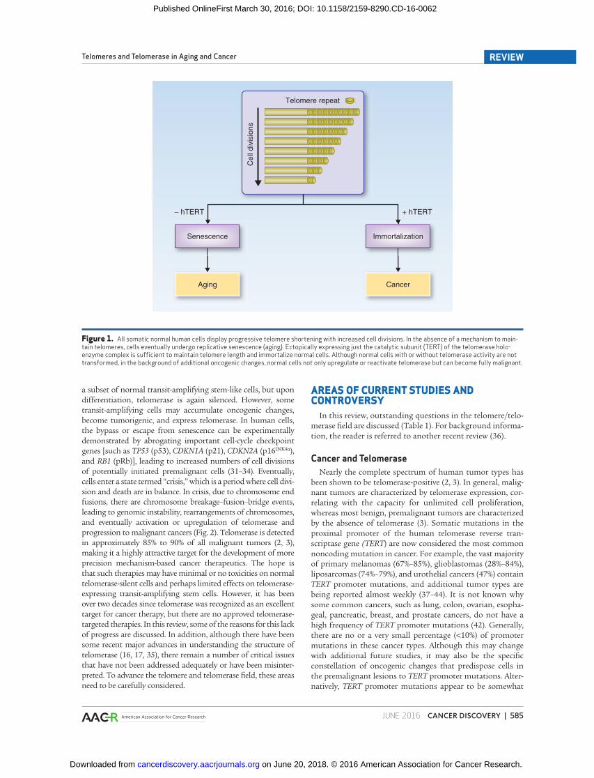
Figure 1. All somatic normal human cells display progressive telomere shortening with increased cell divisions. In the absence of a mechanism to main-tain telomeres, cells eventually undergo replicative senescence (aging). Ectopically expressing just the catalytic subunit (TERT) of the telomerase holo-enzyme complex is suffi cient to maintain telomere length and immortalize normal cells. Although normal cells with or without telomerase activity are not transformed, in the background of additional oncogenic changes, normal cells not only upregulate or reactivate telomerase but can become fully malignant.
Telomere repeat
Senescence Immortalization
Aging Cancer
+ hTERT– hTERT
Cel
l div
isio
ns
on June 20, 2018. © 2016 American Association for Cancer Research. cancerdiscovery.aacrjournals.org Downloaded from
Published OnlineFirst March 30, 2016; DOI: 10.1158/2159-8290.CD-16-0062
586 | CANCER DISCOVERY�JUNE 2016 www.aacrjournals.org
ShayREVIEW
more common in tissues that do not have a high rate of cell turnover (self-renewal; ref. 40 ), but exceptions to this, such as carcinomas of the oral cavity, already exist ( 40 ). Although it is believed that these mutations activate telomerase activity (by
converting conserved regions within the TERT promoter to an ETS transcription factor binding site) to permit the continu-ous cell divisions required for advanced cancers, much of the molecular steps about the causal relationship of promoter mutations to telomere length maintenance remain unknown. Previously, it was reported that some cancers do not have detectable telomerase activity, and these often result in sponta-neous cancer remission ( 45, 46 ). These examples demonstrate that one does not need to have telomerase activity to develop cancer, but a mechanism to maintain telomeres is required for the continuous growth of the advanced tumor ( 45–47 ). In almost all human cancers, immortalization of emergent cancer cells occurs by the reactivation or upregulation of telomerase; however, another mechanism can also reverse telomere attri-tion in order to bypass senescence, which is termed alternative lengthening of telomeres (ALT) and involves DNA recombina-tion between telomeres ( 48 ). The ALT pathway is not common in carcinomas but does appear in soft-tissue sarcomas and some other less common tumor types, but at present there are no directed therapies to the ALT pathway ( 48 ).
In addition, very little telomerase is required to maintain the shortest telomeres. Previously, it was shown that even 1% of the telomerase observed in typical advanced cancer levels is suf-fi cient to maintain the shortest telomeres ( 49 ), and that short-term ( ∼ 2 weeks) expression of telomerase in normal cells is suffi cient to double the proliferative lifespan of cells ( 50 ). These studies indicate that telomerase is recruited to the shortest tel-omeres and that very little telomerase enzyme activity is required to maintain these short telomeres. These issues have important
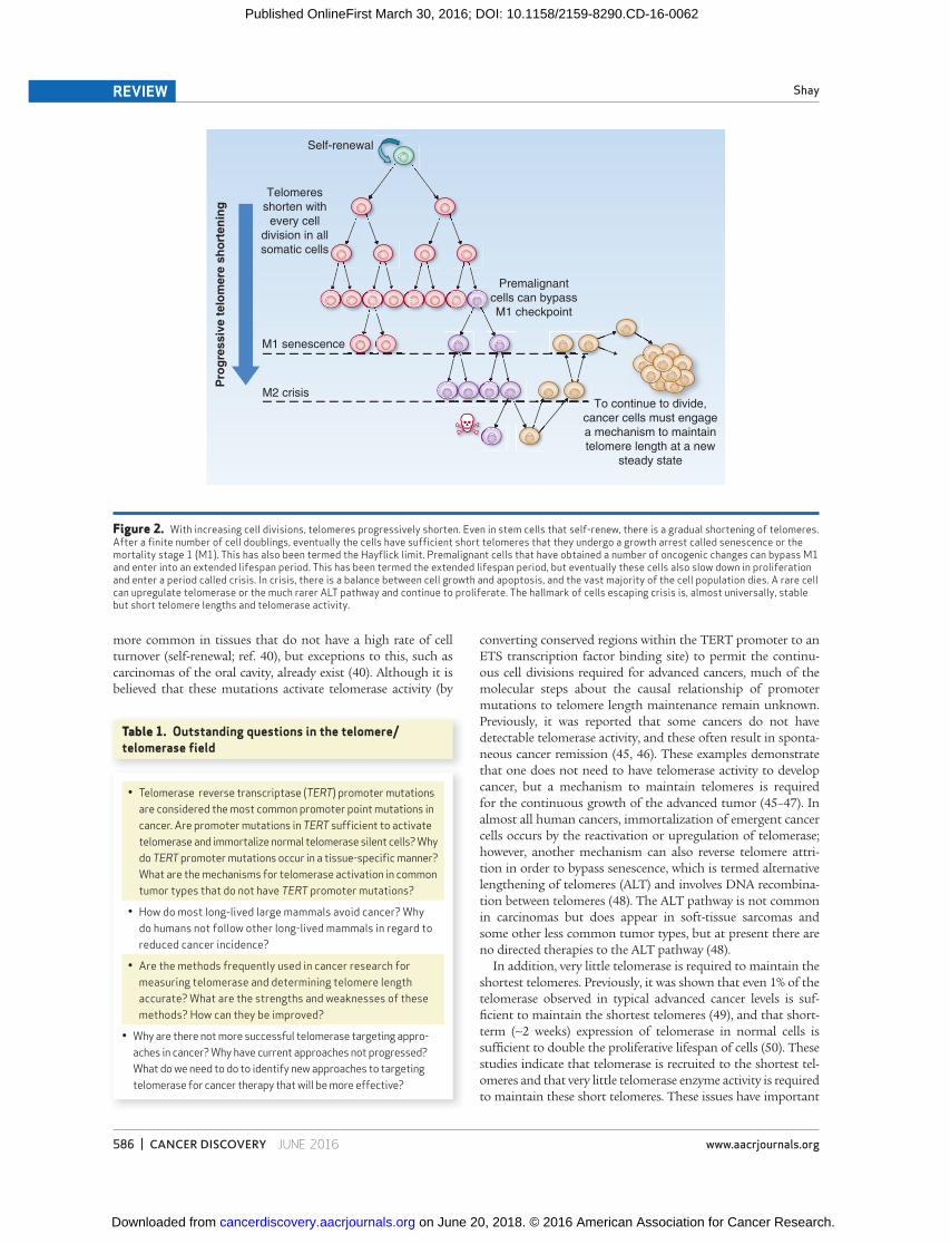
Figure 2. With increasing cell divisions, telomeres progressively shorten. Even in stem cells that self-renew, there is a gradual shortening of telomeres. After a fi nite number of cell doublings, eventually the cells have suffi cient short telomeres that they undergo a growth arrest called senescence or the mortality stage 1 (M1). This has also been termed the Hayfl ick limit. Premalignant cells that have obtained a number of oncogenic changes can bypass M1 and enter into an extended lifespan period. This has been termed the extended lifespan period, but eventually these cells also slow down in proliferation and enter a period called crisis. In crisis, there is a balance between cell growth and apoptosis, and the vast majority of the cell population dies. A rare cell can upregulate telomerase or the much rarer ALT pathway and continue to proliferate. The hallmark of cells escaping crisis is, almost universally, stable but short telomere lengths and telomerase activity.
Self-renewal
Telomeresshorten with
every celldivision in allsomatic cells
M2 crisis
Premalignantcells can bypassM1 checkpoint
To continue to divide,cancer cells must engagea mechanism to maintaintelomere length at a new
steady state
Pro
gre
ssiv
e t
elo
mere
sh
ort
en
ing
M1 senescence
Table 1. Outstanding questions in the telomere/telomerase fi eld
• Telomerase reverse transcriptase ( TERT ) promoter mutations are considered the most common promoter point mutations in cancer. Are promoter mutations in TERT suffi cient to activate telomerase and immortalize normal telomerase silent cells? Why do TERT promoter mutations occur in a tissue-specifi c manner? What are the mechanisms for telomerase activation in common tumor types that do not have TERT promoter mutations?
• How do most long-lived large mammals avoid cancer? Why do humans not follow other long-lived mammals in regard to reduced cancer incidence?
• Are the methods frequently used in cancer research for measuring telomerase and determining telomere length accurate? What are the strengths and weaknesses of these methods? How can they be improved?
• Why are there not more successful telomerase targeting appro-aches in cancer? Why have current approaches not progressed? What do we need to do to identify new approaches to targeting telomerase for cancer therapy that will be more effective?
on June 20, 2018. © 2016 American Association for Cancer Research. cancerdiscovery.aacrjournals.org Downloaded from
Published OnlineFirst March 30, 2016; DOI: 10.1158/2159-8290.CD-16-0062
JUNE 2016�CANCER DISCOVERY | 587
Telomeres and Telomerase in Aging and Cancer REVIEW
implications for the development of telomerase therapeutics (discussed in a later section). Thus, one possibility is that high levels of telomerase and signifi cant elongation of telomeres may not be required for the sustained growth of emerging malignant cells. Indeed, almost all malignant tumors have very short telom-eres. One could speculate that if telomerase was expressed at very high levels, then telomeres might elongate greatly and this could have detrimental consequences. Alternatively, if telomerase was not activated suffi ciently, then telomeres would continue to shorten with continuing cell divisions, and the cells would eventually stop dividing. Thus, there is unlikely to be a selective advantage to having more than suffi cient telomerase to work on a very small number of the shortest telomeres. In addition, there may be other mechanisms to activate telomerase, such as genomic amplifi cations, rearrangements ( 51 ), or alterations in TERT splicing ( 52 ). Finally, there is the possibility that the TERT gene may have functions independent of maintaining telomeres.
Recently, it was shown that active chromatin marks in cells with TERT promoter mutations correlate with TERT expression ( 44 ). It was reported that mutant TERT promoters exhibit the H3K4me2/3 mark of active chromatin and recruit the GABPA/B1 transcription factor, although the wild-type TERT allele retains the H3K27me3 mark of epigenetic silencing and does not recruit the GABPA/B1 transcription factor. Interestingly, TERT promoter mutations in telomerase-expressing normal human embryonic stem cells (hESC) only modestly increase telomerase activity ( 39 ). Although wild-type hESCs silence telomerase activ-ity when induced to differentiate, telomerase remains active in hESCs with TERT promoter mutations under differentiation conditions ( 39 ). Thus, monoallelic TERT promoter mutations must provide a selective advantage in specifi c tumor types, such as glioblastomas, urothelial carcinomas, and melanomas, possi-bly by retaining an active chromatin state ( 44 ), to perhaps bypass telomere-based senescence, permitting extra cell divisions for other oncogenic changes to occur. In contrast, many common solid tumor types do not have frequent TERT promoter muta-
tions, and very little is presently known about why there are such large variations in frequencies of promoter mutations or if TERT promoter mutations are suffi cient for the formation of tumors. Most, but not all, carcinomas undergo dramatic telomere short-ening prior to telomerase activation, so one possibility is that the greatly shortened telomeres also change the chromatin state in the TERT promoter (which is about 1.2 Mb from the 5p telomere) in cancers that do not contain TERT promoter muta-tions. There still remain many fundamental questions that are unresolved about telomerase in cancer:
• Are TERT promoter mutations suffi cient for cell immortal-ity in normal human cells that are silenced for telomerase activity (e.g., normal fi broblasts)?
• What is the basis of tissue specifi city for TERT promoter mutations?
• At what stage of cancer development do TERT promoter mutations activate telomerase? Does it depend on telomere length, rate of cell turnover, or other genomic rearrange-ments at the time of telomerase activation?
Role of Telomerase in Malignant Transformation Almost all preneoplastic lesions have critically shortened
telomeres, and this may be an initial protective mechanism limiting the maximum number of divisions human cells can undergo. Thus, a short telomere senescence-based mecha-nism would be a potent initial tumor suppressor mechanism because a large number of genetic and epigenetic alterations are required for a normal cell to become malignant. One can imagine, however, that limiting the maximal number of cel-lular divisions in human cells would eventually result in a preneoplastic proliferative growth arrest state referred to as replicative aging or senescence ( Fig. 3 ). Thus, senescence may have evolved as an anticancer molecular mechanism in large long-lived mammals to avoid cancer at an early age ( 53, 54 ). In cells that acquire a series of oncogenic changes, replicative
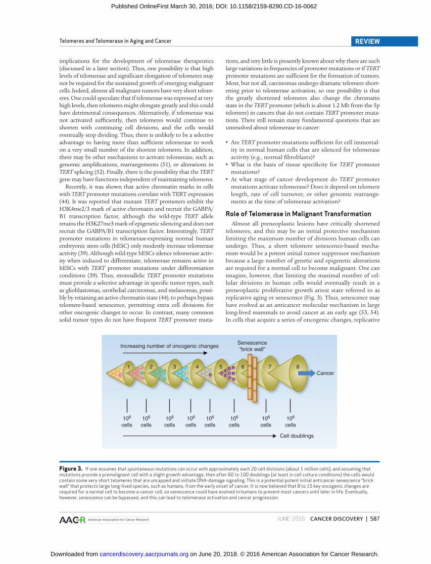
Figure 3. If one assumes that spontaneous mutations can occur with approximately each 20 cell divisions (about 1 million cells), and assuming that mutations provide a premalignant cell with a slight growth advantage, then after 60 to 100 doublings (at least in cell culture conditions) the cells would contain some very short telomeres that are uncapped and initiate DNA-damage signaling. This is a potential potent initial anticancer senescence “brick wall” that protects large long-lived species, such as humans, from the early onset of cancer. It is now believed that 8 to 15 key oncogenic changes are required for a normal cell to become a cancer cell, so senescence could have evolved in humans to prevent most cancers until later in life. Eventually, however, senescence can be bypassed, and this can lead to telomerase activation and cancer progression.
106
cells106
cells106
cells106
cells106
cells106
cells106
cells106
cells
Senescence“brick wall”
1 2 3 4 5 6 7 8Cancer
Increasing number of oncogenic changes
Cell doublings
on June 20, 2018. © 2016 American Association for Cancer Research. cancerdiscovery.aacrjournals.org Downloaded from
Published OnlineFirst March 30, 2016; DOI: 10.1158/2159-8290.CD-16-0062
588 | CANCER DISCOVERY�JUNE 2016 www.aacrjournals.org
ShayREVIEW
senescence can be bypassed, and eventually cells enter a state known as crisis ( 31, 55 ). In crisis, telomeres are so short that end–end chromosome fusions occur followed by bridge–breakage–fusion cycles and then rarely in humans ( 55 ) a cell engages a mechanism to escape from crisis. The molecular mechanisms to bypass crisis are not well understood, and in some instances a DNA recombination mechanism is engaged instead of telo merase ( 48, 56 ). In addition, it is likely that what is often being called replicative (telomere-based) senes-cence is in fact a DNA-damage response that may be due not to terminally shortened telomeres but perhaps to inadequate cell culture conditions ( 57 ).
Multiple mechanisms have been proposed for engaging telomerase activity. These include mutations/deletions in the TERT promoter ( 36–44 ), engagement of TERT alternative splicing ( 58, 59 ), TERT gene amplifi cation ( 60 ), and epigenetic changes ( 44 ). Another possibility is that the human TERT gene may autoregulate itself because it is located very close to the telomere end of chromosome 5 ( 61 ). In most large long-lived species, TERT is also close to a telomere, but in small short-lived species such as mice, TERT is not located near a telomere. Telomerase by necessity would have to be carefully regulated in large long-lived species to avoid the early onset of cancer, whereas in smaller mammals, such as mice, telomerase is known to be more promiscuous and most inbred strains of mice have very long telomeres compared with humans, but the reasons for this are not well understood. One could speculate that the TERT gene being located near a telomere in large and long-lived species may have been selected for over evolutionary time to regulate telomerase and thus the maxi-mal telomere length permitted during human development
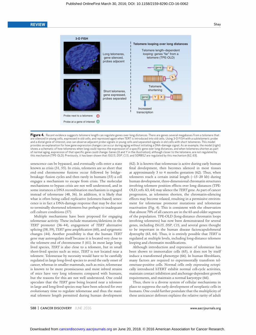
( 62 ). It is known that telomerase is active during early human fetal development, then becomes silenced in most tissues at approximately 3 to 4 months gestation ( 62 ). Thus, when telomeres reach a certain initial length ( ∼ 15–20 kb) during human development, three-dimensional chromatin structures involving telomere position effects over long distances (TPE-OLD; refs. 63, 64 ) may silence the TERT gene. As part of cancer progression, as telomeres shorten, the chromatin-silencing effects may become relaxed, resulting in a permissive environ-ment for telomerase promoter mutations and telomerase reactivation ( Fig. 4 ). This is consistent with the observation that almost 70% of all cancers are in the 65-and-older segment of the population. TPE-OLD (long-distance chromatin loops involving telomeres) has now been demonstrated for several genes, including ISG15 , DSP , C1S , and several genes thought to be important in the human disease facioscapulohmeral dystrophy ( 63, 64 ). Thus, it is entirely possible that TERT is regulated at multiple levels, including long-distance telomere looping and chromatin modifi cations.
Although introduction and expression of telomerase has been shown to immortalize cells ( 65 ), it does not by itself induce a transformed phenotype ( 66 ). In human fi broblasts, many factors are required to experimentally transform tel-omerase-positive cells. Normal cells only expressing ectopi-cally introduced hTERT exhibit normal cell-cycle activities, maintain contact inhibition and anchorage-dependent growth requirements, and maintain a normal karyotype ( 66 ).
Thus, there is a diverse system of cellular mechanisms in place to suppress the early development of neoplastic cells in humans. One could further postulate that the multiplicity of these anticancer defenses explains the relative rarity of adult
Figure 4. Recent evidence suggests telomere length can regulate genes over long distances. There are genes several megabases from a telomere that are silenced in young cells, expressed in old cells, and repressed again when TERT is introduced into old cells. Using 3-D FISH with a subtelomeric probe and a distal gene of interest, one can observe adjacent probe signals in young cells and separated signals in old cells with short telomeres. This model provides an explanation for how gene expression changes can occur during aging without initiating a DNA-damage signal. As an example, the model (right) shows a schematic of how telomeres when long could repress the expression of a specifi c gene over long distances, and when telomeres shorten as part of normal aging, expression of that specifi c gene could change. Genes (X and Y in the illustration), although closer to the telomere, are not regulated by this mechanism (TPE-OLD). Previously, it has been shown that ISG15 , DSP , C1S , and SORBS2 are regulated by this mechanism ( 62, 63 ).
Long telomeres,gene silenced,
probes adjacent
Telomere length–dependentlooping: genes “far” from a
telomere (TPE-OLD)
Increasedtranscription
gene
gene
Y
Y
X
X
Telomereshortening
Probe next to a telomere
Probe at a gene of interest
Short telomeres,gene expressed,probes separated
3-D FISH
Telomere looping over long distances
on June 20, 2018. © 2016 American Association for Cancer Research. cancerdiscovery.aacrjournals.org Downloaded from
Published OnlineFirst March 30, 2016; DOI: 10.1158/2159-8290.CD-16-0062
JUNE 2016�CANCER DISCOVERY | 589
Telomeres and Telomerase in Aging and Cancer REVIEW
human cancers in the fi rst four decades of life. Given that human cancer incidence increases with age, older individu-als, whose telomeres in somatic cells are shorter than those in younger ones, should have an increased propensity to major cancers. Although this is correlative, and certainly does not prove a cause-and-effect relationship, these fi ndings suggest that individuals with inherently short telomeres should be at increased risk for cancer. It is widely believed that short telomeres in combination with other oncogenic changes lead to genomic instability, which is typically observed in most human cancers. However, recent studies have shown that in the general population individuals with inherently long telomeres are also at a higher risk for major cancers ( 67–71 ). How do we explain this apparent paradox?
Peto’s Paradox: Why do Most Large Long-Lived Species Not Get Cancer at a Higher Frequency Compared to Small Short-Lived Species?
It is well established that most large mammals also have more cells and generally longer lifespans that require more cell replications, which theoretically should increase the mutational burden and augment cancer risk. However, Peto pointed out that cancer risk does not always scale with size ( 72 ). Large, long-living mammals show no increase in cancer risk compared with small, short-lived ones. Known as Peto’s paradox ( 73, 74 ), these fi ndings suggest a role of evolution-ary forces, part of which might be mediated through tel-omere biology. Large, long-living mammals typically repress telomerase in somatic tissues and have short telomeres compared with small, short-lifespan mammals (e.g., tel-omerase activity inversely correlates with body mass, not necessarily lifespan; refs. 75, 76 ). Repressed telomerase and short telomeres would thus diminish the maximal number of replication-mediated mutations that would occur prior to engaging telomere-based senescence. It was reported that short telomere length correlated with increased lifespan and that telomerase repression correlated with increased body size (mass) in over 50 mammalian species covering most of the mammalian radiation ( 75 ). This paradigm has led to the concept of evolutionary tradeoffs. Cancer resist-ance due to repressed telomerase and short telomeres might limit regenerative capacity, thus increasing the likelihood of age-dependent degenerative diseases, particularly as animals get older and their telomeres undergo further shortening. However, there are exceptions, such as the small long-lived mole rat ( 77 ), that show increases in tumor suppressor p15/p16 variants, decreased infl ammation, and increases in high–molecular-mass hyaluronan, perhaps infl uencing cell adhesion. In contrast to somewhat rare exceptions, the concept of shorter inherited telomere length being an anticancer protection mechanism has been experimentally tested in a large series of mammals and remains a viable explanation ( 75 ).
Thus, the overarching question is: How do large mammals reduce their risk of cancer? In two recent papers on the elephant ( 78 ) and the bowhead whale ( 79 ), there are emerging fi ndings that mechanisms to reduce cancer risk in large mammals may have evolved. For example, in the African and Asian elephant approximately 20 TP53 -related sequences (p53) are detected by DNA sequencing ( 78 ), and although some of these may be pseu-
dogenes, others produce functional protein. Thus, cancer-free longevity in the elephant may be due to acquiring extra copies of functional ancestral TP53 ( 78 ). Although p53 protein is generally thought of as a tumor suppressor pathway, it is more diffi cult to understand in evolutionary terms how these extra copies could have been selected for to protect against cancer. TP53 is also a cell stressor responsive gene, and this could possibly explain the evolutionary acquisition of extra copies of TP53 . One pos-sibility that was recently demonstrated ( 80 ) is that an ancestral function of wild-type p53, but not mutant p53, is to restrain retrotransposon mobility, and thus extra copies of wild-type p53 could serve as a tumor suppressor mechanism by reduc-ing transposable elements from moving around in the normal genome. In contrast, the bowhead whale, which lives almost 200 years and is believed to be the longest-living mammal, has ∼ 1,000 times more cells compared with humans. Similar to elephants, whales are rarely found to develop cancer. The bowhead whale genome was also recently sequenced, and the investigators proposed that increased copies or variants in DNA-damage repair genes (mutations in ERCC1 and PCNA and FEN1 duplications) may account for cancer-free longevity in whales ( 79 ).
Man versus Mouse Cancer Paradox If whales and elephants have evolved anticancer protec-
tion mechanisms, what occurs in humans? An average human weighs about 60 to 80 kg and lives about 75 to 80 years compared with inbred strains of mice that weigh about 20 to 25 grams and live approximately 2 to 3 years. Yet humans and mice get about the same incidence of cancer. For this to make sense, humans would have to be at least 100,000 times more resistant to cancer compared with mice ( 53 ). Perhaps humans have better DNA repair mechanisms, or perhaps inbred strains of mice are inap-propriate to compare with wild-type mice. In addition, inbred strains of mice have probably been inadvertently selected for fast growth, big litter sizes, and rapid matura-tion, which may have discarded slow-aging genes, including anticancer genes. Indeed, wild-type mice in captivity have been shown to live longer than inbred strains. Many wild-type mouse strains also have somewhat shorter telo meres compared with inbred strains, which generally have very long telo meres. Finally, if one deletes Tert or Terc (encoding the functional RNA template component of telomerase) from inbred strains of mice ( 81 ), telomeres do progressively shorten and, in later generations, mice develop aging phe-notypes (stem cell dysfunction, cardiomyopathies, insulin resistance, diminished stress responses, and only a modest increase in cancer) similar to humans ( 81 ). Thus, inbred strains of mice in a normal lifespan probably do not use telomere-based replicative aging as an anticancer protec-tion mechanism ( 53 ). Although this large difference in protection from cancer may be true when comparing inbred mice to humans, it is not true for humans when compared to elephants and whales. Thus, although humans may have evolutionarily evolved more effi cient DNA repair or other mechanisms to reduce cancer incidence, humans still appear to be less protected from developing can-cer when compared to other large long-lived mammalian species.
on June 20, 2018. © 2016 American Association for Cancer Research. cancerdiscovery.aacrjournals.org Downloaded from
Published OnlineFirst March 30, 2016; DOI: 10.1158/2159-8290.CD-16-0062
590 | CANCER DISCOVERY�JUNE 2016 www.aacrjournals.org
ShayREVIEW
Why Are Humans More Susceptible to Cancer Compared to Elephants and Whales?
So, one could ask, why are humans especially vulnerable to cancer? Although some anticancer mechanisms may have evolved in evolutionary terms, such as dark pigmented skin to protect against UVB-induced cancers, humans in the modern era have a reasonably large tumor incidence (some estimate close to 50% in more developed, Western societies; refs. 82, 83 ). One explanation is that humans historically died in child-birth, of accidents, infectious diseases, and/or starvation and have never had the evolutionary pressures to develop even bet-ter anticancer protection mechanisms. With the improvement in sanitation, the development of vaccines and antibiotics, safer working environments, and improved medicines and surgical procedures, humans have essentially doubled their average lifespan in the last 150 years. In addition, humans have also dramatically changed their lifestyles from our ances-tral hunter-gatherer, low-fat, and active environment, to a more sedentary, high-fat, smoking, sun-exposed, polluted environment. Some have estimated that the vast majority of human cancers are indeed associated with lifestyle factors that do not occur in other large long-lived mammals ( 83 ). Thus, because humans are living longer and most cancers occur in the 65-year-old and older segment of the population (e.g., post-reproduction), evolutionary adaptations have yet to occur in humans to the extent they have occurred in elephants and whales, even though humans have shorter telomeres and repress telomerase in somatic tissues similarly to elephants and whales. Because humans now live in a vastly different environment, it is possible that infl ammatory responses are driving human cells past senescence into an extended lifespan phase so cells have additional divisions to engage additional oncogenic changes. When cells then enter crisis, in combina-tion with other genetic and epigenetic changes, instead of engaging senescence, cells develop genomic instability and an increased risk of cancer and activation of telomerase. One way to think about this is that the rapid lifespan increases have most likely put most humans out of balance with evolution.
Are the Commonly Used Methods for Measuring Telomerase and Telomere Length Being Interpreted Correctly?
Although it is well established that the vast majority of human tumors express telomerase activity, assays for measuring this activity are varied, making comparisons between studies difficult. Telomerase can be assayed using a variety of methods, some more reliable and reproducible than others. For example, telomeric repeat amplification protocol (TRAP), which uses PCR to amplify the extension products of the telomerase enzyme, is quite sensitive and can detect as few as 0.01% positive cells ( 2, 49 ). Recently, more quantitative telomerase assays using droplet digital PCR (ddPCR) have been described ( 84 ), and ddTRAP can potentially provide more exact numbers of molecules of telomerase per cell instead of semiquantitative informa-tion using other methods. Indeed, the standard TRAP assay can vary widely in semiquantitating telomerase activ-ity levels in tumor specimens, so most studies indicating that telomerase activity levels are prognostic indicators
of outcome may be suspect. Many investigators also use mRNA for TERT as a surrogate for telomerase enzyme activity, but because there is now evidence that mRNA for TERT does exist in normal cells, caution is needed in using indirect methods for assuming enzyme activity.
There are also many methods to measure telomere length, including terminal restriction fragment (TRF) analysis ( 6 ), in situ Q-FISH ( 85, 86 ), fl ow-FISH ( 87 ), Q-PCR ( 88 ), chromo-some-specifi c single telomere length analysis (STELA; ref. 89 ), and universal STELA ( 90, 91 ). In addition, there is now whole genome sequencing (TelSeq) to estimate average or mean telomere length that is quantitative but somewhat still expen-sive compared with other methods ( 92 ). All these methods for measuring telomere lengths have their strengths and limita-tions. For example, depending on the number of restriction enzymes used for the TRF Southern blot analysis, one gets very different ranges of average telomere sizes, and no stand-ardizations in the fi eld exist. Perhaps the most popular and widely used method for determining average telomere length is the Q-PCR method, because it is quite easy to conduct and provides an average telomere length compared with a single copy gene ( 88 ). The problem in using this technique in cancer cells as opposed to normal diploid cells is the global ane-uploidy that exists in cancers, raising the very real possibility that the single copy reference gene may not be accurate, and almost nothing is mentioned about this in published studies.
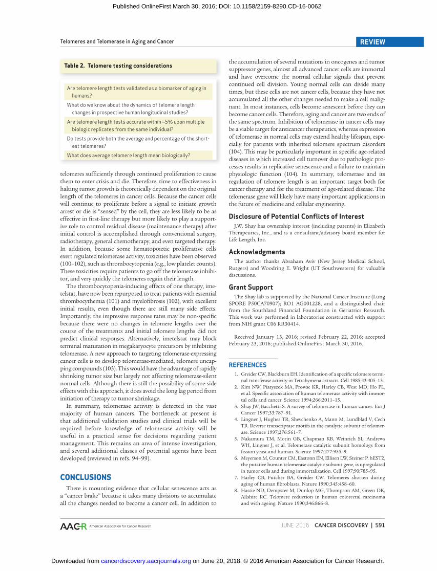
Perhaps, even more importantly, it is not certain what average telomere length actually means when it is well estab-lished that the shortest telomeres lead to senescence and genomic instability ( 93 ). Although in situ Q-FISH and fl ow-FISH can provide information about the shorter telomere lengths in normal and tumor cells, both methods rely on probe hybridization kinetics to the telomeres that may not hybridize to the very shortest telomeres. For example, signal-free ends using in situ telomere Q-FISH does not mean these chromosome ends do not have telomeric repeats. Thus, quantitation of the very shortest telomeres requires more sensitive assays. Both single-chromosome and universal STELA are methods to identify the percentage of telomeres that are the very shortest (e.g., less than 1 to 2 kb). Single-chromosome STELA is perhaps less useful in cancer because there is a great variation and losses in chromosome numbers. Universal STELA ( 90, 91 ) has recently emerged to measure the shortest telomeres on all chromosomes, but neither single-chromosome nor universal STELA are high-through-put methods, so large-scale studies would be more diffi cult. Issues to consider when conducting telomere testing for dis-ease susceptibility and aging are provided in Table 2 .
Targeting Telomerase: Therapeutic Potential Although there have been several comprehensive reviews
on the approaches being considered to inhibit telomerase in cancer ( 94–99 ), there have yet to be any antitelo merase thera-pies approved for any indication. This is certainly not from lack of trying, and some approaches have recently led to phase II clinical trials ( 100–102 ). Telomerase inhibitors remain an attractive approach to targeting cancer cells, largely because of the specifi city of the activity in tumor cells. However, a key to understanding the role for this class of agents is that the inhibi-tory effects are apparent only after the cancer cells shorten their
on June 20, 2018. © 2016 American Association for Cancer Research. cancerdiscovery.aacrjournals.org Downloaded from
Published OnlineFirst March 30, 2016; DOI: 10.1158/2159-8290.CD-16-0062
JUNE 2016�CANCER DISCOVERY | 591
Telomeres and Telomerase in Aging and Cancer REVIEW
telomeres suffi ciently through continued proliferation to cause them to enter crisis and die. Therefore, time to effectiveness in halting tumor growth is theoretically dependent on the original length of the telo meres in cancer cells. Because the cancer cells will continue to proliferate before a signal to initiate growth arrest or die is “sensed” by the cell, they are less likely to be as effective in fi rst-line therapy but more likely to play a support-ive role to control residual disease (maintenance therapy) after initial control is accomplished through conventional surgery, radiotherapy, general chemotherapy, and even targeted therapy. In addition, because some hematopoietic proliferative cells exert regulated telomerase activity, toxicities have been observed ( 100–102 ), such as thrombocytopenia (e.g., low platelet counts). These toxicities require patients to go off the telomerase inhibi-tor, and very quickly the telomeres regain their length.
The thrombocytopenia-inducing effects of one therapy, ime-telstat, have now been repurposed to treat patients with essential thrombocythemia ( 101 ) and myelofi brosis ( 102 ), with excellent initial results, even though there are still many side effects. Importantly, the impressive response rates may be non-specifi c because there were no changes in telomere lengths over the course of the treatments and initial telomere lengths did not predict clinical responses. Alternatively, imetelstat may block terminal maturation in megakaryocyte precursors by inhibiting telomerase. A new approach to targeting telomerase-expressing cancer cells is to develop telomerase-mediated, telomere uncap-ping compounds ( 103 ). This would have the advantage of rapidly shrinking tumor size but largely not affecting telomerase-silent normal cells. Although there is still the possibility of some side effects with this approach, it does avoid the long lag period from initiation of therapy to tumor shrinkage.
In summary, telomerase activity is detected in the vast majority of human cancers. The bottleneck at present is that additional validation studies and clinical trials will be required before knowledge of telomerase activity will be useful in a practical sense for decisions regarding patient management. This remains an area of intense investigation, and several additional classes of potential agents have been developed (reviewed in refs. 94–99 ).
CONCLUSIONS
There is mounting evidence that cellular senescence acts as a “cancer brake” because it takes many divisions to accumulate all the changes needed to become a cancer cell. In addition to
the accumulation of several mutations in oncogenes and tumor suppressor genes, almost all advanced cancer cells are immortal and have overcome the normal cellular signals that prevent continued cell division. Young normal cells can divide many times, but these cells are not cancer cells, because they have not accumulated all the other changes needed to make a cell malig-nant. In most instances, cells become senescent before they can become cancer cells. Therefore, aging and cancer are two ends of the same spectrum. Inhibition of telomerase in cancer cells may be a viable target for anticancer therapeutics, whereas expression of telomerase in normal cells may extend healthy lifespan, espe-cially for patients with inherited telomere spectrum disorders ( 104 ). This may be particularly important in specifi c age-related diseases in which increased cell turnover due to pathologic pro-cesses results in replicative senescence and a failure to maintain physiologic function ( 104 ). In summary, telomerase and its regulation of telomere length is an important target both for cancer therapy and for the treatment of age-related disease. The telomerase gene will likely have many important applications in the future of medicine and cellular engineering.
Disclosure of Potential Confl icts of Interest J.W. Shay has ownership interest (including patents) in Elizabeth
Therapeutics, Inc., and is a consultant/advisory board member for Life Length, Inc .
Acknowledgments The author thanks Abraham Aviv (New Jersey Medical School,
Rutgers) and Woodring E. Wright (UT Southwestern) for valuable discussions.
Grant Support The Shay lab is supported by the National Cancer Institute (Lung
SPORE P50CA70907); RO1 AG001228, and a distinguished chair from the Southland Financial Foundation in Geriatrics Research. This work was performed in laboratories constructed with support from NIH grant C06 RR30414.
Received January 13, 2016; revised February 22, 2016; accepted February 23, 2016; published OnlineFirst March 30, 2016.
REFERENCES 1. Greider CW , Blackburn EH . Identifi cation of a specifi c telomere termi-
nal transferase activity in Tetrahymena extracts . Cell 1985 ; 43 : 405 – 13 . 2. Kim NW , Piatyszek MA , Prowse KR , Harley CB , West MD , Ho PL ,
et al. Specifi c association of human telomerase activity with immor-tal cells and cancer . Science 1994 ; 266 : 2011 – 15 .
3. Shay JW , Bacchetti S . A survey of telomerase in human cancer . Eur J Cancer 1997 ; 33 : 787 – 91 .
4. Lingner J , Hughes TR , Shevchenko A , Mann M , Lundblad V , Cech TR . Reverse transcriptase motifs in the catalytic subunit of telomer-ase . Science 1997 ; 276 : 561 – 7 .
5. Nakamura TM , Morin GB , Chapman KB , Weinrich SL , Andrews WH , Lingner J , et al. Telomerase catalytic subunit homologs from fi ssion yeast and human . Science 1997 ; 277 : 955 – 9 .
6. Meyerson M , Counter CM , Eastonn EN , Ellisen LW , Steiner P . hEST2, the putative human telomerase catalytic subunit gene, is upregulated in tumor cells and during immortalization . Cell 1997 ; 90 : 785 – 95 .
7. Harley CB , Futcher BA , Greider CW . Telomeres shorten during aging of human fi broblasts . Nature 1990 ; 345 : 458 – 60 .
8. Hastie ND , Dempster M , Dunlop MG , Thompson AM , Green DK , Allshire RC . Telomere reduction in human colorectal carcinoma and with ageing . Nature 1990 ; 346 : 866 – 8 .
Table 2. Telomere testing considerations
Are telomere length tests validated as a biomarker of aging in humans?
What do we know about the dynamics of telomere length changes in prospective human longitudinal studies?
Are telomere length tests accurate within ∼ 5% upon multiple biologic replicates from the same individual?
Do tests provide both the average and percentage of the short-est telomeres?
What does average telomere length mean biologically?
on June 20, 2018. © 2016 American Association for Cancer Research. cancerdiscovery.aacrjournals.org Downloaded from
Published OnlineFirst March 30, 2016; DOI: 10.1158/2159-8290.CD-16-0062
592 | CANCER DISCOVERY�JUNE 2016 www.aacrjournals.org
ShayREVIEW
9. Lindsey J , McGill NI , Lindsey LA , Green DK , Cooke HJ . In vivo loss of telomeric repeats with age in humans . Mut Res 1991 ; 256 : 45 – 8 .
10. de Lange T , Shiue L , Myers RM , Cox DR , Naylor SL , Killery AM, et al. Structure and variability of human chromosome ends . Mol Cell Biol 1990 ; 10 : 518 – 27 .
11 Shay JW , Wright WE . The reactivation of telomerase activity in can-cer progression . Trends Genet 1996 ; 12 : 129 – 31 .
12. Kim N-W , Harley CB , Prowse KR , Weinrich SL , Piatyszek MA , Wright WE , et al. Telomeres, telomerase and cancer . Science 1995 ; 268 : 1115 – 7 .
13. Wright WE , Pereira-Smith OM , Shay JW . Reversible cellular senes-cence: A two-stage model for the immortalization of normal human diploid fi broblasts . Mol Cell Biol 1989 ; 9 : 3088 – 92 .
14. Counter CM. Telomere shortening associated with chromosome instability is arrested in immortal cells which express telomerase activity . EMBO J 1992 ; 11 : 1921 – 9 .
15. Blackburn E . Telomerases . Annual Rev Biochem 1992 ; 61 : 113 – 29 . 16. Jiang J , Chan H , Cash DD , Miracco EJ , Ogorzalek RR , Loo HE , et al.
Structure of Tetrahymena telomerase reveals previously unknown subunits, functions and interactions . Science 2015 ; 350 : 529 – 34 .
17. Wu RA , Dagdas YS , Yilmaz ST , Yildiz A , Collins K . Single-molecule imaging of telomerase reverse transcriptase in human telomerase holoenzyme and minimal RNP complexes . eLife 2015 ; 4 : e08363 .
18. Muller HJ. The remaking of chromosomes . In: Studies of genetics: The selected papers of HJ Muller , Indiana University Press, Bloom-ington 1962 ; 384 – 408 .
19. McClintock B. The stability of broken ends of chromosomes in Zea mays . Genetics 1941 ; 26 : 234 – 82 .
20. Blackburn EH , Gall JG . A tandemly repeated sequence at the termini of the extrachromosomal ribosomal RNA genes in Tetrahymena . J Mol Biol 1978 ; 120 : 33 – 53 .
21. Moyzis RK , Buckingham JM , Cram LS , Dani M , Deaven LL , Jones MD , et al. A highly conserved repetitive DNA sequence (TTAGGG)n, present at the telomeres of human chromosomes . Proc Natl Acad Sci U S A 1988 ; 85 : 6622 – 6 .
22. de Lange T . Shelterin: the protein complex that shapes and safe-guards human telomeres . Genes Dev 2005 ; 19 : 2100 – 10 .
23. Palm W , de Lange T . How shelterin protects mammalian telomeres . Ann Rev Genet 2008 ; 42 : 301 – 34 .
24. Griffi th JD , Comeau L , Rosenfi eld S , Standel RM , Bianchi A , Moss H , et al. Mammalian telomeres end in a large duplex loop . Cell 1999 ; 97 : 503 – 14 .
25. Watson JD . Origin of concatemeric T7 DNA . Nature, New Biol 1972 ; 239 : 197 – 201 .
26. Olovnikov AM. A theory of margintomy: The incomplete copying of template margin in enzymes synthesis of polynucleotides and bio-logical signifi cance of the problem . J Theoret Biol 1973 ; 41 : 181 – 90 .
27. Zou Y , Sfeir A , Gryaznov SM , Shay JW , Wright WE . Does a sentinel or groups of short telomere determine replicative senescence? Mol Biol Cell 2004 ; 15 : 3709 – 18 .
28. Fumagalli M , Rossiello F , Clerici M , Barozzi S , Cittaro D , Kaplunov JM , et al. DNA damage is irreparable and causes persistent DNA-damage-response activation . Nat Cell Bio 2012 ; 14 : 355 – 65 .
29. Von Zglinicki T , Saretzki G , Ladhoff J , d’Adda di Fagagna F , Jackson SP . Human cell senescence as a DNA damage responses . Mechan Ageing Devel 2005 ; 26 : 111 – 7 .
30. Tchkonia T , Zhu Y , van Deursen J , Campisi J , Kirkland JL . Cellu-lar senescence and the senescent secretory phenotype: therapeutic opportunities . J Clin Invest 2013 ; 123 : 966 – 72 .
31. Shay JW , Wright WE . Historical claims and current interpretations of replicative aging . Nat Biotech 2002 ; 20 : 682 – 8 .
32. Wright WE , Shay JW . The two-stage mechanism controlling cellular senescence and immortalization . Exp Gerontol 1992 ; 27 ; 383 – 9 .
33. Shay JW , Wright WE , Werbin H . Defi ning the molecular mechanism of human cell immortalization . Biochim Biophys Acta . 1991 ; 1072 : 1 – 7 .
34. Harley CB. Telomere loss: mitotic clock or genetic time bomb? Mut Res 1991 ; 256 : 271 – 82 .
35. Sandin S , Rhodes D . Telomerase Structure . Curr Opin Struct Biol 2014 ; 25 : 104 – 10 .
36. Blackburn EH , Epel ES , Lin J . Human telomere biology: A contribu-tory and interactive factor in aging, disease risks, and protection . Science . 2015 : 350 ; 1193 – 8 .
37. Bojesen SE , Pooley KA , Johnatty SE , Beesley J , Michailidou K , Tyrer JP , et al. Multiple independent variants at the TERT locus are asso-ciated with telomere length and risks of breast and ovarian cancer . Nat Genet 2013 ; 45 ; 371 – 384 .
38. Heidenreich B , Rachakonda PS , Hemminki K , Kumar R . TERT promoter mutations in cancer development . Current Opin Genet Develop 2014 ; 24 : 30 – 7 .
39. Huang FW , Hodis E , Xu MJ , Kryukov GV , Chin L , Garraway LA . Highly recurrent TERT promoter mutations in human melanoma . Science 2013 ; 339 : 957 – 9 .
40. Chiba K , Johnson JZ , Vogan JM , Wagner T , Boyle JM , Hockemeyer D . Cancer-associated TERT promoter mutations abrogate telomer-ase silencing . eLife 2015 ; 4e07918 .
41. Killela PJ , Reitman ZJ , Jiao Y , Bettegowda C , Agrawal N , Diaz LA Jr , et al. TERT promoter mutations occur frequently in gliomas and a subset of tumors derived from cells with low rates of self-renewal . Proc Natl Acad Sci U S A 2013 ; 110 : 6021 – 6 .
42. Vinagre J , Almeida A , Pópulo H , Batista R , Lyra J , Pinto V , et al. Frequency of TERT promoter mutations in human cancers . Nature Comm 2013 ; 4 : 2185 .
43. Horn S , Figl A , Rachakonda PS , Fischer C , Sucker A , Gast A , et al. TERT promoter mutations in familial and sporadic melanoma . Science 2013 ; 339 : 959 – 61 .
44. Stern JL , Theodorescu D , Vogelstein B , Papadopoulos N , Cech TR . Mutation of the TERT promoter switch to active chromatin, and monoallelic TERT expression in multiple cancers. Genes Dev 2015 ; 29 : 1 – 6 .
45. Hiyama E , Hiyama K , Yokoyama T , Matsuura Y , Piatyszek MA , Shay JW . Correlating telomerase activity levels with human neuroblas-toma outcomes . Nature Med 1995 ; 1 : 249 – 57 .
46. Tabori U , Vukovic B , Zielenska M , Hawkins C , Braude I , Rutka J , et al. The role of telomere maintenance in the spontaneous growth arrest of pediatric low-grade gliomas . Neoplasia 2006 ; 8 : 136 – 42 .
47. Hiyama E , Hiyman K , Ohtsu K , Yamaoka H , Ichikawa T , Shay JW , et al. Telomerase activity in neuroblastoma: Is it a prognostic indica-tor of clinical behavior? Eur J Cancer 1997 ; 33 : 1932 – 36 .
48. Shay JW , Reddel RR , Wright WE . Cancer and telomerase: An ALTer-native to telomerase . Science 2012 ; 336 : 1388 – 1390 .
49. Ouellette MM , Liao M , Herbert B-S , Johnson M , Holt SE , Liss HS , et al. Senescent telomere lengths in fi broblasts immortalized by limiting amounts of telomerase . J Biol Chem 2000 ; 275 : 10072 – 6 .
50. Steinert S , Shay JW , Wright WE . Transient expression of human tel-omerase extends the lifespan of normal human fi broblasts . Biochem Biophys Res Commun 2000 ; 273 : 1095 – 8 .
51. Peifer M , Hertwig F , Roel F , Dreidax D , Gartlgruber M , Menon R , et al. Telomerase activation by genomic rearrangements in high-risk neuroblastoma. Nature 2015 ; 526 : 700 – 4.
52. Wong MS , Wright WE , Shay JW . Alternative splicing regulation of telomerase: a new paradigm? Trends Genet 2015 ; 30 ; 10 : 430 – 8 .
53. Shay JW , Wright WE . When do telomeres matter? Science 2001 ; 291 : 839 – 40 .
54. Wright WE , Shay JW . Cellular senescence as a tumor-protection mechanism: the essential role of counting . Curr Opin Genet Develop 2001 ; 11 : 98 – 103 .
55. Shay JW , Wright WE . Quantitation of the frequency of immortaliza-tion of normal diploid fi broblasts by SV40 large T-antigen . Exp Cell Res 1989 ; 184 : 109 – 18 .
56. Cesare AJ , Reddel RR . Alternative lengthening of telomeres: models, mechanisms and implications . Nat Rev Genet 2010 ; 11 : 319 – 30 .
57. Ramirez R , Morales CP , Herbert B-S , Rhode JM , Passon C , Shay JW , et al. Putative telomere-independent mechanisms of replicative aging refl ect inadequate growth conditions . Gen Develop 2001 ; 15 : 398 – 403 .
58. Wong MS , Chen L , Foster C , Kainthla R , Shay JW , Wright WE . Regu-lation of hTERT (telomerase) alternative splicing: a new target for chemotherapy . Cell Rep 2013 ; 3 : 1028 – 35 .
on June 20, 2018. © 2016 American Association for Cancer Research. cancerdiscovery.aacrjournals.org Downloaded from
Published OnlineFirst March 30, 2016; DOI: 10.1158/2159-8290.CD-16-0062
JUNE 2016�CANCER DISCOVERY | 593
Telomeres and Telomerase in Aging and Cancer REVIEW
59. Wong MS , Shay JW , Wright WE . Regulation of human telomerase splicing by RNA:RNA pairing . Nature Commun 2014 ; 5 : 3306 .
60. Cao Y , Bryan TM , Reddel RR . Increased copy number of the TERT and TERC telomerase subunit genes in cancer cells . Cancer Sci 2008 ; 99 : 1092 – 9 .
61. Shay JW , Wright WE . Implications of mapping the human telom-erase genes (hTERT) as the most distal gene on chromosome 5p . Neoplasia 2000 ; 2 : 195 – 6 .
62. Wright WE , Piatyszek MA , Rainey WE , Byrd W , Shay JW . Telomerase activity in human germline and embryonic tissues and cells . Dev Genet 1996 ; 18 : 173 – 9 .
63. Robin JD , Ludlow AT , Chen M , Magdinier F , Batten K , Holohan B , et al. Telomere looping: a new paradigm for the regulation of gene expression with progressive telomere shortening . Genes Dev 2014 ; 28 : 2464 – 76 .
64. Robin JD , Ludlow AT , Batten K , Gaillard M-C , Stadler G , Magdinier F , et al. SORBS2 transcription is activated by telomere position effect-over long distance upon telomere shortening in muscle cells from patients with fasciscapulohumeral dystrophy . Genome Res 2015 ; 25 : 1781 – 90 .
65. Bodnar AG , Ouellette M , Frolkis M , Holt SE , Chiu CP , Morin GB , et al. Extension of life-span by introduction of telomerase into nor-mal human cells . Science 1998 ; 279 : 349 – 52 .
66. Morales CP , Holt SE , Ouellette M , Kaur KJ , Wilson KS , White MA , et al. Absence of cancer-associated changes in human fi broblasts immortalized with telomerase . Nat Genet 1999 ; 21 : 115 – 8 .
67. Julin B , Shui I , Heaphy CM , Joshu CE , Meeker AK , Giovannucci E , et al. Circulating leukocyte telomere length and risk of overall and aggressive prostate cancer . Br J Cancer 2015 ; 112 : 769 – 76 .
68. Nan H , Du M , De Vivo I , Manson JE , Liu S , McTiernan A , et al. Shorter telomeres associate with a reduced risk of melanoma devel-opment . Cancer Res 2011 ; 71 : 6758 – 63 .
69. Lynch SM , Major JM , Cawthon R , Weinstein SJ , Virtamo J , Lan Q , et al. A prospective analysis of telomere length and pancreatic cancer in the alpha-tocopherol beta-carotene cancer (ATBC) prevention study . Int J Cancer 2013 ; 133 : 2672 – 80 .
70. Qu S , Wen W , Shu XO , Chow WH , Xiang YB , Wu J , et al. Association of leukocyte telomere length with breast cancer risk: nested case-control fi ndings from the Shanghai Women’s Health Study . Am J Epidemiol 2013 ; 177 : 617 – 24 .
71. Seow WJ , Cawthon RM , Purdue MP , Hu W , Gao YT , Huang WY , et al. Telomere length in white blood cell DNA and lung cancer: a pooled analysis of three prospective cohorts . Cancer Res 2014 ; 74 : 4090 – 8 .
72. Peto R. Quantitative implications of the approximate irrelevance of mammalian body size and lifespan to lifelong cancer risk . Philos Trans R Soc Lond B Biol Sci 2015 ; 370 : 2015.0198 .
73. Schiffman J , Maley CC , Nunney L , Hochberg M , Breen M , eds. Cancer across life: Peto’s paradox and the promise of comparative oncology . Philos Trans R Soc Lond B Biol Sci. (theme issue) 2015 ; 370 :1673.
74. Caulin AF , Maley CC . Peto’s paradox: evolution’s prescription for cancer prevention . Trends Ecol Evol 2011 ; 26 : 175 – 82 .
75. Gomes NMV , Ryder OA , Houck ML , Charter SJ , Walker W , Forsyth NR , et al. The comparative biology of mammalian telomeres: ances-tral states and functional transitions . Aging Cell 2011 ; 10 : 761 – 8 .
76. Seluanov A , Chen Z , Hine C , Sasahara TH , Ribeiro AA , Catania KC , et al. Telomerase activity coevolves with body mass not lifespan . Aging Cell 2007 ; 6 : 45 – 52 .
77. Gorbunova V , Seluanov A , Zhang Z , Gladyshev VN , Vijg J . Com-parative genetics of longevity and cancer: insights from long-lived rodents . Nat Rev Genet 2014 ; 15 : 531 – 40 .
78. Abegglen LM , Caulin AF , Chan A , Lee K , Robinson R , Campbell MS , et al. Potential mechanisms for cancer resistance in elephants and comparative cellular response to DNA damage in humans . JAMA 2015 ; 314 : 1850 – 1860 .
79. Keane M , Semeiks J , Webb AE , Li YI , Quesada V , Craig T , et al. Insights into the evolution of longevity from the bowhead whale genome . Cell Rep 2015 ; 10 : 112 – 22 .
80. Wylie A , Jones AE , D’Brot A , Lu W-J , Kurtz P , Moran JV , et al. p53 genes function to restrain mobile elements . Genes Dev 2015 ; 30 : 1 – 14 .
81. Sahin E , DePinho RA . Linking functional decline of telomeres, mitochondria and stem cells during ageing . Nature 2010 ; 464 : 520 – 8 .
82. Greaves M. Evolutionary determinants of cancer . Cancer Discov 2015 ; 5 : 806 – 20 .
83. Greaves M , Ermini L. Evolutionary adaptation to risk of cancer: evi-dence from cancer resistance in elephants . JAMA 2015 ; 314 : 1806 – 7 .
84. Ludlow AT , Robin JD , Sayed M , Litterst CM , Shelton DN , Shay JW , et al. Quantitative telomerase enzyme activity determination using droplet digital PCR with single cell resolution . Nucleic Acids Res , 2014 ; 42 : e104 .
85. Meeker AK , Gage WR , Hicks JL , Simon I , Coffman JR , Platz EA, et al. Telomere length assessment in human archival tissues: combined telomere fl uorescence in situ hybridization and immu-nostaining . Am J Pathol 2002 ; 160 : 1259 – 68 .
86. Canela A , Vera E , Klatt P , Blasco MA . High-throughput telomere length quantifi cation by FISH and its application to human popula-tions studies . Proc Natl Acd Sci U S A 2006 ; 104 : 5300 – 5 .
87. Rufer N , Dragowska W , Thornbury G , Roosnek E , Lansdorp PM . Telomere length dynamics in human lymphocytes subpopulations measured by fl ow cytometry . Nature Biotech 1998 ; 16 : 743 – 7 .
88. Cawthon RM . Telomere measurement by quantitative PCR . Nucleic Acids Res 2002 ; 30 : e47 .
89. Baird DM , Rowson J , Wynford-Thomas D , Kipling D . Extensive allelic variation and ultrashort telomeres in senescent human cells . Nature Genet 2003 ; 33 : 203 – 7 .
90. Bendix L , Horn PB , Jensen UB , Rubelj I , Kolvraa S . The load of short telomeres, estimated by a new method, Universal STELA, correlates with number of senescent cells . Aging Cell 2010 ; 9 : 383 – 97 .
91. Holohan B , Hagiopian MM , Lai TP , Huang E , Friedman DR , Wright WE, et al. Perifosine as a potential novel anti-telomerase therapy . Oncotarget 2015 .
92. Ding Z , Mangino M , Aviv A , Spector T , Durbin R , UK10K Consor-tium . Estimating telomere length from whole genome sequence data . Nucl Acids Res 2014 ; 42 : e75 .
93. Hemann MT , Strong MA , Hao LY , Greider CW . The shortest tel-omere, not average telomere length, is critical for cell viability and chromosome stability . Cell 2001 ; 107 : 67 – 77 .
94. Artandi SE , DePinho RA . Telomeres and telomerase in cancer . Carcinogenesis 2009 ; 31 : 9 – 18 .
95. Buseman CM , Wright WE , Shay JW . Is telomerase a viable target in cancer? Mutat Res 2012 ; 730 : 90 – 7 .
96. White LK , Wright WE , Shay JW . Telomerase inhibitors . Trends Bio-technol 2001 ; 19 : 114 – 20 .
97. Harley CB. Telomerase and cancer therapeutics . Nat Rev Cancer 2008 ; 8 : 167 – 79 .
98. Autexier C , Greider CW . Telomerase and cancer: revisiting the tel-omere hypothesis . Trend Bioch Sci 1996 ; 21 : 387 – 91 .
99. Agrawal A , Dang S , Gabrani R . Recent patents on anti-telomerase cancer therapy . Recent Pat Anticancer Drug Discov 2012 ; 7 : 102 – 17 .
100. Chiappori AA , Kolevska T , Spigel DR , Hager S , Rarick M , Gadgeel S , et al. A randomized phase II study of the telomerase inhibitor imetelstat as maintenance therapy for advanced non-small-cell lung cancer . Annal Oncol 2015 ; 26 : 354 – 62 .
101. Baerlocker GM , Leibundgut EO , Ottmann OG , Spitzer G , Odenike O , McDivitt MA , et al. Telomerase inhibitor imetelstat in patients with essential thrombocythemia . New Engl J Med 2015 ; 373 : 920 – 8 .
102. Tefferi A , Lasho TL , Begna KH , Patnaik MM , Zblewski DL , Finke CM , et al. A pilot study of the telomerase inhibitor imetelstat for myelofi brosis . New Engl J Med 2015 ; 373 : 908 – 19 .
103. Mender I , Gryaznov S , Dikmen ZG , Wright WE , Shay JW . Induc-tion of telomere dysfunction mediated by the telomerase sub-strate precursor, 6-thio-2-deoxyguanosine . Cancer Discov 2015 ; 5 : 82 – 95 .
104. Holohan B , Wright WE , Shay JW . Impaired telomere maintenance spectrum disorders . J Cell Biology 2014 ; 205 : 289 – 99 .
on June 20, 2018. © 2016 American Association for Cancer Research. cancerdiscovery.aacrjournals.org Downloaded from
Published OnlineFirst March 30, 2016; DOI: 10.1158/2159-8290.CD-16-0062
2016;6:584-593. Published OnlineFirst March 30, 2016.Cancer Discov Jerry W. Shay Role of Telomeres and Telomerase in Aging and Cancer
Updated version
10.1158/2159-8290.CD-16-0062doi:
Access the most recent version of this article at:
Cited articles
http://cancerdiscovery.aacrjournals.org/content/6/6/584.full#ref-list-1
This article cites 100 articles, 27 of which you can access for free at:
Citing articles
http://cancerdiscovery.aacrjournals.org/content/6/6/584.full#related-urls
This article has been cited by 15 HighWire-hosted articles. Access the articles at:
E-mail alerts related to this article or journal.Sign up to receive free email-alerts
Subscriptions
Reprints and
To order reprints of this article or to subscribe to the journal, contact the AACR Publications Department at
Permissions
Rightslink site. Click on "Request Permissions" which will take you to the Copyright Clearance Center's (CCC)
.http://cancerdiscovery.aacrjournals.org/content/6/6/584To request permission to re-use all or part of this article, use this link
on June 20, 2018. © 2016 American Association for Cancer Research. cancerdiscovery.aacrjournals.org Downloaded from
Published OnlineFirst March 30, 2016; DOI: 10.1158/2159-8290.CD-16-0062