role of km23-1 in rhoa/actin-based cell migration
TRANSCRIPT

Biochemical and Biophysical Research Communications 428 (2012) 333–338
Contents lists available at SciVerse ScienceDirect
Biochemical and Biophysical Research Communications
journal homepage: www.elsevier .com/locate /ybbrc
Role of km23-1 in RhoA/actin-based cell migration
Qunyan Jin a,1, Nageswara R. Pulipati a,1, Weidong Zhou b,1, Cory M. Staub a, Lance A. Liotta b,Kathleen M. Mulder a,⇑a Department of Biochemistry and Molecular Biology, Penn State Hershey College of Medicine, PA 17033, United Statesb Center for Applied Proteomics and Molecular Medicine, George Mason University, Manassas, VA 20110, United States
a r t i c l e i n f o
Article history:Received 6 October 2012Available online 15 October 2012
Keywords:km23-1Cell migrationActinRhoDynein
0006-291X/$ - see front matter � 2012 Elsevier Inc. Ahttp://dx.doi.org/10.1016/j.bbrc.2012.10.047
⇑ Corresponding author. Address: Department ofBiology-MC H171, Penn State Hershey College of MeHershey, PA 17033, United States. Fax: +1 717 531 09
E-mail address: [email protected] (K.M. Mulder).1 These authors contributed equally.
a b s t r a c t
km23-1 was originally identified as a TGFß receptor-interacting protein that plays an important role inTGFß signaling. Moreover, km23-1 is actually part of an ancient superfamily of NTPase-regulatory pro-teins, widely represented in archaea and bacteria. To further elucidate the function of km23-1, we iden-tified novel protein interacting partners for km23-1 by using tandem affinity purification (TAP) andtandem mass spectrometry (MS). Here we show that km23-1 interacted with a class of proteins involvedin actin-based cell motility and modulation of the actin cytoskeleton. We further showed that km23-1modulates the formation of a highly organized stress fiber network. More significantly, we demonstratedthat knockdown (KD) of km23-1 decreased RhoA activation in Mv1Lu epithelial cells. Finally, our resultsdemonstrated for the first time that depletion of km23-1 inhibited cell migration of human colon carci-noma cells (HCCCs) in wound-healing assays. Overall, our findings demonstrate that km23-1 regulatesRhoA and motility-associated actin modulating proteins, suggesting that km23-1 may represent a noveltarget for anti-metastatic therapy.
� 2012 Elsevier Inc. All rights reserved.
1. Introduction
Cell migration is an important aspect of the tumor metastaticprocess that transforms tumor cells from local, noninvasive, con-fined cells to the migrating, metastatic cancer cells [1]. Cell migra-tion is a highly integrated multistep process that is initiated by theprotrusion of the cell membrane [2,3]. Protrusive structuresformed by migrating and invading cells are termed filopodia,lamellipodia, and invadopodia/podosomes, dependent on theirmorphological, structural, and functional characteristics [3,4]. For-mation of these protrusive structures is driven by spatially- andtemporally-regulated actin polymerization at the leading edge [4].
To date, several important proteins that regulate reorganizationof the actin cytoskeleton have been identified and found to beoverexpressed in several types of cancers [3]. For example, theWiskott–Aldrich syndrome protein (WASP) family/actin-relatedprotein 2 and 3 (Arp2/3) complex, LIM-kinase (LIMK)/cofilin, andcortactin pathways have been studied extensively due to their rec-ognized importance in cell migration and invasion [3]. In addition,the Rho family of GTPase proteins Rho, Rac, Cdc42 are well-estab-
ll rights reserved.
Biochemistry and Moleculardicine, 500 University Drive,39.
lished regulators of cell migration, and have been implicated in theprocess of tumor cell invasion and metastasis [5,6]. In particular,RhoA activity plays a key role in protrusion in addition to its pre-viously reported role in cell retraction during cell motility [7,8].For example, disruption of RhoA activity has been shown to leadto invasive properties in human pancreatic cancer [7]. As a result,there has been considerable interest in the possibility that specificproteins in the signal transduction pathways mediating these cyto-skeletal events could be potential targets for cancer therapy [9].
km23-1 (also referred as km23 [10–12], Robl1 [13], DNLC2A [14],mLC7-1 [15], DYNLRB1 [16]) was originally identified as a TGFßreceptor-interacting protein that is also a dynein light chain [15].More recently, we have shown that Ras and km23-1 form a TGFß-regulated complex in vivo [17]. However, km23-1 is actually partof an ancient superfamily, widely represented in archaea and bacte-ria, which appears to be involved in regulating NTPase activity [18].One of the members of this superfamily (MglB) displays consider-able structural correspondence with km23-1. Further, in bacteria,MglB interacts with a Ras-like small G protein, MglA [19–21], andplays a critical role in the spatial control of directed motility [21].By analogy to the bacterial model, km23-1 would be expected toregulate the activity and biological functions of Ras family membersof the Rho type, in the control of actin-based cell motility.
In the current report, using TAP and MS methods, we haveidentified km23-1-binding proteins that are involved in actin-based cell motility and modulation of the actin cytoskeleton.
334 Q. Jin et al. / Biochemical and Biophysical Research Communications 428 (2012) 333–338
Further, we have demonstrated a role for km23-1 in RhoA/actin-based cell migration.
2. Materials and methods
2.1. Reagents
The pGEX2T-RBD plasmid (15247) was from Addgene. The piL-enti km23-1 siRNA-GFP (i006555) and piLenti NC siRNA-GFP vec-tors were from Applied Biological Materials Inc. (Canada).InterPlay TAP purification kit was from Agilent technologies (LaJolla, CA, USA). Anti-dynein intermediate chain (DIC) (MAB 1685)was from Chemicon (Temecula, CA). Anti-Flag (M2) mAb was formSigma. Anti-SBP and -CBP epitope tag antibodies (Abs) (07-482)were from Upstate/Millipore. Anti-actin mAb was from Sigma.Anti-RhoA (ARH03-A) mAb was from Cytoskeleton. GST-RBD plas-mid (15247) was from Addgene.
2.2. Construction of expression vectors
The full-length coding region of human km23-1 was digestedfrom the pcDNA 3.1-km23-1 vector by BamH1 and Xho1 and wascloned into the pCTAP expression vector (Stratagene, La Jolla, CA,USA).
2.3. Cell culture
293T cells were grown in Dulbecco’s modified Eagle’s medium(DMEM) supplemented with 10% fetal bovine serum (FBS) at37 �C, 5% CO2. Madin Darby canine kidney (MDCK) cells (CCL-34)were grown in minimal essential medium-a, supplemented with10% FBS. HCT116 cells were cultured in McCoy’s 5A, supplementedwith amino acids, pyruvate, and antibiotics (streptomycin, penicil-lin) with 10% FBS as described previously [22]. Cells were routinelyscreened for mycoplasma using Hoechst 33258 staining.
2.4. TAP and Western blot analyses
Protein complexes were purified by the InterPlay TAP purifica-tion kit (Agilent technologies La Jolla, CA, USA) according to themanufacturer’s instructions. Proteins were separated on 5–20%SDS–PAGE and visualized by Sypro Ruby Red staining (Invitrogen)according to the manufacturer’s instructions. For Westerns, theTAP purified samples were resolved by 4–15% Bis-Tris SDS–PAGEgels and transferred to a PVDF membrane.
2.5. In-gel digestion, MS, and MS data analysis
The Sypro Ruby stained bands were excised from the gel and in-gel digested with trypsin. The proteins in the gel bands were iden-tified by liquid chromatography (LC) tandem MS [23].
Stable transfections were performed as described previously[15] except that MDCK cells were used. Expression of km23-1was verified by Western blot analysis, and stably transfected poolsof km23-1-FLAG or (empty vector) EV were used for actinimmunostaining.
2.6. Immunofluorescence microscopy analyses
Stably transfected pools of km23-1-FLAG or EV cells weregrown in 6-well plates on top of coverslips. Immunofluorescencestaining was performed as described previously [24]. Subse-quently, these cells were incubated with an anti-actin Ab for 1 h.The bound primary Abs were visualized with FITC-conjugated goat
anti-mouse IgG (green). Images were collected by fluorescentmicroscopy (Phase contrast-2, Diaphot, Nikon, Japan).
2.7. RhoA GTPase activation assays
The amount of activated, GTP-bound RhoA protein was mea-sured using a technique similar to the method described [25].
2.8. Preparation of viruses/shRNA production and cell infection
Lentiviruses were generated by transfecting appropriate piLentikm23-1 shRNA green fluorescent protein (GFP) constructs withpackaging plasmids into 293T cells using lipofectamine 2000 trans-fection reagent according to the manufacturer’s protocol. Viralsupernatants were harvested 48 h after transfection and were fil-tered through a 0.45-mm filter. The virus was subsequently ali-quoted (100 ll) and supernatants were stored at �80 �C. HCT116cells were infected with lentivirus in the presence of 8 lg/mlpolybrene.
2.9. Wound-healing assays
HCT116 cells were plated in 12-well plates and were infectedwith piLenti NC siRNA-GFP and piLenti km23-1 siRNA-GFP accord-ing to the manufacturer’s instructions. 24 h after infection, cellswere grown to confluence: Wound-healing assays were then per-formed as described previously [26].
The statistical significance was determined using the Student’st-test. The results are expressed as the mean ± SE.
3. Results
To validate the TAP protocol employed using our system, wepurified the km23-1 protein complexes from 293T cells over-expressing km23-1-TAP. DIC was used as a positive control fordetection of known km23-1-binding partners, since it is knownto bind km23-1 [15]. As shown in Fig. 1A, in the EV-transfectedcells, no band was detected (lane 1, 1st and 2nd panels) as ex-pected. However, in the km23-1-TAP-transfected cells, DIC was de-tected after both the SBP pulldown (lane 2, 1st panel) and the SBP/CBP tandem pulldown (lane 2, 2nd panel). The expression of km23-1-TAP was confirmed by Western blot analyses (lane 2, 3rd panel),and GAPDH was used as a loading control (bottom panel). Our re-sults demonstrate that the two-step tandem purification procedurecan efficiently co-purify km23-1-TAP and its interacting partnerDIC from cultured 293T cells.
To identify the proteins that were co-purified with km23-1-TAP,protein mixtures obtained from two successive affinity purificationsteps were fractionated on a 5–20% SDS–PAGE gradient gel andvisualized by Sypro Ruby Red staining (Fig. 1B). Visible bands wereexcised and subjected to tryptic digestion. The extracted trypticpeptides were analyzed by the highly sensitive nanospray LC–MS/MS using an LTQ-Orbitrap mass spectrometer. The results weresearched against the human NCBI database. The criteria used foridentifying a protein interacting with km23-1-TAP were the iden-tification of two and more independent peptides from a proteinwith a probability score cut-off of 1.0 x 10⁻⁷. This means that thebest match is the one with the smallest score [27]. The dyneincomplex proteins that met these criteria, such as the dynein heavychain (DHC), DIC, km23-1 itself, and km23-2, are shown in Table 1.Since km23-1 is a dynein light chain (DLC), subunits of the dyneincomplex would be expected in the MS results, as would km23-1 it-self and km23-2, which are known to form homodimers or hetro-dimers [10]. Other dynein complex proteins (such as dynein lightintermediate chain, DLC1, and DLC LC8-type 2) were identified

Fig. 1. TAP of km23-1 interacting protein complexes. (A) 293T cells weretransiently transfected with either EV or km23-1-TAP, and protein complexes werepurified by the TAP protocol described in Section 2. Aliquots were analyzed byWestern blotting. All data are representative of three independent experiments, (B)Protein complexes obtained as in A were resolved by 5–20% SDS–PAGE. The gel wasvisualized with Sypro Ruby Red stain. The indicated bands were excised from thegel and identified by MS. The results are representative of two independentexperiments.
Table 2Motility-related km23-1-interacting proteins identified by LC–MS/MS from km23-1-TAP purification.
NCBI Accno.
Protein p-Score Peptide
7656991 Coronin 1C 1.11E�10 321361358 Serine/threonine kinase 25 (STK25) 1.29E�10 24504789 1D-myo-inositol-trisphosphate 3-kinase
(ITPKA)1.94E�09 2
67906814 Rho GTPase activating protein (GAP)19 2.38E�08 233946278 Cofilin 2 3.91E�08 24507913 Wiskott–Aldrich syndrome protein family
member (WASP) 11.42E�07 2
Table 1Subunits of the cytoplasmic dynein complex identified by LC–MS/MS from the km23-1-TAP purification.
NCBI Accno.
Protein p-Score Peptides
7661822 km23-1/DYNLRB1 1.63E�11 7118702323 km23-2/DYNLRB2 1.85E�08 633350932 Cytoplasmic dynein 1 heavy chain 1 1.61E�08 1224307879 Cytoplasmic dynein 1 intermediate
chain 25.55E�15 3
5453634 Dynein light intermediate polypeptide2
5.60E�07 1
18087855 Dynein light chain, LC8-type 2 7.71E�06 183267866 Dynein light chain 1 1.11E�12 1
Q. Jin et al. / Biochemical and Biophysical Research Communications 428 (2012) 333–338 335
with one peptide (Table 1), possibly due to the low abundance ofthese proteins in our TAP sample. Overall, our TAP-MS resultsconfirmed previous findings regarding the other subunits of thedynein motor complex [10], and validated the methodologiesemployed in our study.
In addition to the subunits of the dynein complex identified inTable 1, several other classes of km23-1-binding partners wereidentified, including proteins involved in vesicle transport/proteintrafficking and transcriptional regulation. Of particular interest,various km23-1-binding partners that were related to actin-basedmotility met the criteria defined above and are shown in Table 2.Since these km23-1-interacting proteins have been shown to be in-volved in regulating reorganization of the actin cytoskeleton [3,28],we anticipated that forced expression of km23-1 would affect theactin cytoskeleton. Accordingly, we developed MDCK cell clonesstably expressing EV or km23-1-FLAG, and confirmed km23-1expression by Western blot analysis. As shown in Fig. 2, MDCKcontrol cells displayed a polygonal shape, with actin staining more
prominent at the cell cortex. In contrast, km23-1 MDCK pool cellsexhibited an enlongated morphology, with actin staining demon-strating increased actin polymerization and a more highly orga-nized stress fiber network. Thus, our results suggest that km23-1may modulate the actin cytoskeleton, and in particular, stress fiberformation.
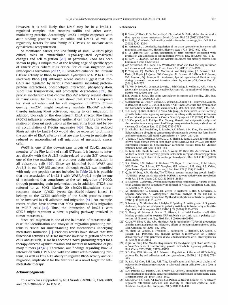
It is known that the RhoA signaling cascade plays an importantrole in regulating actin cytoskeletal organization and dynamics,inducing actin stress fiber and focal adhesion formation [29]. Sincewe have shown that overexpression of km23-1 significantly in-duces a highly organized actin stress fiber network, it was conceiv-able that KD of km23-1 might affect RhoA activation. Accordingly,we performed RhoA GTPase activation assays using Mv1Lu cellstransient transfected with either NC siRNA or km23-1 siRNA(km23-1KD) in the absence or presence of TGFß. TGFß was usedas the extracellular stimulus because it was previously shown toactivate RhoA and induce actin stress fiber formation [30,31]. Asexpected, in the NC siRNA-transfected cells, TGFb treatment dem-onstrated an accumulation of RhoA-GTP (Fig. 3., lane 1 vs. 2), con-sistent with a previous report [32]. In contrast, in km23-1KD cells,basal RhoA-GTP activity was significantly inhibited compared toNC siRNA-expressing cells (lane 3 vs. 1). While TGFß also activatedRhoA in the km23-1KD cells (lanes 3, 4), TGFß induction of RhoA-GTP was reduced compared to the NC siRNA-expressingcells + TGFß (lane 2 vs. 4). Thus, our results demonstrate thatkm23-1 is required for RhoA activity in epithelial cells.
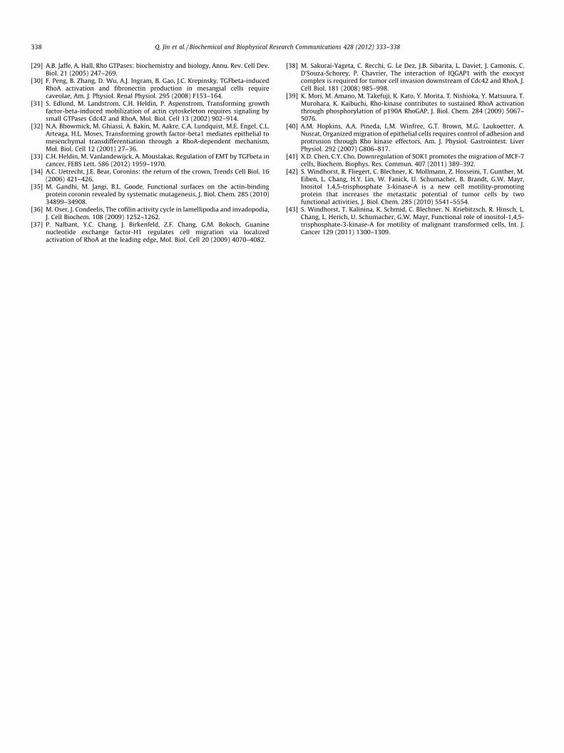
It was of interest that km23-1 depletion dramatically affectedRhoA activity even in the absence of TGFß treatment. Since it iswell-established that aggressive cancer cells with invasive proper-ties are generally resistant to TGFß [33], it was conceivable thatkm23-1 depletion might reduce the migratory properties of ininvasive, TGFß-resistant tumor cells. Toward this end, we exam-ined whether km23-1KD would affect the cell migration of TGFb-resistant HCT116 HCCCs using wound-healing assays. In the NCsiRNA-infected cells, we observed an almost complete closure ofthe wound area within 24 h. In contrast, km23-1KD cells failed toclose the wound area as effectively as control cells. As shown inFig. 4, quantification for multiple wounds from multiple experi-ments revealed that km23-1KD cells exhibited a 43% decrease inmigration compared to parental control cells. GFP expression wasdetectable in approximately 50% of both NC siRNA- and km23-1KD-infected cells, suggesting that the results plotted representan underestimate of the actual effects of km23-1 depletion ontumor cell migration. Overall, however, our results demonstratethat km23-1 is required for the cell migration of highly invasiveHCCCs.
4. Discussion
In the present study, we identified km23-1-interacting proteinsthat were involved in actin-based cell motility and modulation ofthe actin cytoskeleton, including cofilin, coronin, RhoGAP, serine/
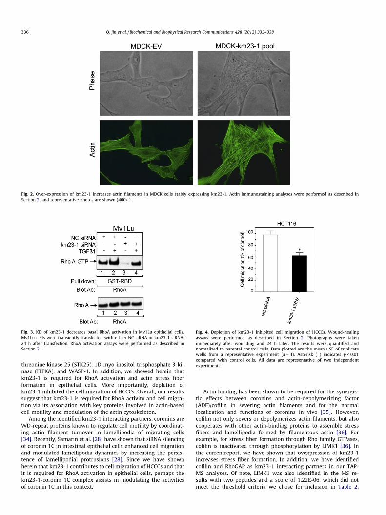
Fig. 2. Over-expression of km23-1 increases actin filaments in MDCK cells stably expressing km23-1. Actin immunostaining analyses were performed as described inSection 2, and representative photos are shown (400�).
Fig. 3. KD of km23-1 decreases basal RhoA activation in Mv1Lu epithelial cells.Mv1Lu cells were transiently transfected with either NC siRNA or km23-1 siRNA.24 h after transfection, RhoA activation assays were performed as described inSection 2.
Fig. 4. Depletion of km23-1 inhibited cell migration of HCCCs. Wound-healingassays were performed as described in Section 2. Photographs were takenimmediately after wounding and 24 h later. The results were quantified andnormalized to parental control cells. Data plotted are the mean ± SE of triplicatewells from a representative experiment (n = 4). Asterisk (�) indicates p < 0.01compared with control cells. All data are representative of two independentexperiments.
336 Q. Jin et al. / Biochemical and Biophysical Research Communications 428 (2012) 333–338
threonine kinase 25 (STK25), 1D-myo-inositol-trisphosphate 3-ki-nase (ITPKA), and WASP-1. In addition, we showed herein thatkm23-1 is required for RhoA activation and actin stress fiberformation in epithelial cells. More importantly, depletion ofkm23-1 inhibited the cell migration of HCCCs. Overall, our resultssuggest that km23-1 is required for RhoA activity and cell migra-tion via its association with key proteins involved in actin-basedcell motility and modulation of the actin cytoskeleton.
Among the identified km23-1 interacting partners, coronins areWD-repeat proteins known to regulate cell motility by coordinat-ing actin filament turnover in lamellipodia of migrating cells[34]. Recently, Samarin et al. [28] have shown that siRNA silencingof coronin 1C in intestinal epithelial cells enhanced cell migrationand modulated lamellipodia dynamics by increasing the persis-tence of lamellipodial protrusions [28]. Since we have shownherein that km23-1 contributes to cell migration of HCCCs and thatit is required for RhoA activation in epithelial cells, perhaps thekm23-1-coronin 1C complex assists in modulating the activitiesof coronin 1C in this context.
Actin binding has been shown to be required for the synergis-tic effects between coronins and actin-depolymerizing factor(ADF)/cofilin in severing actin filaments and for the normallocalization and functions of coronins in vivo [35]. However,cofilin not only severs or depolymerizes actin filaments, but alsocooperates with other actin-binding proteins to assemble stressfibers and lamellipodia formed by filamentous actin [36]. Forexample, for stress fiber formation through Rho family GTPases,cofilin is inactivated through phosphorylation by LIMK1 [36]. Inthe currentreport, we have shown that ovexpression of km23-1increases stress fiber formation. In addition, we have identifiedcofilin and RhoGAP as km23-1 interacting partners in our TAP-MS analyses. Of note, LIMK1 was also identified in the MS re-sults with two peptides and a score of 1.22E-06, which did notmeet the threshold criteria we chose for inclusion in Table 2.
Q. Jin et al. / Biochemical and Biophysical Research Communications 428 (2012) 333–338 337
However, it is still likely that LIMK may be in a km23-1-regulated complex that contains cofilin and other actin-modulating proteins. Accordingly, km23-1 might cooperate withactin-binding proteins such as cofilin and LIMK1, as well asregulatory factors for Rho family of GTPases, to mediate actincytoskeletal reorganization.
As mentioned earlier, the Rho family of small GTPases playscritical roles in extracellular signal-regulated cytoskeletalchanges and cell migration [29]. In particular, RhoA has beenshown to play a unique role at the leading edge of specific typesof cancer cells, where it is critical for cellular migration andinvadopodia formation [29,37,38]. RhoGAPs enhance the intrinsicGTPase activity of RhoA to promote hydrolysis of GTP to GDP toinactivate RhoA [39]. Although recent studies suggest that Rho-GAPs are regulated by various mechanisms, including protein–protein interactions, phospholipid interaction, phosphorylation,subcellular translocation, and proteolytic degradation [39], theprecise mechanisms that control RhoGAP activity remain elusivein many cases. Our results have shown that km23-1 is requiredfor RhoA activation and for cell migration of HCCCs. Conse-quently, km23-1 might negatively regulate RhoGAP activity,thereby inducing RhoA activation, leading to cell migration. Inaddition, blockade of the downstream RhoA effector Rho kinase(ROCK) influences coordinated epithelial cell motility by the for-mation of aberrant protrusions at the migrating front and by ba-sal accumulation of F-actin aggregates [40]. Thus, the reducedRhoA activity by km23-1KD would also be expected to diminishthe activity of RhoA effectors that are also known to mediate thereduced or uncoordinated cell migration of metastatic cancercells.
WASP is one of the downstream targets of Cdc42, anothermember of the Rho family of small GTPases. It is known to inter-act directly with the Arp2/3 (actin-related protein 2/3) complex,one of the two machines that promotes actin polymerization inall eukaryotic cells [29]. Since we identified both WASP andArp2/3 in our TAP-MS analyses, although Arp2/3 was identifiedwith only one peptide (so not included in Table 2), it is possiblethat the association of km23-1 with WASP/Arp2/3 might be oneof mechanisms that contributes to the cell migration of HCCCsthrough promoting actin polymerization. In addition, STK25 alsoreferred to as SOK1 (Sterile 20 (Ste20)-like/oxidant stress-response kinase 1)/YSK1 (yeast Sps1/Ste20-related kinase 1)belongs to the GCKIII subfamily of the Ste20 proteins knownto be involved in cell adhesion and migration [41]. For example,recent studies have shown that SOK1 promotes cells migrationin MCF-7 cells [41]. Thus, the interaction of km23-1 withSTK25 might represent a novel signaling pathway involved intumor metastasis.
Since cell migration is one of the hallmarks of metastatic dis-ease, the identification and analysis of migration promoting pro-teins is crucial for understanding the mechanisms underlyingmetastasis formation [1]. Previous results have shown that twofunctional activities of ITPKA increase invasive migration of tumorcells, and as such, ITPKA has become a novel promising target for atherapy directed against invasion and metastasis formation of pri-mary tumors [42,43]. Therefore, our findings regarding km23-1interaction with ITPKA and with the other actin-modulating pro-teins, as well as km23-1’s ability to regulate RhoA activity and cellmigration, implicate it for the first time as a novel target for anti-metastatic therapy.
Acknowledgments
This work was supported by NIH Grants CA090765, CA092889,and CA092889-08S1 to K.M.M.
References
[1] D. Spano, C. Heck, P. De Antonellis, G. Christofori, M. Zollo, Molecular networksthat regulate cancer metastasis, Semin. Cancer Biol. 22 (2012) 234–249.
[2] M. Bailly, J. Condeelis, Cell motility insights from the backstage, Nat. Cell Biol. 4(2002) E292–E294.
[3] H. Yamaguchi, J. Condeelis, Regulation of the actin cytoskeleton in cancer cellmigration and invasion, Biochim. Biophys. Acta 1773 (2007) 642–652.
[4] C. Le Clainche, M.F. Carlier, Regulation of actin assembly associated withprotrusion and adhesion in cell migration, Physiol. Rev. 88 (2008) 489–513.
[5] M. Parri, P. Chiarugi, Rac and Rho GTPases in cancer cell motility control, CellCommun. Signal 8 (2010) 23.
[6] A.P. Struckhoff, M.K. Rana, R.A. Worthylake, RhoA can lead the way in tumorcell invasion and metastasis, Front. Biosci. 16 (2011) 1915–1926.
[7] P. Timpson, E.J. McGhee, J.P. Morton, A. von Kriegsheim, J.P. Schwarz, S.A.Karim, B. Doyle, J.A. Quinn, N.O. Carragher, M. Edward, M.F. Olson, M.C. Frame,V.G. Brunton, O.J. Sansom, K.I. Anderson, Spatial regulation of RhoA activityduring pancreatic cancer cell invasion driven by mutant p53, Cancer Res. 71(2011) 747–757.
[8] Y.I. Wu, D. Frey, O.I. Lungu, A. Jaehrig, I. Schlichting, B. Kuhlman, K.M. Hahn, Agenetically encoded photoactivatable Rac controls the motility of living cells,Nature 461 (2009) 104–108.
[9] M.F. Olson, E. Sahai, The actin cytoskeleton in cancer cell motility, Clin. Exp.Metastasis 26 (2009) 273–287.
[10] U. Ilangovan, W. Ding, Y. Zhong, C.L. Wilson, J.C. Groppe, J.T. Trbovich, J. Zuniga,B. Demeler, Q. Tang, G. Gao, K.M. Mulder, A.P. Hinck, Structure and dynamics ofthe homodimeric dynein light chain km23, J. Mol. Biol. 352 (2005) 338–354.
[11] H.C. Kang, I.J. Kim, K. Kim, H.J. Yoon, S.G. Jang, J.G. Park, km23, a transforminggrowth factor-beta signaling component, is infrequently mutated in humancolorectal and gastric cancers, Cancer Genet Cytogenet 175 (2007) 173–174.
[12] I.G. Campbell, W.A. Phillips, D.Y. Choong, Genetic and epigenetic analysis ofthe putative tumor suppressor km23 in primary ovarian, breast, and colorectalcancers, Clin. Cancer Res. 12 (2006) 3713–3715.
[13] K. Nikulina, R.S. Patel-King, S. Takebe, K.K. Pfister, S.M. King, The roadblocklight chains are ubiquitous components of cytoplasmic dynein that form homoand heterodimers, Cell Motil. Cytoskeleton 57 (2004) 233–245.
[14] J. Jiang, L. Yu, X. Huang, X. Chen, D. Li, Y. Zhang, L. Tang, S. Zhao, Identificationof two novel human dynein light chain genes DNLC2A and DNLC2B and theirexpression changes in hepatocellular carcinoma tissues from 68 Chinesepatients, Gene 281 (2001) 103–113.
[15] Q. Tang, C.M. Staub, G. Gao, Q. Jin, Z. Wang, W. Ding, R.E. Aurigemma, K.M.Mulder, A novel transforming growth factor-beta receptor-interacting proteinthat is also a light chain of the motor protein dynein, Mol. Biol. Cell 13 (2002)4484–4496.
[16] K.K. Pfister, E.M. Fisher, I.R. Gibbons, T.S. Hays, E.L. Holzbaur, J.R. McIntosh,M.E. Porter, T.A. Schroer, K.T. Vaughan, G.B. Witman, S.M. King, R.B. Vallee,Cytoplasmic dynein nomenclature, J. Cell Biol. 171 (2005) 411–413.
[17] Q. Jin, W. Ding, K.M. Mulder, The TGFbeta receptor-interacting protein km23-1/DYNLRB1 plays an adaptor role in TGFbeta1 autoinduction via its associationwith Ras, J. Biol. Chem. 287 (2012) 26453–26463.
[18] E.V. Koonin, L. Aravind, Dynein light chains of the Roadblock/LC7 group belongto an ancient protein superfamily implicated in NTPase regulation, Curr. Biol.10 (2000) R774–R776.
[19] M. Miertzschke, C. Koerner, I.R. Vetter, D. Keilberg, E. Hot, S. Leonardy, L.Sogaard-Andersen, A. Wittinghofer, Structural analysis of the Ras-like Gprotein MglA and its cognate GAP MglB and implications for bacterial polarity,EMBO J. 30 (2011) 4185–4197.
[20] S. Leonardy, M. Miertzschke, I. Bulyha, E. Sperling, A. Wittinghofer, L. Sogaard-Andersen, Regulation of dynamic polarity switching in bacteria by a Ras-likeG-protein and its cognate GAP, EMBO J. 29 (2010) 2276–2289.
[21] Y. Zhang, M. Franco, A. Ducret, T. Mignot, A bacterial Ras-like small GTP-binding protein and its cognate GAP establish a dynamic spatial polarity axisto control directed motility, PLoS Biol. 8 (2010) e1000430.
[22] G. Liu, W. Ding, X. Liu, K.M. Mulder, c-Fos is required for TGFbeta1 productionand the associated paracrine migratory effects of human colon carcinoma cells,Mol. Carcinog. 45 (2006) 582–593.
[23] W. Zhou, M. Capello, C. Fredolini, L. Racanicchi, L. Piemonti, L.A. Liotta, F.Novelli, E.F. Petricoin, MS analysis reveals O-methylation of L-lactatedehydrogenase from pancreatic ductal adenocarcinoma cells, Electrophoresis33 (2012) 1850–1854.
[24] Q. Jin, W. Ding, K.M. Mulder, Requirement for the dynein light chain km23-1 ina Smad2-dependent transforming growth factor-beta signaling pathway, J.Biol. Chem. 282 (2007) 19122–19132.
[25] X.D. Ren, W.B. Kiosses, M.A. Schwartz, Regulation of the small GTP-bindingprotein Rho by cell adhesion and the cytoskeleton, EMBO J. 18 (1999) 578–585.
[26] H. Yan, A.J. Choi, B.H. Lee, A.H. Ting, Identification and functional analysis ofepigenetically silenced microRNAs in colorectal cancer cells, PloS One 6 (2011)e20628.
[27] D.N. Perkins, D.J. Pappin, D.M. Creasy, J.S. Cottrell, Probability-based proteinidentification by searching sequence databases using mass spectrometry data,Electrophoresis 20 (1999) 3551–3567.
[28] S.N. Samarin, S. Koch, A.I. Ivanov, C.A. Parkos, A. Nusrat, Coronin 1C negativelyregulates cell-matrix adhesion and motility of intestinal epithelial cells,Biochem. Biophys. Res. Commun. 391 (2010) 394–400.
338 Q. Jin et al. / Biochemical and Biophysical Research Communications 428 (2012) 333–338
[29] A.B. Jaffe, A. Hall, Rho GTPases: biochemistry and biology, Annu. Rev. Cell Dev.Biol. 21 (2005) 247–269.
[30] F. Peng, B. Zhang, D. Wu, A.J. Ingram, B. Gao, J.C. Krepinsky, TGFbeta-inducedRhoA activation and fibronectin production in mesangial cells requirecaveolae, Am. J. Physiol. Renal Physiol. 295 (2008) F153–164.
[31] S. Edlund, M. Landstrom, C.H. Heldin, P. Aspenstrom, Transforming growthfactor-beta-induced mobilization of actin cytoskeleton requires signaling bysmall GTPases Cdc42 and RhoA, Mol. Biol. Cell 13 (2002) 902–914.
[32] N.A. Bhowmick, M. Ghiassi, A. Bakin, M. Aakre, C.A. Lundquist, M.E. Engel, C.L.Arteaga, H.L. Moses, Transforming growth factor-beta1 mediates epithelial tomesenchymal transdifferentiation through a RhoA-dependent mechanism,Mol. Biol. Cell 12 (2001) 27–36.
[33] C.H. Heldin, M. Vanlandewijck, A. Moustakas, Regulation of EMT by TGFbeta incancer, FEBS Lett. 586 (2012) 1959–1970.
[34] A.C. Uetrecht, J.E. Bear, Coronins: the return of the crown, Trends Cell Biol. 16(2006) 421–426.
[35] M. Gandhi, M. Jangi, B.L. Goode, Functional surfaces on the actin-bindingprotein coronin revealed by systematic mutagenesis, J. Biol. Chem. 285 (2010)34899–34908.
[36] M. Oser, J. Condeelis, The cofilin activity cycle in lamellipodia and invadopodia,J. Cell Biochem. 108 (2009) 1252–1262.
[37] P. Nalbant, Y.C. Chang, J. Birkenfeld, Z.F. Chang, G.M. Bokoch, Guaninenucleotide exchange factor-H1 regulates cell migration via localizedactivation of RhoA at the leading edge, Mol. Biol. Cell 20 (2009) 4070–4082.
[38] M. Sakurai-Yageta, C. Recchi, G. Le Dez, J.B. Sibarita, L. Daviet, J. Camonis, C.D’Souza-Schorey, P. Chavrier, The interaction of IQGAP1 with the exocystcomplex is required for tumor cell invasion downstream of Cdc42 and RhoA, J.Cell Biol. 181 (2008) 985–998.
[39] K. Mori, M. Amano, M. Takefuji, K. Kato, Y. Morita, T. Nishioka, Y. Matsuura, T.Murohara, K. Kaibuchi, Rho-kinase contributes to sustained RhoA activationthrough phosphorylation of p190A RhoGAP, J. Biol. Chem. 284 (2009) 5067–5076.
[40] A.M. Hopkins, A.A. Pineda, L.M. Winfree, G.T. Brown, M.G. Laukoetter, A.Nusrat, Organized migration of epithelial cells requires control of adhesion andprotrusion through Rho kinase effectors, Am. J. Physiol. Gastrointest. LiverPhysiol. 292 (2007) G806–817.
[41] X.D. Chen, C.Y. Cho, Downregulation of SOK1 promotes the migration of MCF-7cells, Biochem. Biophys. Res. Commun. 407 (2011) 389–392.
[42] S. Windhorst, R. Fliegert, C. Blechner, K. Mollmann, Z. Hosseini, T. Gunther, M.Eiben, L. Chang, H.Y. Lin, W. Fanick, U. Schumacher, B. Brandt, G.W. Mayr,Inositol 1,4,5-trisphosphate 3-kinase-A is a new cell motility-promotingprotein that increases the metastatic potential of tumor cells by twofunctional activities, J. Biol. Chem. 285 (2010) 5541–5554.
[43] S. Windhorst, T. Kalinina, K. Schmid, C. Blechner, N. Kriebitzsch, R. Hinsch, L.Chang, L. Herich, U. Schumacher, G.W. Mayr, Functional role of inositol-1,4,5-trisphosphate-3-kinase-A for motility of malignant transformed cells, Int. J.Cancer 129 (2011) 1300–1309.