rheumatic heart disease oleh dr robert
TRANSCRIPT

Rheumatic Heart DiseaseRobert E Saragih SpJP FIHA
SMF Jantung Pembuluh Darah RSU UKI

Learning Objectives:• To understand the pathogenesis of acute rheumatic fever and
rheumatic heart disease
• To appreciate the burden of disease
• To recognize the features of a streptococcal sore throat
• To know the treatment regimens of a streptococcal sore throat
• To be aware of secondary prevention measures
Performance Objectives:• Examine the burden of disease within own communities
• Timely recognition of a streptococcal sore throat with correcttreatment
• Institute secondary prevention programme
• Join the global community fighting Rheumatic fever andrheumatic heart disease

• Rheumatic fever is an immunologically mediatedinflammatory disease, that occurs as a delayed sequel togroup A streptococcal throat infection,in geneticallysusceptible individuals.
• Rheumatic heart disease is the most serious complication ofrheumatic fever.
• Acute rheumatic fever and rheumatic heart disease arethought to result from an autoimmune response, but theexact pathogenesis remains unclear.

Epidemiology of group A streptococci,rheumatic fever and rheumatic heart disease
• Rheumatic fever (RF) and rheumatic heart disease (RHD) arenonsuppurative complications of Group A streptococcalpharyngitis due to a delayed immune response.
• RF and RHD are rare in developed countries, but they are stillmajor public health problems among children and youngadults in developing countries.
• The economic effects of the disability and premature deathcaused by these diseases are felt at both the individual andnational levels.
Group A streptococcal infections• Beta-haemolytic streptococci can be divided into a number of
sero-logical groups on the basis of their cell-wallpolysaccharide antigen.
• Those in serological group A (Streptococcus pyogenes ) can befurther subdivided into more than 130 distinct M types, andare responsible for the vast majority of infections in humans.
• Only pharyngitis caused by group A streptococci has beenlinked with the etiopathogenesis of RF and RHD.
• Other streptococcal groups (e.g. B, C, G and F) have beenisolated from human subjects and are some-times associatedwith infection; and streptococci in groups C and G can produceextracellular antigens (including streptolysin-O) with similarcharacteristics to that produced by group A streptococci .

• Pharyngitis and skin infection (impetigo) are the mostcommon infections caused by group A streptococci.
• Group A streptococci are the most common bacterial cause ofpharyngitis, with a peak incidence in children 15 years of age .
• Streptococcal pharyngitis is less frequent among chil-dren inthe first three years of life and among adults.
• That most children develop at least one episode of pharyn-gitis per year,20% of which are caused by group A streptococciand nearly 80% by viral pathogens .
• The incidence of pharyngeal beta-haemolytic streptococcalinfections can vary, depending upon season,age group,socioeconomic conditions, environmental factors and thequality of health care .
• Surveys of healthy school children 6–10 years of age, forexample, found anti-streptolysin-O titres >200 Todd units in15–70% of the children , while other studies reported beta-haemolytic streptococci carrier rates of 10–50% forasymptomatic schoolchildren .

• The presence of group A streptococci in the upper respiratorytract (URT) may reflect either true infection or a carrier state.
• Only in the case of a true infection does the patient show arising antibody response.
• In the carrier state there is no rising antibody response.
• Patient with a true infection is at risk of developing RF and ofspreading the organism to close contacts, while this is notthought to be the case with carriers .
• Therefore, many professionals feel that only patients with trueinfections need to be given antibiotics state .

• Group A streptococci are highly transmissible and spreadrapidly in families and communities,with the predominant Mtypes constantly changing.
• However, in publications about RF outbreaks, including recentones in the United States of America (USA), it was reportedthat only a limited number of streptococcal stereotypes (i.e. Mserotypes 1, 3, 5, 6, 18, 19, 24) were obtained from the throatcultures of children in the affected communities.

Examples of presence of group A beta-haemolyticstreptococci in children withsymptomatic pharyngitis
• In the USA, despite a remarkable reduction in theincidence of RF since the 1950s, the incidence of URTinfections caused by group A streptococci has notdeclined .
• In the mid-1980s, the virulence, severity and sequelae ofthese infections also appear to have changed remarkably.
• Outbreaks of acute RF have been described from widelyseparated areas of the country, and complications ofstreptococcal infections have been reported, includingnecrotising fascitis, streptococcal myositis, streptococcalbacteremia and streptococcal toxic shock syndrome.

Rheumatic fever and rheumatic heart disease
• In 1994, it was estimated that 12 million individuals sufferedfrom RF and RHD worldwide, and at least 3 million hadcongestive heart failure (CHF) that required repeatedhospitalisation .
• A large proportion of the individuals with CHF required cardiacvalve surgery within 5–10 years .
• The mortality rate per 100 000 population varied from 1.8 inthe WHO Region of the Americas, to 7.6 in WHO South-EastAsia Region.
• An estimated 6.6 million DALYs are lost per year worldwide.
• Data from developing countries suggest that mortality due toRF and RHD remains a problem and that children and youngadults still die from acute RF.

Estimated deaths and DALYs lost to rheumatic heartdisease in 2000, by WHORegion

• The annual incidence of RF in developedcountries began to decrease in the 20th century,with a marked decrease after the 1950s; it isnow below 1.0 per 100 000
• The surveys results showed there was widevariation between countries .
• The prevalence of RF and RHD and the mortalityrates varied widely between countries andbetween population groups in the same country.

• Based on such data, RHD accounts for 12–65% ofhospital admissions related to cardiovasculardisease, and for 2.0–9.9% of all hospitaldischarges in some developing countries .
• There has been a marked decrease in themortality, incidence, prevalence, hospitalmorbidity and severity of RF and RHD in someplaces that have implemented preventionprogrammes .

Examples of reported prevalence of rheumatic heart disease in schoolchildren

Determinants of the disease burden of rheumatic fever and rheumatic heart disease
• It is well known that socioeconomic andenvironmental factors play an indirect, butimportant, role in the magnitude and severity ofRF and RHD.
• Factors such as a shortage of resources forproviding quality health care, inadequateexpertise of health-care providers, and a lowlevel of awareness of the disease in thecommunity can all impact the expression of thedisease in populations.

Direct and indirect results of environmental and health-system determinants onrheumatic fever and rheumatic heart disease

Pathogenesis of rheumatic fever• The epidemiological association between group A b-
haemolytic streptococcal infections and the subsequentdevelopment of acute rheumatic fever (RF) has been wellestablished.
• RF is a delayed autoimmune response to Group Astreptococcal pharyngitis, and the clinical manifestationof the response and its severity in an individual isdetermined by host genetic susceptibility, the virulenceof the infecting organism, and a conducive environment.
• In temperate regions, serogroup A is the predominantisolate (50–60%), with serogroups C and G together lessthan 30% of isolates.
• Nonsuppurative sequel, such as RF and RHD, are seenonly after group A streptococcal infection of the upperrespiratory tract.

• Chronic streptococcal “carrier” states do nottrigger the development of RF .
• The precise pathogenetic mechanism of RF hasnot been defined.
• Histocompatibiltiy antigens, potential tissue-specific antigens, and antibodies developedduring and immediately after a streptococcalinfection are being investigated as potential riskfactors in the pathogenesis of the disease.
• T-cell lymphocytes play an important role in thepathogenesis of rheumatic carditis.
• Particular M types of group A streptococci haverheumatogenic potential.
• Such serotypes are usually heavily encapsulated,and form large, mucoid colonies that are rich inM-protein.
Streptococcal M-protein
• M-protein is one of the best-defineddeterminants of bacterial virulence.
• The streptococcal M-protein extends from thesurface of the streptococcal cell as coiled coildimer, and shares structural homology withcardiac myosin and other alpha-helical coiled coilmolecules, such as tropomyosin, keratin andlaminin.
• This homology is responsible for the pathologicalfindings in acute rheumatic carditis.
• The C repeat regions contain highly conserved epitopes,and streptococci are often classified into Class I or II .
• The majority of Class I strains (with reactive Mprotein)are implicated in RF.
• More than 130 M-protein types identified, M types suchas 1, 3, 5, 6, 14, 18, 19 and 24 have been associated withRF.
• Individuals may have multiple streptococcal infectionsthroughout their lifetime, but reinfections with the sameserological M type are relatively less common becauseindividuals acquire circulating homologous anti-Mantibodies following an infection.

Streptococcal superantigens
• The role of the superantigen-like activity of M-proteinfragments , as well as the streptococcal pyrogenicexotoxin, in the pathogenesis of RF.
• Superantigenic activation is not limited to the T-cellcompartment alone.
• Streptococcal erythrogenic toxin may behave like asuperantigen for the B-cell, leading to the production ofautoreactive antibodies.
• Extracellular products and cell-wall componentsrepresent the virulence of group A streptococci.

• GRAB (an alpha-2 macroglobulin-binding proteinexpressed by Streptococcus pyogenes),streptococcal fibronectin-binding protein 1(sfb1), which mediates streptococcal adherenceand invasion into human epithelial cells,.
• Streptococcal C5a peptidase (SCPA), whichinactivates complement chemotaxin C5a andallows streptococci to adhere to tissues, are allsubjects in the pathogenesis of streptococcalinfections.

The role of the human host in the development of rheumatic fever and rheumatic heart disease
• An autoimmune response to streptococcal antigensmediates the development of RF and RHD in asusceptible host.
• Genetically-programmed determinants of hostsusceptibility to RF only 0.3–3% of individuals with acutestreptococcal pharyngitis go on to develop RF .
• Pedigree studies suggested that this immune response isgenetically controlled, with high responsiveness to thestreptococcal cell-wall antigen .
• The gene controlling the low-level response tostreptococcal antigen is closely linked to the Class IIhuman leukocyte antigen( HLA ).

Host-pathogen interaction
• Infection by streptococci begins with the binding ofbacterial surface ligands to specific receptors on hostcells, and subsequently involves specific processes ofadherence, colonization and invasion.
• The binding of bacterial surface ligands to host surfacereceptors is the most crucial event in the colonization ofthe host, and it is initiated by fibronectin and bystreptococcal fibronectin-binding proteins .
• Streptococcal lipoteichoic acid and M-protein also play amajor role in bacterial adherence .

The role of environmental factors in RF and RHD
• Trends in RF and RHD , towards environmental factors such aspoor living conditions, overcrowding and access to health careas the most significant determinants of disease distribution.
• Indeed, the global distributions of RF and RHD are stillinfluenced by socioeconomic indices.
• Crowded living conditions, contribute to the rapid spread andpersistence of virulent streptococcal strains.
• Seasonal variations in the incidence of RF (i.e. high incidencesin early fall, late winter and early spring) closely mimicvariations in streptococcal infections.
• These variations are particularly pronounced in temperateclimates, but are not significant in the tropics.
• Initial streptococcal infection in a genetically predisposed hostin a susceptible environment leads to the activation of T-celland B-cell lymphocytes by streptococcal antigens andsuperantigens, which results in the production of cytokinesand antibodies directed against streptococcal carbohydrateand myosin.
• Injury to the valvular endothelium by the anti-carbohydrateantibodies leads to an up-regulation of VCAM1 and otheradhesion molecules .
• VCAM1 expression is a hallmark of inflammation and it heraldscellular infiltration.
• VCAM1 interacts with VLA4 on activated lymphocytes andleads to an influx of activated CD4+ and CD8+ T-cells.

• A break in the endothelial continuity of a heart valve wouldexpose subendothelial structures (vimentin, laminin andvalvular interstitial cells) and lead to a “chain reaction” ofvalvular destruction.
• Once valve leaflets are inflamed through the valvular surfaceendothelium and new vascularization occurs, the newlyformed microvasculature allows T-cells to infiltrate andperpetuate the cycle of valvular damage.
• The presence of T-cell infiltration, is indicative of persistentand progressive disease in the valves.
• Valvular interstitial cells and other valvular constituents underthe influence of inflammatory cytokines perpetuate aberrantrepair.

• One group A streptococcal serotype can be rapidly andcompletely replaced by another erotype in a stable populationwith adequate access to health care .
• The ability of streptococci to infect the host after a priorinfection by a different M serotype strain, suggests there is nobroad.
• Non-type-specific immunity directed against conserved M-protein epitopes or their extracellular products, whichcomplicates the development of a RF vaccine aimed atconserved M-protein sequences.

Diagnosis of rheumatic fever
• The Jones criteria were introduced in 1944 as a set of clinicalguidelines for the diagnosis of rheumatic fever (RF) andcarditis .
• The clinical features of RF were divided into major and minorcategories, based on the prevalence and specificity ofmanifestations .
• Major manifestations were included carditis, joint symptoms,subcutaneous nodules, and chorea.
• A history of RF or preexisting rheumatic heart disease (RHD)was considered to be a major criterion .

• Minor manifestations were considered to be suggestive,but not sufficient, for a diagnosis of RF.
• The minor manifestations comprised clinical findings(such as fever and erythema marginatum, abdominalpain, epistaxis and pulmonary findings), and laboratorymarkers of acute inflammation, such as leukocytosis(WBC), and elevated erythrocyte sedimentation rate(ESR) or C-reactive protein (CRP) .

• It was proposed that the presence of two major, or one majorand two minor, manifestations offered reasonable clinicalevidence of rheumatic activity.
• Carditis is the single most important prognostic factor in RF;only valvulitis leads to permanent damage and its presencedetermines the prophylactic strategy.
• The clinical diagnosis of carditis in an index attack of RF isbased on the presence of significant murmurs (suggestive ofmitral and/or aortic regurgitation), pericardial rub, or anunexplained cardiomegaly with CHF.
• A diagnosis of recurring carditis requires the demonstration ofvalvular damage or involvement, with or without pericardial ormyocardial involvement.
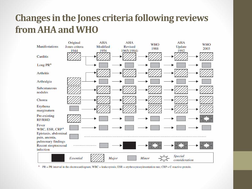
Changes in the Jones criteria following reviews from AHA and WHO

2002–2003 WHO criteria for the diagnosis of rheumatic fever andrheumatic heart disease

2002–2003 WHO criteria for the diagnosis of rheumatic fever andrheumatic heart disease
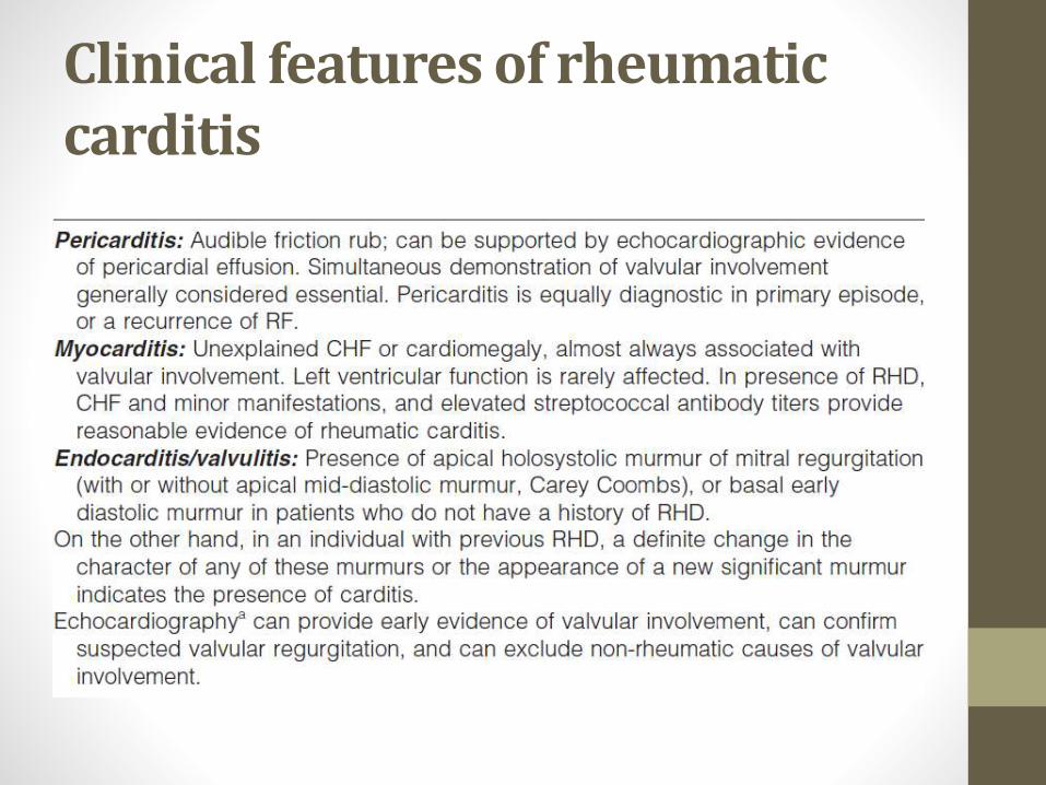
Clinical features of rheumatic carditis

Differential diagnosis of polyarthritis and fevera

New diagnostic techniques for rheumatic carditis• Echocardiography
• Endomyocardial biopsy
• Radionuclide imaging

Variation in normal antibody titres with age and/or geography
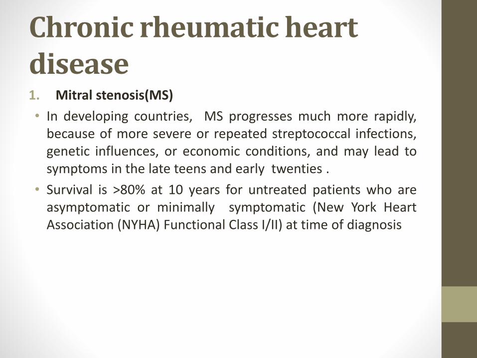
Chronic rheumatic heart disease1. Mitral stenosis(MS)
• In developing countries, MS progresses much more rapidly,because of more severe or repeated streptococcal infections,genetic influences, or economic conditions, and may lead tosymptoms in the late teens and early twenties .
• Survival is >80% at 10 years for untreated patients who areasymptomatic or minimally symptomatic (New York HeartAssociation (NYHA) Functional Class I/II) at time of diagnosis

• Mean survival time falls to less than three yearsif severe pulmonary hypertension has .
• The mortality of untreated patients with MS isattributable to progressive heart failure in 60–70% of patients, systemic embolism in 20–30%,pulmonary embolism in 10%, and infection in 1–5%.

• The initial presentation of patients with evenmild-to-moderate mitral stenosis (mitral valvearea 1.5–2.0cm2) may be precipitated byexercise, emotional upset, fever, pregnancy, orAF, especially with a rapid ventricular response.
• Late stages of MS, as pulmonary vascularresistance rises and cardiac output falls, fatigueor effort intolerance may play a dominant role.
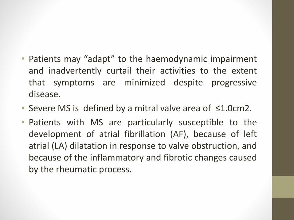
• Patients may “adapt” to the haemodynamic impairmentand inadvertently curtail their activities to the extentthat symptoms are minimized despite progressivedisease.
• Severe MS is defined by a mitral valve area of ≤1.0cm2.
• Patients with MS are particularly susceptible to thedevelopment of atrial fibrillation (AF), because of leftatrial (LA) dilatation in response to valve obstruction, andbecause of the inflammatory and fibrotic changes causedby the rheumatic process.

• There is an abrupt loss of the atrial contribution to ventricularfilling and as much as a 30% reduction in cardiac output(CO).
• There is the potential for a sudden increase in LA pressure,especially with rapid ventricular rates due to a criticaldecrease in diastolic filling times, and the potential for asignificant increase in the associated risk ofthromboembolism.
• AF is more common among older patients , has been relatedto the severity of the stenosis and to the LA pressure.

• MS is associated with the highest risk of systemicthromboembolism.
• The incidence of systemic embolization, including stroke,estimated at 1.5–4.7% per year.
• Patients who suffer a first embolus are at increased risk for asecond, particularly within the next six months.
• Significant reductions in the incidence of recurrent emboliamong patients treated long-term with warfarinanticoagulation, approximately 5% per year in untreatedpatients, to 0.7–0.8% per year in those receiving warfarin.

Mitral stenosis


Mitral regurgitation(MR)
• The volume load of chronic MR can be well tolerated forseveral years.
• Symptoms and/or signs of left ventricular systolic dysfunction(defined by an ejection fraction <0.60, or an end-systolicdimension ≥4.5 cm) are indications for surgery .
• Compared with patients with predominant stenosis, patientswith isolated MR are less susceptible to thromboembolismwith AF, but are more prone to infective endocarditis.


Aortic stenosis(AS)
• The well-known natural history of AS has long dictated thatsurgery be undertaken once symptoms appear.
• Survival without valve replacement after the onset of angina,syncope, or heart failure is generally measured at five, three,and two years, respectively .
• For patients with severe AS (valve area ≤1.0cm2) who developheart failure and who are not candidates for surgery, diureticscan be provided to alleviate congestion.

• AF is an uncommon complication of isolated aortic stenosis
• Patients with heart failure and AS with “low gradient/lowoutput” should undergo referral and additional testing todetermine if the depressed left ventricular function is due tosevere, uncorrected AS (afterload mismatch) or to a primarycardiomyopathy


• Physical activity need not be restricted in patients with mildAS (valve area >1.5cm2).
• Patients with moderate AS (valve area 1.0–1.5cm2) should beadvised to avoid strenuous activity and competitive sports.
• Severe AS usually mandates a reduction in physical activitiesto low levels

Aortic regurgitation(AR)
• Patients with chronic, severe AR usually enjoy a long,yetvariable compensated phase characterized by an increase inleft ventricular end-diastolic volume, an increase in chambercompliance,and a combination of both eccentric andconcentric hypertrophy.
• Preload reserve is maintained, ejection performance remainsnormal, and the enormous increase in stroke volume allowspreservation of forward output .
• AR thus leads both to volume and pressure overload .
• Preoperative left ventricular function is the most importantpredictor of postoperative survival

Medical management of rheumatic feverGENERAL MEASURES
• Hospital admission
• Initial tests should a throat culture (or in some circunstancesrapid streptococcal detection test), a measurement ofstreptococcal antibody titres (eg ASO or anti DNase B), anassessment of acute-phase reactants (eg ESR or CRP), a chestX-ray, an electrocardiogram, and an echocardiogram (iffacilities are available).
• A blood culture may help to exclude infective endocarditis
• All patients with acute RF should be placed on bed–chair restand monitored closely for the onset of carditis. I
• In patients with carditis, a rest period of at least four weeks isrecommended , although physicians should make this decisionon an individual basis.
• Patients with chorea must be placed in a protectiveenvironment so they do not injure themselves.

Antimicrobial therapy
• Eradication of the pharyngeal streptococcal infection ismandatory to avoid chronic repetitive exposure tostreptococcal antigens .
• Ideally, two throat cultures should be performed beforestarting antibiotics.
• However, antibiotic therapy is warranted even if the throatcultures are negative.
• The eradication of pharyngeal streptococci should be followedby long-term secondary prophylaxis to guard against recurrentpharyngeal streptococcal infections.

Suppression of the inflammatory process
• Aspirin, 100 mg/kg-day divided into 4–5 doses, is the first lineof therapy
• In children, the dose may be increased to 125 mg/kg-day, andto 6–8g/day in adults.
• After achieving the desired initial steady-state concentrationfor two weeks, the dosage can be decreased to 60–70mg/kg-day for an additional 3–6 weeks .
• However, in patients who are intolerant or allergic to aspirin,naproxen (10–20mg/kg-day) has been used .

• Corticosteroids are also advisable in patients who do notrespond to salicylates and who continue to worsen anddevelop heart failure despite anti-inflammatory therapy .
• Prednisone (1–2mg/kg-day, to a maximum of 80mg/day givenonce daily, or in divided doses)
• In life-threatening circumstances, therapy may be initiatedwith intravenous methyl prednisolone .
• After 2–3 weeks of therapy the dosage may be decreased by20–25% each week .
• While reducing the steroid dosage, a period of overlap withaspirin is recommended to prevent rebound of diseaseactivity.

Surgery for rheumatic heart disease• Surgery is usually performed for chronic rheumatic valve
disease.
• The necessity for surgical treatment is determined by theseverity of the patient’s symptoms and/or evidence thatcardiac function is significantly impaired.
• Important to prevent irreversible damage to the left ventricleand irreversible pulmonary hypertension, since bothconsiderably increase the risk of surgical treatment, impairlong-term results and render surgery contra-indicated.

The indications for surgical treatment are as follows:
• In the presence of MS, patients with moderate or
severe MS (mitral valve area 1.5cm2) and NYHA class
II/IV symptoms
• In the presence of MR, patients with NYHA functional classsymptoms II/III/IV
• In the presence of AS, symptomatic patients with severe AS orin the presence of LV dysfunction, ventricular tachycardia,>15mm LV hypertrophy, valve area <0.6cm2.
• In the presence of AR, with NYHA functional class symptomsII/III/IV
Contra-indications to surgery• Relative contra-indications include manifestations of end-
stage valve disease, such as very poor LV function inassociation with a regurgitant lesion, severe fixed pulmonaryhypertension or extensive extraannular tissue destruction dueto uncontrolled endocarditis

Treatment options
1. Balloon valvotomy (commissurotomy)
2. Surgical treatment

Primary prevention of rheumatic fever
• The adequate antibiotic therapy of group A streptococcalupper respiratory tract (URT) infections to prevent an initialattack of acute RF .
• Primary prevention is administered only when there is group Astreptococcal URT infection.
• The therapy is therefore intermittent, in contrast to thetherapy used for the secondary prevention of RF, whereantibiotics are administered continuously.

Antibiotic therapy of group A streptococcal pharyngitis

Secondary prevention of rheumatic fever

Several factors can influence the risk of RF recurrence, including:
— the age of the patient
— the presence of RHD
— the time elapsed from the last attack
— the number of previous attacks
— the degree of crowding in the family
— a family history of RF/RHD
— the socioeconomic and educational status of the individual
— the risk of streptococcal infection in the area
— whether a patient is willing to receive injections
— the occupation and place of employment of the patient (school
teachers, physicians, employees in crowded areas).

Suggested duration of secondary prophylaxis

TERIMAKASIH