rheumatic fever - a forgotten disease - laaap.org filerheumatic fever acute rheumatic fever is a...
TRANSCRIPT

RHEUMATIC FEVER A FORGOTTEN DISEASE
Vickren Pillay, MDPediatrics LSU Health Shreveport
Louisiana Chapter AAPAcadiana Potpourri 2017

Disclosure
■ Presenter: Vickren Pillay, MD
■ I have nothing to disclose regarding this topic.

Objectives
■ Expand the pediatricians perspective on the clinical presentation of rheumatic fever and associated complications
■ Impart skills for the clinician to recognize uncommon presentations of rheumatic fever
■ Discuss the updated Echocardiographic recommendations for Rheumatic Heart disease diagnosis

Case
6 year old African American female presented to ED with fever that began earlier that day. T-Max was 103 and ED arrival temperature was 101.
Associated symptoms were headache and refusal to swallow. Physical exam revealed a well appearing child with mild pharyngeal erythema. A grade 3/6 systolic murmur was auscultated that had not previously been documented.
A rapid strep test was performed and was negative. Reflex throat culture was done and negative. She was diagnosed with viral pharyngitis, given Tylenol and discharged home.
An appointment was scheduled for the follow day in order for patient’s new heart murmur to be addressed.

CaseDespite scheduled appointments, patient did not show up for approximately 2 months.
When she returned to clinic, her mother noted that for the past month she had been falling asleep more frequently in class and her activity level had decreased. Mother also reported that patient has had intermittent fever treated with antipyretics for the past month. On exam, her harsh systolic murmur was still present
She was then sent for an immediate cardiac ECHO that was significant for severe mitral valve regurgitation with nodular appearance of the anterior leaflet, moderate to severe aortic insufficiency with nodular appearance of the leaflets. Moderate dilation of the left ventricle with normal systolic function was also noted


Past Medical History
Past Medical Hx: No significant PMH. Never been treated for streptococcal pharyngitis
Past Surgical Hx: surgical repair of umbilical hernia in 09/2014, labial adhesion lysis
Birth Hx: Born term approximately 38 weeks via NSVD with normal newborn nursery stay. Mother denied pregnancy or birth complications
Developmental Hx: Normal development. Met milestone appropriately.
Family Hx: Maternal Uncle with MI at age 38. Maternal grandmother with CAD
Social Hx: Lives with mother and 3 brothers in a house. No pets
Immunization Hx: Up to date on immunizations

Hospital Course
Physical examination on admission to hospital was significant for the following:
- well appearing child who was active and an playful and in no acute distress.
- mucous membranes moist, normal, non erythematous tonsils and posterior pharynx, normal lymphadenopathy
- Cardiac auscultation significant for normal S1 and S2. No S3 or S4. 2/4 diastolic murmur loudest at the mid sternal border and a 3/6 holosystolic murmur at the apex. PMI was slightly displaced to the left. No rubs or gallops, lifts, thrills or heaves. 3+ Bounding pulses noted in all extremities
- No arthropathy with full range of motion in both upper and lower extremities
- No rashes or nodules on skin. She had no splinter hemorrhages, Janeway lesions or Osler nodes

Hospital Course
Pediatric cardiology and Infectious disease were consulted. Patient was started on Oral prednisone 20 mg BID and high dose aspirin (25 mg/kg Q6H) to resolve any potential inflammation from a possible rheumatic fever.
Lisinopril was also added to help reduce the afterload on the left ventricle
Blood cultures were drawn at 0, 6, and 12 hours and she was started on Vancomycin 20 mg/kg and Gentamicin 1 mg/kg Q8H for synergy. Antibiotics were started after the third blood culture was drawn.
In addition, the following laboratory investigations were obtained: CBC w differential, CMP, ESR, CRP, ASO Titer, Anti-DNAase, ANA, BNP
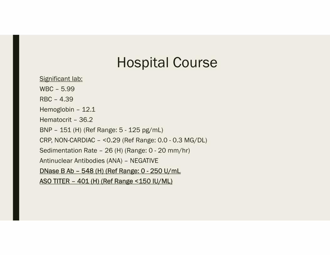
Hospital CourseSignificant lab:
WBC – 5.99
RBC – 4.39
Hemoglobin – 12.1
Hematocrit – 36.2
BNP – 151 (H)( (Ref Range: 5 - 125 pg/mL)
CRP, NON-CARDIAC – <0.29 (Ref Range: 0.0 - 0.3 MG/DL)
Sedimentation Rate – 26 (H) (Range: 0 - 20 mm/hr)
Antinuclear Antibodies (ANA) – NEGATIVE
DNase B Ab – 548 (H) (Ref Range: 0 - 250 U/mL)
ASO TITER – 401 (H) (Ref Range <150 IU/ML)

Hospital Course
Patient remained in hospital for approximately 4 days. She remained afebrile with no symptoms of cardiac dysfunction and no change in her clinical status
Vancomycin and Gentamicin were discontinued after blood cultures remained negative for 72 hours.
Prior to discharge a repeat ECHO was done that showed trivial improvement in mitral regurgitation and minimal improvement in aortic regurgitation.
Patient was started on Penicillin VK 250 mg BID. Parents were counseled on the need for Penicillin therapy until age 40, and possibly lifelong due to increased risk of recurrent attack of rheumatic fever
She was discharged from hospital with Pediatric Cardiology follow up.

Rheumatic Fever
Acute rheumatic fever is a non-suppurative sequela that occurs two to four weeks following a Group A Streptococcus pharyngitis (GAS).
GAS pharyngitis is a disease primarily of children aged 5 to 15 years of age
Clinical findings suggestive of GAS pharyngitis:
- Sudden onset of sore throat, pain on swallowing, fever (101F-104F), scarlet fever rash, headache, nausea/vomiting, tonsilopharyngeal erythema/exudates, soft palate petechiae, Tender and enlarged anterior cervical lymph nodes and presentation in the winter or early spring.
Clinical features suggestive of viral origin:
- Coryza, conjuncitivitis, cough, hoarseness, diarrhea, and viral exanthems

Diagnosing Rheumatic Fever
The Jones Criteria, published in 1944, modified in 1992 and reaffirmed in 2002, has been used for guidance in the diagnosis of Acute Rheumatic Fever
5 Major Jones Criteria■ Carditis (50%-70%)
■ Arthritis (35%-66%)
■ Sydenham Chorea (10%-30%)
■ Subcutaneous Nodules (0%-10%)
■ Erythema Marginatum (<6%)
4 Minor Jones Criteria■ Arthralgia
■ Fever
■ Elevated acute phase reactants (ESR, CRP)
■ Prolonged PR interval on EKG

Problems with Traditional Jones Criteria■ Disproportion in the global Distribution of Rheumatic fever and Rheumatic heart
disease
■ Declining incidence of acute rheumatic fever in Europe and North America
■ Decline in the prevalence of Rheumatic Heart disease in Europe and North America
■ Limited diagnostic role of Echocardiography
■ Previous guidelines did not categorize recommendations based on classification of recommendations and level of evidence categories
■ Thus the Jones criteria was revised in 2015

Using Classification of Recommendations and Level of Evidence
■ Revision of jones criteria using classification of recommendations and level of evidence provides 2 sets of criteria for low risk and high risk populations (class IIa; Level of evidence C)
■ Monoarthritis as part of the acute rheumatic fever spectrum, should be limited to patients from moderate to high risk populations (Class I; Level of evidence C)
■ The inclusion of polyarthralgia as a major manifestation is applicable only for moderate or high risk populations and only after careful consideration and exclusion of other causes of arthralgia (Class IIb; Level of evidence C)

Role of Echocardiography
■ Classically carditis as a major manifestation of acute rheumatic fever has been a clinical diagnosis based on the auscultation of murmurs that indicate mitral or aortic valve regurgitation
■ Subclinical carditis refers to the circumstance in which classic auscultatory findings of valvar dysfunction are not present or not recognized but ECHO studies reveal valvulitis.
■ Several studies have reported echocardiographic evidence of mitral or aortic valve regurgitation in patients with acute rheumatic fever despite the absence of classic auscultatory findings
■ All of the studies reviewed overwhelmingly support the use of echocardiography as part of the diagnostic criteria for confirmation of the presence of carditis in patients with suspected acute rheumatic fever

Role of Echocardiography
■ Echocardiography with Doppler should be performed in all cases of confirmed and suspected acute rheumatic fever (class I; level of evidence B)
■ It is reasonable to consider performing serial ECHO/Doppler studies in any patient with diagnosed or suspected acute rheumatic fever even if documented carditis is not present on diagnosis (class IIa; level of evidence C)
■ ECHO/Doppler testing should be performed to assess whether carditis is present in the absence of auscultatory findings, particularly in moderate to high risk populations andwhen acute rheumatic fever is considered likely (class I; level of evidence B)
■ ECHO/Doppler findings not consistent with carditis should exclude that diagnosis in patients with a heart murmur otherwise thought to indicate rheumatic carditis (Class I; level of evidence B)

Case Update2 days after starting Penicillin therapy patient developed a erythemic, pruritic rash of the extremities. Penicillin was switched to sulfadiazine and the rash improved.
She was seen in Allergy & immunology clinic and penicillin skin testing was scheduled. Sulfadiazine was switched to azithromycin.
Patient did not make appointment for penicillin skin testing. She was switched back to Penicillin without any hypersensitivity reaction.
Her most recent ECHO continues to show severe valvar dysfunction however clinically patient remains asymptomatic.
She will likely require valve replacement in the future if her valvar dysfunction does not improve.
She continues to take Lisinopril and Penicillin. Her penicillin therapy will likely be lifelong

References■ Gerber, M.A., et al (2009). Prevention of Rheumatic Fever and Diagnosis and
Treatment of Acute Streptococcal Pharyngitis. Circulation. 2009;119:1541-1551
■ Gewitz, M.H., et al. (2015). Revision of the Jones Criteria for the Diagnosis of Acute Rheumatic Fever in the Era of Doppler Echocardiography, A scientific statement from the American Heart Association, Circulation. 2015
■ Remenyi, B. et al. Position statement of the World Heart Federation on the prevention and control of rheumatic heart diseaseNat. Rev. Cardiol. 10, 284-292 (2013

Questions?