revisão imaging procedures in adrenal pathology - … adrenal glands are paired retroperitoneal...
TRANSCRIPT

592 Arq Bras Endocrinol Metab vol 48 nº 5 Outubro 2004
ABSTRACT
Imaging plays a vital role in the evaluation of adrenal pathology. Themost widely used modalities are computed tomography and magneticresonance imaging. Alone or in conjunction with appropriate clinicaland biochemical data, imaging can provide specific diagnoses thatpreclude the need for tissue sampling. This article reviews imaging fea-tures of normal and diseased adrenals, from both benign and malig-nant causes. (Arq Bras Endocrinol Metab 2004;48/5:592-611)
Keywords: Adrenal glands; Imaging; Computed tomography; Magnet-ic resonance imaging
RESUMO
Procedimentos de Imagem na Patologia Adrenal.Procedimentos de imagem têm um papel vital na avaliação dapatologia adrenal. As modalidades mais amplamente empregadassão a tomografia computadorizada e a imagem por ressonânciamagnética. Isoladas ou em combinação com dados clínicos e bio-químicos apropriados, a imagem pode prover diagnósticos específi-cos que dispensam a necessidade de amostras de tecido por biopsia.Este artigo revisa os achados de imagem da adrenal normal epatológica, incluindo tanto causas benignas como malignas. ( A r qBras Endocrinol Metab 2004;48/5:592-611)
Descritores: Glândulas adrenais; Imagem; Tomografia computadoriza-da; Ressonância magnética
INTRODUCTION AND IMAGING TECHNIQUE
THE ADRENAL GLANDS CAN BE imaged through ultrasound, computedtomography or magnetic resonance imaging.
Sonography (US) of the adrenal glands is not performed routinely,either because of the deep and often times inaccessible location of thegland or because of the lack of specific imaging patterns, leading to non-diagnostic studies. Adrenal pathology is therefore usually an incidentalfinding in an US study performed for other reasons, and patients arereferred for further evaluation.
Computed Tomography (CT) is the primary diagnostic imagingmethod for evaluation of adrenal disorders. When optimal CT scan-ning technique is used, the normal or pathologic adrenal glands can bewell visualized in virtually 100% of patients (1). Large and small mass-es and hyperplasia can be readily detected when present. With modernCT scanners, imaging of the adrenals can be achieved even in the mostemaciated patients, and in patients who are unable to suspend respira-t i o n .
revisão Imaging Procedures in Adrenal Pathology
Suzan M. GoldmanRafael Darahem CoelhoEdison de O. Freire FilhoNitamar AbdalaDenis SzejnfeldJuliano FariaPaola L.P. JudiceViviane Vieira FranciscoPhilip J. KenneyJacob Szejnfeld
Genitourinary Imaging Service,Department of DiagnosticImaging, Universidade Federalde São Paulo, São Paulo, SP,Brasil; and Department ofRadiology, University ofAlabama (PJK), Birmingham,Alabama, USA
Recebido em 09/06/04Aceito em 14/06/04

Imaging Procedures in Adrenal PathologyGoldman et al.
593Arq Bras Endocrinol Metab vol 48 nº 5 Outubro 2004
The adrenals can be well imaged with axialslices. Most large adrenal masses can be detected withrapid scanning with 5mm contiguous slice technique.However, thinner slices (2.5 – 3mm, contiguous)should be made if small lesions are suspected or if ini-tial screening is negative in a patient strongly suspect-ed of having adrenal disease (figure 1). Oral contrastshould be used routinely as it helps delineate the gas-trointestinal structures of the upper abdomen, and ithelps avoid mistaking a bowel structure for an adrenalmass. The imaging protocol must be tailored to theclinical indication. The conditions that require differ-ent techniques will be discussed later. For mostpatients, the following technique for a helical CT studyis appropriate: 2.5 – 3mm slice thickness, with 1.5 –3mm reconstruction interval through the adrenalregion, following oral contrast, performed before andafter intravenous injection 100 – 150ml low osmolariodinated contrast medium. Imaging delays after con-trast injection are routine “portal venous phase” (60 –90 seconds) and “delayed-phase” (15 minutes), whenappropriate. With a 4 multidetector-row CT scanner,using HS mode (pitch 1.5 or “6”) with 1.25mmdetectors, 2.5mm slice thickness with table speed7.5mm/rotation (1.5 pitch x 1.25mm detector x 4row). Alternatively, in HQ mode (pitch 0.75 or “3”),2.5mm detectors, slice thickness of 2.5mm with tablespeed 7.5mm/rotation (0.75 pitch x 2.5mm detectorx 4 row). Reformatted images (in sagital or coronalplane) can sometimes be useful in determiningwhether a mass arises from the adrenal, liver or kidney.
Great strides have been made in magnetic reso-nance imaging (MRI) of the abdomen; and with cur-rent techniques, the adrenals also can be delineated innearly all patients with MRI (2-4). Patients who areclaustrophobic, are unstable or erratic breathers,exceed the size limits, or have pacemakers cannot beevaluated with MRI. State-of-the-art MRI scanners
now produce thin sections (3 to 5mm) with good spa-tial resolution and signal-to-noise ratio. The use of res-piratory compensation techniques, or rapid imagingsequences that can be performed during suspendedrespiration, produces images without significantmotion artifacts. Fat suppression makes normal adren-als very conspicuous on T2-weighted images: they arebright, in contrast to surrounding retroperitoneal fat.Intravenous contrast agents now available for MRIprovide information about enhancement patterns ofmasses that is similar to that obtained with contrast-enhanced CT. Techniques sensitive to the presence oflipid (such as in/out-phase and fat suppression) aremost useful in differentiating between subacute hem-orrhage and fat-containing adrenal masses, both ofwhich can appear as high signal lesions on T1-weight-ed spin echo images.
An MRI evaluation of the adrenals usually con-sists of: T1 gradient refocused echo (GRE) weightedsequences in and out of phase in the axial and coronalplanes, 3 to 5mm slice thickness; a T2-weighted TSEsequence with fat saturation in the axial plane, 3 to5mm slice thickness; and a T2-weighted HASTEsequence in the coronal plane, slice thickness of 5mm.The use of intravenous contrast agents should be lim-ited to those cases where there rests a doubt, being, inthose cases, used to differentiate solid lesions fromhemorrhages, and in cases of pheochromocytoma. Theuse of such contrast media is not yet established in thediagnosis of adenomas.
Despite the many improvements in MRI thatallow more rapid data acquisition, an MRI examina-tion of the adrenals is still a relatively lengthy proce-dure compared to CT. Therefore, it is most useful asan alternative to CT in patients who cannot tolerateintravenous iodinated contrast, in cases of suspectedpheochromocytoma and to confirm a diagnosis ofhemorrhage.
Figure 1. Use of thinner collimation. A: CT with 7mm contiguous sections demonstrates an equivocal right adrenal nodule.B: Repeat with contiguous 3mm sections clearly shows a small nodule (arrow).

Imaging Procedures in Adrenal PathologyGoldman et al.
594 Arq Bras Endocrinol Metab vol 48 nº 5 Outubro 2004
NORMAL ANATOMY
The adrenal glands are paired retroperitoneal organsthat lie in a suprarenal location and are enclosed with-in the perinephric fascia. The right adrenal is usuallyseen directly superior to the upper pole of the rightkidney with its most caudal portion just anterior to theupper pole. The right adrenal is directly posterior tothe inferior vena cava and insinuates between the rightlobe of the liver and the right crus of the diaphragm.The left adrenal may be seen at the same level as theright, but it often is slightly more caudal. It is antero-medial to the upper pole of the left kidney and is usu-ally seen on CT in the same slice as the left kidney. Theleft adrenal lies lateral to the aorta and the leftdiaphragmatic crus, and superior to the left renal vein.A portion of the pancreas or the splenic vessels may beimmediately anterior to the left adrenal. When per-forming a dedicated (thin section) CT of the adrenals,it is important to obtain a sufficient number of images,because the adrenals each extend about 2 to 4cm incraniocaudal direction, and because masses may pro-trude superiorly or inferiorly.
The adrenal glands have medial and laterallimbs extending posteriorly from central ridges.Inverted V, Y, L, and other configurations may beseen. On precontrast CT, the adrenals have a soft-tis-sue density similar to that of the liver. If very earlypost-contrast scans are obtained, there is considerableenhancement, which fades quickly to moderateenhancement – slightly less than that of liver. Theadrenal cortex and medulla cannot be reliably distin-guished by either CT or MRI. On T1-weighted MRIimages, the adrenals have intermediate signal intensity,similar to that of the liver, somewhat greater than thediaphragmatic crus but much less than the surround-ing fat (5). On standard T2-weighted images, theadrenals are hypointense to fat and isointense to liver,but they are hyperintense to the crus. There is less dif-ference between adrenal and fat signal intensities onT2-weighted images, and significant chemical shiftartifact may obscure details of the normal adrenals. Onfat-suppressed T2-weighted images, however, the nor-mal adrenals appear somewhat brighter than liver, andare much brighter than the suppressed fat. Thus, nor-mal adrenals and small masses are best seen on T1-weighted or fat-suppressed T2-weighted images.
There is considerable variability in the lengthsof the limbs. The surface of the normal adrenal glandsshould be quite smooth, without protruding nodules,and the limbs should have uniform thickness.Although no strict measurements have been standard-
ized, any area thicker than 10mm is abnormal. A use-ful rough estimate of normal size is that the thicknessof the gland’s limbs should not exceed the thickness ofthe ipsilateral diaphragmatic crus, at the same level. Itmust be recognized that in the face of stress (as may beseen in severely ill patients), the adrenals may becomeenlarged in response to physiologically high circulatingadrenocorticotropic hormone (ACTH) levels.
PSEUDOTUMORS
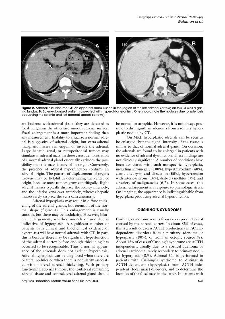
A variety of normal structures may simulate an adren-al mass if meticulous technique is not used. The rou-tine use of oral contrast and rapid scanning after bolusintravenous contrast will usually allow these pseudotu-mors to be distinguished from true adrenal masses.Pseudotumors are less common on the right, becausethere are fewer adjacent organs. Rarely, the duode-num, stomach or colon may produce a mass-likeappearance (figure 2a). One should be careful wheninterpreting images of previously splenectomizedpatients, some of which develop splenosis and thesmall splenic tissue nodules may mimic tumors whenlocated near to the adrenal space (figure 2b). Otherpotential mimickers of adrenal masses on the left arethe pancreatic tail, tortuous vessels and small bowel.
PATHOLOGY
A variety of pathologic processes can affect the adrenalglands. Some cause endocrine disorders (functional dis-eases), either hyperfunction or adrenal insufficiency,and some do not produce biochemical abnormalities.Although both CT and MRI provide accurate depic-tion of the adrenal morphology, both functional andnonfunctional disorders may produce similar appear-ances, so correlation of imaging findings with theendocrine status is usually necessary for diagnosis. Inpatients with biochemical evidence of adrenal hyper-function, a CT examination is valuable because of itsability to differentiate between a focal mass, hyperplas-tic glands, and normal adrenals. A pathologic diagnosiscan usually be made based on the imaging appearanceof an adrenal mass and the clinical history, even whenthere is no adrenal dysfunction. The accuracy of CT fordiagnosis of adrenal masses has been reported as beingbetter than 90% (1). With proper technique, massessmaller than 5mm can be detected. A normal appear-ance of the adrenal effectively excludes the presence ofan adrenal tumor. Because many small adrenal masses
Imaging Procedures in Adrenal PathologyGoldman et al.
595Arq Bras Endocrinol Metab vol 48 nº 5 Outubro 2004
are isodense with adrenal tissue, they are detected asfocal bulges on the otherwise smooth adrenal surface.Focal enlargement is a more important finding thanany measurement. Inability to visualize a normal adre-nal is suggestive of adrenal origin, but extra-adrenalmalignant masses can engulf or invade the adrenal.Large hepatic, renal, or retroperitoneal tumors maysimulate an adrenal mass. In these cases, demonstrationof a normal adrenal gland essentially excludes the pos-sibility that the mass is adrenal in origin. Conversely,the presence of adrenal hyperfunction confirms anadrenal origin. The pattern of displacement of organslikewise may be helpful in determining the center oforigin, because most tumors grow centrifugally. Rightadrenal masses typically displace the kidney inferiorly,and the inferior vena cava anteriorly, whereas hepaticmasses rarely displace the vena cava anteriorly.
Adrenal hyperplasia may result in diffuse thick-ening of the adrenal glands, but retention of the nor-mal shape (figure 3). This enlargement is usuallysmooth, but there may be nodularity. However, bilat-eral enlargement, whether smooth or nodular, isindicative of hyperplasia. A significant number ofpatients with clinical and biochemical evidence ofhyperplasia will have normal adrenals with CT. In part,this is because there may be significant hyperfunctionof the adrenal cortex before enough thickening hasoccurred to be recognizable. Thus, a normal appear-ance of the adrenals does not exclude hyperplasia.Adrenal hyperplasia can be diagnosed when there arebilateral nodules or when there is nodularity associat-ed with bilateral adrenal thickening. With primaryfunctioning adrenal tumors, the ipsilateral remainingadrenal tissue and contralateral adrenal gland should
be normal or atrophic. However, it is not always pos-sible to distinguish an adenoma from a solitary hyper-plastic nodule by CT.
On MRI, hyperplastic adrenals can be seen tobe enlarged, but the signal intensity of the tissue issimilar to that of normal adrenal gland. On occasion,the adrenals are found to be enlarged in patients withno evidence of adrenal dysfunction. These findings arenot clinically significant. A number of conditions havebeen associated with such nonspecific hyperplasia,including acromegaly (100%), hyperthyroidism (40%),aortic aneurysm and dissection (55%), hypertensionwith arteriosclerosis (16%), diabetes mellitus (3%), anda variety of malignancies (6,7). In some cases, thisadrenal enlargement is a response to physiologic stress.On imaging, the appearance is indistinguishable fromhyperplasia producing adrenal hyperfunction.
CUSHING’S SYNDROME
Cushing’s syndrome results from excess production ofcortisol by the adrenal cortex. In about 85% of cases,this is a result of excess ACTH production (an ACTH-dependent disorder) from a pituitary adenoma orhyperplasia (80%), or from an ectopic source (8).About 15% of cases of Cushing’s syndrome are ACTHindependent, usually due to a cortical adenoma oradrenal carcinoma, rarely secondary to primary nodu-lar hyperplasia (8,9). Adrenal CT is performed inpatients with Cushing’s syndrome to distinguishACTH-dependent (hyperplasia) from ACTH-inde-pendent (focal mass) disorders, and to determine thelocation of the focal mass in the latter. In patients with
Figure 2. Adrenal pseudotumor. A: An apparent mass is seen in the region of the left adrenal (arrow) on this CT was a gas-tric fundus. B: Splenectomized patient suspected with hyperaldosteronism. One should note the nodules due to splenosisoccupying the splenic and left adrenal spaces (arrows).

Imaging Procedures in Adrenal PathologyGoldman et al.
596 Arq Bras Endocrinol Metab vol 48 nº 5 Outubro 2004
an adenoma or a carcinoma, the remaining ipsilateraland the contralateral adrenals are normal or atrophic.Surgical removal of an adenoma (or of a resectable car-cinoma) is curative. Biochemical testing can beextremely useful.
Computed tomography is virtually 100% accu-rate for detection of adrenal adenomas resulting inhypercortisolism. These adenomas are almost alwaysover 2cm at the time of presentation, are usually in therange 2 to 5cm (10), and are readily seen on CT, espe-cially because these patients have abundant retroperi-toneal fat. The masses are smooth, round or oval, andrelatively homogeneous, with little enhancement afterintravenous contrast and rapid washout at 15 minutes,in excess of 60% from baseline attenuation (11). Mostoften they have a soft-tissue-attenuation value, butthey may have near water attenuation because of rela-tively high lipid content. Calcification can be found inadenomas, although it is rare (12) (figure 4).
The contralateral adrenal is commonly visiblythinner than normal, indicating atrophy from ACTHsuppression (figure 5). With these findings, and in theproper clinical setting, no further investigation is need-ed. It should be noted that no CT characteristic hasbeen reported that distinguishes whether an adenomaproduces excess cortisol or aldosterone, or whether itis not hyperfunctioning.
Magnetic resonance imaging is equally accuratein patients with cortisol producing adenomas, becauseof their size (9). Typically, the signal intensity of the ade-nomas is similar to that of liver on T1-weighted images,and similar or only slightly greater on T2-weightedimages (3) (figure 6). The presence of intracellular lipidcan be documented by opposed-phase imaging (17).Although adrenal adenomas and cysts may have similar
attenuation values on CT, they are readily distinguishedon MRI, because cysts are very hyperintense on T2-weighted images and do not enhance.
A normal CT or MRI appearance does notexclude hyperplasia. In fact, in the absence of a focalmass, a patient with biochemical evidence of hypercor-tisolism and normal adrenals can confidently be giventhe diagnosis of hyperplasia as long as exogenoussteroid use is excluded. Most often, both adrenalsbecome smoothly thickened due to excess ACTH.The thickening may become massive, especially withectopic ACTH production (10).
Adrenal nodules ranging from 6mm to 7cmmay be seen in 12% to 15% of patients with ACTH-dependent Cushing’s syndrome (10). Although usual-ly bilateral, such nodules may be unilateral. Carefulexamination will usually show some evidence of bila-teral enlargement. Primary pigmented nodularadrenocortical disease is a rare disorder that causesCushing’s syndrome. It tends to present in youngerpatients than the other types of Cushing’s syndrome.Elevated cortisol is found, with very low ACTH levels.On CT, multiple bilateral nodules up to 3cm are typi-cal. Unlike macronodular hyperplasia due to excessACTH, the cortex between the nodules is atrophic.On MRI, the nodules are relatively low signal on bothT1- and T2-weighted images.
PRIMARY ALDOSTERONISM
Primary aldosteronism (Conn’s syndrome) resultsfrom excess adrenal production of the mineralocorti-coid aldosterone. It is characterized by reduced plasmarenin levels, hypokalemia, and hypertension, with as
Figure 3. Nodular hyperplasia – Diffuse nodular thickening of the adrenal glands (arrows).

Imaging Procedures in Adrenal PathologyGoldman et al.
597Arq Bras Endocrinol Metab vol 48 nº 5 Outubro 2004
many as 95% of cases resulting from an autonomouscortical adenoma (aldosteronoma). Most of theremaining cases result from primary idiopathic bilater-al hyperplasia. A few rare cases have been reported as aresult of bilateral adenomas, unilateral hyperplasia, andadrenal carcinoma. Correct diagnosis is important,because surgical removal of an aldosteronoma is cura-tive, but partial and even bilateral total adrenalectomycommonly fails to cure hypertension in patients withhyperplasia. Adrenal CT should be performed first toevaluate a patient with biochemical evidence of prima-ry hyperaldosteronism. Certain biochemical featuresare strongly indicative of an aldosteronoma, and CTshould be done to locate the tumor before surgical
removal. If the imaging study is inconclusive, adrenalvenous sampling should be done and all diagnosticstudies correlated prior to surgery.
On CT, aldosteronomas appear as round oroval lesions, similar to other adenomas, but typical-ly smaller than cortisol-producing adenomas. Theyare rarely larger than 3cm, with a median size of 16to 17mm. Because of relatively high lipid content,they often (50%) have an attenuation value similarto that of water (-10 to +10). This propensity forlow attenuation should be recognized, so that alow-attenuation mass discovered in a patient withdocumented hyperaldosteronism is not misdiag-nosed as a cyst.
Figure 4. Calcification on a right adrenal adenoma(arrow).
Figure 5. Cushing’s syndrome due to a cortical adenoma.The left adrenal (arrowhead) is atrophic in this patient withhypercortisolism. There is a 3cm right adrenal mass (arrow).
Figure 6. Cushing’s syndrome due to a cortical adenoma. A: T1-weighted GRE image done during suspended respirationshows a homogeneous 3.0cm mass in the right adrenal (arrow) with signal intensity slightly less than that of liver. B: On theopposed phase image the mass lost signal.

Imaging Procedures in Adrenal PathologyGoldman et al.
598 Arq Bras Endocrinol Metab vol 48 nº 5 Outubro 2004
Because aldosteronomas are typically small,they present a greater challenge than cortisol-pro-ducing adenomas. Early publications reported asensitivity of 70% with several false negative CTsbecause of inability to detect small aldosteronomas.With better quality, thin-section CT, a sensitivityof 80% to 90% has been achieved. With the 5mm-section technique, false negatives are uncommon(12% to 14%). Primary adrenal hyperplasia causingaldosteronism may be micronodular or macron-odular. The adrenals may appear normal or diffuse-ly thickened on CT. One or more discrete nodulesranging from 7 to 16mm may be seen. Diagnosticerrors may occur because of a unilateral nodule thatsimulates an adenoma on CT. Conversely, tinybilateral nodules that are present in 25% of patientswith aldosteronomas may result in an erroneousdiagnosis of hyperplasia. An accuracy of 80% forCT showing lack of lateralization (either bothglands are normal, both are enlarged, or there arebilateral nodules) has been reported. Because ofthese potential difficulties, adrenal venous sam-pling still can be useful in certain patients.Although aldosteronomas can be shown with MRI,there is no advantage of MRI over CT. Becausemany aldosteronomas are small, the lesser spatialresolution of MRI is a theoretical disadvantage.Aldosteronomas are not distinguishable from otheradenomas by any MRI feature. In fact, as with CT,no MRI feature has been described that indicateswhether an adenoma is hyperfunctioning or non-h y p e r f u n c t i o n i n g .
ADRENAL CARCINOMA
Adrenal carcinoma is a highly malignant neoplasm thatarises in the adrenal cortex. It is rare, with an incidenceestimated at two cases per million people. It can occurat any age, with a median age of about 40 years. About50% of adrenal carcinomas will produce an endocrinedisorder. Cushing’s syndrome is commonest, seen inabout 50% of adrenal carcinoma patients, and itaccounts for 65% of the functional disorders. It may beseen alone or in combination with virilization. Viril-ization alone, feminization, and aldosteronism may beseen, in order of decreasing frequency.
Adrenal carcinomas are often very large when firstdetected. This is especially true of nonfunctioningtumors, which remain clinically silent until advancedstages, they may be discovered because of flank pain,fatigue, palpable mass, or evidence of metastases. Evenfunctioning tumors are usually large at presentation,which may be a result of relatively inefficient hormoneproduction. Average size at presentation is 12cm (range,3 to 30cm). Nowadays, however, with widespread use ofimaging, some of these neoplasms are discovered inci-dentally and are smaller than previously reported. Ade-nomas are typically small, round or ovoid, homoge-neous, with less than 10 Hounsfield units attenuation onprecontrast CT and signal loss on opposed phase imageson MRI and show very slow growth. Adrenal carcinomatypically shows rapid growth (figure 7). As a result, ifthere is concern, a single 6-month follow-up CT canreadily distinguish these entities; lack of growth effec-tively excludes adrenal carcinoma.
Figure 7. Carcinoma of the Right Adrenal. A: Initial examination shows small nodule of the right adrenal gland (arrow), erro-neously diagnosed as an adenoma. B: Repeat CT 9 months later shows rapid growth of the mass (arrow), a characteristicof adrenal carcinoma.

Imaging Procedures in Adrenal PathologyGoldman et al.
599Arq Bras Endocrinol Metab vol 48 nº 5 Outubro 2004
The histology of adrenocortical carcinoma isvariable. It can be difficult to distinguish a well-differ-entiated carcinoma from an adenoma, even with aresected specimen. Needle biopsy may be nondiagnos-tic. Correlation of histology with radiological features,and sometimes with biologic behavior, is needed fordiagnosis. The overall prognosis is very poor, with 5-year survival of 20% to 25%. However, prognosis is bet-ter (42% to 57%) for localized (stage I) adrenal carcino-mas if complete surgical resection can be accomplished.
CT readily detects adrenal carcinomas. Mostoften they are seen as large, irregularly shaped, hetero-geneous masses in the adrenal region. Bilateral diseaseis seen in less than 10% of cases. Necrosis is common.Calcification is found in about 40% (12) (figure 8).Heterogeneous enhancement after intravenous con-trast is typical, with strong peripheral and little centralenhancement. The tumors may be poorly marginated,or they may show local invasion. Invasion of the infe-rior vena cava, liver metastases, and retroperitoneallymphadenopathy may be seen. In general, these fea-tures allow differentiation from adenoma or hyperpla-sia. However, an incidentally found carcinoma may bemore difficult to discriminate from an adenoma onCT, as both may be less than 5cm, well circumscribed,and homogeneous.
On MRI, adrenal carcinomas are easily seen aslarge heterogeneous masses in the adrenal bed, withareas isointense or hypointense to liver on T1-weight-ed images, and isointense or hyperintense to fat onT2-weighted images (2) (figure 9). Areas of hemor-rhage may result in variable signal intensity dependingon the age of the hemorrhage. With multiplanar capa-
bility and the high tissue contrast of T2-weightedimages, MRI can be useful to define the adrenal originand the extent of disease (figure 9). After injection ofgadolinium, bright heterogeneous enhancement isseen (18). The high sensitivity of MRI for venousinvolvement and liver disease make it helpful for stag-ing; venous extension is a poor prognostic sign, and ifmetastases are present surgery is not indicated.
PHEOCHROMOCYTOMA
A pheochromocytoma is a neoplasm of the adrenalmedulla that contains chromaffin cells and causesexcess catecholamine production. When such a tumorarises outside the adrenal, it is labeled a paragan-glioma. Most are benign, although about 10% aremalignant. Sporadic cases are usually unilateral, affect-ing the right adrenal slightly more frequently; about5% are bilateral (figure 10). Most patients are hyper-tensive. However, pheochromocytoma is found in lessthan 1% of the hypertensive population, and in 0.3% ofautopsies. There is an increased likelihood of pheo-chromocytoma in patients with neurofibromatosis,von Hippel-Lindau disease, and multiple endocrineneoplasia (MEN) syndromes (50% in MEN 2 and 90%in MEN 2b). In such syndromes and in children, mul-tiple or bilateral cases are more likely. In MEN 2b,bilateral tumors are so common that bilateral adrena-lectomy is recommended, because lesions may recurafter unilateral surgery.
In most cases, diagnosis of pheochromocytomacan be established with biochemical testing. However,
Figure 8. Adrenal carcinoma. A: Precontrast CT shows a heterogeneous 8cm left adrenal mass (*) containing a smallamount of calcification (arrow). B: The mass shows moderate, heterogeneous enhancement after contrast.

Imaging Procedures in Adrenal PathologyGoldman et al.
600 Arq Bras Endocrinol Metab vol 48 nº 5 Outubro 2004
biochemical tests are expensive, time consuming andfraught with difficulty, because such factors as episodiccatecholamine production, concurrent medication,stress, inadequate urine collection for 24-hour samples,and other factors can contribute to both false positiveand false negative results. Detection and localization areimportant because surgical resection is curative, andbecause there is no effective medical therapy.
Although 90% of pheochromocytomas arise inthe adrenal, up to 10% are extra-adrenal (figure 11),with many such lesions (7%) in the infrarenal portionof the retroperitoneum, arising in the organ of Zuck-
erkandl (figure 11). Paragangliomas can be single ormultiple, and they may have greater malignant poten-tial. Paragangliomas also can be found in the neck, themediastinum, and the wall of the urinary bladder.
Pheochromocytomas are usually over 3cm atpresentation and should be invariably identified byCT. When small, the tumors are round and havehomogeneous soft-tissue attenuation values (figure12). Because pheochromocytomas are hypervascularneoplasms, they have a propensity to undergo hemor-rhagic necrosis even when benign, accounting for thecentral low attenuation seen in large neoplasms. Cen-
Figure 9. Cushing’s syndrome due to adrenal carcinoma. A: Post-contrast CT shows a heterogeneous right adrenal mass(arrow) with non-enhancing area. B: T1-weighted GRE sequence. C: On T2-weighted sequence (with fat suppression) mostof the mass is moderately hyperintense, with a very hyperintense area (arrow) corresponding to the non-enhancing areaon CT, representing an area of necrosis. D: Coronal image following intravenous administration of Gd-DTPA, there is het-erogeneous enhancement (arrow).

Imaging Procedures in Adrenal PathologyGoldman et al.
601Arq Bras Endocrinol Metab vol 48 nº 5 Outubro 2004
tral necrosis may be so extensive as to simulate a cyst.Calcification is uncommon; when present, it may havean eggshell pattern. After intravenous administrationof iodinated contrast medium, pheochromocytomasexhibit heterogeneous enhancement, a pattern indis-tinguishable from a malignant adrenal neoplasm. Cor-relation with biochemical function is required toestablish the correct diagnosis.
Because pheochromocytomas are large, theycan be detected even with unenhanced CT. Some con-cern has been raised about the use of intravenous con-trast in patients with pheochromocytoma. Plasma cat-echolamine levels can be raised by intravenous injec-tion of iodinated contrast medium, but symptomaticblood pressure elevations do not usually result. Only ifa patient has had hypertensive episodes and no ade-quate pharmacologic adrenergic blockade is it neces-sary to avoid contrast. Contrast is especially useful fordetection of extra-adrenal lesions. Although paragan-gliomas can usually be identified on CT (figure 11),
they have a nonspecific appearance. The CT features ofmalignant paragangliomas in particular overlap withthose of other retroperitoneal malignancies. Radionu-clide metaiodobenzylguanidine (MIBG) scintigraphycan be useful to document whether a retroperitonealmass is in fact a paraganglioma.
Pheochromocytomas have a rather characteris-tic appearance on MRI (2,3). They are readily detect-ed (with a sensitivity of 100% in one report), becausethey are several centimeters in diameter. When small,they are usually homogeneous and isointense to mus-cle, hypointense to liver on T1-weighted images, andhave a distinctive marked signal hyperintensity relativeto fat on T2-weighted images (3) (figures 12 and 13).As they grow and develop central necrosis, there maybe central areas that are hyperintense on both T1- andT2-weighted images.
Persistent enhancement after intravenousgadolinium is typical (18). Because no lipid is found inpheochromocytoma, there is no decrease in signal onopposed phase images. Paragangliomas have similardistinctive imaging characteristics; as a result, MRI issuperior to CT for diagnosis of paragangliomas, andnearly as sensitive as MIBG. Because most suchtumors lie in the adrenal or retroperitoneum, coronalMRI can quickly and effectively show the area ofabnormality.
Biopsy of a mass suspected to be a pheochro-mocytoma is not recommended, especially if adequatehypertensive control has not been achieved, as severalepisodes of severe hemorrhage and even death haveresulted following percutaneous biopsy. MIBG hasboth high sensitivity and high specificity, and it candetect a paraganglioma in any part of the body. How-ever, it is an expensive test that requires up to 72 hoursto complete and is not widely available. Furthermore,it does not provide sufficient anatomic detail for surgi-cal planning. It is most useful in evaluating patients
Figure 10. Multiple pheochromocytomas in MEN2b. A and B: CT shows bilateral heterogeneous adrenal tumors (arrows).
Figure 11. Organ of Zuckerkandl paraganglioma.Enhanced CT shows a mass at the aortic bifurcation (arrow).

Imaging Procedures in Adrenal PathologyGoldman et al.
602 Arq Bras Endocrinol Metab vol 48 nº 5 Outubro 2004
with a strong clinical suspicion and in whom CT orMRI is normal or equivocal, or for follow-up of malig-nant lesions.
The follow up for these patients should be veryjudicious and information on clinical presentations andsurgical data should be included and taken intoaccount during analysis of exam results, be they froma CT or from an MR. The presence of hypertension inthis group of patients is not always indicative of persis-tence or recurrence of the disease. Other causes ofhypertension should be evaluated, such as renalchanges due to chronic disease.
NONHYPERFUNCTIONING NEOPLASMS
Nonhyperfunctioning adrenal neoplasms are clini-cally silent until they become very large, althoughthey may present with pain if they bleed. Current-ly, most such masses are found incidentally onstudies performed for other reasons. About 30% of
all adrenal masses are incidentally detected by CT.An adrenal mass is seen in about 4% of all abdomi-nal CT scans, with one third being serendipitousfindings; the remainder are either metastases inpatients with known malignancies, or they arefunctioning lesions. Most incidental adrenal massesare benign and of no clinical significance, especial-ly in patients with no known malignancy. In twolarge series, only 6.7% and 9% of serendipitousadrenal masses were subsequently proven malig-nant. Although historically size has been consi-dered an important factor, larger tumors having agreater likelihood of malignancy, size is an imper-fect criterion. Although malignant neoplasms wereall larger than 6.5cm in one study, and mostbenign masses are less than 5cm, there is conside-rable overlap. The majority of incidental massesgreater than 5cm are still benign in patients withno history of malignancy, and lesions as small as1cm may be metastases (13). Thus, imaging crite-ria are required for differentiation.
Figure 12. Pheochromocytoma in a man with episodic hypertensive crises. A: Homogeneous right adrenal mass on CTimage (arrow). B: On T1-weighted image (366/26), the mass (arrow) has signal intensity similar to liver. C: On T2-weightedimage (2500/80), the mass (arrow) is homogeneous and more intense than fat.
Figure 13. Pheochromocytoma. A: Post-contrast CT shows a right adrenal mass (arrow) with marked heterogeneousenhancement. B: On fat-suppressed T2-weighted image (2500/70), the mass (arrow) is markedly hyperintense.
Imaging Procedures in Adrenal PathologyGoldman et al.
603Arq Bras Endocrinol Metab vol 48 nº 5 Outubro 2004
NONHYPERFUNCTIONING ADENOMAS
Nonhyperfunctioning adrenal adenomas are not infre-quent, being found in 2% to 8% of autopsies (1,65)and in 1% to 2% of abdominal CT scans. They aremost commonly unilateral, although bilateral adeno-mas do occur. Although nonhyperfunctioning adeno-mas may be 6cm or larger (13), the majority is 3cm orless, with only 5% exceeding 5cm. Calcification maybe present (12).
Nonhyperfunctioning adenomas have a CTappearance indistinguishable from other adenomas,except that contralateral atrophy is not present. Theyare smooth, round or oval, with a well-defined margin.
CT densitometry can be used to accurately dif-ferentiate benign from malignant adrenal masses. Leeet al. reported the use of nonenhanced CT attenua-tion values for the characterization of adrenal masseswhere most adenomas had attenuation values lowerthan those of malignant masses (15). Korobkin et al.and Boland et al., confirmed these findings. Bolandpooled the data from 10 articles and showed that asensitivity of 71% and a specificity of 98% result fromchoosing a threshold value of 10HU for the diagno-sis of adrenal adenoma. Ninety-eight percent ofhomogeneous adrenal masses with a nonenhancedCT attenuation value of 10HU or less will be benign(most will be adenomas), whereas 29% of adenomaswill have an attenuation value of more than 10HUand will be indistinguishable from most nonadeno-mas, including metastases.
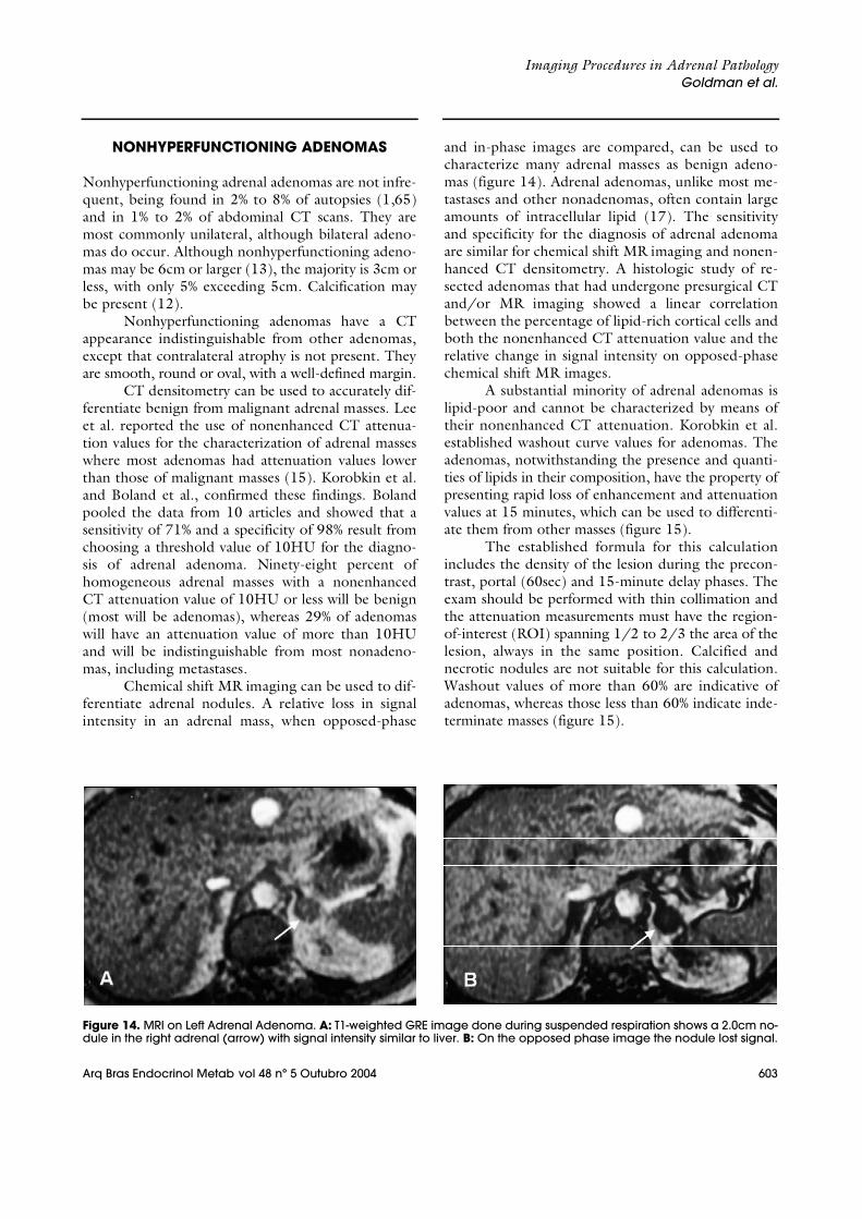
Chemical shift MR imaging can be used to dif-ferentiate adrenal nodules. A relative loss in signalintensity in an adrenal mass, when opposed-phase
and in-phase images are compared, can be used tocharacterize many adrenal masses as benign adeno-mas (figure 14). Adrenal adenomas, unlike most me-tastases and other nonadenomas, often contain largeamounts of intracellular lipid (17). The sensitivityand specificity for the diagnosis of adrenal adenomaare similar for chemical shift MR imaging and nonen-hanced CT densitometry. A histologic study of re-sected adenomas that had undergone presurgical CTand/or MR imaging showed a linear correlationbetween the percentage of lipid-rich cortical cells andboth the nonenhanced CT attenuation value and therelative change in signal intensity on opposed-phasechemical shift MR images.
A substantial minority of adrenal adenomas islipid-poor and cannot be characterized by means oftheir nonenhanced CT attenuation. Korobkin et al.established washout curve values for adenomas. Theadenomas, notwithstanding the presence and quanti-ties of lipids in their composition, have the property ofpresenting rapid loss of enhancement and attenuationvalues at 15 minutes, which can be used to differenti-ate them from other masses (figure 15).
The established formula for this calculationincludes the density of the lesion during the precon-trast, portal (60sec) and 15-minute delay phases. Theexam should be performed with thin collimation andthe attenuation measurements must have the region-of-interest (ROI) spanning 1/2 to 2/3 the area of thelesion, always in the same position. Calcified andnecrotic nodules are not suitable for this calculation.Washout values of more than 60% are indicative ofadenomas, whereas those less than 60% indicate inde-terminate masses (figure 15).
Figure 14. MRI on Left Adrenal Adenoma. A: T1-weighted GRE image done during suspended respiration shows a 2.0cm no-dule in the right adrenal (arrow) with signal intensity similar to liver. B: On the opposed phase image the nodule lost signal.

Imaging Procedures in Adrenal PathologyGoldman et al.
604 Arq Bras Endocrinol Metab vol 48 nº 5 Outubro 2004
The percentage enhancement washout is calcu-lated as follows:
% enhancement washout= (E-D/E-U) x 100,where:
E= Enhancement: attenuation at portal phaseD= delayed enhanced attenuation at 15minU= attenuation at pre contrastIn summary, the evaluation of a known adrenal
mass starts by using nonenhanced CT. If the attenua-tion of the mass is 10HU or less the diagnosis is, mostof the times, a lipid-rich adrenal adenoma and a smallfraction of these will be cysts. In such a case, there isno need for further evaluation. If the attenuation ismore than 10HU, the mass is considered to be inde-terminate and an enhanced and 15-minute-delayedenhanced CT scan should be done. If the enhance-ment washout is more than 60%, the most likely diag-nosis is of a lipid-poor adenoma. Again, there is noneed for further evaluation. If the enhancementwashout is less than 60%, the mass is considered inde-terminate. Percutaneous adrenal biopsy is recom-mended if the patient has a primary neoplasm with noother evidence of metastases. In a patient without can-cer, surgery is recommended if the mass measuresmore than 4 – 5cm. Follow-up CT, or adrenal scintig-raphy with the use of radioiodinated norcholesterol(NP-59) can also be done in this group of patients.
METASTATIC DISEASE
Metastases to the adrenals are common from a varietyof primary malignancies, including thyroid, renal, gas-tric, colon, pancreatic, and esophageal carcinomas, andmelanoma. Lung and breast cancer, however, are themost common sources, with adrenal metastases foundon CT in approximately 19% of lung cancer patients.Because adrenal metastases are so common in lung
cancer and the adrenals may be the only site of metas-tasis, they should be included in the CT examinationof all patients presenting with lung cancer. Even inpatients with lung cancer, about one third of adrenalmasses are benign. Thus, imaging features must beused to help make the correct diagnosis. If the imagingfindings are equivocal, a percutaneous CT-guidedbiopsy should be performed to establish a histologicdiagnosis. A baseline CT at the time of presentation ofpatients with lung cancer is a useful aid in follow-up.Detection of a new small adrenal mass on follow-up isclear evidence of metastasis if the baseline showed nor-mal adrenals.
Adrenal metastases can vary considerably onCT. Size can range from less than a centimeter to largemasses; the size, however, overlaps with that of ade-nomas (13) (figure 16). Adrenal metastases may beunilateral or bilateral. When small (< 5cm), they com-monly are fairly well circumscribed, round or oval, andof soft-tissue density. They may have smooth or irreg-ular, lobulated contours. They may show local inva-sion, a sign of malignancy. Calcification is rare (12),and they may bleed (14).
Small adrenal metastases are solid tumors andthus usually have homogeneous soft-tissue-attenua-tion values, similar to or higher than that of muscle onnoncontrast scans. Even if there is central necrosis,however, the density is not lower than that of water,because malignant tumors do not produce lipid (15).
Following intravenous administration of iodi-nated contrast material, there may be homogeneousenhancement, but commonly enhancement is hetero-geneous, especially with larger tumors. A thick, nodu-lar enhancing rim also may be seen (16).
Adrenal metastases can vary in size and appear-ance on MRI. On T1-weighted images, metastasesusually have signal intensity similar to or lower thanthat of normal liver tissue, not distinctly different from
Figure 15. Right Adrenal Adenoma. CT images on precontrast phase (A), portal post-contrast phase (B) and 15-minute-delayed post-contrast phase (C). Washout values of more than 60% are indicative of adenoma (E-D/E-U x 100 =115,3–54,6/115,3-18,0 x 100 = 64%).
Imaging Procedures in Adrenal PathologyGoldman et al.
605Arq Bras Endocrinol Metab vol 48 nº 5 Outubro 2004
that of adenomas. They may be heterogeneous (2,3).On T2-weighted images, they are often heteroge-neous, and they are usually hyperintense compared tonormal liver, often similar to or of higher intensitythan fat, unlike the typical adenoma (2,3). Numerouscalculations based on signal intensity ratios, or calcu-lated T2 values, have been investigated, but none havebeen found to be reliable in practice for distinguishingmetastases from adenomas (2,5). Because metastasesdo not produce lipid, there is no decrease in signal onopposed phase images (17). This has been shown tobe a more consistent finding than T2 values. Afterintravenous administration of gadolinium compounds,metastases exhibit exuberant and heterogeneousenhancement that persists for several minutes, anenhancement pattern quite different from that of ade-nomas (18). MRI is readily able to demonstrate orexclude local invasion because of the great contrastbetween neoplastic and normal tissue, especially onfat-suppressed T2-weighted images.
Computed tomography is the most cost-effec-tive method for screening and following patients withmalignancies. In most cases, adrenal metastases can bediagnosed or excluded by a well-performed CT. MRIcan be valuable in cases in which CT could not be per-formed. Percutaneous biopsy under CT guidance canbe very effective, with accuracy and negative predictivevalue of over 90% (19). However, complications canoccur, and biopsy is not needed if the imaging findingsare diagnostic. Follow-up by CT can be diagnostic,because adenomas are very slow growing and will notchange in size over a period of a few months, whilemetastases will show growth.
Neoplastic patients frequently show a diffuseenlargement of the adrenal glands but with no massesor changes in contours. Biochemical studies carriedout with this group of patients have demonstrated anumber of changes in the gland function, which areshown to be consistent with hyperplasia. A definiteassociation between malignant neoplasms and adrenalgland hyperplasia is observed; there is a higher preva-lence of adrenal gland hyperplasia in tumors patientswhen compared to the general population (20).
ADRENAL LYMPHOMA
Lymphoma occasionally involves the adrenal glands,with diffuse non-Hodgkin’s being the commonesttype. This may be found at presentation or at follow-up, with adrenal lymphoma reported in 1% to 4% ofpatients being followed up for lymphoma. Adrenalinvolvement is most commonly seen in conjunctionwith an extra-adrenal disease site. Primary adrenal lym-phoma is rare and is believed to arise from hematopoi-etic cells in the adrenal (21).
Lymphomatous adrenal masses are bilateral inone third of cases; when bilateral, the patient maydevelop Addison’s disease.
On CT, adrenal lymphomas usually are seen aslarge soft-tissue masses replacing the adrenal. Mild tomoderate enhancement is seen after intravenousadministration of iodinated contrast (figure 17). Thelesions may be homogeneous, but they are often het-erogeneous with low attenuation areas even beforetherapy (21). Sometimes the growth pattern can sug-
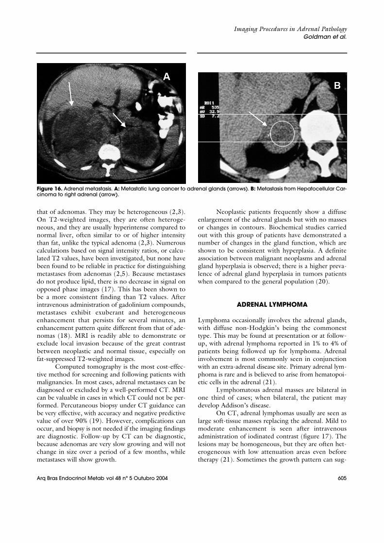
Figure 16. Adrenal metastasis. A: Metastatic lung cancer to adrenal glands (arrows). B: Metastasis from Hepatocellular Car-cinoma to right adrenal (arrow).

Imaging Procedures in Adrenal PathologyGoldman et al.
606 Arq Bras Endocrinol Metab vol 48 nº 5 Outubro 2004
gest lymphoma, as it is more likely to infiltrate orinsinuate around the upper pole of the kidney thandisplace it, as would be typical of carcinoma. Theremay be hemorrhage, and calcification can be found,especially after chemotherapy (21). On MRI, adrenallymphomas are indistinguishable from other malig-nancies. They are usually heterogeneous, with low sig-nal on T1-weighted images (less intense than normalliver, but more intense than muscle) and more intensethan fat on T2-weighted images (2,22).
MYELOLIPOMA
Myelolipoma is an uncommon, benign, nonfunction-ing neoplasm of the adrenal, found in less than 1% ofautopsies. It is composed of variable amounts of fatand hematopoietic tissue, including myeloid and ery-throid cells and megakaryocytes. It affects men andwomen equally. Although this is a nonfunctioningtumor, in 10% of cases it is associated with endocrinedisorders, including Cushing’s syndrome, congenitaladrenal hyperplasia, and Conn’s syndrome. Mostmyelolipomas (80%) are asymptomatic and of no clin-ical significance. Some (10%) become large and causevague symptoms or pain. Large myelolipomas mayhemorrhage, which can be the cause of pain. Sizeranges from 1 to 15cm, with a mean of about 4cm.
On CT, most myelolipomas are well-circum-scribed masses, sometimes with a discrete thin appar-ent capsule (figure 18). Occasionally, the mass mayappear to extend into the retroperitoneum. Nearly allcontain some definite fat density (< -20HU). Howe-
ver, the amount of fat is widely variable, ranging fromnearly all fat, to more than half fat (50%), to only a fewtiny foci of fat in a soft-tissue mass (10%) (23). Occa-sionally, the mass has an attenuation value betweenthat of fat and water because the fat and myeloid ele-ments are diffusely mixed. Calcification is seen in 30%,often punctate. With hemorrhage, high-density areascan be seen. Bilateral myelolipomas occur in about10% (23).
The presence of fat in an adrenal mass also canbe recognized on MRI, because fat is typically brighton both T1- and T2-weighted images (figure 18).Decrease in signal with fat suppression or phase can-cellation is confirmatory (17) (figure 18). However, ifthe mass is nearly all mature fat, there will not be lossof signal with opposed phase images, because the lossof signal occurs only with phase cancellation in areaswith an admixture of fat and water protons (24).Overall, myelolipomas are often heterogeneousbecause the nonfatty areas will have signal intensitysimilar to that of hematopoietic bone marrow. Thelesions enhance brightly after intravenous administra-tion of gadolinium.
The presence of fat in an adrenal mass is the keyto the diagnosis of myelolipoma, because virtually noother adrenal lesion contains fat. Teratoma andliposarcoma of the adrenals are extraordinarily rare. Anangiomyolipoma of the upper pole of a kidney may bemistaken to be an adrenal myelolipoma. However, thisis of no clinical significance, because both these lesionsare benign. If necessary, diagnosis can be confirmed bypercutaneous needle biopsy. If the biopsy reveals bonemarrow elements and the mass contains fat, the diag-
Figure 17. Adrenal lymphoma. A: Bilateral lobular adrenal masses (40HU) are present on noncontrast CT in this patient pre-senting with adrenal insufficiency (arrows). B: Contrast-enhanced scan shows slightly heterogeneous, mild enhancement(88HU). The patient was Addisonian.

Imaging Procedures in Adrenal PathologyGoldman et al.
607Arq Bras Endocrinol Metab vol 48 nº 5 Outubro 2004
nosis is assured, because extramedullary hematopoiesisdoes not contain fat. The presence of megakaryocytesalso is an important diagnostic histologic feature. Adefinite diagnosis is important because surgical resec-tion is not indicated unless there has been significanthemorrhage. In nearly all cases, a diagnosis of adrenalmyelolipoma can be made confidently based on CT orMRI findings alone.
ADRENAL CYSTS
Adrenal cysts are rare, found in only 1 of 1,400 autop-sies. They are nonfunctional and usually found inci-dentally. The commonest (45%) type is endothelialcyst. These are predominantly lymphangiomatouscysts, typically small and asymptomatic. Thirty-ninepercent are pseudocysts, which lack an endothelial lin-ing and are most often a sequelae of remote adrenal
hemorrhage. These are the type most often detectedby CT. These can be quite large and may producesymptoms because of their size. The remaining cysticlesions of the adrenals are parasitic cysts (7%), causedby Echinococcus, and true epithelial cysts (9%).
On CT, adrenal cysts are large masses (5 to20cm) that are well circumscribed and round. Theyare suprarenal in location, but it may be difficult onCT to recognize that they arise in the adrenal ratherthan the kidney or liver. Sonography or MRI may bet-ter show their true origin. They usually are of near-water attenuation, but they can have higher or mixedattenuation values resulting from old hemorrhage.The wall may be thick and can show contrast enhance-ment. Calcification is common (75%) (12), being usu-ally curvilinear in shape and often limited to the infe-rior aspect of the wall. (12). Although MRI may showtypical cystic features, homogeneous low intensity onT1-weighted images and extreme hyperintensity on
Figure 18. Patient with adrenal hyperplasia due to bilateral nodules, one at the right side is calcified (arrowhead) and alarge left mass with predominant fat content (-25HU) characteristic of a myelolipoma (arrow) (A). T1 in phase (B), T1 out ofphase (C) and T2 with fat saturation (D) MRI images. Out of phase shows no significant signal decrease compared to inphase. Fat saturation shows a significant decrease of signal, compatible with extra cellular lipid.
Imaging Procedures in Adrenal PathologyGoldman et al.
608 Arq Bras Endocrinol Metab vol 48 nº 5 Outubro 2004
T2-weighted images, signal intensity can vary depend-ing on the age of hemorrhage. There is no centralenhancement on either CT or MRI. Adrenal cysts areof no clinical significance, and surgical removal isunnecessary if the diagnosis can be established byimaging. Concern about malignancy is reasonable onlyin complicated cysts, which may have high attenuationvalues, a thick enhancing wall, and septations. In suchcases, percutaneous needle aspiration may be helpfulfor both diagnosis and treatment.
INFLAMMATORY DISEASE
Inflammatory processes in the adrenals are uncom-mon. Adrenal abscesses occur rarely; they sometimesrepresent adrenal hematomas that became infected.More often, adrenal inflammation is due to chronicgranulomatous disease, with tuberculosis (TB) andhistoplasmosis most common, although North Ame-rican blastomycosis has been reported to involve theadrenals. Paracoccidioidomycosis, or South Americanblastomycosis, is the most common systemic mycosisin South America, with Brazil responsible for about70% of total world cases. Its etiologic agent is Para-coccidioides brasiliensis. This disease chiefly involvesthe respiratory tract and its presentation can vary fromacute and self-limited to a progressive pulmonary dis-ease or extra pulmonary dissemination. Diagnosis ismade by lesion biopsy and fungal culture, serologicaltests and chest radiographs. The organs that constitutethe reticuloendothelial system, including the adrenals,are the most affected ones.
Granulomatous disease of the adrenals cancause a variety of appearances depending on the dis-ease stage. In most cases, there is bilateral enlargementof the adrenals (25). Bilateral adrenal enlargement in a
patient with a reactive tuberculin skin test, or withchest radiographic changes of TB, paracoccidioidomy-cosis or histoplasmosis, should suggest the diagnosis,even if pulmonary cultures are nondiagnostic.
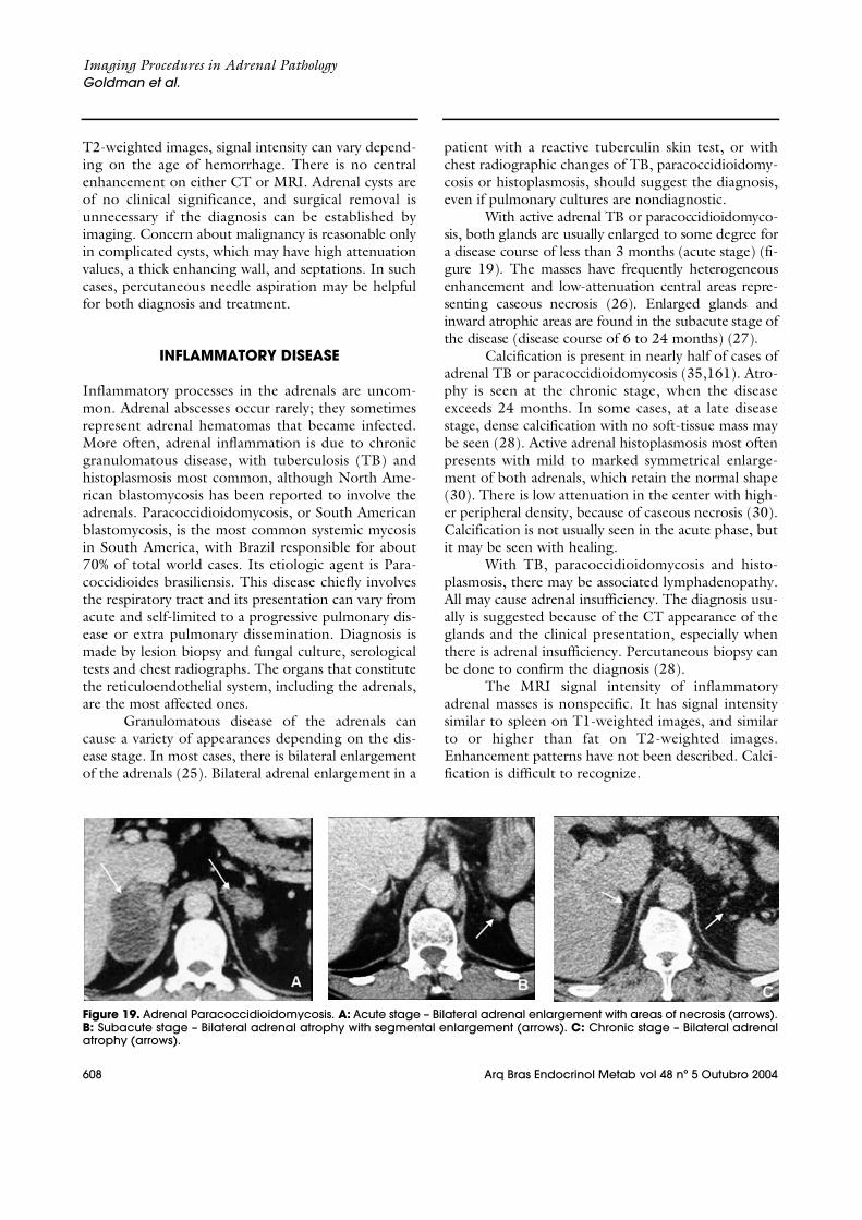
With active adrenal TB or paracoccidioidomyco-sis, both glands are usually enlarged to some degree fora disease course of less than 3 months (acute stage) (fi-gure 19). The masses have frequently heterogeneousenhancement and low-attenuation central areas repre-senting caseous necrosis (26). Enlarged glands andinward atrophic areas are found in the subacute stage ofthe disease (disease course of 6 to 24 months) (27).
Calcification is present in nearly half of cases ofadrenal TB or paracoccidioidomycosis (35,161). Atro-phy is seen at the chronic stage, when the diseaseexceeds 24 months. In some cases, at a late diseasestage, dense calcification with no soft-tissue mass maybe seen (28). Active adrenal histoplasmosis most oftenpresents with mild to marked symmetrical enlarge-ment of both adrenals, which retain the normal shape(30). There is low attenuation in the center with high-er peripheral density, because of caseous necrosis (30).Calcification is not usually seen in the acute phase, butit may be seen with healing.
With TB, paracoccidioidomycosis and histo-plasmosis, there may be associated lymphadenopathy.All may cause adrenal insufficiency. The diagnosis usu-ally is suggested because of the CT appearance of theglands and the clinical presentation, especially whenthere is adrenal insufficiency. Percutaneous biopsy canbe done to confirm the diagnosis (28).
The MRI signal intensity of inflammatoryadrenal masses is nonspecific. It has signal intensitysimilar to spleen on T1-weighted images, and similarto or higher than fat on T2-weighted images.Enhancement patterns have not been described. Calci-fication is difficult to recognize.
Figure 19. Adrenal Paracoccidioidomycosis. A: Acute stage – Bilateral adrenal enlargement with areas of necrosis (arrows).B: Subacute stage – Bilateral adrenal atrophy with segmental enlargement (arrows). C: Chronic stage – Bilateral adrenalatrophy (arrows).
Imaging Procedures in Adrenal PathologyGoldman et al.
609Arq Bras Endocrinol Metab vol 48 nº 5 Outubro 2004
ADRENAL HEMORRHAGE
Adrenal hemorrhage occurs in three distinct settings:neonatal hemorrhage, spontaneous (atraumatic) he-morrhage in the adult and severe trauma. Neonatalhemorrhage is the most common, resulting partlyfrom the large fetal adrenal that is prone to injury du-ring birth trauma. Because it is primarily the regressingfetal adrenal tissue that is involved, such patients donot develop adrenal insufficiency, and in the adult theonly sequelae is calcification of the adrenal without anassociated mass (12).
Adrenal hemorrhage in the adult may be seen inthe setting of severe illness, such as sepsis, including butnot limited to meningococcemia, burns, hypotension,and other life threatening illnesses. In these circum-stances, it is likely that the stress-related hyperplasiamakes the adrenal prone to spontaneous rupture.About one third of cases of adrenal hemorrhage areassociated with anticoagulant therapy. Commonly, thebleeding occurs in the first 3 weeks of anticoagulation.On CT, adrenal hemorrhage results in unilateral orbilateral adrenal masses that are usually ovoid, about3cm in diameter, and of about muscle density or high-er with poor enhancement, if any, after intravenousadministration of iodinated contrast. With larger hem-orrhages, the masses may be heterogeneous and mayhave ill-defined margins. The size and attenuation valuedecrease over time if followed with CT. Calcificationmay develop in several weeks to months. If the hemor-rhage is bilateral, adrenal insufficiency may occur.
The adrenal glands are well protected in theretroperitoneum. Posttraumatic adrenal hemorrhage is
uncommon, reported in 2% of patients having CT forsevere abdominal injury (31). The lack of specific clin-ical signs makes acute traumatic adrenal hemorrhage ausually incidental finding in CT studies performed forevaluation of acute blunt trauma. It has long been rec-ognized that the right adrenal is more prone to trau-matic hemorrhage (31). However bilateral and isolat-ed left adrenal hematomas occur.
Metastases to the adrenal can bleed, but onlyrarely. This usually can be distinguished from otheradrenal hemorrhages because there is a large hetero-geneous mass, with more extensive infiltrativeretroperitoneal bleeding (14). Because these patientsusually have advanced disease, neoplasm elsewhere isalso usually evident.
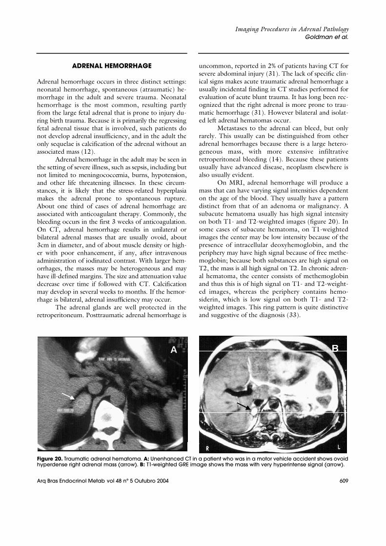
On MRI, adrenal hemorrhage will produce amass that can have varying signal intensities dependenton the age of the blood. They usually have a patterndistinct from that of an adenoma or malignancy. Asubacute hematoma usually has high signal intensityon both T1- and T2-weighted images (figure 20). Insome cases of subacute hematoma, on T1-weightedimages the center may be low intensity because of thepresence of intracellular deoxyhemoglobin, and theperiphery may have high signal because of free methe-moglobin; because both substances are high signal onT2, the mass is all high signal on T2. In chronic adren-al hematoma, the center consists of methemoglobinand thus this is of high signal on T1- and T2-weight-ed images, whereas the periphery contains hemo-siderin, which is low signal on both T1- and T2-weighted images. This ring pattern is quite distinctiveand suggestive of the diagnosis (33).
Figure 20. Traumatic adrenal hematoma. A: Unenhanced CT in a patient who was in a motor vehicle accident shows ovoidhyperdense right adrenal mass (arrow). B: T1-weighted GRE image shows the mass with very hyperintense signal (arrow).
Imaging Procedures in Adrenal PathologyGoldman et al.
610 Arq Bras Endocrinol Metab vol 48 nº 5 Outubro 2004
ADDISON’S DISEASE
Adrenal insufficiency may result from a variety of caus-es including bilateral hemorrhage, inflammatory dis-ease, and idiopathic autoimmune primary Addison’sdisease. Although metastases to the adrenal are com-mon, they rarely lead to Addison’s disease. When itoccurs, the disease is either in advanced stages or isassociated with spontaneous hemorrhage. Anotherrare cause is hemochromatosis, which can be recog-nized on CT, as the adrenals are normal or small insize but have increased attenuation values (28).
CT is indicated in evaluation of patients withAddison’s disease. In one study, all cases of idiopathicAddison’s disease could be distinguished from otheretiologies (29).
CT can also determine the stage of the dis-ease: acute, subacute and chronic, as discussed pre-viously (27).
Adrenal atrophy that results from autoimmunedisease will result in small glands without calcification(28,29). This must be distinguished from adrenal atro-phy due to exogenous steroids, which has a similarappearance, by careful history taking. Conversely,either adrenal hemorrhage, neoplasms, or inflammato-ry disease will show either adrenal masses or calcifica-tion, as discussed previously. Small adrenals that arepartly or completely calcified suggest old granuloma-tous disease (particularly TB and Pb), whereas verydense calcifications with no soft tissue component sug-gest remote adrenal hemorrhage.
CT AND OTHER IMAGING METHODS
Except in a pediatric population, ultrasound is notused as a primary imaging method for adrenal disease,because it has both lower sensitivity and specificitythan CT or MRI. Adrenal angiography and venogra-phy have been replaced by CT and MRI. Adrenalvenous sampling remains useful in certain cases.
Adrenal scintigraphy is useful in patients sus-pected of having primary extra-adrenal or malignantmetastatic pheochromocytomas.
MRI is extremely useful in selected cases, par-ticularly in the evaluation of pheochromocytomas andparagangliomas. MRI is equivalent to CT in evaluationof Cushing’s syndrome. However, CT is preferred forevaluation of hyperaldosteronism or adrenal insuffi-ciency. MRI can be very useful to further characterizelesions detected on CT. It can demonstrate moreclearly the extent of a mass, its exact organ of origin,
and vascular compromise. MRI can be useful in evalu-ating an adrenalectomy bed for possible recurrence ofa malignancy, because spin echo images show lessdegradation by clip artifact than CT. Last, pregnantpatients suspected of having adrenal disease can bemore safely studied with MRI than with CT.
REFERENCES
1. Abrams HI, Siegelman SS, Adams DF. Computed tomog-raphy versus ultrasound of the adrenal gland: aprospective study. Radiology 1982;143:121-8.
2. Chang A, Glazer HS, Lee JKT, et al. Adrenal gland: MRimaging. Radiology 1987;163:123-8.
3. Falke TH, te-Strake L, Shaff MI, et al. MR imaging of theadrenals: correlation with computed tomography. JComput Assist Tomogr 1986;10:242-53.
4. Schultz CL, Haaga JR, Fletcher BD, et al. Magnetic reso-nance imaging of the adrenal glands: a comparisonwith computed tomography. AJR 1984;143:1235-40.
5. Chezmar JL, Robbins SM, Nelson RC, et al. Adrenalmasses: characterization with T1-weighted MR imaging.Radiology 1988;166:357-9.
6. Vincent JM, Morison ID, Armstrong P, et al. Computertomography of diffuse, non-metastatic enlargement ofthe adrenal glands in patients with malignant disease.Clin Radiol 1994;49:456-60.
7. Goldman SM, Palacio G, Borri ML, et al. Prevalence ofadrenal gland enlargement in patients with aorticaneurysm or aortic dissection. Presented at SUR Annu-al Meeting, 2000.
8. Kamilaris TC, Chrousos GP. Adrenal diseases. In: MooreWT, Eastman RC, eds. Diagnostic endocrinology.Philadelphia: BC Becker, 1990. p. 79-109.
9. Perry RR, Nieman LK, Cutler GB, et al. Primary adrenalcauses of Cushing’s syndrome: diagnosis and surgicalmanagement. Ann Surg 1989;210:59-68.
10. Doppman JL, Miller DL, Dwyer AJ, et al. Macronodularadrenal hyperplasia in Cushing’s disease. Radiology1988;166:347-52.
11. Korobkin M. Combined unenhanced and delayedenhanced CT for characterization of adrenal masses.Radiology 2002;222:629-33.
12. Kenney PJ, Stanley RJ. Calcified adrenal masses. Uro-logic Radiol 1987;9:9-15.
1 3 . Kobayshi S, Seki T, Nonomura K, et al. Clinical experienceof incidentally discovered adrenal tumor with particularreference to cortical function. J Urol 1993; 1 5 0 : 8 - 1 2 .
14. Shah HR, Love L, Williamson MR, et al. Hemorrhagicadrenal metastases: CT findings. J Comput AssistTomogr 1989;13:77-81.
15. Lee MJ, Hahn PF, Papanicolaou N, et al. Benign andmalignant adrenal masses: CT distinction with attenua-tion coefficients, size, and observer analysis. Radiology1991;179:415-8.

Imaging Procedures in Adrenal PathologyGoldman et al.
611Arq Bras Endocrinol Metab vol 48 nº 5 Outubro 2004
16. Berland LL, Koslin DB, Kenney PJ, et al. Differentiationbetween small benign and malignant adrenal masseswith dynamic incremented CT. AJR 1988;151:95-101.
17. Mitchell DG, Crovello M, Matteucci T, et al. Benignadrenocortical masses: diagnosis with chemical shiftMR imaging. Radiology 1992;185:345-51.
18. Krestin GP, Stenbrich W, Friedman G. Adrenal masses:evaluation with fast gradient-echo MR imaging and Gd-DTPA-enhanced dynamic studies. R a d i o l o g y1989;171:675-80.
19. Silverman SG, Mueller PR, Pinkney LP, et al. Predictivevalue of image-guided adrenal biopsy: analysis ofresults of 101 biopsies. Radiology 1993;187:715-8.
20. Goldman SM, Borri M L, Abbehusen C, Faiçal S, Szenjn-feld J, Ajzen S. Prevalence of non-metastatic adrenalglands’ enlargement in patients with malignant neo-plasms evaluated by Computed Tomography (CT).Presented at SUR/SGR meeting 2000-Hawaii).
21. Falchook FS, Allard JC. Case report. CT of primaryadrenal lymphoma. J Comput Assist Tomogr1991;15:1048-50.
22. Lee FT, Thornbury JR, Grist TM, et al. MR imaging ofadrenal lymphoma. Abdom Imaging 1993;18:95-6.
23. Rao P, Kenney PJ, Wagner BJ, et al. Imaging and patho-logic features of myelolipoma. R a d i o G r a p h i c s1997;17:1373-85.
24. Tsushima Y, Ishizaka H, Matsumoto M. Adrenal masses:differentiation with chemical shift, fast low-angle shotMR imaging. Radiology 1993;186:705-9.
25. Hauser H, Gurret JP. Miliary tuberculosis associated withadrenal enlargement: CT appearance. J ComputAssist Tomogr 1986;10:254-6.
26. Leal AMO, Bellucci AD, Muglia VF, Lucchesi FR. Uniqueadrenal gland imaging features in Addison’s diseasecaused by paracoccidioidis brasiliensis. A J R2003;181:1433-4.
27. Goldman SM, Palacios GG, Falcão MA. Determiningphase of Addison’s disease: Value of computed tomog-raphy. Eur Radiol 2002;12(P):D6.
2 8 . Doppman JL, Gill JR Jr, Nienhuis AW, et al. CT findings inAddison’s disease. J Comput Assist Tomogr 1982; 6 : 7 5 7 -6 1 .
29. Sun ZH, Nomura K, Toraya S, et al. Clinical significanceof adrenal computed tomography in Addison’s dis-ease. Endocrinol Japan 1992;39:563-9.
30. Levine E. CT evaluation of active adrenal histoplasmo-sis. Urol Radiol 1991;13:103-6.
31. Burks DW, Mirvis SE, Shanmuganathan K. Acute adrenalinjury after blunt abdominal trauma: CT findings. AJR1992;158:503-7.
3 2 . Bowen A, Keslar PJ, Newman B, et al. Adrenal hemor-rhage after liver transplantation. Radiology 1990; 1 7 6 : 8 5 -8 .
33. Itoh K, Yamashita K, Satoh Y, et al. Case report. MRimaging of bilateral adrenal hemorrhage. J ComputAssist Tomogr 1988;12:1054-6.
Endereço para correspondência:
Suzan M. GoldmanRua Escobar Ortiz 604, 12º andar04512-051 São Paulo, SP, Brasile-mail: [email protected]