review caspases: a molecular switch node in the crosstalk
TRANSCRIPT
Int. J. Biol. Sci. 2014, Vol. 10
http://www.ijbs.com
1072
IInntteerrnnaattiioonnaall JJoouurrnnaall ooff BBiioollooggiiccaall SScciieenncceess 2014; 10(9): 1072-1083. doi: 10.7150/ijbs.9719
Review
Caspases: A Molecular Switch Node in the Crosstalk between Autophagy and Apoptosis Haijian Wu1*, Xiaoru Che2*, Qiaoli Zheng3, An Wu1, Kun Pan4, Anwen Shao1, Qun Wu1, Jianmin Zhang1, and Yuan Hong1
1. Department of Neurosurgery, Second Affiliated Hospital, School of Medicine, Zhejiang University, Hangzhou, China; 2. Department of Cardiology, Zhejiang Provincial People’s Hospital, Hangzhou, China; 3. Clinical Research Center, Second Affiliated Hospital, School of Medicine, Zhejiang University, Hangzhou, China. 4. Department of Neurological Surgery, Weill Cornell Medical College, New York, New York, USA.
*These authors provided equal contribution to this work.
Corresponding author: Dr. Yuan Hong Or Dr. Jianmin Zhang, Department of Neurosurgery, Second Affiliated Hospital, School of Med-icine, Zhejiang University, Hangzhou, Zhejiang 310009, China; Tel: +86-571-87784785; Fax: +86-571-87784755; E-mail: [email protected].
© Ivyspring International Publisher. This is an open-access article distributed under the terms of the Creative Commons License (http://creativecommons.org/ licenses/by-nc-nd/3.0/). Reproduction is permitted for personal, noncommercial use, provided that the article is in whole, unmodified, and properly cited.
Received: 2014.05.22; Accepted: 2014.08.20; Published: 2014.09.13
Abstract
Autophagy and apoptosis are two important catabolic processes contributing to the maintenance of cellular and tissue homeostasis. Autophagy controls the turnover of protein aggregates and damaged organelles within cells, while apoptosis is the principal mechanism by which unwanted cells are dismantled and eliminated from organisms. Despite marked differences between these two pathways, they are highly interconnected in determining the fate of cells. Intriguingly, caspases, the primary drivers of apoptotic cell death, play a critical role in mediating the complex crosstalk between autophagy and apoptosis. Pro-apoptotic signals can converge to activate caspases to execute apoptotic cell death. In addition, activated caspases can degrade autophagy proteins (i.e., Beclin-1, Atg5, and Atg7) to shut down the autophagic response. Moreover, caspases can convert pro-autophagic proteins into pro-apoptotic proteints to trigger apoptotic cell death instead. It is clear that caspases are important in both apoptosis and autophagy, thus a detailed deciphering of the role of caspases in these two processes is still required to clarify the functional relationship between them. In this article, we provide a current overview of caspases in its interplay between autophagy and apoptosis. We emphasized that defining the role of caspases in autophagy-apoptosis crosstalk will provide a framework for more precise manipulation of these two processes during cell death.
Key words: autophagy; apoptosis; caspases; Atg proteins; crosstalk; cell death.
Introduction Macroautophagy (hereafter refer to as autopha-
gy) is an intracellular catabolic process by which por-tions of cytoplasmic components are delivered to au-tolysosomes for degradation [1]. Apoptosis is a well-studied form of programmed cell death that dis-plays typical morphological characteristics, such as chromatin condensation, nuclear fragmentation, and membrane blebbing [2, 3]. Both autophagy and apoptosis are highly orchestrated cascades that play a
vital role in embryonic development and tissue ho-meostasis [4, 5]. Dysregulation of them have been associated with a number of pathologies, such as cancer, autoimmune diseases, and neurodegenerative diseases [6]. It’s known that autophagy extensively communicates with apoptosis during cell fate decision in a myriad of physiological and pathological condi-tions [7, 8]. In low-stress situations, cells initiate au-tophagy as a pro-survival mechanism to combat
Ivyspring
International Publisher

Int. J. Biol. Sci. 2014, Vol. 10
http://www.ijbs.com
1073
apoptotic cell death. However, as stress increases to-wards a point of no return where cells are doomed to die, cells block autophagy and begin initiating apop-totic cascades [9]. Thus, a better knowledge of those interactions between autophagy and apoptosis is of great importance, in order to optimally manipulate these two crucial pathways for therapeutic purposes.
Caspases, a family of cysteinyl aspar-tate-requiring proteases, play a central role in the transduction of apoptotic signals [10]. Apoptotic caspases can be divided into two general categories: the initiator caspases consisting of caspase-2, -8, -9 and -10; and the effector caspases consisting of caspases-3, -6 and -7 [11, 12]. Caspases normally ap-pear as inactive zymogenic precursors. They can be activated to mediate apoptosis in response to diverse pro-apoptotic stimuli from outside and inside of the cells [13]. Emerging evidences demonstrate that caspases can also influence the non-apoptotic signal-ing events, in particular, the “self-eating” autophagy [14, 15]. Further research shows that caspases can in-hibit autophagy by cleaving and destroying the pro-autophagic activity of autophagy-related (Atg)
proteins, such as Atg3 and Beclin-1 [16, 17]. Whereas, caspase-9 can facilitate autophagosome formation by promoting the Atg7-dependent conversion process of microtubule-associated protein 1 light chain 3 (LC3) [18]. On the other hand, the cytoprotective autophagy counter-balances apoptosis by continuous sequestra-tion of active caspase-8 into autophagosomes for its subsequent degradation in Bax-/- Hct116 colon carci-noma cells [19]. However, autophagosomal mem-brane also serves as a platform for intracellular death-inducing signaling complex (DISC)-mediated caspase-8 activation and apoptosis [20]. Therefore, elucidating these unexpected functions of caspases in linking the autophagic and apoptotic signaling path-ways helps decipher the molecular basis underlying autophagy-apoptosis crosstalk, which can drive therapeutic development by optimally manip-ulating these two important processes.
In this review article, we focus on the biological effects and underlying mechanisms of caspases in directing the conversation between autophagy and apoptosis in the mammalian systems (unless other-wise stated). In particular, we emphasize that further
insights into the role of caspases in mediating au-tophagy-apoptosis cross-talk will guide future therapeutic strategies during cell death control.
The autophagic process
Autophagy is a cata-bolic process which in-volves the turnover of cy-tosolic protein aggregates and damaged organelles in the lysosomes via dou-ble-membrane autopha-gosomes (Figure 1) [21]. The autophagic process consists of initiation, elongation, maturation, fusion, and degradation [22]. It begins with the nu-cleation and expansion of an isolated membrane called the phagophore, which can sequester por-tions of cytoplasmic mate-rials to form an autopha-gosome [23]. Subsequent-ly, the autophagosome fuses with a lysosome to form an autolysosome,
Figure 1. An overview of the cellular and molecular events during autophagy.
Int. J. Biol. Sci. 2014, Vol. 10
http://www.ijbs.com
1074
where the captured cargos are degraded by hydro-lytic enzymes for recycling [24].
The autophagy machinery is tightly controlled by a set of Atg proteins that can assemble into several different complexes to produce autophagosomes [25, 26]. Among these core multimolecular complexes, the ULK1 complex, made of ULK1, Atg13, Atg101, and FIP200 (focal adhesion kinase family-interacting pro-tein of 200 kD), plays a critical role in the initiation of autophagy [27]. Under nutrient-rich conditions, the serine/threonine kinase mammalian Target of ra-pamycin (mTOR) acts in concert with other proteins of the mTOR complex 1 (mTORC1) to suppress au-tophagy through direct interaction with the ULK1 complex [27]. In contrast, in response to stress condi-tions such as starvation, mTOR is inhibited and dis-sociates from the ULK1 complex [28]. This can result in the activation of ULK1 and ULK1-catalyzed phos-phorylation of Atg13, FIP200, and ULK1 itself, which is essential for initiating autophagy [29]. Also, AMP activated protein kinase (AMPK) can activate ULK1 via a direct phosphorylation mechanism [30]. AMPK interacts with ULK1 and phosphorylates it on Ser 317 and Ser 777, which can lead to activation of ULK1 kinase and autophagy induction in response to glu-cose starvation [29]. Downstream of the ULK1 com-plex, the class III phosphatidylinositol 3-kinase (PI3K) complex exerts an essential role in the nucleation and assembly of the initial phagophore membrane [31]. Beclin-1 functions as a platform by binding several cofactors such as barkor (Beclin-1-associated autoph-agy-related key regulator), p150, and UVRAG (UV irradiation resistance-associated gene) to assemble the class III PI3K complex during autophagosome for-mation [32-36]. Of note, the phosphorylated ULK1 can promote the class III PI3K complex-mediated phago-phore nucleation [37], possible through phosphoryla-tion of key molecules such as AMBRA (activating molecule in Beclin-1-regulated autophagy protein 1) [38], and Beclin-1 [39]. The subsequent elongation process requires the involvement of two ubiqui-tin-like conjugation reactions: the Atg5-Atg12 conju-gation and LC3-PE (phosphatidylethanolamine) con-jugation [40]. Analogous to ubiquitination, Atg12 is conjugated to Atg5 to form Atg12-Atg5 conjugate, with the help of the E1-like enzyme Atg7 and the E2-like enzyme Atg10. Similarly, the conjugation of LC3 to PE is mediated by the E1-like enzyme Atg7 and the E2-like enzyme Atg3. The Atg12-Atg5 then conjugates with Atg16L (Atg16-like protein) to form an approximately 800-kDa protein complex contain-ing Atg12-Atg5-Atg16L [41]. This complex serves as a platform for stimulating the LC3-PE conjugation re-action [42, 43], which is essential for the elongation of the pre-autophagosomal membranes [44, 45]. Note-
worthily, although molecules such as Beclin-1 inter-acting proteins (i.e., UVRAG and Rubicon) have been proven to be involved in the maturation of autopha-gosomes [46-48], the role of Atg proteins in the mat-uration and fusion step of autophagosomes, however, remains unclear and requires further characterization.
The apoptotic cascades Apoptosis is a self-destructive process with
carefully choreographed steps (Figure 2) [49]. During the apoptotic process, a series of intracellular events come into play to decommission the unwanted and dangerous cells [50, 51]. Apoptosis is crucial for em-bryonic development, organogenesis, as well as tissue homeostasis [3, 52]. Aberrant regulation of apoptosis can lead to either pathological cell accumulation or inappropriate cell loss, which eventually results in the pathogenesis of various human disorders, including cancer, infection, neurodegeneration, and autoim-mune diseases [5, 50, 53].
Caspases, both the initiators and the effectors, play a major role in execution of apoptotic cascades [54]. Mechanically, two major apoptotic signaling pathways, including the extrinsic and intrinsic sig-nals, converge onto these caspases to initiate cell death [3, 55]. The extrinsic apoptotic cascade is trig-gered by ligation of cell-surface death receptors of the tumor necrosis factor (TNF) receptor superfamily, such as CD95/Fas or TNF-related apoptosis-inducing ligand (TRAIL) receptors [56, 57]. Upon ligand bind-ing, the receptors become oligomerized forms, re-cruiting Fas-associated death domain (FADD) and procaspase-8 to form the death-inducing signaling complex (DISC) [58]. Recruitment of procaspase-8 to the DISC facilitates its self-cleavage into active caspase-8 enzyme that later cleaves effector caspases [59]. These effector caspases can further cleave a number of cytosolic and nuclear substrates to execute apoptotic cascades that leads to morphological and biochemical features of apoptosis [60]. Alternatively, caspase-8 can propagate apoptotic death signals through proteolysis of the Bcl-2 homology-3 (BH3)-only protein Bid. The truncated Bid (tBid) translocates to the outer mitochondrial membrane to orchestrate a process that mediating mitochondrial outer membranes permeabilization (MOMP) [61, 62]. MOMP is a crucial event during apoptosis [63]. As an outcome of this process, pores are formed in the outer mitochondrial membrane, membrane integrity is compromised, and apoptogenic proteins (i.e., cyto-chrome c) within the intermembrane space are re-leased into the cytosol to induce apoptotic cell death.
The intrinsic pathway of apoptosis is triggered by a wide range of intracellular death signals, such as DNA damage, cytotoxic stress, and growth factor

Int. J. Biol. Sci. 2014, Vol. 10
http://www.ijbs.com
1075
withdrawal [64]. In response to these apoptotic stim-uli, the integrity of the outer mitochondrial membrane is lost, and the permeabilization of outer mitochon-drial membranes is culminated [65]. This will thereby permit release of many pro-apoptotic proteins that are normally confined to the intermembrane space, in particular, cytochrome c, into the cytoplasm to pro-mote caspase activation [66, 67]. In the cytoplasm, the apoptogenic factor cytochrome c associates with apoptotic protease-activating factor 1 (Apaf-1) and initiator caspase-9 to form a multi-protein complex known as apoptosome [65, 68]. The apoptosome serves as a molecular platform for caspase-9 pro-cessing and activation, which can subsequently acti-vate downstream executioner caspases to orchestrate apoptotic cell death [69].
Caspases: key orchestrators of autopha-gy-apoptosis crosstalk
Autophagy and apoptosis have been proven to extensively cross talk with each other in cell fate deci-sion [70-73]. With series of key findings, molecular mechanisms underlying the complex coun-ter-regulation between autophagy and apoptosis are emerging. Of much interest, caspases, a family of
cysteine proteases that are instrumental to the execution of apoptosis, serve as important play-ers in directing these conversations between autophagy and apopto-sis. Multiple pro-apoptotic signals, both inside and outside the cells, can converge to activate caspases to ini-tiate apoptosis. In addi-tion, activated caspases can degrade essential autophagy proteins (i.e., Beclin-1, Atg5, and Atg7) to shut down the au-tophagic response in-stead [74-76]. Moreover, after been cleaved by caspases, some pro-autophagic proteins can be even converted into pro-apoptotic ones to trigger apoptotic cell death. On the other hand, autophagy can influence apoptotic cas-cades by regulating the amount and activity of
caspases. And autophagic elimination of caspases, such as caspase-8, can hamper their involvement in the apoptotic pathways [19].
Initiator caspases (Figure 3) Caspase-8 is a well-known initiator caspase im-
plicated in the death receptor-triggered apoptosis [77]. As aforementioned above, in the context of ex-trinsic apoptosis, caspase-8 is activated by dimeriza-tion and autoproteolysis inside the DISC, and subse-quently released into the cytoplasm to activate effec-tor caspases for efficient apoptosis execution [57, 78-81].
Apart from its pro-apoptotic properties, caspa-se-8 plays an important role in the modulation of au-tophagy. Data from in vitro studies demonstrated that caspase-8 can rescue T cells from hyperactive au-tophagy [15, 82]. In response to energetic demands for rapid clonal expansion, the autophagic responses in-side T cells are stimulated [82]. The autophagic sig-naling can induce an interaction among Atg5-Atg12-Atg16L, FADD, caspase-8, and receptor interacting protein kinase 1 (RIPK1), which promotes the activation of caspase-8 in mitogenically stimulated
Figure 2. Scheme of the extrinsic and intrinsic apoptotic pathways.

Int. J. Biol. Sci. 2014, Vol. 10
http://www.ijbs.com
1076
live T cells [83]. Activated caspase-8 can directly cleave the serine/threonine kinase RIPK1 as part of a negative feedback loop to limit autophagic signaling [82, 84]. In fact, RIPK1 also has emerged as a key up-stream regulator which controls of apoptosis and necroptosis [85-87]. Besides, RIPK1 is required for MAPK8 activation and induction of protective au-tophagy to blunt apoptosis in TRAIL-treated cancer cells [88]. Whereas, RIPK1 can be cleaved by the active caspase-8 during apoptotic cascades [84]. These evi-dences indicate that the caspase-8/RIPK1 is involved in the crosstalk between autophagy, apoptosis and
necroptosis, which is of interest for future investiga-tion. Atg3 is a critical regulatory component for au-tophagosome biogenesis. It is conceived as a new substrate of caspase-8 during receptor-mediated cell death [16]. By targeting the evolutionary conserved LETD sequence (Atg3 amino acids 166–169) of this protein, caspase-8 mediates the cleavage of Atg3. As a result, the pro-autophagic activity of Atg3 is abolished after it has been cleaved, which is a critical event in autophagy inactivation during death recep-tor-triggered apoptosis [16].
Figure 3. Scenarios for initiator caspases-mediated crosstalk between apoptosis and autophagy.
On the other hand, autophagy contributes to the
regulation of caspase-8-mediated apoptosis under the condition of proteasome inhibition [82, 89]. In re-sponse to proteasome inhibition, the autophagy flux can increase as a compensatory mechanism for pro-tein degradation [90, 91]. Moreover, it has been shown that autophagosomes serve as a platform for intra-cellular DISC formation for caspase-8 self-processing, which is responsible for initiation of the caspase cas-cades and execution of apoptosis [20]. The association of FADD with the Atg12-Atg5 complex facilitates caspase-8 recruitment to the autophagosomal mem-brane to form a proper higher-order oligomer struc-
ture for caspase-8 activation [20]. Also, the adapter protein p62 can facilitate the recruitment of self-associated caspase-8 to autophagosomal mem-branes via its interaction with LC3-II [20]. This event contributes to the formation of proper oligomer structures for caspase-8 self-processing and self-activation [20, 92, 93]. It is important to note that the functional domains of p62, including UBA (ubiq-uitin-associated) which binds ubiquitinated proteins, PB1 (Phox and Bem1p) that mediates dimeriza-tion/oligomerization, and LC3 interaction region, are required for p62 in mediating caspase-8 activation [94-97]. In particular, the UBA domain at the
Int. J. Biol. Sci. 2014, Vol. 10
http://www.ijbs.com
1077
C-terminus of p62 is an important domain for binding and aggregation of polyubiquitinated caspase-8 for its final activation [98, 99]. As a consequence, the en-hancement of caspase-8 self-aggregation and activa-tion due to p62 upregulation in the setting of au-tophagy inhibition can sensitize human colon carci-noma cells to the BH3 mimetic agent ABT-263-induced apoptosis [97]. Consequently, blockage of autophagolysosomal degradation path-way can result in an increase in the association of ag-gregated caspase-8 with p62 and LC3, which contrib-utes to caspase-8 oligomerization, activation, and apoptotic signal transduction in the mammalian de-generin homolog G430F mutant-triggered cells [93]. Conversely, the autophagic degradation of caspase-8 precursor or a subunit of the active caspase-8 enzyme can hinder caspase-8-induced apoptosis [19]. In Bax-/- Hct116 colon carcinoma cells that are resistant to TRAIL-mediated apoptosis, TRAIL signaling medi-ates a protective autophagic response. This keeps the caspase-8-dependent apoptotic response at bay, probably through continuous elimination of the caspase-8 precursor and/or the active caspase-8 large subunit [19]. And autophagy inhibition due to Be-clin-1 RNA interference allows for a significant in-crease in caspase-8 enzymatic activity, resulting in downstream caspase processing and apoptosis acti-vation [19]. Summarizing it all together, caspase-8 functions as a switch point in polarization between autophagy and apoptosis during cell-fate determina-tion.
Caspase-9 is another essential initiator caspase that is involved in the intrinsic apoptosis pathway [100, 101]. Once activated, caspase-9 is able to process downstream effector caspases, including caspase-3, -6, and -7, all of which can target key structural and reg-ulatory proteins to bring about apoptotic cell death [102]. Intriguingly, Jeong and colleagues demon-strated the involvement of caspase-9 in promoting autophagy-mediated cell survival of breast cancer MCF-7 cells [103]. Inhibition of caspase-9 can nega-tively regulate lysosomal pH and acid-dependent cathepsin activities that causes blockage of cytopro-tective autophagy and augment of cell death follow-ing the exposure of the non-steroidal an-ti-inflammatory drug FR122047 [103]. In addition, Han and coworkers described that caspase-9 acts as a novel co-regulator of autophagy and apoptosis through its mutually regulation with Atg7 [18]. Caspase-9 can interact with Atg7 via the C-terminal region of the large subunit caspase-9, which facilitates the Atg7-dependent formation of autophagosomal LC3-II and autophagic function. The interaction of caspase-9 with Atg7 can potentially interfere with the recruitment and processing of caspase-9 in the apop-
tosome. The interference can ultimately inhibit caspase-9 activation and apoptotic signal transduction [18]. Although the cross-regulation between Atg7 and caspase-9 is a possible mechanism that determines the participation of caspase-9 either in autophagy or apoptosis, additional molecular scenarios should be still considered. For instance, a phosphorylation mechanism may serve as a potential determinant in the involvement of caspase-9 in either apoptotic or autophagic cascades [18, 104, 105]. However, the exact molecular mechanisms that forces caspase-9 into ei-ther autophagic or apoptotic mode should be ad-dressed.
Caspase-2, one of the most conserved members of the caspase family, has been proven to be a crucial mediator of apoptotic cascades in a con-text-dependent manner. Examples include genotoxic stress [106], heat shock [107, 108], and cytoskeletal disruption [109]. In addition to its role in apoptosis, evidence suggests that caspase-2 is involved in the negative regulation of autophagy [110]. Loss of caspase-2 has been shown to lead to an enhanced au-tophagy in mouse embryonic fibroblast. Increased production of reactive oxygen species and oxidative stress in cells lacking caspase-2 contribute to upregu-lation of AMPK, activation of MAPK1/3, downregu-lation of mTOR induction, and inactivation of MAPK14, which play a role autophagy induction [110]. Additionally, when the neurons cultured from young adult mice cannot undergo apoptosis due to lack of caspase-2, the pro-survival autophagy can be induced at an early stage in response to rote-none-mediated mitochondrial oxidative stress [111]. These evidences suggested that caspase-2 can be a molecular link between autophagy and apoptosis.
Caspase-10, the closest homolog of caspase-8, functions as an initiator caspase of extrinsic apoptotic signaling pathway [112, 113]. Of interest, Lamy et al. discovered that caspase-10 also plays an important role in maintaining the proper autophagic activities in myeloma cells [114]. Caspase-10 can cleave and inac-tivate Bcl-2-associated transcription factor 1 (BCLAF1), a potent autophagy inducer that can in-duce autophagy by displacing Beclin-1 from Bcl-2 complex, thus preventing myeloma cells from uncon-trolled autophagy [114]. The enhanced association between the caspase-like protein cFLIPL and caspa-se-10 in myeloma cells promotes caspase-10-mediated BCLAF1 cleavage, thus limits BCLAF1-induced au-tophagic cell death of myeloma cells [114].
Effector caspases (Figure 4) Caspase-3 is a predominant player in the execu-
tion of apoptotic cell death. However, recent studies indicated that capase-3 plays a role in autophagic

Int. J. Biol. Sci. 2014, Vol. 10
http://www.ijbs.com
1078
processes [115]. In human apoptotic endothelial cells under conditions of nutrient shortage, activated caspase-3 favors the extracellular export of au-tophagic vacuoles via rerouting autophagic vacuoles toward the cell membrane [116]. The externalization of large autophagic vacuoles may contribute to apoptotic decrease in volume, a geometric determi-nant for cell dismantling into apoptotic bodies [117]. These evidences represent caspase-3 as a potential molecular switch in mediating crosstalk between the autophagic and apoptotic programs [116]. Zhu and coworkers reported that caspase-3 can cleave au-tophagy-associated protein Beclin-1 at positions 124 and 149 during staurosporine-induced apoptosis, thus abrogating the pro-autophagic effect of Beclin-1 in HeLa cells [118]. Moreover, the cleavage of Beclin-1 disrupts its interaction with Bcl-2, which leads to the exposure of the BH3 domain of Beclin-1 to other an-ti-apoptotic Bcl-2 family members [119-121]. The competitive binding of truncated Beclin-1 with an-ti-apoptotic Bcl-2 family members allows pro-apoptotic BH3-only molecules released from the Bcl-2/Bcl-xL complex to initiate intrinsic apoptotic cascades [121, 122]. Also, Atg4D, one member of the Atg4 family in mammalian cells that contributes to starvation-induced autophagy, can be cleaved by caspase-3 at the DEVD63K motif [123]. More im-portantly, cleavage of Atg4D by caspase-3 enables it to stimulate the priming and delipidation of the LC3 paralogue, GABARAP-L1 (γ-aminobutyric acid re-ceptor-associated protein (GABARAP)-like 1), indi-cating that caspase-3 promotes Atg4D-coordinated
autophagy [123]. It is noteworthy that caspase-cleaved ΔN63 Atg4D can induce cell death when overex-pressed in human cells [123]. The cleaved Atg4D-mediated cell toxicity is associated with its recruitment to mitochondria to induction of apoptosis [123]. Unfortunately, whether Atg4D can cause the release of proapoptotic mitochondrial factors by tar-geting Bax or Bak is still unknown.
Other effector caspases, including caspase-6 and caspase-7, not only play a role in the execution phase of apoptosis [124, 125], but also affect the autophagic pathway. When melanoma cell lines undergo arginine deprivation, TRAIL-initiated caspase-6 activation contributes to disruption of pro-survival autophagy by cleavage of two key autophagic proteins (Beclin-1 and Atg5) [74]. And evidence from an in vitro study demonstrated that proteins such as Atg3 and p62 can be cleaved by caspase-6, suggesting its potential role in the regulation of autophagy [126]. By using knock-in mice, the effects of the Atg16L1 T300A (a common threonine to alanine coding variant at posi-tion 300 in Atg16L) polymorphism on the autophagy pathway were studied by Lassen et al. They found that Atg16L is a substrate for caspase-7, while the Atg16L1 T300A is more susceptible to caspa-se-7-mediated cleavage compared with the wild-type protein, which is associated with the reduced anti-bacterial autophagy in cells of Atg16L T300A mice [76]. Nevertheless, other mechanisms underlying these two caspases in linking autophagy and apopto-sis are warranted to be further elucidated.
Figure 4. Paradigms of effector caspases-mediated conversation between autophagy and apoptosis.
Int. J. Biol. Sci. 2014, Vol. 10
http://www.ijbs.com
1079
The calpain system Calpains represent a well-conserved family of
calcium-sensitive cysteine proteases localized to the cytosol and mitochondria [127]. They exert important roles in a number of fundamental physiological pro-cesses, including the execution of apoptosis [128-130]. Evidence demonstrates calpains are involved in the apoptotic process at numerous steps. For instance, calpain is capable of processing and activation of several caspases, such as caspase-7 and caspase-12, which can sensitize cells to Ca2+-dependent apoptosis [131-133]. Calpain is also able to cleave pro-apoptotic Bcl-2 family proteins including Bid and Bax, which in turn mediates cytochrome c release and initiates the apoptotic execution [134-136].
Not surprisingly, similar to the caspase family proteases, the calpain plays a role in linking autoph-agy to apoptosis [137]. In sphingosine-1-phosphate phosphohydrolase 1 (SPP1)-depleted cells, doxorubi-cin switches protective autophagy to apoptosis via calpain-mediated Atg5 cleavage [138]. The truncated Atg5 cleaved by calpain translocates from the cytosol to mitochondria and associates with the an-ti-apoptotic Bcl-xL to trigger cytochrome c release and caspase activation, and triggers apoptotic cell death [139]. Taken together, these experimental data suggest the potential importance of calpain in the crosstalk between autophagy and apoptosis and the need for further investigation of the cellular functions of these cysteine proteases in the switch between autophagy and apoptosis.
Caspase-mediated autophagy-apoptosis crosstalk in other model systems
The role of caspase in the cross-regulation be-tween apoptosis and autophagy has also been sur-veyed in other model systems, in particular, the Dro-sophila system. As an effector caspase, death caspase-1 (Dcp-1) plays a role in maintaining the balance be-tween autophagy and apoptosis during Drosophila melanogaster oogenesis [140-142]. DeVorkin et al., found that Dcp-1 is a positive regulator of starva-tion-induced autophagic flux during Drosophila mi-doogenesis [143]. Dcp-1 promotes autophagy by neg-atively regulating the levels of mitochondrial adenine nucleotide translocase stress-sensitive B and adeno-sine triphosphate, suggesting a novel mechanism of caspase-mediated regulation of autophagy [143]. By performing a large-scale in vivo genetic screen, Kim and coworkers found that full-length Dcp-1 can in-duce autophagy, whereas the truncated active Dcp-1 can result in apoptosis instead [144]. It is also hy-pothesized that the levels of Dcp-1 activity determine the sensitivity thresholds of autophagic and apoptotic
responses [142]. Low levels of Dcp-1 activity due to starvation initially induce a pro-survival autophagy for Drosophila melanogaster cells [145]. Whereas, higher levels of Dcp-1 activity in response to prolonged starvation signals will result in the cleavage and acti-vation of another effector caspase, drICE [146], which triggers apoptotic death of these cells. Nevertheless, the molecular details of Dcp-1 in governing the au-tophagy-apoptosis switch are warranted to be eluci-dated [145].
Concluding Remarks and Perspectives Autophagy and apoptosis are two evolutionarily
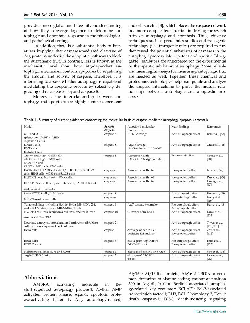
conserved processes that play a crucial role in deter-mining cell fate. Despite of the marked differences between these two catabolic processes, autophagy and apoptosis are intimately connected with each other. In the majority of cases, autophagy and apop-tosis tend to be mutually inhibitory. Caspases have a cardinal role in apoptotic cell death and plays a criti-cal role in directing autophagy-apoptosis crosstalk (Table 1). Processed caspases can shut off the au-tophagic response by degradation of Atg proteins (i.e., Beclin-1, Atg5, and Atg7). In some special cases, the pro-autophagic proteins can be even converted into pro-apoptotic ones to mediate apoptotic cell death after having been cleaved by caspases. On the other hand, autophagy can influence the apoptotic cascades by regulating the amount and activity of caspases. Undoubtedly, these key findings have renewed our knowledge of the unique roles of caspases in directing autophagy-apoptosis conversation.
However, molecular mechanisms underlying caspase-mediated autophagy-apoptosis crosstalk are still incomplete and fragmented. Current evidence emphasized that direct protein–protein interactions between caspases and autophagy-associated proteins enable the multiple layers of communications be-tween autophagy and apoptosis during cell fate deci-sion [9, 147]. For instance, most cases merely illustrate a functional outcome for the given interacting pro-teins (i.e., cleavage of Beclin-1 by caspase-3 abolishs its pro-autophagic function) in the in vitro settings. However, these binary protein interactions of specific caspases and Atg proteins exhibit clear directionality that does suggest the effects of these given caspases on autophagic and apoptotic processes in an in vitro assay under specific conditions. It remains a pending conundrum whether such binary interactions actually reflect the nature of the crosstalk between autophagy and apoptosis in an in vivo circumstance. Thus, further research is necessary to evaluate that the functions of the protein-protein interactions between caspases and Atg proteins in the autophagy-apoptosis crosstalk in animal models and human clinical models. This will
Int. J. Biol. Sci. 2014, Vol. 10
http://www.ijbs.com
1080
provide a more global and integrative understanding of how they converge together to determine au-tophagic and apoptotic response in the physiological and pathological settings.
In addition, there is a substantial body of liter-atures implying that caspases-mediated cleavage of Atg proteins underlies the apoptotic pathway to block the autophagic flux. In contrast, less is known at the mechanistic level about how Atg-dependent au-tophagic mechanism controls apoptosis by regulating the amount and activity of caspase. Therefore, it is interesting to assess whether autophagy is capable of modulating the apoptotic process by selectively de-grading other caspases beyond caspase-8.
Moreover, the interrelationship between au-tophagy and apoptosis are highly context-dependent
and cell-specific [8], which places the caspase network in a more complicated situation in driving the switch between autophagy and apoptosis. Thus, effective techniques such as proteomics studies and transgenic technology (i.e., transgenic mice) are required to fur-ther reveal the potential substrates of caspases in the autophagic process. More potent and specific “drug-gable” inhibitors are anticipated for the experimental or therapeutic inhibition of autophagy. More reliable and meaningful assays for measuring autophagic flux are needed as well. Together, these chemical and proteomics technologies help manipulate and analyze the caspase interactome to probe the mutual rela-tionships between autophagic and apoptostic pro-cesses.
Table 1. Summary of current evidences concerning the molecular basis of caspase-mediated autophagy-apoptosis crosstalk.
Model Specific caspases
Associated molecular mechanisms
Main findings References
OT1 and OT-II splenocytes; FADD−/− MEFs; caspse8−/− T cells
caspase-8 RIPK1 cleavage Anti-autophagic effect Bell et al., [82]
Jurkat T cells; U937 cells; HEK293T cells
caspase-8
Atg3 cleavage (Atg3 amino acids 166–169)
Anti-autophagic effect Oral et al., [16]
Atg5+/+ and Atg5−/− MEF cells; Atg3+/+ and Atg3−/− MEF cells; FADD+/+ and FADD−/− MEF cells; KG-1 cells
caspase-8 Association with FADD:Atg12-Atg5 complex
Pro-apoptotic effect
Young et al., [20]
H460 cells; HEK293T cells; Bax+/− HCT116 cells; HT29 cells; JHH6 cells; MG63 cells; U2OS cells
caspase-8 Association with p62 Pro-apoptotic effect
Jin et al., [92]
HEK293T cells; bax−/−bak−/− BMK cells caspase-8 Association with p62 Pro-apoptotic effect Pan et al., [93]
HCT116 Bax−/−cells; caspase-8-deficient, FADD-deficient,
and parental Jurkat cells caspase-8 Association with p62 Pro-apoptotic effect Huang et al.,
[97]
Bax-/- HCT116 cells; Jurkat cells caspase-8 Anti-apoptotic effect Hou et al., [19]
MCF-7 breast cancer cells caspase-9 Pro-autophagic effect Jeong et al., [103]
Tumor cell lines, including Hct116, HeLa, MB-MDA-231, and RKO, VP-16-resistant MDA-MB-231 cells
caspase-9 Atg7-caspase-9 complex Pro-autophagic effect Anti-apoptotic effect
Han et al., [18]
Myeloma cell lines, lymphoma cell lines, and the human
stromal cell line HS-5 caspase-10 Cleavage of BCLAF1 Anti-autophagic effect Lamy et al.,
[114]
Neurons, astrocytes, osteoclasts, and embryonic fibroblasts cultured from caspase-2 knockout mice
caspase-2 Anti-autophagic effect Tiwari et al., [110, 111]
HeLa cells caspase-3 cleavage of Beclin-1 at positions 124 and 149
Anti-autophagic effect Pro-apoptotic effect
Zhu et al., [118]
HeLa cells; HEK293 cells
caspase-3 cleavage of Atg4D at the DEVD63K motif
Pro-autophagic effect Pro-apoptotic effect
Betin et al., [123]
Melanoma cell lines A375 and A2058 caspase-6 cleavage of Beclin-1 and Atg5 Anti-autophagic effect You et al., [74] Atg16L1 T300A mice caspase-7 cleavage of ATG16L1
T300A Anti-autophagic effect Lassen et al.,
[76]
Abbreviations AMBRA: activating molecule in Be-
clin1-regulated autophagy protein 1; AMPK: AMP activated protein kinase; Apaf-1: apoptotic prote-ase-activating factor 1; Atg: autophagy-related;
Atg16L: Atg16-like protein; Atg16L1 T300A: a com-mon threonine to alanine coding variant at position 300 in Atg16L; barkor: Beclin-1-associated autopha-gy-related key regulator; BCLAF1: Bcl-2-associated transcription factor 1; BH3, BCL-2 homology-3; Dcp-1: death caspase-1; DISC: death-inducing signaling

Int. J. Biol. Sci. 2014, Vol. 10
http://www.ijbs.com
1081
complex; FADD: Fas-associated death domain; FIP200: focal adhesion kinase family-interacting pro-tein of 200 kD; GABARAP: γ-aminobutyric acid re-ceptor-associated protein; LC3: the protein microtu-bule-associated protein 1 light chain 3; MOMP: mito-chondrial outer membranes permeabilization; mTOR: mammalian Target of rapamycin; PE: phosphatidyl-ethanolamine; PI3K: phosphatidylinositol 3-kinase; RIPK1: receptor interacting protein kinase 1; SPP1: sphingosine-1-phosphate phosphohydrolase 1; TNF: tumor necrosis factor; TRAIL: TNF-related apopto-sis-inducing ligand; UBA: ubiquitin-associated; UVRAG: UV irradiation resistance-associated gene.
Acknowledgements This study was supported by National Natural
Science Foundation of China (grant. 81171096, 81371433, 81271273, and 81371369), Research Fund for the Doctoral Program of Higher Education of China (20120101120030), Zhejiang Provincial Medical Sci-ence and Technology Planning Project (grant. 2012RCA030 and 2013KYA088), Zhejiang Provincial Education Planning Project (Y201226287) and Zhejiang Provincial Natural Science Foundation of China (grant Y13H090007 and LY13H090002).
Competing Interests The authors have declared that no competing
interest exists.
References 1. Boya P, Reggiori F, Codogno P. Emerging regulation and functions of autophagy.
Nature cell biology. 2013; 15: 713-20. doi:10.1038/ncb2788. 2. Hengartner MO. The biochemistry of apoptosis. Nature. 2000; 407: 770-6.
doi:10.1038/35037710. 3. Taylor RC, Cullen SP, Martin SJ. Apoptosis: controlled demolition at the cellular
level. Nature reviews Molecular cell biology. 2008; 9: 231-41. doi:10.1038/nrm2312. 4. Mizushima N, Levine B. Autophagy in mammalian development and differentiation.
Nature cell biology. 2010; 12: 823-30. doi:10.1038/ncb0910-823. 5. Thompson CB. Apoptosis in the pathogenesis and treatment of disease. Science.
1995; 267: 1456-62. 6. Caroppi P, Sinibaldi F, Fiorucci L, Santucci R. Apoptosis and human diseases:
mitochondrion damage and lethal role of released cytochrome C as proapoptotic protein. Current medicinal chemistry. 2009; 16: 4058-65.
7. Moretti L, Cha YI, Niermann KJ, Lu B. Switch between apoptosis and autophagy: radiation-induced endoplasmic reticulum stress? Cell cycle. 2007; 6: 793-8.
8. Marino G, Niso-Santano M, Baehrecke EH, Kroemer G. Self-consumption: the interplay of autophagy and apoptosis. Nature reviews Molecular cell biology. 2014; 15: 81-94. doi:10.1038/nrm3735.
9. Rubinstein AD, Kimchi A. Life in the balance - a mechanistic view of the crosstalk between autophagy and apoptosis. Journal of cell science. 2012; 125: 5259-68. doi:10.1242/jcs.115865.
10. Nunez G, Benedict MA, Hu Y, Inohara N. Caspases: the proteases of the apoptotic pathway. Oncogene. 1998; 17: 3237-45. doi:10.1038/sj.onc.1202581.
11. Riedl SJ, Shi Y. Molecular mechanisms of caspase regulation during apoptosis. Nature reviews Molecular cell biology. 2004; 5: 897-907. doi:10.1038/nrm1496.
12. Tait SW, Green DR. Mitochondria and cell death: outer membrane permeabilization and beyond. Nature reviews Molecular cell biology. 2010; 11: 621-32. doi:10.1038/nrm2952.
13. Parrish AB, Freel CD, Kornbluth S. Cellular mechanisms controlling caspase activation and function. Cold Spring Harbor perspectives in biology. 2013; 5. doi:10.1101/cshperspect.a008672.
14. Stupack DG. Caspase-8 as a therapeutic target in cancer. Cancer letters. 2013; 332: 133-40. doi:10.1016/j.canlet.2010.07.022.
15. Yu L, Alva A, Su H, Dutt P, Freundt E, Welsh S, et al. Regulation of an ATG7-beclin 1 program of autophagic cell death by caspase-8. Science. 2004; 304: 1500-2. doi:10.1126/science.1096645.
16. Oral O, Oz-Arslan D, Itah Z, Naghavi A, Deveci R, Karacali S, et al. Cleavage of Atg3 protein by caspase-8 regulates autophagy during receptor-activated cell death. Apoptosis : an international journal on programmed cell death. 2012; 17: 810-20. doi:10.1007/s10495-012-0735-0.
17. Djavaheri-Mergny M, Maiuri MC, Kroemer G. Cross talk between apoptosis and autophagy by caspase-mediated cleavage of Beclin 1. Oncogene. 2010; 29: 1717-9. doi:10.1038/onc.2009.519.
18. Han J, Hou W, Goldstein LA, Stolz DB, Watkins SC, Rabinowich H. A Complex between Atg7 and Caspase-9: A NOVEL MECHANISM OF CROSS-REGULATION BETWEEN AUTOPHAGY AND APOPTOSIS. The Journal of biological chemistry. 2014; 289: 6485-97. doi:10.1074/jbc.M113.536854.
19. Hou W, Han J, Lu C, Goldstein LA, Rabinowich H. Autophagic degradation of active caspase-8: a crosstalk mechanism between autophagy and apoptosis. Autophagy. 2010; 6: 891-900. doi:10.4161/auto.6.7.13038.
20. Young MM, Takahashi Y, Khan O, Park S, Hori T, Yun J, et al. Autophagosomal membrane serves as platform for intracellular death-inducing signaling complex (iDISC)-mediated caspase-8 activation and apoptosis. The Journal of biological chemistry. 2012; 287: 12455-68. doi:10.1074/jbc.M111.309104.
21. Nixon RA. The role of autophagy in neurodegenerative disease. Nature medicine. 2013; 19: 983-97. doi:10.1038/nm.3232.
22. Harris H, Rubinsztein DC. Control of autophagy as a therapy for neurodegenerative disease. Nature reviews Neurology. 2012; 8: 108-17. doi:10.1038/nrneurol.2011.200.
23. Mizushima N, Levine B, Cuervo AM, Klionsky DJ. Autophagy fights disease through cellular self-digestion. Nature. 2008; 451: 1069-75. doi:10.1038/nature06639.
24. Vinod V, Padmakrishnan CJ, Vijayan B, Gopala S. 'How can I halt thee?' The puzzles involved in autophagic inhibition. Pharmacological research : the official journal of the Italian Pharmacological Society. 2014; 82C: 1-8. doi:10.1016/j.phrs.2014.03.005.
25. Levine B, Klionsky DJ. Development by self-digestion: molecular mechanisms and biological functions of autophagy. Developmental cell. 2004; 6: 463-77.
26. Tanida I. Autophagosome formation and molecular mechanism of autophagy. Antioxidants & redox signaling. 2011; 14: 2201-14. doi:10.1089/ars.2010.3482.
27. He C, Klionsky DJ. Regulation mechanisms and signaling pathways of autophagy. Annual review of genetics. 2009; 43: 67-93. doi:10.1146/annurev-genet-102808-114910.
28. Wong PM, Puente C, Ganley IG, Jiang X. The ULK1 complex: sensing nutrient signals for autophagy activation. Autophagy. 2013; 9: 124-37. doi:10.4161/auto.23323.
29. Kim J, Kundu M, Viollet B, Guan KL. AMPK and mTOR regulate autophagy through direct phosphorylation of Ulk1. Nature cell biology. 2011; 13: 132-41. doi:10.1038/ncb2152.
30. Egan DF, Shackelford DB, Mihaylova MM, Gelino S, Kohnz RA, Mair W, et al. Phosphorylation of ULK1 (hATG1) by AMP-activated protein kinase connects energy sensing to mitophagy. Science. 2011; 331: 456-61. doi:10.1126/science.1196371.
31. Jaber N, Dou Z, Chen JS, Catanzaro J, Jiang YP, Ballou LM, et al. Class III PI3K Vps34 plays an essential role in autophagy and in heart and liver function. Proceedings of the National Academy of Sciences of the United States of America. 2012; 109: 2003-8. doi:10.1073/pnas.1112848109.
32. Sun Q, Fan W, Chen K, Ding X, Chen S, Zhong Q. Identification of Barkor as a mammalian autophagy-specific factor for Beclin 1 and class III phosphatidylinositol 3-kinase. Proceedings of the National Academy of Sciences of the United States of America. 2008; 105: 19211-6. doi:10.1073/pnas.0810452105.
33. Panaretou C, Domin J, Cockcroft S, Waterfield MD. Characterization of p150, an adaptor protein for the human phosphatidylinositol (PtdIns) 3-kinase. Substrate presentation by phosphatidylinositol transfer protein to the p150.Ptdins 3-kinase complex. The Journal of biological chemistry. 1997; 272: 2477-85.
34. Takahashi Y, Coppola D, Matsushita N, Cualing HD, Sun M, Sato Y, et al. Bif-1 interacts with Beclin 1 through UVRAG and regulates autophagy and tumorigenesis. Nature cell biology. 2007; 9: 1142-51. doi:10.1038/ncb1634.
35. Furuya N, Yu J, Byfield M, Pattingre S, Levine B. The evolutionarily conserved domain of Beclin 1 is required for Vps34 binding, autophagy and tumor suppressor function. Autophagy. 2005; 1: 46-52.
36. Itakura E, Mizushima N. Atg14 and UVRAG: mutually exclusive subunits of mammalian Beclin 1-PI3K complexes. Autophagy. 2009; 5: 534-6.
37. Suzuki K, Kubota Y, Sekito T, Ohsumi Y. Hierarchy of Atg proteins in pre-autophagosomal structure organization. Genes to cells : devoted to molecular & cellular mechanisms. 2007; 12: 209-18. doi:10.1111/j.1365-2443.2007.01050.x.
38. Di Bartolomeo S, Corazzari M, Nazio F, Oliverio S, Lisi G, Antonioli M, et al. The dynamic interaction of AMBRA1 with the dynein motor complex regulates mammalian autophagy. The Journal of cell biology. 2010; 191: 155-68. doi:10.1083/jcb.201002100.
39. Russell RC, Tian Y, Yuan H, Park HW, Chang YY, Kim J, et al. ULK1 induces autophagy by phosphorylating Beclin-1 and activating VPS34 lipid kinase. Nature cell biology. 2013; 15: 741-50. doi:10.1038/ncb2757.
40. Geng J, Klionsky DJ. The Atg8 and Atg12 ubiquitin-like conjugation systems in macroautophagy. 'Protein modifications: beyond the usual suspects' review series. EMBO reports. 2008; 9: 859-64. doi:10.1038/embor.2008.163.
41. Walczak M, Martens S. Dissecting the role of the Atg12-Atg5-Atg16 complex during autophagosome formation. Autophagy. 2013; 9: 424-5. doi:10.4161/auto.22931.
42. Noda NN, Fujioka Y, Hanada T, Ohsumi Y, Inagaki F. Structure of the Atg12-Atg5 conjugate reveals a platform for stimulating Atg8-PE conjugation. EMBO reports. 2013; 14: 206-11. doi:10.1038/embor.2012.208.
43. Hanada T, Noda NN, Satomi Y, Ichimura Y, Fujioka Y, Takao T, et al. The Atg12-Atg5 conjugate has a novel E3-like activity for protein lipidation in autophagy. The Journal of biological chemistry. 2007; 282: 37298-302. doi:10.1074/jbc.C700195200.
44. Romanov J, Walczak M, Ibiricu I, Schuchner S, Ogris E, Kraft C, et al. Mechanism and functions of membrane binding by the Atg5-Atg12/Atg16 complex during autophagosome formation. The EMBO journal. 2012; 31: 4304-17. doi:10.1038/emboj.2012.278.
45. Kaufmann A, Beier V, Franquelim HG, Wollert T. Molecular mechanism of autophagic membrane-scaffold assembly and disassembly. Cell. 2014; 156: 469-81. doi:10.1016/j.cell.2013.12.022.
46. Liang C, Lee JS, Inn KS, Gack MU, Li Q, Roberts EA, et al. Beclin1-binding UVRAG targets the class C Vps complex to coordinate autophagosome maturation and endocytic trafficking. Nature cell biology. 2008; 10: 776-87. doi:10.1038/ncb1740.
Int. J. Biol. Sci. 2014, Vol. 10
http://www.ijbs.com
1082
47. Matsunaga K, Saitoh T, Tabata K, Omori H, Satoh T, Kurotori N, et al. Two Beclin 1-binding proteins, Atg14L and Rubicon, reciprocally regulate autophagy at different stages. Nature cell biology. 2009; 11: 385-96. doi:10.1038/ncb1846.
48. Zhong Y, Wang QJ, Li X, Yan Y, Backer JM, Chait BT, et al. Distinct regulation of autophagic activity by Atg14L and Rubicon associated with Beclin 1-phosphatidylinositol-3-kinase complex. Nature cell biology. 2009; 11: 468-76. doi:10.1038/ncb1854.
49. Mattson MP. Apoptosis in neurodegenerative disorders. Nature reviews Molecular cell biology. 2000; 1: 120-9. doi:10.1038/35040009.
50. Reed JC. Apoptosis-based therapies. Nature reviews Drug discovery. 2002; 1: 111-21. doi:10.1038/nrd726.
51. Fuchs Y, Steller H. Programmed cell death in animal development and disease. Cell. 2011; 147: 742-58. doi:10.1016/j.cell.2011.10.033.
52. White E. Life, death, and the pursuit of apoptosis. Genes & development. 1996; 10: 1-15.
53. Yuan J, Yankner BA. Apoptosis in the nervous system. Nature. 2000; 407: 802-9. doi:10.1038/35037739.
54. Boyce M, Degterev A, Yuan J. Caspases: an ancient cellular sword of Damocles. Cell death and differentiation. 2004; 11: 29-37. doi:10.1038/sj.cdd.4401339.
55. Strasser A, O'Connor L, Dixit VM. Apoptosis signaling. Annual review of biochemistry. 2000; 69: 217-45. doi:10.1146/annurev.biochem.69.1.217.
56. Sayers TJ. Targeting the extrinsic apoptosis signaling pathway for cancer therapy. Cancer immunology, immunotherapy : CII. 2011; 60: 1173-80. doi:10.1007/s00262-011-1008-4.
57. Kaufmann T, Strasser A, Jost PJ. Fas death receptor signalling: roles of Bid and XIAP. Cell death and differentiation. 2012; 19: 42-50. doi:10.1038/cdd.2011.121.
58. Fulda S, Debatin KM. Extrinsic versus intrinsic apoptosis pathways in anticancer chemotherapy. Oncogene. 2006; 25: 4798-811. doi:10.1038/sj.onc.1209608.
59. Fulda S, Debatin KM. Exploiting death receptor signaling pathways for tumor therapy. Biochimica et biophysica acta. 2004; 1705: 27-41. doi:10.1016/j.bbcan.2004.09.003.
60. Mahalingam D, Szegezdi E, Keane M, de Jong S, Samali A. TRAIL receptor signalling and modulation: Are we on the right TRAIL? Cancer treatment reviews. 2009; 35: 280-8. doi:10.1016/j.ctrv.2008.11.006.
61. Shamas-Din A, Brahmbhatt H, Leber B, Andrews DW. BH3-only proteins: Orchestrators of apoptosis. Biochimica et biophysica acta. 2011; 1813: 508-20. doi:10.1016/j.bbamcr.2010.11.024.
62. Wang Y, Tjandra N. Structural insights of tBid, the caspase-8-activated Bid, and its BH3 domain. The Journal of biological chemistry. 2013; 288: 35840-51. doi:10.1074/jbc.M113.503680.
63. Bender T, Martinou JC. Where killers meet--permeabilization of the outer mitochondrial membrane during apoptosis. Cold Spring Harbor perspectives in biology. 2013; 5: a011106. doi:10.1101/cshperspect.a011106.
64. Franklin JL. Redox regulation of the intrinsic pathway in neuronal apoptosis. Antioxidants & redox signaling. 2011; 14: 1437-48. doi:10.1089/ars.2010.3596.
65. Tait SW, Green DR. Mitochondrial regulation of cell death. Cold Spring Harbor perspectives in biology. 2013; 5. doi:10.1101/cshperspect.a008706.
66. Pradelli LA, Beneteau M, Ricci JE. Mitochondrial control of caspase-dependent and -independent cell death. Cellular and molecular life sciences : CMLS. 2010; 67: 1589-97. doi:10.1007/s00018-010-0285-y.
67. Harris MH, Thompson CB. The role of the Bcl-2 family in the regulation of outer mitochondrial membrane permeability. Cell death and differentiation. 2000; 7: 1182-91. doi:10.1038/sj.cdd.4400781.
68. Zou H, Yang R, Hao J, Wang J, Sun C, Fesik SW, et al. Regulation of the Apaf-1/caspase-9 apoptosome by caspase-3 and XIAP. The Journal of biological chemistry. 2003; 278: 8091-8. doi:10.1074/jbc.M204783200.
69. Zentgraf H, Scheer U, Franke WW. Characterization and localization of the RNA synthesized in mature avian erythrocytes. Experimental cell research. 1975; 96: 81-95.
70. Yang Y, Xing D, Zhou F, Chen Q. Mitochondrial autophagy protects against heat shock-induced apoptosis through reducing cytosolic cytochrome c release and downstream caspase-3 activation. Biochemical and biophysical research communications. 2010; 395: 190-5. doi:10.1016/j.bbrc.2010.03.155.
71. Mohseni N, McMillan SC, Chaudhary R, Mok J, Reed BH. Autophagy promotes caspase-dependent cell death during Drosophila development. Autophagy. 2009; 5: 329-38.
72. Kim R, Emi M, Tanabe K. Role of mitochondria as the gardens of cell death. Cancer chemotherapy and pharmacology. 2006; 57: 545-53. doi:10.1007/s00280-005-0111-7.
73. Joubert PE, Werneke SW, de la Calle C, Guivel-Benhassine F, Giodini A, Peduto L, et al. Chikungunya virus-induced autophagy delays caspase-dependent cell death. The Journal of experimental medicine. 2012; 209: 1029-47. doi:10.1084/jem.20110996.
74. You M, Savaraj N, Kuo MT, Wangpaichitr M, Varona-Santos J, Wu C, et al. TRAIL induces autophagic protein cleavage through caspase activation in melanoma cell lines under arginine deprivation. Molecular and cellular biochemistry. 2013; 374: 181-90. doi:10.1007/s11010-012-1518-1.
75. Cho DH, Jo YK, Hwang JJ, Lee YM, Roh SA, Kim JC. Caspase-mediated cleavage of ATG6/Beclin-1 links apoptosis to autophagy in HeLa cells. Cancer letters. 2009; 274: 95-100. doi:10.1016/j.canlet.2008.09.004.
76. Lassen KG, Kuballa P, Conway KL, Patel KK, Becker CE, Peloquin JM, et al. Atg16L1 T300A variant decreases selective autophagy resulting in altered cytokine signaling and decreased antibacterial defense. Proceedings of the National Academy of Sciences of the United States of America. 2014; 111: 7741-6. doi:10.1073/pnas.1407001111.
77. Fulda S. Caspase-8 in cancer biology and therapy. Cancer letters. 2009; 281: 128-33. doi:10.1016/j.canlet.2008.11.023.
78. Zhao Y, Sui X, Ren H. From procaspase-8 to caspase-8: revisiting structural functions of caspase-8. Journal of cellular physiology. 2010; 225: 316-20. doi:10.1002/jcp.22276.
79. Li H, Zhu H, Xu CJ, Yuan J. Cleavage of BID by caspase 8 mediates the mitochondrial damage in the Fas pathway of apoptosis. Cell. 1998; 94: 491-501.
80. Gross A, Yin XM, Wang K, Wei MC, Jockel J, Milliman C, et al. Caspase cleaved BID targets mitochondria and is required for cytochrome c release, while BCL-XL prevents this release but not tumor necrosis factor-R1/Fas death. The Journal of biological chemistry. 1999; 274: 1156-63.
81. Ferreira KS, Kreutz C, Macnelly S, Neubert K, Haber A, Bogyo M, et al. Caspase-3 feeds back on caspase-8, Bid and XIAP in type I Fas signaling in primary mouse hepatocytes. Apoptosis : an international journal on programmed cell death. 2012; 17: 503-15. doi:10.1007/s10495-011-0691-0.
82. Bell BD, Leverrier S, Weist BM, Newton RH, Arechiga AF, Luhrs KA, et al. FADD and caspase-8 control the outcome of autophagic signaling in proliferating T cells. Proceedings of the National Academy of Sciences of the United States of America. 2008; 105: 16677-82. doi:10.1073/pnas.0808597105.
83. Bell BD, Walsh CM. Coordinate regulation of autophagy and apoptosis in T cells by death effectors: FADD or foundation. Autophagy. 2009; 5: 238-40.
84. Lin Y, Devin A, Rodriguez Y, Liu ZG. Cleavage of the death domain kinase RIP by caspase-8 prompts TNF-induced apoptosis. Genes & development. 1999; 13: 2514-26.
85. Orozco S, Yatim N, Werner MR, Tran H, Gunja SY, Tait SW, et al. RIPK1 both positively and negatively regulates RIPK3 oligomerization and necroptosis. Cell death and differentiation. 2014. doi:10.1038/cdd.2014.76.
86. Duprez L, Bertrand MJ, Vanden Berghe T, Dondelinger Y, Festjens N, Vandenabeele P. Intermediate domain of receptor-interacting protein kinase 1 (RIPK1) determines switch between necroptosis and RIPK1 kinase-dependent apoptosis. The Journal of biological chemistry. 2012; 287: 14863-72. doi:10.1074/jbc.M111.288670.
87. Kaiser WJ, Daley-Bauer LP, Thapa RJ, Mandal P, Berger SB, Huang C, et al. RIP1 suppresses innate immune necrotic as well as apoptotic cell death during mammalian parturition. Proceedings of the National Academy of Sciences of the United States of America. 2014; 111: 7753-8. doi:10.1073/pnas.1401857111.
88. He W, Wang Q, Xu J, Xu X, Padilla MT, Ren G, et al. Attenuation of TNFSF10/TRAIL-induced apoptosis by an autophagic survival pathway involving TRAF2- and RIPK1/RIP1-mediated MAPK8/JNK activation. Autophagy. 2012; 8: 1811-21. doi:10.4161/auto.22145.
89. Laussmann MA, Passante E, Dussmann H, Rauen JA, Wurstle ML, Delgado ME, et al. Proteasome inhibition can induce an autophagy-dependent apical activation of caspase-8. Cell death and differentiation. 2011; 18: 1584-97. doi:10.1038/cdd.2011.27.
90. Pandey UB, Nie Z, Batlevi Y, McCray BA, Ritson GP, Nedelsky NB, et al. HDAC6 rescues neurodegeneration and provides an essential link between autophagy and the UPS. Nature. 2007; 447: 859-63. doi:10.1038/nature05853.
91. Zhu K, Dunner K, Jr., McConkey DJ. Proteasome inhibitors activate autophagy as a cytoprotective response in human prostate cancer cells. Oncogene. 2010; 29: 451-62. doi:10.1038/onc.2009.343.
92. Jin Z, Li Y, Pitti R, Lawrence D, Pham VC, Lill JR, et al. Cullin3-based polyubiquitination and p62-dependent aggregation of caspase-8 mediate extrinsic apoptosis signaling. Cell. 2009; 137: 721-35. doi:10.1016/j.cell.2009.03.015.
93. Pan JA, Fan Y, Gandhirajan RK, Madesh M, Zong WX. Hyperactivation of the mammalian degenerin MDEG promotes caspase-8 activation and apoptosis. The Journal of biological chemistry. 2013; 288: 2952-63. doi:10.1074/jbc.M112.441063.
94. Seibenhener ML, Babu JR, Geetha T, Wong HC, Krishna NR, Wooten MW. Sequestosome 1/p62 is a polyubiquitin chain binding protein involved in ubiquitin proteasome degradation. Molecular and cellular biology. 2004; 24: 8055-68. doi:10.1128/MCB.24.18.8055-8068.2004.
95. Moscat J, Diaz-Meco MT, Albert A, Campuzano S. Cell signaling and function organized by PB1 domain interactions. Molecular cell. 2006; 23: 631-40. doi:10.1016/j.molcel.2006.08.002.
96. Moscat J, Diaz-Meco MT, Wooten MW. Signal integration and diversification through the p62 scaffold protein. Trends in biochemical sciences. 2007; 32: 95-100. doi:10.1016/j.tibs.2006.12.002.
97. Huang S, Okamoto K, Yu C, Sinicrope FA. p62/sequestosome-1 up-regulation promotes ABT-263-induced caspase-8 aggregation/activation on the autophagosome. The Journal of biological chemistry. 2013; 288: 33654-66. doi:10.1074/jbc.M113.518134.
98. Pankiv S, Clausen TH, Lamark T, Brech A, Bruun JA, Outzen H, et al. p62/SQSTM1 binds directly to Atg8/LC3 to facilitate degradation of ubiquitinated protein aggregates by autophagy. The Journal of biological chemistry. 2007; 282: 24131-45. doi:10.1074/jbc.M702824200.
99. Zhang YB, Zhao W, Zeng RX. Autophagic degradation of caspase-8 protects U87MG cells against H2O2-induced oxidative stress. Asian Pacific journal of cancer prevention : APJCP. 2013; 14: 4095-9.
100. Wurstle ML, Laussmann MA, Rehm M. The central role of initiator caspase-9 in apoptosis signal transduction and the regulation of its activation and activity on the apoptosome. Experimental cell research. 2012; 318: 1213-20. doi:10.1016/j.yexcr.2012.02.013.
101. Reubold TF, Eschenburg S. A molecular view on signal transduction by the apoptosome. Cellular signalling. 2012; 24: 1420-5. doi:10.1016/j.cellsig.2012.03.007.
102. Bratton SB, Salvesen GS. Regulation of the Apaf-1-caspase-9 apoptosome. Journal of cell science. 2010; 123: 3209-14. doi:10.1242/jcs.073643.
103. Jeong HS, Choi HY, Lee ER, Kim JH, Jeon K, Lee HJ, et al. Involvement of caspase-9 in autophagy-mediated cell survival pathway. Biochimica et biophysica acta. 2011; 1813: 80-90. doi:10.1016/j.bbamcr.2010.09.016.
104. Allan LA, Clarke PR. Apoptosis and autophagy: Regulation of caspase-9 by phosphorylation. The FEBS journal. 2009; 276: 6063-73. doi:10.1111/j.1742-4658.2009.07330.x.
105. Cardone MH, Roy N, Stennicke HR, Salvesen GS, Franke TF, Stanbridge E, et al. Regulation of cell death protease caspase-9 by phosphorylation. Science. 1998; 282: 1318-21.
106. Tinel A, Tschopp J. The PIDDosome, a protein complex implicated in activation of caspase-2 in response to genotoxic stress. Science. 2004; 304: 843-6. doi:10.1126/science.1095432.
107. Tu S, McStay GP, Boucher LM, Mak T, Beere HM, Green DR. In situ trapping of activated initiator caspases reveals a role for caspase-2 in heat shock-induced apoptosis. Nature cell biology. 2006; 8: 72-7. doi:10.1038/ncb1340.
108. Bonzon C, Bouchier-Hayes L, Pagliari LJ, Green DR, Newmeyer DD. Caspase-2-induced apoptosis requires bid cleavage: a physiological role for bid in heat shock-induced death. Molecular biology of the cell. 2006; 17: 2150-7. doi:10.1091/mbc.E05-12-1107.
Int. J. Biol. Sci. 2014, Vol. 10
http://www.ijbs.com
1083
109. Ho LH, Read SH, Dorstyn L, Lambrusco L, Kumar S. Caspase-2 is required for cell death induced by cytoskeletal disruption. Oncogene. 2008; 27: 3393-404. doi:10.1038/sj.onc.1211005.
110. Tiwari M, Sharma LK, Vanegas D, Callaway DA, Bai Y, Lechleiter JD, et al. A nonapoptotic role for CASP2/caspase 2: modulation of autophagy. Autophagy. 2014; 10: 1054-70. doi:10.4161/auto.28528.
111. Tiwari M, Lopez-Cruzan M, Morgan WW, Herman B. Loss of caspase-2-dependent apoptosis induces autophagy after mitochondrial oxidative stress in primary cultures of young adult cortical neurons. The Journal of biological chemistry. 2011; 286: 8493-506. doi:10.1074/jbc.M110.163824.
112. Fischer U, Stroh C, Schulze-Osthoff K. Unique and overlapping substrate specificities of caspase-8 and caspase-10. Oncogene. 2006; 25: 152-9. doi:10.1038/sj.onc.1209015.
113. Milhas D, Cuvillier O, Therville N, Clave P, Thomsen M, Levade T, et al. Caspase-10 triggers Bid cleavage and caspase cascade activation in FasL-induced apoptosis. The Journal of biological chemistry. 2005; 280: 19836-42. doi:10.1074/jbc.M414358200.
114. Lamy L, Ngo VN, Emre NC, Shaffer AL, 3rd, Yang Y, Tian E, et al. Control of autophagic cell death by caspase-10 in multiple myeloma. Cancer cell. 2013; 23: 435-49. doi:10.1016/j.ccr.2013.02.017.
115. Sadasivan S, Waghray A, Larner SF, Dunn WA, Jr., Hayes RL, Wang KK. Amino acid starvation induced autophagic cell death in PC-12 cells: evidence for activation of caspase-3 but not calpain-1. Apoptosis : an international journal on programmed cell death. 2006; 11: 1573-82. doi:10.1007/s10495-006-7690-6.
116. Sirois I, Groleau J, Pallet N, Brassard N, Hamelin K, Londono I, et al. Caspase activation regulates the extracellular export of autophagic vacuoles. Autophagy. 2012; 8: 927-37. doi:10.4161/auto.19768.
117. Nunez R, Sancho-Martinez SM, Novoa JM, Lopez-Hernandez FJ. Apoptotic volume decrease as a geometric determinant for cell dismantling into apoptotic bodies. Cell death and differentiation. 2010; 17: 1665-71. doi:10.1038/cdd.2010.96.
118. Zhu Y, Zhao L, Liu L, Gao P, Tian W, Wang X, et al. Beclin 1 cleavage by caspase-3 inactivates autophagy and promotes apoptosis. Protein & cell. 2010; 1: 468-77. doi:10.1007/s13238-010-0048-4.
119. Oberstein A, Jeffrey PD, Shi Y. Crystal structure of the Bcl-XL-Beclin 1 peptide complex: Beclin 1 is a novel BH3-only protein. The Journal of biological chemistry. 2007; 282: 13123-32. doi:10.1074/jbc.M700492200.
120. Erlich S, Mizrachy L, Segev O, Lindenboim L, Zmira O, Adi-Harel S, et al. Differential interactions between Beclin 1 and Bcl-2 family members. Autophagy. 2007; 3: 561-8.
121. Ku B, Woo JS, Liang C, Lee KH, Jung JU, Oh BH. An insight into the mechanistic role of Beclin 1 and its inhibition by prosurvival Bcl-2 family proteins. Autophagy. 2008; 4: 519-20.
122. Czabotar PE, Lessene G, Strasser A, Adams JM. Control of apoptosis by the BCL-2 protein family: implications for physiology and therapy. Nature reviews Molecular cell biology. 2014; 15: 49-63. doi:10.1038/nrm3722.
123. Betin VM, Lane JD. Caspase cleavage of Atg4D stimulates GABARAP-L1 processing and triggers mitochondrial targeting and apoptosis. Journal of cell science. 2009; 122: 2554-66. doi:10.1242/jcs.046250.
124. Slee EA, Adrain C, Martin SJ. Executioner caspase-3, -6, and -7 perform distinct, non-redundant roles during the demolition phase of apoptosis. The Journal of biological chemistry. 2001; 276: 7320-6. doi:10.1074/jbc.M008363200.
125. Eguchi R, Tone S, Suzuki A, Fujimori Y, Nakano T, Kaji K, et al. Possible involvement of caspase-6 and -7 but not caspase-3 in the regulation of hypoxia-induced apoptosis in tube-forming endothelial cells. Experimental cell research. 2009; 315: 327-35. doi:10.1016/j.yexcr.2008.10.041.
126. Norman JM, Cohen GM, Bampton ET. The in vitro cleavage of the hAtg proteins by cell death proteases. Autophagy. 2010; 6: 1042-56.
127. Goll DE, Thompson VF, Li H, Wei W, Cong J. The calpain system. Physiological reviews. 2003; 83: 731-801. doi:10.1152/physrev.00029.2002.
128. Kar P, Samanta K, Shaikh S, Chowdhury A, Chakraborti T, Chakraborti S. Mitochondrial calpain system: an overview. Archives of biochemistry and biophysics. 2010; 495: 1-7. doi:10.1016/j.abb.2009.12.020.
129. Storr SJ, Carragher NO, Frame MC, Parr T, Martin SG. The calpain system and cancer. Nature reviews Cancer. 2011; 11: 364-74. doi:10.1038/nrc3050.
130. Sharma AK, Rohrer B. Calcium-induced calpain mediates apoptosis via caspase-3 in a mouse photoreceptor cell line. The Journal of biological chemistry. 2004; 279: 35564-72. doi:10.1074/jbc.M401037200.
131. Gafni J, Cong X, Chen SF, Gibson BW, Ellerby LM. Calpain-1 cleaves and activates caspase-7. The Journal of biological chemistry. 2009; 284: 25441-9. doi:10.1074/jbc.M109.038174.
132. Tan Y, Dourdin N, Wu C, De Veyra T, Elce JS, Greer PA. Ubiquitous calpains promote caspase-12 and JNK activation during endoplasmic reticulum stress-induced apoptosis. The Journal of biological chemistry. 2006; 281: 16016-24. doi:10.1074/jbc.M601299200.
133. Martinez JA, Zhang Z, Svetlov SI, Hayes RL, Wang KK, Larner SF. Calpain and caspase processing of caspase-12 contribute to the ER stress-induced cell death pathway in differentiated PC12 cells. Apoptosis : an international journal on programmed cell death. 2010; 15: 1480-93. doi:10.1007/s10495-010-0526-4.
134. Wood DE, Thomas A, Devi LA, Berman Y, Beavis RC, Reed JC, et al. Bax cleavage is mediated by calpain during drug-induced apoptosis. Oncogene. 1998; 17: 1069-78. doi:10.1038/sj.onc.1202034.
135. Gao G, Dou QP. N-terminal cleavage of bax by calpain generates a potent proapoptotic 18-kDa fragment that promotes bcl-2-independent cytochrome C release and apoptotic cell death. Journal of cellular biochemistry. 2000; 80: 53-72.
136. Chen M, He H, Zhan S, Krajewski S, Reed JC, Gottlieb RA. Bid is cleaved by calpain to an active fragment in vitro and during myocardial ischemia/reperfusion. The Journal of biological chemistry. 2001; 276: 30724-8. doi:10.1074/jbc.M103701200.
137. Shi M, Zhang T, Sun L, Luo Y, Liu DH, Xie ST, et al. Calpain, Atg5 and Bak play important roles in the crosstalk between apoptosis and autophagy induced by influx of extracellular calcium. Apoptosis : an international journal on programmed cell death. 2013; 18: 435-51. doi:10.1007/s10495-012-0786-2.
138. Lepine S, Allegood JC, Edmonds Y, Milstien S, Spiegel S. Autophagy induced by deficiency of sphingosine-1-phosphate phosphohydrolase 1 is switched to apoptosis
by calpain-mediated autophagy-related gene 5 (Atg5) cleavage. The Journal of biological chemistry. 2011; 286: 44380-90. doi:10.1074/jbc.M111.257519.
139. Yousefi S, Perozzo R, Schmid I, Ziemiecki A, Schaffner T, Scapozza L, et al. Calpain-mediated cleavage of Atg5 switches autophagy to apoptosis. Nature cell biology. 2006; 8: 1124-32. doi:10.1038/ncb1482.
140. Song Z, Guan B, Bergman A, Nicholson DW, Thornberry NA, Peterson EP, et al. Biochemical and genetic interactions between Drosophila caspases and the proapoptotic genes rpr, hid, and grim. Molecular and cellular biology. 2000; 20: 2907-14.
141. Lee G, Wang Z, Sehgal R, Chen CH, Kikuno K, Hay B, et al. Drosophila caspases involved in developmentally regulated programmed cell death of peptidergic neurons during early metamorphosis. The Journal of comparative neurology. 2011; 519: 34-48. doi:10.1002/cne.22498.
142. Hou YC, Hannigan AM, Gorski SM. An executioner caspase regulates autophagy. Autophagy. 2009; 5: 530-3.
143. DeVorkin L, Go NE, Hou YC, Moradian A, Morin GB, Gorski SM. The Drosophila effector caspase Dcp-1 regulates mitochondrial dynamics and autophagic flux via SesB. The Journal of cell biology. 2014; 205: 477-92. doi:10.1083/jcb.201303144.
144. Kim YI, Ryu T, Lee J, Heo YS, Ahnn J, Lee SJ, et al. A genetic screen for modifiers of Drosophila caspase Dcp-1 reveals caspase involvement in autophagy and novel caspase-related genes. BMC cell biology. 2010; 11: 9. doi:10.1186/1471-2121-11-9.
145. Hou YC, Chittaranjan S, Barbosa SG, McCall K, Gorski SM. Effector caspase Dcp-1 and IAP protein Bruce regulate starvation-induced autophagy during Drosophila melanogaster oogenesis. The Journal of cell biology. 2008; 182: 1127-39. doi:10.1083/jcb.200712091.
146. Fraser AG, McCarthy NJ, Evan GI. drICE is an essential caspase required for apoptotic activity in Drosophila cells. The EMBO journal. 1997; 16: 6192-9. doi:10.1093/emboj/16.20.6192.
147. Eisenberg-Lerner A, Bialik S, Simon HU, Kimchi A. Life and death partners: apoptosis, autophagy and the cross-talk between them. Cell death and differentiation. 2009; 16: 966-75. doi:10.1038/cdd.2009.33.