review article rare diseases leading to childhood...
TRANSCRIPT
Review ArticleRare Diseases Leading to Childhood Glaucoma:Epidemiology, Pathophysiogenesis, and Management
Solmaz Abdolrahimzadeh,1 Valeria Fameli,2 Roberto Mollo,1
Maria Teresa Contestabile,3 Andrea Perdicchi,3 and Santi Maria Recupero3
1Ophthalmology Unit, DAI Head/Neck, Umberto I Policlinic, University of Rome “Sapienza”,Viale del Policlinico 155, 00161 Rome , Italy2Ophthalmology Unit, Department of Sense Organs, University of Rome “Sapienza”, Viale del Policlinico 155, 00161 Rome, Italy3Ophthalmology Unit, St. Andrea Hospital, NESMOS Department, University of Rome “Sapienza”,via di Grottarossa 1035-1039, 00189 Rome, Italy
Correspondence should be addressed to Andrea Perdicchi; [email protected]
Received 26 March 2015; Accepted 22 April 2015
Academic Editor: Flavio Mantelli
Copyright © 2015 Solmaz Abdolrahimzadeh et al. This is an open access article distributed under the Creative CommonsAttribution License, which permits unrestricted use, distribution, and reproduction in any medium, provided the original work isproperly cited.
Noteworthy heterogeneity exists in the rare diseases associated with childhood glaucoma. Primary congenital glaucoma is mostlysporadic; however, 10% to 40% of cases are familial. CYP1B1 gene mutations seem to account for 87% of familial cases and 27% ofsporadic cases. Childhood glaucoma is classified in primary and secondary congenital glaucoma, further divided as glaucomaarising in dysgenesis associated with neural crest anomalies, phakomatoses, metabolic disorders, mitotic diseases, congenitaldisorders, and acquired conditions. Neural crest alterations lead to thewide spectrumof iridocorneal trabeculodysgenesis. Systemicdiseases associated with childhood glaucoma include the heterogenous group of phakomatoses where glaucoma is frequentlyencountered in the Sturge-Weber syndrome and its variants, in phakomatosis pigmentovascularis associated with oculodermalmelanocytosis, and more rarely in neurofibromatosis type 1. Childhood glaucoma is also described in systemic disorders ofmitotic and metabolic activity. Acquired secondary glaucoma has been associated with uveitis, trauma, drugs, and neoplasticdiseases. A database research revealed reports of childhood glaucoma in rare diseases, which do not include glaucoma in theirmanifestation. These are otopalatodigital syndrome, complete androgen insensitivity, pseudotrisomy 13, Brachmann-de Langesyndrome, acrofrontofacionasal dysostosis, caudal regression syndrome, and Wolf-Hirschhorn syndrome.
1. Introduction
Childhood glaucoma is a rare disorder that occurs frombirth until teenage years. It usually results in grave visualdeterioration [1]. In the pediatric population approximately8% of blindness has been attributed to the condition [2].
The European Glaucoma Society discriminates primarycongenital glaucoma (birth to 2 years of age) and early juvenileglaucoma (2 years of age to puberty), due to incomplete devel-opment of trabecular meshwork, from secondary childhoodglaucoma [3].
Secondary childhood glaucoma is defined as associ-ated with congenital ocular anomalies such as conditions
associated with mesodermal dysgenesis of the neural crest,phakomatoses characterized by hamartomas, metabolic dis-orders, mitotic disorders, and other congenital disorders andassociated with acquired conditions such as tumors, uveitis,and trauma [3]. Finally, there are rare case reports of diseasesthat do not normally present glaucoma in their manifestationwhere the heterogeneity and sporadic nature of the casesmake correlations with systemic syndromes and diseasescomplex and difficult. The purpose of this paper is to reviewthe major conditions associated with secondary childhoodglaucoma, providing epidemiological data and notions onpathophysiogenesis, and to assess rare diseases that usually donot include congenital glaucoma among theirmanifestations.
Hindawi Publishing CorporationBioMed Research InternationalVolume 2015, Article ID 781294, 11 pageshttp://dx.doi.org/10.1155/2015/781294
2 BioMed Research International
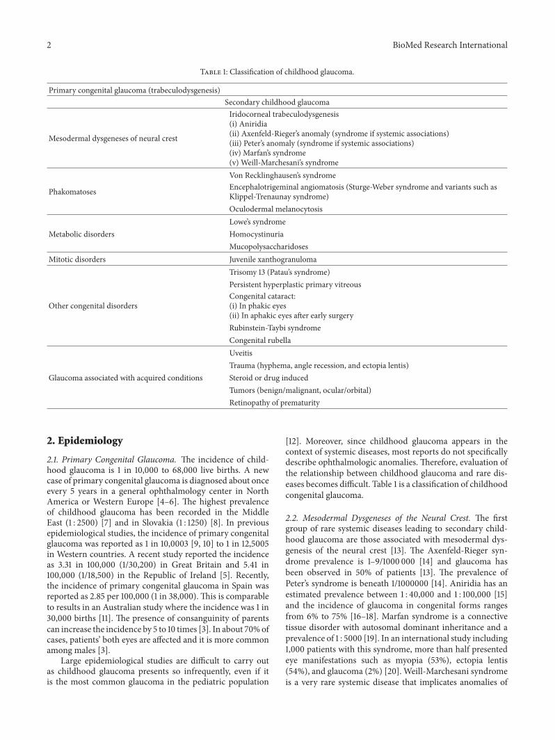
Table 1: Classification of childhood glaucoma.
Primary congenital glaucoma (trabeculodysgenesis)Secondary childhood glaucoma
Mesodermal dysgeneses of neural crest
Iridocorneal trabeculodysgenesis(i) Aniridia(ii) Axenfeld-Rieger’s anomaly (syndrome if systemic associations)(iii) Peter’s anomaly (syndrome if systemic associations)(iv) Marfan’s syndrome(v) Weill-Marchesani’s syndromeVon Recklinghausen’s syndrome
Phakomatoses Encephalotrigeminal angiomatosis (Sturge-Weber syndrome and variants such asKlippel-Trenaunay syndrome)Oculodermal melanocytosisLowe’s syndrome
Metabolic disorders HomocystinuriaMucopolysaccharidoses
Mitotic disorders Juvenile xanthogranulomaTrisomy 13 (Patau’s syndrome)Persistent hyperplastic primary vitreous
Other congenital disordersCongenital cataract:(i) In phakic eyes(ii) In aphakic eyes after early surgeryRubinstein-Taybi syndromeCongenital rubellaUveitisTrauma (hyphema, angle recession, and ectopia lentis)
Glaucoma associated with acquired conditions Steroid or drug inducedTumors (benign/malignant, ocular/orbital)Retinopathy of prematurity
2. Epidemiology
2.1. Primary Congenital Glaucoma. The incidence of child-hood glaucoma is 1 in 10,000 to 68,000 live births. A newcase of primary congenital glaucoma is diagnosed about onceevery 5 years in a general ophthalmology center in NorthAmerica or Western Europe [4–6]. The highest prevalenceof childhood glaucoma has been recorded in the MiddleEast (1 : 2500) [7] and in Slovakia (1 : 1250) [8]. In previousepidemiological studies, the incidence of primary congenitalglaucoma was reported as 1 in 10,0003 [9, 10] to 1 in 12,5005in Western countries. A recent study reported the incidenceas 3.31 in 100,000 (1/30,200) in Great Britain and 5.41 in100,000 (1/18,500) in the Republic of Ireland [5]. Recently,the incidence of primary congenital glaucoma in Spain wasreported as 2.85 per 100,000 (1 in 38,000). This is comparableto results in an Australian study where the incidence was 1 in30,000 births [11]. The presence of consanguinity of parentscan increase the incidence by 5 to 10 times [3]. In about 70%ofcases, patients’ both eyes are affected and it is more commonamong males [3].
Large epidemiological studies are difficult to carry outas childhood glaucoma presents so infrequently, even if itis the most common glaucoma in the pediatric population
[12]. Moreover, since childhood glaucoma appears in thecontext of systemic diseases, most reports do not specificallydescribe ophthalmologic anomalies. Therefore, evaluation ofthe relationship between childhood glaucoma and rare dis-eases becomes difficult. Table 1 is a classification of childhoodcongenital glaucoma.
2.2. Mesodermal Dysgeneses of the Neural Crest. The firstgroup of rare systemic diseases leading to secondary child-hood glaucoma are those associated with mesodermal dys-genesis of the neural crest [13]. The Axenfeld-Rieger syn-drome prevalence is 1–9/1000 000 [14] and glaucoma hasbeen observed in 50% of patients [13]. The prevalence ofPeter’s syndrome is beneath 1/1000000 [14]. Aniridia has anestimated prevalence between 1 : 40,000 and 1 : 100,000 [15]and the incidence of glaucoma in congenital forms rangesfrom 6% to 75% [16–18]. Marfan syndrome is a connectivetissue disorder with autosomal dominant inheritance and aprevalence of 1 : 5000 [19]. In an international study including1,000 patients with this syndrome, more than half presentedeye manifestations such as myopia (53%), ectopia lentis(54%), and glaucoma (2%) [20]. Weill-Marchesani syndromeis a very rare systemic disease that implicates anomalies of
BioMed Research International 3
connective tissue [21] and its prevalence has been estimatedat 1 : 100,000 [22]. The main ocular anomalies of Weill-Marchesani syndrome are microspherophakia, ectopia lentis,glaucoma, and myopia. Microspherophakia occurs in 84%of cases, ectopia lentis in 73%, and glaucoma in 80%; severemyopia and cataract are also reported [21].
2.3. Phakomatoses. The second group of rare diseases linkedwith secondary childhood glaucoma are the phakomatosescharacterized by hamartomas. Von Recklinghausen’s diseasesor neurofibromatosis type 1 is characterized by autosomicdominant inheritance and an incidence of about 1 in 3000[23]. Glaucoma has been reported in 1/300 patients with thiscondition [24]. However, a study on 95 patients, with neurofi-bromatosis type 1, revealed that 23% of 56 patients presentedorbitofacial involvement and had ipsilateral glaucoma [25].
Sturge-Weber syndromehas a frequency of approximately1 in 50,000 births [26]. Glaucoma affects 50% to 70%of patients with this syndrome and its variants (such asKlippel-Trenaunay syndrome) and can be early- or late-onsetdepending on the age of manifestation [27]. In about 60%of cases the cause is due to anterior chamber abnormalitieswhile in approximately 40% of cases the cause is raisedepiscleral venous pressure [28]. Oculodermal melanocytosisis a rare condition more frequently encountered in theAsian population with a prevalence of 0.014%–0.034% [29].Glaucoma is found in about 10% of patients and the patho-genesis may be congenital, developmental or associated withthe diffuse characteristic oculodermal hyperpigmentation ofthe disease which leads to melanocytic glaucoma [30–32].Phakomatosis pigmentovascularis is a rare combination ofoculodermal melanocytosis and Sturge-Weber syndrome (orKlippel-Trenaunay syndrome). In a review of the literatureon 281 Asian patients with oculodermal melanocytosis, 9patients had phakomatosis pigmentovascularis and glaucomawas present in all cases [30].
2.4. Metabolic Disorders. The metabolic disorders leading tochildhood glaucoma include mucopolysaccharidoses, Lowe’ssyndrome, and classical homocystinuria. Lowe’s syndrome(oculocerebrorenal syndrome) has a prevalence of 1 in500,000 [33]. Several cases of secondary congenital glaucomaassociated with this disease have been reported; Walton et al.reviewed 7 patients and found that 71% of patients had glau-coma [34]. Classical homocystinuria is caused by cystathio-nine b-synthase (CbS) deficiency [35–37]. The detection rateof CbS deficiency is 1 in 344,000 [38]. Ectopia lentis, whichis often associated with classical homocystinuria [39, 40],occurs in 85% of classical homocystinuria [39, 41, 42]. In areport on 294 patients with mucopolysaccharidoses, 27 eyesof 14 patients had glaucoma and the prevalence of glaucomawas reported from 2.1% to 12.5% [43].
2.5. Mitotic Disorders. Thegroup ofmitotic disorders leadingto childhood glaucoma includes juvenile xanthogranuloma,the prevalence of which is unknown. It occurs in the pediatricage and 10% of patients present ocular anomalies withoccasional glaucoma [44].
2.6. Other Congenital Conditions. The prevalence of trisomy13 or Patau’s syndrome is 1/6500 where glaucoma has beenreported among the ocular complications [45].
In a study of 62 cases of persistent hyperplastic primaryvitreous, Haddad et al. reported 26 patients where otherocular abnormalities such as leukocoria, microphthalmia,cataract, retinal detachment, glaucoma, and phthisis bulbiwere observed [46].
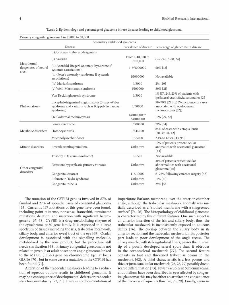
Five to twenty percent of pediatric blindness is attribu-table to congenital cataract, which occurs in 3-4 per 10 000births [47]. Although new surgical techniques have beenintroduced, the incidence of secondary glaucoma followingcataract operations in the pediatric age is still high, varyingfrom 6% to 26% [48]. If the surgical procedure is performedbefore the 9th month of life, the incidence of glaucoma mayrise to 50% [48, 49]. Rubinstein-Taybi syndrome as wellas congenital rubella appears among congenital disordersassociated with glaucoma. Rubinstein-Taybi syndrome has anincidence of 1 in 100 000 newborns [50]. van Genderen etal. reviewed 207 patients with Rubinstein-Taybi syndrome,and 117 had ocular disorders. Congenital glaucoma wasdescribed in 31 cases [51]. Epidemiological assessments ofocular disease in large populations of congenital rubella arefew. In France just 21 cases of rubella during pregnancy werereported in 2001 and only one child presented congenitalrubella syndrome at birth [52]. Various ocular disorders suchas retinopathy, cataract, and glaucoma may occur in thiscondition [53]. In a prospective study byO’Neill in 34 patientswith rubella, glaucoma was observed in 29% (11 eyes) andwas in the context of microphthalmic eyes in 4 patients [54].Table 2 is a summary of the epidemiology and percentageof glaucoma in rare diseases leading to secondary childhoodglaucoma.
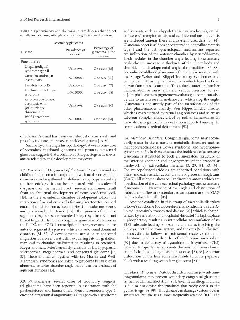
2.7. Rare Diseases. Cases reports of congenital glaucoma havebeen reported in the context of rare systemic syndromessuch as otopalatodigital syndrome type II (OPD II) [55],complete androgen insensitivity [56], pseudotrisomy 13 [57],Brachmann-de Lange syndrome [58], autosomal recessiveacrofrontofacionasal dysostosis [59], caudal regression syn-drome [60], and Wolf-Hirschhorn syndrome [61] (Table 3).
3. Genetics and Pathophysiology
3.1. Primary Congenital Glaucoma. While most cases ofprimary congenital glaucoma are sporadic, 10% to 40% arefamilial and are often associated with consanguinity [62].Familial forms have autosomal recessive inheritance withvariable expression in 40 to 100% of cases [63]. However,the autosomal recessive inheritance modality has been chal-lenged due to the discrepancy between gender distribution,transmission of childhood glaucoma to offspring, and lowerpredicted afflicted siblings in familial cases [63]. Linkagestudies confirm the heterogeneity of primary congenitalglaucoma, so these discrepancies can be explained [63, 64].
Primary congenital glaucoma has been associated with3 chromosomal loci: chromosomes 2p21 (GLC3A), 1p36(GLC3B), and 14q24 (GLC3C) [62, 63, 65, 66].
4 BioMed Research International
Table 2: Epidemiology and percentage of glaucoma in rare diseases leading to childhood glaucoma.
Primary congenital glaucoma 1 in 10,000 to 68,000Secondary childhood glaucoma
Disease Prevalence of disease Percentage of glaucoma in disease
Mesodermaldysgeneses of neuralcrest
Iridocorneal trabeculodysgenesis
(i) Aniridia From 1/40,000 to1/100,000 6–75% [16–18, 24]
(ii) Axenfeld-Rieger’s anomaly (syndrome ifsystemic associations) 1–9/1000000 50% [13]
(iii) Peter’s anomaly (syndrome if systemicassociations) 1/1000000 Not available
(iv) Marfan’s syndrome 1/5000 2% [20](v) Weill-Marchesani syndrome 1/100000 80% [21]
Von Recklinghausen’s syndrome 1/3000 1% [17, 24], 23% of patients withipsilateral craniofacial anomalies [25]
PhakomatosesEncephalotrigeminal angiomatosis (Sturge-Webersyndrome and variants such as Klippel-Trenaunaysyndrome)
1/5000050–70% [27] (100% incidence in casesassociated with oculodermalmelanocytosis [32])
Oculodermal melanocytosis 14/100000 to34/100000 10% [29, 32]
Lowe’s syndrome 1/500000 71% [34]
Metabolic disorders Homocystinuria 1/344000 85% of cases with ectopia lentis[38, 39, 41, 42]
Mucopolysaccharidoses 1/25000 2.1% to 12.5% [43, 95]
Mitotic disorders Juvenile xanthogranuloma Unknown10% of patients present ocularanomalies with occasional glaucoma[44]
Other congenitaldisorders
Trisomy 13 (Patau’s syndrome) 1/6500 Not available
Persistent hyperplastic primary vitreous Unknown31% of patients present ocularabnormalities with occasionalglaucoma [46]
Congenital cataract 1–6/10000 6–26% following cataract surgery [48]Rubinstein-Taybi syndrome Unknown 15% [51]Congenital rubella Unknown 29% [54]
The mutation of the CYP1B1 gene is involved in 87% offamilial and 27% of sporadic cases of congenital glaucoma[63]. Currently 147 mutations of this gene have been found,including point missense, nonsense, frameshift, terminatormutations, deletion, and insertion with significant hetero-geneity [67, 68]. CYP1B1 is a drug-metabolizing enzyme ofthe cytochrome p450 gene family. It is expressed in a largespectrum of tissues including the iris, trabecular meshwork,ciliary body, and anterior uveal tract of the eye [69]. Oculardevelopment is associated with the signalling molecule,metabolized by the gene product, but the procedure stillneeds clarification [68]. Primary congenital glaucoma is notrelated to juvenile or adult onset open angle glaucoma linkedto the MYOC (TIGR) gene on chromosome 1q25 at locusGLCIA [70], but in some cases a mutation in the CYP1B1 hasbeen found [71].
Alteration of the trabecularmeshwork leading to a reduc-tion of aqueous outflow results in childhood glaucoma. Itmay be a consequence of developmental defects or trabecularstructure immaturity [72, 73]. There is no documentation of
imperforate Barkan’s membrane over the anterior chamberangle, although the trabecular meshwork anomaly was ini-tially described as a “clothed membrane with a shagreenedsurface” [74–76]. The histopathology of childhood glaucomais characterized by five different features. One such aspect isan anterior insertion of the iris and ciliary body; thus, thetrabecular meshwork is inconsistently exposed to aqueousdeflux [76]. The overlap between the ciliary body in itsanterior section and the trabecular meshwork in its posteriorpart leads to poor development of the angle recess. Theciliary muscle, with its longitudinal fibers, passes the internaltip of a poorly developed scleral spur; thus, it obtrudesin the corneoscleral meshwork [77]. The second featureconsists in taut and thickened trabecular beams in themeshwork [62]. A third characteristic is a less porous andthicker juxtacanalicularmeshwork [76, 78, 79] possibly due toscarce differentiation [73]. Fewer vacuoles in Schlemm’s canalendothelium have been described in eyes affected by congen-ital glaucoma; this may be either an artefact or a consequenceof the decrease of aqueous flow [76, 78, 79]. Finally, agenesis
BioMed Research International 5
Table 3: Epidemiology and glaucoma in rare diseases that do notusually include congenital glaucoma among their manifestations.
Secondary glaucoma
Disease Prevalence ofdisease
Percentage ofglaucoma in the
diseaseRare diseases
Otopalatodigitalsyndrome type II Unknown One case [55]
Complete androgeninsensitivity 1–9/1000000 One case [56]
Pseudotrisomy 13 Unknown One case [57]Brachmann-de Langesyndrome 1–9/100000 One case [58]
Acrofrontofacionasaldysostosis withgenitourinaryabnormalities
Unknown One case [59]
Wolf-Hirschhornsyndrome 1–9/1000000 One case [61]
of Schlemm’s canal has been described; it occurs rarely andprobably indicates more severe maldevelopment [73, 80].
Similarity of the angle histopathology between some casesof secondary childhood glaucoma and primary congenitalglaucoma suggests that a common pathophysiogeneticmech-anism related to angle development may exist.
3.2. Mesodermal Dysgeneses of the Neural Crest. Secondarychildhood glaucoma in conjunction with ocular or systemicdisorders can be gathered in different subgroups accordingto their etiology. It can be associated with mesodermaldysgenesis of the neural crest. Several syndromes resultfrom an abnormal development of neuroectodermal cells[13]. In the eye, anterior chamber development follows themigration of neural crest cells forming keratocytes, cornealendothelium, iris stroma,melanocytes, trabecularmeshwork,and juxtacanalicular tissue [13]. The gamma of anteriorsegment dysgeneses, or Axenfeld-Rieger syndrome, is notlinked to genetic factors in congenital glaucoma.Mutations inthe PITX2 and FOXC1 transcription factor genes account foranterior segment dysgeneses, which are autosomal dominantdisorders [81, 82]. A developmental arrest or an abnormalmigration of neural crest cells, occurring late in gestation,may lead to chamber malformation resulting in Axenfeld-Rieger anomaly, Peter’s anomaly, aniridia or iris hypoplasia,sclerocornea, megalocornea, and congenital glaucoma [13,83]. These anomalies together with the Marfan and Weil-Marchesani syndromes are linked to glaucoma because of anabnormal anterior chamber angle that effects the drainage ofaqueous humour [13].
3.3. Phakomatoses. Several cases of secondary congeni-tal glaucoma have been reported in association with thephakomatoses and hamartomas. Neurofibromatosis type 1,encephalotrigeminal angiomatosis (Sturge-Weber syndrome
and variants such as Klippel-Trenaunay syndrome), retinaland cerebellar angiomatosis, and oculodermal melanocytosisare included among these multisystem disorders [3, 84].Glaucoma onset is seldom encountered in neurofibromatosistype 1 and the pathophysiological mechanisms reportedare infiltration of the anterior chamber by neurofibromas,Lisch nodules in the chamber angle leading to secondaryangle closure, increase in thickness of the ciliary body andchoroid, and developmental angle abnormalities [85–88].Secondary childhood glaucoma is frequently associated withthe Sturge-Weber and Klippel-Trenaunay syndromes andwith phakomatosis pigmentovascularis which have the facialnaevus flammeus in common.This is due to anterior chambermalformation or raised episcleral venous pressure [30, 89–91]. In phakomatosis pigmentovascularis glaucoma can alsobe due to an increase in melanocytes which clog the angle.Glaucoma is not strictly part of the manifestations of theother phakomatoses, namely, Von Hippel-Lindau disease,which is characterized by retinal angiomatosis and sclerosistuberous complex characterized by retinal hamartomas. Inthese diseases glaucoma has only been reported among thecomplications of retinal detachment [92].
3.4. Metabolic Disorders. Congenital glaucoma may secon-darily occur in the context of metabolic disorders such asmucopolysaccharidoses, Lowe’s syndrome, and hyperhomo-cysteinemia [3]. In these diseases the incidence of secondaryglaucoma is attributed to both an anomalous structure ofthe anterior chamber and engorgement of the trabecularmeshwork by extracellular material [3, 29, 84, 93, 94].The mucopolysaccharidoses are inherited conditions withintra- and extracellular accumulation of glycosaminoglycans(GAG). All subtypes show ocular disorders among which areopacification of the cornea, retinal pathology, and secondaryglaucoma [95]. Narrowing of the angle and obstruction oftrabecular outflow are secondary to an accumulation of GAGwithin trabecular cells. [95].
Another condition in this group of metabolic disordersis Lowe’s syndrome (oculocerebrorenal syndrome), a rare X-linked, recessively transmitted disease [28] which is charac-terized by amutation of phosphatidylinositol 4,5 biphosphate5-phosphatase, resulting in intracellular accumulation of itsPIP2 substrate leading to systemic anomalies involving thekidneys, central nervous system, and the eyes [96]. Classicalhomocystinuria follows an autosomal recessive mode ofinheritance and is a disorder of methionine metabolism[97] due to deficiency of cystathionine b-synthase (CbS)[30–32]. Ectopia lentis represents the most common clinicalanomaly leading to diagnosis in most cases [34, 35]. Anteriordislocation of the lens sometimes leads to acute pupillaryblock with a resulting secondary glaucoma [34].
3.5.Mitotic Disorders. Mitotic disorders such as juvenile xan-thogranuloma may present secondary congenital glaucomaas their ocular manifestation [84]. Juvenile xanthogranulomais due to histiocytic abnormalities that rarely occur in thepediatric age [98, 99].This disease can damage various ocularstructures, but the iris is most frequently affected [100]. The
6 BioMed Research International
onset of secondary glaucoma is frequently related to thepresence of iris nodules that, due to their vascular nature,maybleed causing hyphema [101].
3.6. Other Congenital Conditions. Secondary childhood glau-coma can also be associated with other disorders such aspersistent hyperplastic primary vitreous, congenital cataract,Patau’s syndrome, Rubinstein-Taybi syndrome, or congenitalrubella [84, 102, 103]. Secondary childhood glaucoma isoften associated with congenital cataract and it may occur aspostoperative complication of pediatric cataract surgery [48].Different ocular anomalies are associated with Rubinstein-Taybi syndrome, which results from microdeletions at chro-mosome 16p13.3 or frommutations in the gene for the CREBbinding protein (CBP), located at 16p13.3 [104, 105].
3.7. Acquired Conditions. Uveitis and steroid induced glau-coma are among the major acquired conditions associatedwith childhood glaucoma. Trauma induced glaucoma canbe caused by hyphema, angle recession, and ectopia lentis.Ocular or orbital, benign or malignant, tumors and retinopa-thy of prematurity can also be a cause. Retinopathies andsome drugs can also cause secondary glaucoma and thepathophysiogenetic mechanism is specific to each condition[106–108].
3.8. RareDiseases. There are some rare diseases, which donotnormally present glaucoma in their manifestation.
Kondoh et al. published a case of bilateral glaucoma ina male child presenting otopalatodigital syndrome type II(OPD II) related to a mutation of the gene for filamin A(FLNA). Performing a sequence analysis of the FLNA gene,a missense C to T mutation at position 588, resulting inan Arg196Trp change in the filamin A protein, was found[55]. FLNA gene encodes an actin-binding protein andmutations of the FLNA gene have been found in OPD II,Melnick-Needles syndrome, or frontometaphyseal dysplasia.The association between OPD II, congenital heart defects,and ocular disorder, as presented by Kondoh et al., has notbeen previously reported. Therefore, additional factors areprobably involved in the pathogeneticmechanism behind theOPD-group alterations [55].
Gad et al. reported an association of complete androgeninsensitivity with hypertrophic pyloric stenosis and congeni-tal glaucoma in an Egyptian newborn. Performing a sequenceof the five exons of the 5a-reductase type 2 gene, no evidenceof mutation was obtained while a C to T mutation, whichresulted in substitution of the phenylalanine residue by aleucine at position 804, was identified in exon 6. The father’sfamily had a history of glaucoma, ruling out the causativeeffect of the receptor gene in the pathogenesis of glaucomaand exon 6 of the androgen receptor gene of the motherwas normal; therefore, the mutation was considered de novo.It is possible that defective androgen action determinedthe congenital glaucoma. Moreover, it is interesting to notethat CYP1B1 gene, which is the most common early-onsetglaucoma gene, is related to steroids. The CYP1B1 gene liesnext to the SRD5A2 gene on 2p21 and both the androgen
receptor and SRD5A2 genes are expressed in eye structures.However, the consequences of defective signalling pathwayson the structure and function of ocular tissues and thecorrelated gene are still unknown [56].
Sandal et al. reported the only case of congenital glau-coma in a female infant affected by pseudotrisomy 13.Detailed genetic examination, conventional karyotype, andmicroarray studies are necessary to assess anomalies that arenot usually related to this syndrome [57].
Barry Lee et al. first reported a case of buphthalmosand childhood glaucoma associated with Brachmann-deLange syndrome. Chromosome 3q16 is probably related toBrachmann-de Lange syndrome, while some forms of herita-ble childhood glaucoma have been mapped to chromosomes1 and 2. Further genetic studies could be useful to assess acorrelation between these disorders [58].
Chaabouni et al. reported the first case of congenitalglaucoma in a patient who suffered from acrofrontofacionasaldysostosis with genitourinary abnormalities [59]. As far as weknow, this is one of just three case reports in this condition,which has autosomic recessive transmission. The paucity ofcases precludes our understanding of the ophthalmologicalanomalies. Guirgis et al. first reported a case of childhoodglaucoma associated with caudal regression syndrome in a10-week-old infant. The child also had punctal atresia andsignificant myopia. Embryonic defect of caudal mesodermaldevelopment seems to be the cause of caudal regressionsyndrome [109]. However, goniodysgenesis, caused by neuralcrest alterations, probably led to childhood glaucoma. Afaulty development of surface ectoderm is related to punctualatresia. The embryopathic nature of both caudal regressionsyndrome and childhood glaucoma, as well as their lowincidence, implies that theremight be a common cause ratherthan a coincidence. Furthermore, concomitant punctualatresia validates the notion of embryopathic linkage [110].
Dickmann et al. tried to analyze the relationship betweenWolf-Hirschhorn syndrome and ocular defects, specificallyto correlate ocular findings with the extension of deletionon chromosome 4, so they investigated a population of 10patients affected by Wolf-Hirschhorn syndrome [61]. Thiscondition is a genetic disorder that occurs as a consequence ofpartial deletion of the short arm of chromosome 4; the prox-imal breakpoint occurs between 4p15.1 and 4p16.3, and thesmallest and largest deletion sizes recorded were 2.6Mb and20Mb, respectively [61, 111, 112]. Ocular anomalies appearedin all patients, but just one patient was affected by childhoodglaucoma. Comparing genotype with ocular findings, severeocular abnormalities were associated with large 4p deletionswhile mild ocular disorders were independent of the deletionsize [61]. The only case of childhood glaucoma was observedat birth; it appeared with concomitant corneal clouding, iriscoloboma, and cataract. It was associated with a large 4pdeletion [61, 113].
4. Management
The signs and symptoms of childhood glaucoma are closelylinked to the age of onset and the gravity of the disease;
BioMed Research International 7
however, some children are asymptomatic. Corneal alter-ations can be edema, Descemet’s membrane breaks, andopacification.This leads to the typical manifestations of pho-tophobia, epiphora, and consequent blepharospasm. In somecases a misinterpretation of these symptoms delays diagnosis[62]. Improved instrumental technology with optical coher-ence tomography and ultrabiomicroscopy can provide detailson the anterior chamber and ciliary body which is helpfultowards diagnosis [114, 115]. The management of childhoodglaucoma is also facilitated by new instrumentation to mea-sure intraocular pressure and visual field defects [116–119].
Any type of glaucoma leads to irreversible vision lossin the long term; therefore, the goal of both medical andsurgical management is to prevent visual deterioration [62].Treatment should be carefully tailored, aiming to controlintraocular pressure in the course of time. Surgical pro-cedures represent the mainstay of therapy for childhoodglaucoma. Although medical therapy alone is rarely effective,it plays an important role as adjunctive treatment to surgery;moreover, it can also be used temporarily before surgery toclear the cornea [62]. Ophthalmologists who use medicationshould be aware of different risk and benefit profiles ininfants and children compared with adults. Allergic reactionsand ocular surface toxicity may complicate repeated topicaltreatment initiated at an early age in childhood [62].
4.1. Medical Treatment. Beta-blockers are usually well toler-ated and severe systemic complications are rare in pediatricpatients treated with timolol, but bradycardia and bron-chospasm can occur especially with the concentration of0.5%. Cautious initial treatment with low concentrationsof timolol is wise. Punctal occlusion after topical timololdetermines a decrement of beta-blocker plasma levels, sothe use of punctual occlusion after every drop application isadvised [62].
Oral carbonic anhydrase inhibitors represent the mostefficacious drugs in the reduction of intraocular pressurein childhood glaucoma. Nevertheless it is important to beaware that these can cause decreased appetite and hyperpneafrom renal acidosis, dehydration, chronic fatigue, and failureto grow, so prolonged use should be avoided [62]. Topicalcarbonic anhydrase inhibitors such as dorzolamide and brin-zolamide represent a valid alternative to oral administrationwith less risk of systemic side effects.They reduce intraocularpressure by 25% [120, 121]. Carbonic anhydrase inhibitors andbeta-blockers are both suppressors of aqueous productionbut are used in conjunction in order to yield additivebenefit [122]. Brimonidine is a selective alpha-agonist, whichreduces intraocular pressure increasing uveoscleral outflowand decreasing aqueous production [123]. Since brimonidinecrosses the blood brain barrier, it can lead to nervous systemictoxicity [124]. Brimonidine topical therapy has also beenreported to induce bradycardia, hypotension, hypotonia,hypothermia, apnea, and unresponsiveness in children [125–128].
Prostaglandin analogues such as bimatoprost, travo-prost, and latanoprost are not effective in pediatric patients.Although in some cases of children affected by primary
juvenile glaucoma and glaucoma related to Sturge-Webersyndrome a significant intraocular pressure reduction wasfound [129, 130], further research is necessary in order toassess the performance of prostaglandins in the treatment ofchildhood glaucoma.
Miotics such as pilocarpine are not effective for childhoodglaucoma. In older children, symptoms of spasmof the ciliarymuscle and blurring of vision from induced myopia occur.They can be used for aphakic glaucoma and are adoperatedbefore goniosurgery [62]. Most often childhood glaucoma isbetter managed surgically rather than medically [62].
4.2. Surgical Treatment. Surgical treatment can be separatedinto procedures that improve aqueous outflow through thedrainage channels (goniosurgery such as goniotomy and tra-beculotomy), treatments that by destroying the ciliary bodyreduce aqueous humor production (cyclodestruction), orsurgical procedures that drain aqueous through an alternativeway (trabeculectomy, glaucoma drainage devices) [131].
While for primary congenital glaucoma goniotomy rep-resents the procedure of choice, this indication is less certainfor other forms of childhood glaucoma [62]. An alternativesurgical treatment is trabeculotomy [62]; it appears as a moreappropriate choice especially when a cloudy cornea preventsa comfortable visualization of the angle [62]. Filtering surgerymay be indicated if goniosurgery procedures are unsuccessful[3]. Trabeculectomy is indicated if an unacceptable elevationof intraocular pressure persists after an adequate number ofangle incision procedures [62]. Implant surgery representsa valid alternative for patients with conditions proven to berefractory to goniosurgery or filtering surgery, who are poorcandidates to these procedures, or for whom these treatmentshave been revealed unsuccessful [62]. Long tube drainagedevice surgery is needed in severe cases of primary congenitalglaucoma and, sometimes, in secondary glaucomas [3].
Cyclodestructive procedures can be defined as an inter-mediate or an additional procedure, useful when primarytrabecular surgery has failed [3]. Cyclocryotherapy or bothtransscleral and endoscopic diode laser cyclophotocoagu-lation are used for the ablation of ciliary epithelium. Thesurgeon must consider the anatomic improvement, the long-term visual benefit, and the patient’s expectations for theaffected eye when making the choice for management strate-gies [62].
5. Conclusions
There is a wide array of systemic diseases associated withchildhood glaucoma. Some conditions like dysgenesis of theanterior chamber associated with neural crest disorders andthe phakomatoses are better known. However, there are rarediseases which are occasionally associated with glaucoma.Since the examination of pediatric patients is not alwaysstraightforward, ophthalmologists should be updated on theavailable literature and should bewell aware of the conditions,which may lead to visual deterioration due to glaucoma.Treatment should be carefully tailored, in order to controlintraocular pressure in the long term. Surgical treatment is
8 BioMed Research International
the mainstay of management and medical therapy has anadjunctive role. Furthermore, medication should be carefullymonitored due to different risk and benefit profiles in infantsand children compared with adults. A multidisciplinaryapproach to pediatric patients with rare multisystem diseasesis highly advisable in both the diagnosis and management ofthe conditions.
Conflict of Interests
The authors declare that there is no conflict of interestsregarding the publication of this paper.
References
[1] P. Singh, Y. Kumar, M. Tyagi, K. Kuldeep, and P. Das Sharma,“Childhood glaucoma: an overview,” Open Journal of Ophthal-mology, vol. 02, no. 03, pp. 71–77, 2012.
[2] P. Dureau, “Congenital glaucoma and trabeculodysgenesis.Clinical and genetic aspects,” Journal Francais d’Ophtalmologie,vol. 29, no. 2, pp. 198–215, 2006.
[3] European Glaucoma Society, Terminology and Guidelines forGlaucoma, European Glaucoma Society, 4th edition, 2014.
[4] E. P. Aponte, N. Diehl, and B. G. Mohney, “Incidence andclinical characteristics of childhood glaucoma: a population-based study,”Archives of Ophthalmology, vol. 128, no. 4, pp. 478–482, 2010.
[5] M. Papadopoulos, N. Cable, J. Rahi et al., “The British infantileand childhood glaucoma (BIG) eye study,” Investigative Oph-thalmology and Visual Science, vol. 48, no. 9, pp. 4100–4106,2007.
[6] D. S. Walton, “Primary congenital open-angle glaucoma,” inGlaucoma, P. A. Chandler and W. M. Grant, Eds., pp. 329–343,Lea & Febiger, Philadelphia, Pa, USA, 2nd edition, 1979.
[7] M. Jaafar,Care of the Infantile GlaucomaPatient. OphthalmologyAnnual, Raven Press, New York, NY, USA, 1988.
[8] A. Gencık, “Epidemiology and genetics of primary congenitalglaucoma in Slovakia. Description of a formof primary congen-ital glaucoma in gypsies with autosomal-recessive inheritanceand complete penetrance,”Developments inOphthalmology, vol.16, pp. 76–115, 1989.
[9] V. P. deLuise and D. R. Anderson, “Primary infantile glaucoma(congenital glaucoma),” Survey of Ophthalmology, vol. 28, no. 1,pp. 1–19, 1983.
[10] S. J. Miller, “Genetic aspects of glaucoma,” Transactions of theOphthalmological Societies of the United Kingdom, vol. 86, pp.425–434, 1966.
[11] J. R. MacKinnon, A. Giubilato, J. E. Elder, J. E. Craig, andD. A. Mackey, “Primary infantile glaucoma in an Australianpopulation,” Clinical and Experimental Ophthalmology, vol. 32,no. 1, pp. 14–18, 2004.
[12] R. H. Taylor, J. R. Ainsworth, A. R. Evans, and A. V. Levin, “Theepidemiology of pediatric glaucoma: the Toronto experience,”Journal of AAPOS, vol. 3, no. 5, pp. 308–315, 1999.
[13] Z. Tumer and D. Bach-Holm, “Axenfeld-Rieger syndrome andspectrum of PITX2 and FOXC1mutations,” European Journal ofHuman Genetics, vol. 17, no. 12, pp. 1527–1539, 2009.
[14] Orphanet, Prevalence of Rare Diseases: Bibliographic Data,Number 1, 2009, http://www.orpha.net.
[15] B. Singh, A. Mohamed, M. Tech et al., “Clinical manifestationsof congenital aniridia,” Journal of Pediatric Ophthalmology andStrabismus, vol. 51, no. 1, pp. 59–62, 2014.
[16] S. C. Brauner, D. S. Walton, and T. C. Chen, “Aniridia,”International Ophthalmology Clinics, vol. 48, no. 2, pp. 79–85,2008.
[17] W. M. Grant and D. S. Walton, “Progressive changes in theangle in congenital aniridia, with development of glaucoma,”The American Journal of Ophthalmology, vol. 78, no. 5, pp. 842–847, 1974.
[18] J. C. Swanner, D. S. Walton, and T. C. Chen, “Prevention ofaniridic glaucoma with goniosurgery,” International Ophthal-mology Clinics, vol. 44, no. 1, pp. 67–71, 2004.
[19] K. Steindl, “Marfan syndrome and related connective tissuedisorders,” Praxis, vol. 102, no. 24, pp. 1483–1488, 2013.
[20] L. Faivre, G. Collod-Beroud, B. L. Loeys et al., “Effect ofmutation type and location on clinical outcome in 1,013probands with marfan syndrome or related phenotypes andFBN1 mutations: an international study,”The American Journalof Human Genetics, vol. 81, no. 3, pp. 454–466, 2007.
[21] B. S. Chu, “Weill-Marchesani syndrome and secondary glau-coma associated with ectopia lentis,” Clinical and ExperimentalOptometry, vol. 89, no. 2, pp. 95–99, 2006.
[22] E. Tsilou and I. M. MacDonald,Weill-Marchesani Syndrome, R.A. Pagon,M. P. Adam,H. H. Ardinger, etal, Eds., Gene Reviews,Seattle, Wash, USA, 1993–2015.
[23] A. C. Hirbe and D. H. Gutmann, “Neurofibromatosis type 1: amultidisciplinary approach to care,” The Lancet Neurology, vol.13, no. 8, pp. 834–843, 2014.
[24] W.M.Grant andD. S.Walton, “Distinctive gonioscopic findingsin glaucoma due to neurofibromatosis,”Archives of Ophthalmol-ogy, vol. 79, no. 2, pp. 127–134, 1968.
[25] J. Morales, I. A. Chaudhry, and T. M. Bosley, “Glaucoma andglobe enlargement associated with neurofibromatosis type 1,”Ophthalmology, vol. 116, no. 9, pp. 1725–1730, 2009.
[26] C. Di Rocco and G. Tamburrini, “Sturge-Weber syndrome,”Child’s Nervous System, vol. 22, no. 8, pp. 909–921, 2006.
[27] E. Sujansky and S. Conradi, “Sturge-Weber syndrome: age ofonset on seizures and glaucoma and the prognosis for affectedchildren,” Journal of Child Neurology, vol. 10, no. 1, pp. 49–58,1995.
[28] T. J. Sullivan, M. P. Clarke, and J. D. Morin, “The ocular man-ifestations of the Sturge-Weber syndrome,” Journal of PediatricOphthalmology and Strabismus, vol. 29, no. 6, pp. 349–356, 1992.
[29] H. H. L. Chan and T. Kono, “Nevus of Ota: clinical aspects andmanagement,” Skin Med, vol. 2, no. 2, pp. 89–97, 2003.
[30] C. Teekhasaenee and R. Ritch, “Glaucoma in phakomatosispigmentovascularis,”Ophthalmology, vol. 104, no. 1, pp. 150–157,1997.
[31] J. R. Gonder, J. Nichol, J. J. Augsburger, and J. A. Shields,“Ocular and oculodermal melanocytosis,” Canadian Journal ofOphthalmology, vol. 20, no. 5, pp. 176–178, 1985.
[32] C. Teekhasaenee, R. Ritch, U. Rutnin, and N. Leelawongs,“Glaucoma in oculodermal melanocytosis,” Ophthalmology,vol. 97, no. 5, pp. 562–570, 1990.
[33] M. Loi, “Lowe syndrome,” Orphanet Journal of Rare Diseases,vol. 1, no. 1, article 16, 2006.
[34] D. S. Walton, G. Katsavounidou, and C. U. Lowe, “Glaucomawith the oculocerebrorenal syndrome of Lowe,” Journal ofGlaucoma, vol. 14, no. 3, pp. 181–185, 2005.

BioMed Research International 9
[35] T. L. Perry, “Homocystinuria,” in Heritable Disorders of AminoAcid Metabolism, W. L. Nyan, Ed., pp. 395–428, John Wiley &Sons, New York, NY, USA, 1974.
[36] N. A. J. Carson and D. W. Neill, “Metabolic abnormalitiesdetected in a survey of mental backward individuals in North-ern Ireland,” Archives of Disease in Childhood, vol. 37, pp. 505–513, 1962.
[37] T. Gerritsen, J. G. Vaughn, and H. A. Waisman, “The identifica-tion of homocystine in the urine,” Biochemical and BiophysicalResearch Communications, vol. 9, no. 6, pp. 493–496, 1962.
[38] S. H. Mudd, H. L. Levy, and F. Skovby, “Disorders of transsul-furation,” in The Metabolic and Molecular Bases of InheritedDisease, C. R. Scriver, A. L. Beaudet,W. S. Sly, andD. Valle, Eds.,pp. 1279–1327, McGraw-Hill, New York, NY, USA, 7th edition,1995.
[39] S. H.Mudd, F. Skovby, H. L. Levy, and et al, “The natural historyof homocystinura due to cystathionine 𝛽-synthase deficiency,”The American Journal of Human Genetics, vol. 37, no. 1, pp. 1–31,1985.
[40] N. A. J. Carson, “Homocystinuria: clinical and biochemicalheterogeneity,” in Inborn Errors of Metabolism in Humans, F.Cockburn and R. Gitzelmann, Eds., pp. 53–67, MTP Press,Lancaster, UK, 1982.
[41] G. H. J. Boers, T. W. Polder, and J. R. M. Cruysberg, “Homo-cystinuria versus Marfan’s syndrome: the therapeutic relevanceof the differential diagnosis,” Netherlands Journal of Medicine,vol. 27, no. 6, pp. 206–212, 1984.
[42] H. E. Cross and A. D. Jensen, “Ocular manifestations in theMarfan syndrome and homocystinuria,” American Journal ofOphthalmology, vol. 75, no. 3, pp. 405–420, 1973.
[43] J. Ashworth, M. Flaherty, S. Pitz, and A. Ramlee, “Assess-ment and diagnosis of suspected glaucoma in patients withmucopolysaccharidosis,” Acta Ophthalmologica, vol. 93, no. 2,pp. e111–e117, 2015.
[44] H. H. Lau, W. W. Yip, A. Lee, C. Lai, and D. S. Fan, “Three dif-ferent ophthalmic presentations of juvenile xanthogranuloma,”Hong Kong Medical Journal, vol. 20, no. 3, pp. 261–263, 2014.
[45] I. Kroes, S. Janssens, and P. Defoort, “Ultrasound featuresin trisomy 13 (Patau syndrome) and trisomy 18 (Edwardssyndrome) in a consecutive series of 47 cases,” Facts, Views &Vison in obGyn, vol. 6, no. 4, pp. 245–249, 2014.
[46] R. Haddad, R. L. Font, and F. Reeser, “Persistent hyperplasticprimary vitreous. A clinicopathologic study of 62 cases andreview of the literature,” Survey of Ophthalmology, vol. 23, no.2, pp. 123–134, 1978.
[47] L. M. Reis, R. C. Tyler, and E. V. Semina, “Identification ofa novel C-terminal extension mutation in EPHA2 in a familyaffected with congenital cataract,”Molecular Vision, vol. 20, pp.836–842, 2014.
[48] B. N. Swamy, F. Billson, F. Martin et al., “Secondary glaucomaafter paediatric cataract surgery,” British Journal of Ophthalmol-ogy, vol. 91, no. 12, pp. 1627–1630, 2007.
[49] P. K. Rabiah, “Frequency and predictors of glaucoma afterpediatric cataract surgery,”American Journal of Ophthalmology,vol. 137, no. 1, pp. 30–37, 2004.
[50] R. C. M. Hennekam, C. A. Stevens, and J. J. P. Van de Kamp,“Etiology and recurrence risk in Rubinstein-Taybi syndrome,”American Journal of Medical Genetics, no. 6, pp. 56–64, 1990.
[51] M. M. van Genderen, G. F. Kinds, F. C. C. Riemslag, and R. C.M. Hennekam, “Ocular features in Rubinstein-Taybi syndrome:investigation of 24 patients and review of the literature,” BritishJournal of Ophthalmology, vol. 84, no. 10, pp. 1177–1184, 2000.
[52] I. Parent du Chatelet, L. Bouraoui, C. Six, and D. Levy-Bruhl,“La rubeole chez la femme enceinte et le nouveau-ne en Francemetropolitaine en 2002: les donnees du reseau Renarub,” BEH,vol. 1, pp. 2–3, 2004.
[53] E. Robert-Gnansia, Congenital Rubella Syndrome, OrphanetEncyclopedia, 2004.
[54] J. F. O’Neill, “The ocular manifestations of congenital infection:a study of the early effect and long-term outcome of maternallytransmitted Rubella and toxoplasmosis,” Transactions of theAmerican Ophthalmological Society, vol. 96, pp. 813–879, 1998.
[55] T. Kondoh, N. Okamoto, N. Norimatsu, M. Uetani, G.Nishimura, and H. Moriuchi, “A Japanese case of oto-palato-digital syndrome type II: an apparent lack of phenotype-genotype correlation,” Journal of Human Genetics, vol. 52, no.4, pp. 370–373, 2007.
[56] Y. Z. Gad, I. Mazen, S. Lumbroso, S. A. Temtamy, and C. Sultan,“A novel point mutation of the androgen receptor (F804L)in an Egyptian newborn with complete androgen insensitivityassociated with congenital glaucoma and hypertrophic pyloricstenosis,” Clinical Genetics, vol. 63, no. 1, pp. 59–63, 2003.
[57] G. Sandal, L. Tok, and A. R. Ormeci, “A new case ofholoprosencephaly-polydactyly syndrome with alobar holo-prosencephaly, preaxial polydactyly and congenital glaucoma,”Genetic Counseling, vol. 25, no. 1, pp. 49–52, 2014.
[58] W. Barry Lee, J. D. Brandt, M. J. Mannis, C. Q. Huang, and G. J.Rabin, “Aniridia and Brachmann-de Lange syndrome: a reviewof ocular surface and anterior segment findings,” Cornea, vol.22, no. 2, pp. 178–180, 2003.
[59] M. Chaabouni, F. Maazoul, A. B. Hamida, M. Berhouma,Z. Marrakchi, and H. Chaabouni, “Autosomal recessive acro-fronto-facio-nasal dysostosis associated with genitourinaryanomalies: a third case report,” American Journal of MedicalGenetics Part: A, vol. 146, no. 14, pp. 1825–1827, 2008.
[60] M. F. Guirgis, A. M. F. Wong, and L. Tychsen, “Infantileglaucoma and punctal atresia in a child with caudal regressionsyndrome,” Journal of AAPOS, vol. 7, no. 4, pp. 298–299, 2003.
[61] A. Dickmann, R. Parrilla, A. Salerni et al., “Ocular manifesta-tions inWolf-Hirschhorn syndrome,” Journal of AAPOS, vol. 13,no. 3, pp. 264–267, 2009.
[62] L. H. Ching and D. S. Walton, “Primary congenital glaucoma:2004 update,” Journal of Pediatric Ophthalmology and Strabis-mus, vol. 41, no. 5, pp. 271–288, 2004.
[63] M. Sarfarazi and I. Stoilov, “Molecular genetics of primarycongenital glaucoma,” Eye, vol. 14, no. 3, pp. 422–428, 2000.
[64] A. Gencik, A. Gencikova, and A. Gerinec, “Genetic heterogene-ity of congenital glaucoma,” Clinical Genetics, vol. 17, no. 4, pp.241–248, 1980.
[65] A. N. Akarsu, M. E. Turacli, S. G. Aktan et al., “A second locus(GLC3B) for primary congenital glaucoma (Buphthalmos)maps to the 1p36 region,”HumanMolecular Genetics, vol. 5, no.8, pp. 1199–1203, 1996.
[66] I. R. Stoilov andM. Scarfarazi, “The third genetic locus (GLC3B)for primary congenital glaucoma (PCG) maps to chromosome14q24.3. Presented at the annual Meeting of the Association forResearch in Vision and Ophthalmology,” Investigative Ophthal-mology & Visual Science, vol. 43, E-Abstract 3015. ARVO, 2002.
[67] S. G. Panicker, A. K. Mandal, A. B. M. Reddy, V. K. Gothwal,and S. E. Hasnain, “Correlations of genotype with phenotype inIndian patients with primary congenital glaucoma,” Investiga-tive Ophthalmology and Visual Science, vol. 45, no. 4, pp. 1149–1156, 2004.
10 BioMed Research International
[68] N. Li, Y. Zhou, L. Du, M. Wei, and X. Chen, “Overview ofcytochrome P450 1B1 gene mutations in patients with primarycongenital glaucoma,” Experimental Eye Research, vol. 93, no. 5,pp. 572–579, 2011.
[69] M. Sarfarazi, I. Stoilov, and J. B. Schenkman, “Genetics andbiochemistry of primary congenital glaucoma,” OphthalmologyClinics of North America, vol. 16, no. 4, pp. 543–554, 2003.
[70] W. L. M. Alward, J. H. Fingert, M. A. Coote et al., “Clinicalfeatures associated with mutations in the chromosome 1 open-angle glaucoma gene (GLC1A),” The New England Journal ofMedicine, vol. 338, no. 15, pp. 1022–1027, 1998.
[71] A. L. Vincent, G. Billingsley, Y. Buys et al., “Digenic inheritanceof early-onset glaucoma: CYP1B1, a potential modifier gene,”American Journal of Human Genetics, vol. 70, no. 2, pp. 448–460, 2002.
[72] D. R. Anderson, “Pathology of the glaucomas,” British Journalof Ophthalmology, vol. 56, no. 3, pp. 146–156, 1972.
[73] A. Tawara and H. Inomata, “Developmental immaturity ofthe trabecular meshwork in congenital glaucoma,” AmericanJournal of Ophthalmology, vol. 92, no. 4, pp. 508–525, 1981.
[74] O. Barkan, “Pathogenesis of congenital glaucoma: gonioscopicand anatomic observation of the angle of the anterior chamberin the normal eye and in congenital glaucoma,” AmericanJournal of Ophthalmology, vol. 40, no. 1, pp. 1–11, 1955.
[75] J. G. Worst, “Congenital glaucoma. Remarks on the aspect ofchamber angle, ontogenetic and pathogenetic background, andmode of action of goniotomy,” Investigative Ophthalmology, vol.7, no. 2, pp. 127–134, 1968.
[76] D. R. Anderson, “The development of the trabecular meshworkand its abnormality in primary infantile glaucoma,” Transac-tions of the American Ophthalmological Society, vol. 79, pp. 458–485, 1981.
[77] A. Tawara, H. Inomata, and S. Tsukamoto, “Ciliary body bandwidth as an indicator of goniodysgenesis,” American Journal ofOphthalmology, vol. 122, no. 6, pp. 790–800, 1996.
[78] E. Maul, L. Strozzi, C. Munoz, and C. Reyes, “The outflowpathway in congenital glaucoma,”American Journal of Ophthal-mology, vol. 89, no. 5, pp. 667–675, 1980.
[79] A. Tawara and H. Inomata, “Congenital abnormalities of thetrabecular meshwork in primary glaucoma with open angle,”Glaucoma, vol. 9, pp. 28–34, 1987.
[80] A. E. Maumanee, “The pathogenesis of congenital glaucoma:a new theory,” Transactions of the American OphthalmologicalSociety, vol. 56, pp. 507–570, 1958.
[81] W. L. M. Alward, E. V. Semina, J. W. Kalenak et al., “Autosomaldominant iris hypoplasia is caused by a mutation in theRieger syndrome (RIEG/PITX2) gene,” American Journal ofOphthalmology, vol. 125, no. 1, pp. 98–100, 1998.
[82] D. Y. Nishimura, R. E. Swiderski, W. L. M. Alward et al., “Theforkhead transcription factor gene FKHL7 is responsible forglaucoma phenotypes whichmap to 6p25,”Nature Genetics, vol.19, no. 2, pp. 140–147, 1998.
[83] M. T. Contestabile, R. Plateroti, C. Galasso, S. Abodol-rahimzadeh, G. Delorenzi, and F. Rosa, “Peters’ anomaly asso-ciated with central spastic palsy,” Journal of Pediatric Ophthal-mology and Strabismus, vol. 32, no. 6, pp. 395–396, 1995.
[84] M. Yanoff and J. S. Duker, Ophthalmology, Mosby, 3rd edition,2003.
[85] S. Emre, M. Palamar, M. O. Ulusoy, and G. Gencoglan, “Ciliarybody cysts in neurofibromatosis: a new coexistence?” Graefe’sArchive for Clinical and Experimental Ophthalmology, vol. 250,no. 6, pp. 857–861, 2012.
[86] S.H. Al Freihi, D. P. Edward, S. R.Nowilaty,M.A. Abouammoh,and J. Morales, “Iris neovascularization and neovascular glau-coma in neurofibromatosis type 1: report of 3 cases in children,”Journal of Glaucoma, vol. 22, no. 4, pp. 336–341, 2013.
[87] S. M. Recupero, R. Plateroti, S. Abdolrahimzadeh et al.,“Lisch nodules in neurofibromatosis type 1: relationship toage and cutaneous neurofibromas,” Annals of Ophthalmology—Glaucoma, vol. 28, no. 3, pp. 178–183, 1996.
[88] F.Mantelli, S. Abdolrahimzadeh, G.Mannino, andA. Lambiase,“Unusual case of angle closure glaucoma in a patient withneurofibromatosis type 1,” Case Reports in Ophthalmology, vol.5, no. 3, pp. 386–391, 2014.
[89] S. M. Recupero, S. Abdolrahimzadeh, M. de Dominicis, and R.Mollo, “Sturge-Weber syndrome associated with naevus of Ota,”Eye, vol. 12, no. 2, pp. 212–213, 1998.
[90] D. I. Weiss, “Dual origin of glaucoma in encephalotrigem-inal haemangiomatosis. A pathogenetic concept based uponhistopathologic and haemodynamic considerations,” Transac-tions of the Ophthalmological Societies of the United Kingdom,vol. 93, pp. 477–493, 1973.
[91] C. D. Phelps, “The pathogenesis of glaucoma in Sturge-Webersyndrome,” Ophthalmology, vol. 85, no. 3, pp. 276–286, 1978.
[92] E. R. Maher, H. P. H. Neumann, and S. Richard, “Von Hippel–Lindau disease: a clinical and scientific review,” EuropeanJournal of Human Genetics, vol. 19, no. 6, pp. 617–623, 2011.
[93] M. J. Nowaczyk, J. T. R. Clarke, and J. D. Morin, “Glaucoma asan early complication of Hurler’s disease,” Archives of Disease inChildhood, vol. 63, no. 9, pp. 1091–1093, 1988.
[94] L. Tranchina, M. Centofanti, F. Oddone et al., “Levels of plasmahomocysteine in pseudoexfoliation glaucoma,” Graefe’s Archivefor Clinical and Experimental Ophthalmology, vol. 249, no. 3, pp.443–448, 2011.
[95] J. L. Ashworth, S. Biswas, E. Wraith, and I. C. Lloyd,“Mucopolysaccharidoses and the eye,” Survey of Ophthalmol-ogy, vol. 51, no. 1, pp. 1–17, 2006.
[96] M. L. D. A. Maia, M. L. D. M. do Val, C. P. Genzani, F. A. T.Fernandes, M. C. de Andrade, and J. T. D. A. Carvalhaes, “Lowesyndrome: report of five cases,” Jornal Brasileiro de Nefrologia,vol. 32, no. 2, pp. 216–222, 2010.
[97] S. Harvey Mudd, J. D. Finkelstein, F. Irreverre, and L. Laster,“Homocystinuria: an enzymatic defect,” Science, vol. 143, no.3613, pp. 1443–1445, 1964.
[98] L. E. Zimmerman, “Ocular lesions of juvenile xanthogranu-loma. Nevoxanthoedothelioma,” American Journal of Ophthal-mology, vol. 60, no. 6, pp. 1011–1035, 1965.
[99] M. C. Mocan, B. Bozkurt, D. Orhan, G. Kuzey, and M. Irkec,“Juvenile xanthogranuloma of the corneal limbus: report of twocases and review of the literature,”Cornea, vol. 27, no. 6, pp. 739–742, 2008.
[100] A. C. Chu, “Juvenile xanthogranuloma,” in Rook’s Textbook ofDermatology, R. H. Champion, J. L. Burton, D. A. Burn, and S.M. Breathnach, Eds., pp. 2323–2325, Blackwell Science, Oxford,UK, 6th edition, 2004.
[101] Z. Vendal, D. Walton, and T. Chen, “Glaucoma in juvenilexanthogranuloma,” Seminars in Ophthalmology, vol. 21, no. 3,pp. 191–194, 2006.
[102] A. B. Reese, “Persistent hyperplastic primary vitreous,” Ameri-can Journal of Ophthalmology, vol. 40, no. 3, pp. 317–331, 1955.
[103] M. F. Goldberg, “Persistent fetal vasculature (PFV): an inte-grated interpretation of signs and symptoms associated withpersistent hyperplastic primary vitreous (PHPV) LIV Edward
BioMed Research International 11
Jackson Memorial Lecture,” American Journal of Ophthalmol-ogy, vol. 124, no. 5, pp. 587–626, 1997.
[104] M.H. Breuning, H. G.Dauwerse, G. Fugazza et al., “Rubinstein-Taybi syndrome caused by submicroscopic deletions within16p13.3,” The American Journal of Human Genetics, vol. 52, no.2, pp. 249–254, 1993.
[105] F. Petrij, R. H. Giles, H. G. Dauwerse et al., “Rubinstein-Taybi syndrome caused by mutations in the transcriptional co-activator CBP,” Nature, vol. 376, no. 6538, pp. 348–351, 1995.
[106] F. Cruciani, M. Lorenzatti, V. Nazzarro, and S. Abdol-rahimzadeh, “Bilateral acute angle closure glaucoma andmyopia induced by topiramate,” La Clinica Terapeutica, vol. 160,no. 3, pp. 215–216, 2009.
[107] E. Carreno, S. Villaron, A. Portero, J. M. Herreras, J. A. Maquet,and M. Calonge, “Surgical outcomes of uveitic glaucoma,”Journal of Ophthalmic Inflammation and Infection, vol. 1, no. 2,pp. 43–53, 2011.
[108] R. Jones III and D. J. Rhee, “Corticosteroid-induced ocularhypertension and glaucoma: a brief review and update of theliterature,” Current Opinion in Ophthalmology, vol. 17, no. 2, pp.163–167, 2006.
[109] L. F. Escobar and D. D. Weaver, “Caudal regression syndrome,”in Birth Defects Encyclopedia, M. L. Buyse, Ed., pp. 296–297,Blackwell Scientific, Cambridge, Mass, USA, 1990.
[110] M. Boerner, S. R. Seiff, and J. Arroyo, “Congenital absence of thelacrimal puncta,” Ophthalmic Surgery, vol. 26, no. 1, pp. 53–56,1995.
[111] M. Zollino, R. Lecce, R. Fischetto et al., “Mapping theWolf-Hirschhorn syndrome phenotype outside the currentlyacceptedWHS critical region and defining a new critical region,WHSCR-2,”American Journal of Human Genetics, vol. 72, no. 3,pp. 590–597, 2003.
[112] K. Flipsen-ten Berg, P. M. van Hasselt, M. J. Eleveld etal., “Unmasking of a hemizygous WFS1 gene mutation by achromosome 4p deletion of 8.3 Mb in a patient with Wolf-Hirschhorn syndrome,” European Journal of Human Genetics,vol. 15, no. 11, pp. 1132–1138, 2007.
[113] S. Finzi, C. F. Pinto, and J. L. Wiggs, “Molecular and clinicalcharacterization of a patient with a chromosome 4p deletion,Wolf-Hirschhorn syndrome, and congenital glaucoma,” Oph-thalmic Genetics, vol. 22, no. 1, pp. 35–41, 2001.
[114] G. Mannino, R. Malagola, S. Abdolrahimzadeh, G. M. Villani,and S. M. Recupero, “Ultrasound biomicroscopy of the periph-eral retina and the ciliary body in degenerative retinoschisisassociated with pars plana cysts,” British Journal of Ophthalmol-ogy, vol. 85, no. 8, pp. 976–982, 2001.
[115] F. F. Ghasia, S. F. Freedman, A. Rajani, S. Holgado, S. Asrani, andM. El-Dairi, “Optical coherence tomography in paediatric glau-coma: Time domain versus spectral domain,” British Journal ofOphthalmology, vol. 97, no. 7, pp. 837–842, 2013.
[116] S. Kocabeyoglu, S. Uzun, M. C. Mocan, B. Bozkurt, M. Irkec,andM. Orhan, “Comparison of visual field test results obtainedthrough Humphrey matrix frequency doubling technologyperimetry versus standard automated perimetry in healthychildren,” Indian Journal of Ophthalmology, vol. 61, no. 10, pp.576–579, 2013.
[117] G. L. Scuderi, N. C. Cascone, F. Regine et al., “Validity and limitsof the rebound tonometer (ICare): clinical study,” EuropeanJournal of Ophthalmology, vol. 21, pp. 251–257, 2011.
[118] M. H.Mendes, A. J. Betinjane, and V. A. Quiroga, “Correlationsbetween different tonometries and ocular biometric parame-ters in patients with primary congenital glaucoma,” ArquivosBrasileiros de Oftalmologia, vol. 76, no. 6, pp. 354–356, 2013.
[119] G. L. Scuderi, M. Cesareo, A. Perdicchi, and S. M. Recupero,“Standard automated perimetry and algorithms for monitoringglaucoma progression,” Progress in Brain Research, vol. 173, pp.77–99, 2008.
[120] L.-I. Larsson and A. Alm, “Aqueous humor flow in human eyestreated with dorzolamide and different doses of acetazolamide,”Archives of Ophthalmology, vol. 116, no. 1, pp. 19–24, 1998.
[121] M. Portellos, E. G. Buckley, and S. F. Freedman, “Topicalversus oral carbonic anhydrase inhibitor therapy for pediatricglaucoma,” Journal of AAPOS, vol. 2, no. 1, pp. 43–47, 1998.
[122] L. L. Wayman, L.-I. Larsson, T. L. Maus, and R. F. Brubaker,“Additive effect of dorzolamide on aqueous humor flow inpatients receiving long-term treatment with timolol,” Archivesof Ophthalmology, vol. 116, no. 11, pp. 1438–1440, 1998.
[123] C. B. Toris, M. L. Gleason, C. B. Camras, and M. E. Yablonski,“Effects of brimonidine on aqueous humor dynamics in humaneyes,” Archives of Ophthalmology, vol. 113, no. 12, pp. 1514–1517,1995.
[124] M. Juzych, A. Robin, and G. Novack, “Alpha-2 agonists inglaucoma therapy,” in Textbook of Ocular Pharmacology, T.Zimmerman, K. Kooner, M. Sharir, and R. Fechtner, Eds., pp.247–254, Lippincott-Raven, Philadelphia, Pa, USA, 1997.
[125] J. O. Carlsen, N. A. Zabriskie, Y. H. Kwon, M. E. Barbe, andW. E. Scott, “Apparent central nervous system depression ininfants after the use of topical brimonidine,” American Journalof Ophthalmology, vol. 128, no. 2, pp. 255–256, 1999.
[126] E. Korsch, A. Grote, M. Seybold, and V. Soditt, “Systemicadverse effects of topical treatment with brimonidine in aninfant with secondary glaucoma,” European Journal of Pedi-atrics, vol. 158, no. 8, p. 685, 1999.
[127] N. K. Mungan, T. W.Wilson, K. K. Nischal, G. Koren, and A. V.Levin, “Hypotension and bradycardia in infants after the use oftopical brimonidine and beta-blockers,” Journal of AAPOS, vol.7, no. 1, pp. 69–70, 2003.
[128] R. J. Berlin, U. T. Lee, J. R. Samples et al., “Ophhtalmic dropscausing coma in an infant,”The Journal of Pediatrics, vol. 5, pp.281–284, 2001.
[129] L. B. Enyedi and S. F. Freedman, “Latanoprost for the treatmentof pediatric glaucoma,” Survey of Ophthalmology, vol. 47, no. 4,pp. S129–S132, 2002.
[130] L. B. Enyedi, S. F. Freedman, and E. G. Buckley, “The effective-ness of latanoprost for the treatment of pediatric glaucoma,”Journal of AAPOS, vol. 3, no. 1, pp. 33–39, 1999.
[131] H. H. Yeung and D. S. Walton, “Recognizing childhood glau-coma in the primary pediatric setting,” Contemporary Pedi-atrics, vol. 29, no. 5, pp. 32–40, 2012.

Submit your manuscripts athttp://www.hindawi.com
Stem CellsInternational
Hindawi Publishing Corporationhttp://www.hindawi.com Volume 2014
Hindawi Publishing Corporationhttp://www.hindawi.com Volume 2014
MEDIATORSINFLAMMATION
of
Hindawi Publishing Corporationhttp://www.hindawi.com Volume 2014
Behavioural Neurology
EndocrinologyInternational Journal of
Hindawi Publishing Corporationhttp://www.hindawi.com Volume 2014
Hindawi Publishing Corporationhttp://www.hindawi.com Volume 2014
Disease Markers
Hindawi Publishing Corporationhttp://www.hindawi.com Volume 2014
BioMed Research International
OncologyJournal of
Hindawi Publishing Corporationhttp://www.hindawi.com Volume 2014
Hindawi Publishing Corporationhttp://www.hindawi.com Volume 2014
Oxidative Medicine and Cellular Longevity
Hindawi Publishing Corporationhttp://www.hindawi.com Volume 2014
PPAR Research
The Scientific World JournalHindawi Publishing Corporation http://www.hindawi.com Volume 2014
Immunology ResearchHindawi Publishing Corporationhttp://www.hindawi.com Volume 2014
Journal of
ObesityJournal of
Hindawi Publishing Corporationhttp://www.hindawi.com Volume 2014
Hindawi Publishing Corporationhttp://www.hindawi.com Volume 2014
Computational and Mathematical Methods in Medicine
OphthalmologyJournal of
Hindawi Publishing Corporationhttp://www.hindawi.com Volume 2014
Diabetes ResearchJournal of
Hindawi Publishing Corporationhttp://www.hindawi.com Volume 2014
Hindawi Publishing Corporationhttp://www.hindawi.com Volume 2014
Research and TreatmentAIDS
Hindawi Publishing Corporationhttp://www.hindawi.com Volume 2014
Gastroenterology Research and Practice
Hindawi Publishing Corporationhttp://www.hindawi.com Volume 2014
Parkinson’s Disease
Evidence-Based Complementary and Alternative Medicine
Volume 2014Hindawi Publishing Corporationhttp://www.hindawi.com