retroperitoneal leiomyosarcoma associated with an elevated...
TRANSCRIPT
Sarcoma (2000) 4, 179± 181
CASE REPORT
Retroperitoneal leiomyosarcoma associated with an elevated b -HCGserum level mimicking extragonadal germ cell tumor
MICHAEL FROEHNER,1 HANS-JUERGEN GAERTNER,2 ANDREAS MANSECK,1
SVEN OEHLSCHLAEGER1 & MANFRED P.WIRTH1
1Department of Urology and 2Department of Pathology, University Clinics `Carl Gustav Carus’ ,Technical University of
Dresden, Dresden, Germany
AbstractPatient. A 65-year-old man was admitted with a large primary retroperitoneal tumor and an increased b -human chorionicgonadotropin ( b -HCG) serum level. A germ cell tumor was suspected; however, a computed tomography-guided biopsyfailed to enable tumor classi® cation. After two courses of chemotherapy, the b -HCG serum level had returned to the normallevel and a diagnostic laparotomy with incisional biopsy was performed. The immunohistochemical examination of thespecimen identi® ed the tumor as a retroperitoneal pleomorphic leiomyosarcoma.Discussion. Tumor markers play only a marginal role in the work-up of patients with soft tissue sarcomas. In men withsuspected retroperitoneal sarcomas, however, the determination of germ cell tumor markers occasionally enables a preop-erative distinguishing of primary retroperitoneal germ cell tumors with considerable consequences for management. In thissetting, a retroperitoneal tumor associated with a moderately elevated b -HCG is a diagnostic dilemma, and surgeons shouldbe aware of the pitfall of a b -HCG-producing leiomyosarcoma in the differential diagnosis.
Key words: sarcoma, leiomyosarcoma, retroperitoneal, b -human chorionic gonadotropin, germ cell tumor, extragonadal
Introduction
Leiomyosarcomas producing b -human chorionicgonadotropin ( b -HCG) are exceedingly rare with onlythree cases reported in the available literature, oneeach of spermatic cord,1 small2 and large intestinal3
origin.We report, to our knowledge, the ® rst case ofa large primary retroperitoneal leiomyosarcomaassociated with an elevated b -HCG serum levelcausing considerable difficulties in differentialdiagnosis.
Case history
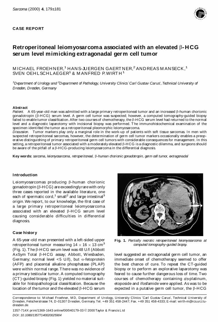
A 65-year-old man presented with a left-sided upperretroperitoneal tumor measuring 14 3 16 3 13 cm3
(Fig. 1).The b -HCG serum level was 48 U/l (AbbottAxSym Total b -HCG assay; Abbott, Wiesbaden,Germany; normal level <5 U/l), but a -fetoprotein(AFP) and placental alkaline phosphatase (PLAP)were within normal range.There was no evidence ofa primary testicular tumor. A computed tomography(CT)-guided biopsy (Fig. 1) yielded no material suit-able for histopathological classi® cation. Because thelocation of the tumor and the elevated b -HCG serum
level suggested an extragonadal germ cell tumor, animmediate onset of chemotherapy seemed to offerthe best chance of cure. To repeat the CT-guidedbiopsy or to perform an explorative laparotomy wasfeared to cause further dangerous loss of time. Twocourses of chemotherapy containing cis-platinum,etoposide and ifosfamide were applied. As was to beexpected in a putative germ cell tumor, the b -HCG
Correspondence to: Michael Froehner, MD, Department of Urology, University Clinics `Carl Gustav Carus’ , Technical University ofDresden, Fetscherstrasse 74, D-01307 Dresden, Germany. Tel. +49 351 458-2447; Fax. +49 351 458-4333; E-mail: [email protected]
Fig. 1. Partially necrotic retroperitoneal leiomyosarcoma atcomputed tomography-guided biopsy.
1357-714X print/1369-1643 online/00/040179-03 ½ 2000 Taylor & Francis Ltd
DOI: 10.1080/13577140020025904
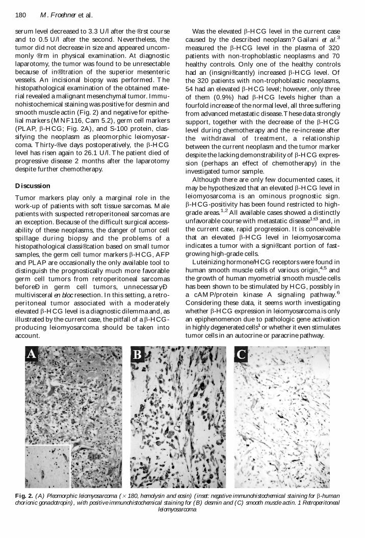
serum level decreased to 3.3 U/l after the ® rst courseand to 0.5 U/l after the second. Nevertheless, thetumor did not decrease in size and appeared uncom-monly ® rm in physical examination. At diagnosticlaparotomy, the tumor was found to be unresectablebecause of in® ltration of the superior mesentericvessels. An incisional biopsy was performed. Thehistopathological examination of the obtained mate-rial revealed a malignant mesenchymal tumor. Immu-nohistochemical staining was positive for desmin andsmooth muscle actin (Fig. 2) and negative for epithe-lial markers (MNF116, Cam 5.2), germ cell markers(PLAP, b -HCG; Fig. 2A), and S-100 protein, clas-sifying the neoplasm as pleomorphic leiomyosar-coma. Thirty-® ve days postoperatively, the b -HCGlevel has risen again to 26.1 U/l. The patient died ofprogressive disease 2 months after the laparotomydespite further chemotherapy.
Discussion
Tumor markers play only a marginal role in thework-up of patients with soft tissue sarcomas. Malepatients with suspected retroperitoneal sarcomas arean exception. Because of the difficult surgical access-ability of these neoplasms, the danger of tumor cellspillage during biopsy and the problems of ahistopathological classi® cation based on small tumorsamples, the germ cell tumor markers b -HCG, AFPand PLAP are occasionally the only available tool todistinguish the prognostically much more favorablegerm cell tumors from retroperitoneal sarcomasbeforeÐ in germ cell tumors, unnecessaryÐmultivisceral en bloc resection. In this setting, a retro-peritoneal tumor associated with a moderatelyelevated b -HCG level is a diagnostic dilemma and, asillustrated by the current case, the pitfall of a b -HCG-producing leiomyosarcoma should be taken intoaccount.
Was the elevated b -HCG level in the current casecaused by the described neoplasm? Gailani et al.3
measured the b -HCG level in the plasma of 320patients with non-trophoblastic neoplasms and 70healthy controls. Only one of the healthy controlshad an (insigni® cantly) increased b -HCG level. Ofthe 320 patients with non-trophoblastic neoplasms,54 had an elevated b -HCG level; however, only threeof them (0.9%) had b -HCG levels higher than afourfold increase of the normal level, all three sufferingfrom advanced metastatic disease.These data stronglysupport, together with the decrease of the b -HCGlevel during chemotherapy and the re-increase afterthe withdrawal of treatment, a relationshipbetween the current neoplasm and the tumor markerdespite the lacking demonstrability of b -HCG expres-sion (perhaps an effect of chemotherapy) in theinvestigated tumor sample.
Although there are only few documented cases, itmay be hypothesized that an elevated b -HCG level inleiomyosarcoma is an ominous prognostic sign.b -HCG-positivity has been found restricted to high-grade areas.1,2 All available cases showed a distinctlyunfavorable course with metastatic disease1± 3 and, inthe current case, rapid progression. It is conceivablethat an elevated b -HCG level in leiomyosarcomaindicates a tumor with a signi® cant portion of fast-growing high-grade cells.
Luteinizing hormone/HCG receptors were found inhuman smooth muscle cells of various origin,4,5 andthe growth of human myometrial smooth muscle cellshas been shown to be stimulated by HCG, possibly ina cAMP/protein kinase A signaling pathway.6
Considering these data, it seems worth investigatingwhether b -HCG expression in leiomyosarcoma is onlyan epiphenomenon due to pathologic gene activationin highly degeneratedcells1 or whether it even stimulatestumor cells in an autocrine or paracrine pathway.
Fig. 2. (A) Pleomorphic leiomyosarcoma ( 3 180, hemolysin and eosin) (inset: negative immunohistochemical staining for b -humanchorionic gonadotropin), with positive immunohistochemical staining for (B) desmin and (C) smooth muscle actin. 1 Retroperitoneal
leiomyosarcoma
180 M. Froehner et al.

References
1 Seidl C, Lippert C, Grouls V, JellinghausW. Leiomyosa-rkom des samenstrangs mit paraneoplastischer b -HCGproduktion. Pathologe 1998; 19:146± 50.
2 Meredith RF,Wagman LD, Piper JA, Mills AS, NeifeldJP. Beta-chain human chorionic gonadotropin-producing leiomyosarcoma of the small intestine.Cancer1986; 58:131± 35.
3 Gailani S, Chu TM, Nussbaum A, Ostrander M,Christoff N. Human chorionic gonadotropins (hCG)in nontrophoblastic neoplasms. Assessment ofabnormalities of hCG and CEA in bronchogenic anddigestive neoplasms. Cancer 1976; 38:1684± 6.
4 Tao YX, Bao S, Ackermann DM, Lei ZM, Rao CV.Expression of luteinizing hormone/human chorionicgonadotropin receptor gene in benign prostatic hyper-plasia and in prostate carcinoma in humans. Biol Reprod1997; 56:67± 72.
5 TaoYX, Heit M, Lei ZM, Rao CV.The urinary bladderof a woman is a novel site of luteinizing hormone-human chorionic gonadotropin receptor gene expres-sion. Am J Obstet Gynecol 1998; 179:1026± 32.
6 Kornyei JL, Lei ZM, Rao CV. Human myometrialsmooth muscle cells are novel targets of direct regula-tion by chorionic gonadotropin. Biol Reprod 1993;49:1149± 57.
Retroperitoneal leiomyosarcoma associated with an elevated b -HCG serum level 181

Submit your manuscripts athttp://www.hindawi.com
Stem CellsInternational
Hindawi Publishing Corporationhttp://www.hindawi.com Volume 2014
Hindawi Publishing Corporationhttp://www.hindawi.com Volume 2014
MEDIATORSINFLAMMATION
of
Hindawi Publishing Corporationhttp://www.hindawi.com Volume 2014
Behavioural Neurology
EndocrinologyInternational Journal of
Hindawi Publishing Corporationhttp://www.hindawi.com Volume 2014
Hindawi Publishing Corporationhttp://www.hindawi.com Volume 2014
Disease Markers
Hindawi Publishing Corporationhttp://www.hindawi.com Volume 2014
BioMed Research International
OncologyJournal of
Hindawi Publishing Corporationhttp://www.hindawi.com Volume 2014
Hindawi Publishing Corporationhttp://www.hindawi.com Volume 2014
Oxidative Medicine and Cellular Longevity
Hindawi Publishing Corporationhttp://www.hindawi.com Volume 2014
PPAR Research
The Scientific World JournalHindawi Publishing Corporation http://www.hindawi.com Volume 2014
Immunology ResearchHindawi Publishing Corporationhttp://www.hindawi.com Volume 2014
Journal of
ObesityJournal of
Hindawi Publishing Corporationhttp://www.hindawi.com Volume 2014
Hindawi Publishing Corporationhttp://www.hindawi.com Volume 2014
Computational and Mathematical Methods in Medicine
OphthalmologyJournal of
Hindawi Publishing Corporationhttp://www.hindawi.com Volume 2014
Diabetes ResearchJournal of
Hindawi Publishing Corporationhttp://www.hindawi.com Volume 2014
Hindawi Publishing Corporationhttp://www.hindawi.com Volume 2014
Research and TreatmentAIDS
Hindawi Publishing Corporationhttp://www.hindawi.com Volume 2014
Gastroenterology Research and Practice
Hindawi Publishing Corporationhttp://www.hindawi.com Volume 2014
Parkinson’s Disease
Evidence-Based Complementary and Alternative Medicine
Volume 2014Hindawi Publishing Corporationhttp://www.hindawi.com