repli-g cell wga & wta handbook
TRANSCRIPT
July 2014
Sample & Assay Technologies
REPLI-g® Cell WGA & WTA Handbook
For simultaneous whole genome
amplification and whole transcriptome
amplification from cells and limited samples

QIAGEN Sample and Assay Technologies QIAGEN is the leading provider of innovative sample and assay technologies, enabling the isolation and detection of contents of any biological sample. Our advanced, high-quality products and services ensure success from sample to result. QIAGEN sets standards in: Purification of DNA, RNA, and proteins Nucleic acid and protein assays microRNA research and RNAi Automation of sample and assay technologies Our mission is to enable you to achieve outstanding success and breakthroughs. For more information, visit www.qiagen.com.
REPLI-g Cell WGA & WTA Handbook 07/2014 3
Contents Kit Contents 4
Storage 5
Intended Use 6
Safety Information 6
Quality Control 6
Introduction 7
Biological Background 8
Target nucleic acids and downstream applications 9
Kit concept and utilization 11
Principle and procedure 12
Unique components of the REPLI-g Cell WGA & WTA Kit 13
Description of protocols 15
Equipment and Reagents to Be Supplied by User 17
Protocol: Lysis of Cell Samples 18
Protocol: Amplification of Genomic DNA 20
Protocol: Amplification of RNA (Total RNA or mRNA) 25
Troubleshooting Guide 29
References 32
Appendix A: Use of Amplified cDNA or gDNA for Next-Generation Sequencing 33
Appendix B: Determining DNA Concentration and Yield 35
Appendix C: PicoGreen Quantification of Amplified DNA 36
Appendix D: Purification of Amplified cDNA or gDNA for use in labeling reactions or OD measurement 39
Ordering Information 40
4 REPLI-g Cell WGA & WTA Handbook 07/2014
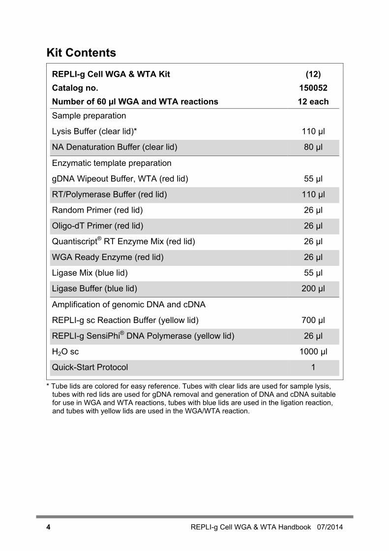
Kit Contents REPLI-g Cell WGA & WTA Kit (12) Catalog no. 150052 Number of 60 µl WGA and WTA reactions 12 each Sample preparation
Lysis Buffer (clear lid)* 110 µl NA Denaturation Buffer (clear lid) 80 µl
Enzymatic template preparation
gDNA Wipeout Buffer, WTA (red lid) 55 µl
RT/Polymerase Buffer (red lid) 110 µl
Random Primer (red lid) 26 µl
Oligo-dT Primer (red lid) 26 µl
Quantiscript® RT Enzyme Mix (red lid) 26 µl
WGA Ready Enzyme (red lid) 26 µl
Ligase Mix (blue lid) 55 µl
Ligase Buffer (blue lid) 200 µl
Amplification of genomic DNA and cDNA
REPLI-g sc Reaction Buffer (yellow lid) 700 µl
REPLI-g SensiPhi® DNA Polymerase (yellow lid) 26 µl
H2O sc 1000 µl
Quick-Start Protocol 1
* Tube lids are colored for easy reference. Tubes with clear lids are used for sample lysis, tubes with red lids are used for gDNA removal and generation of DNA and cDNA suitable for use in WGA and WTA reactions, tubes with blue lids are used in the ligation reaction, and tubes with yellow lids are used in the WGA/WTA reaction.
REPLI-g Cell WGA & WTA Handbook 07/2014 5
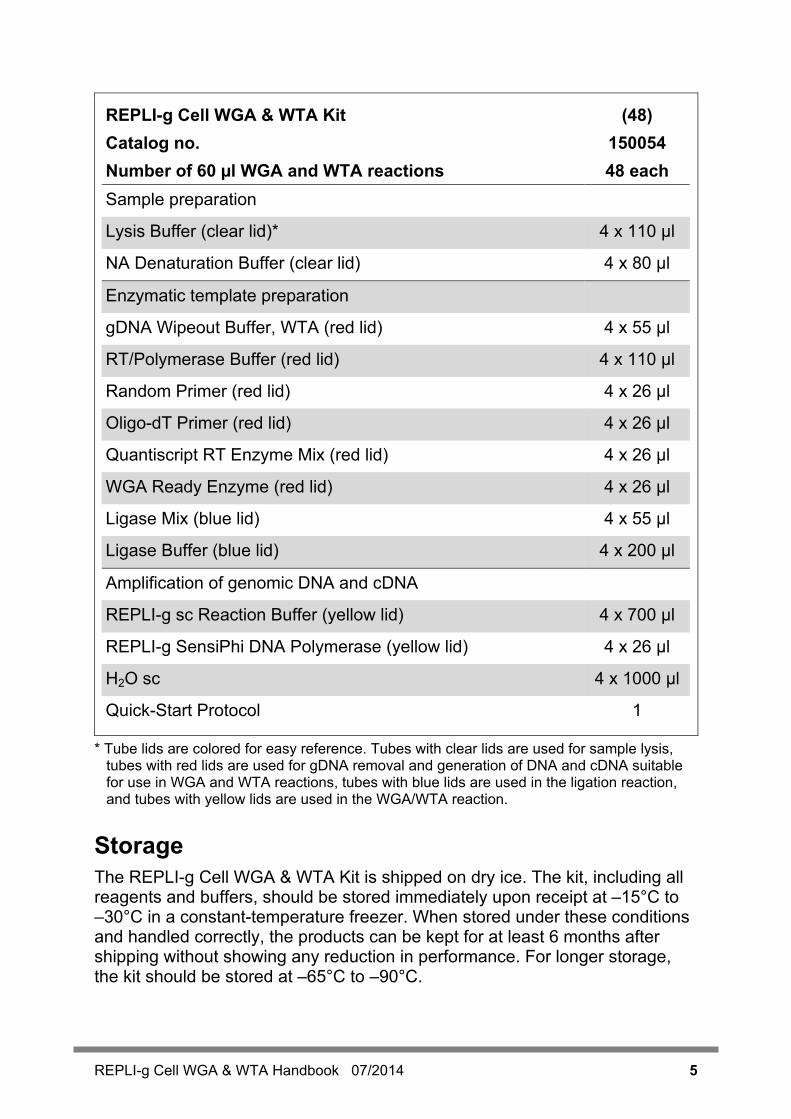
REPLI-g Cell WGA & WTA Kit (48) Catalog no. 150054 Number of 60 µl WGA and WTA reactions 48 each Sample preparation
Lysis Buffer (clear lid)* 4 x 110 µl NA Denaturation Buffer (clear lid) 4 x 80 µl
Enzymatic template preparation
gDNA Wipeout Buffer, WTA (red lid) 4 x 55 µl
RT/Polymerase Buffer (red lid) 4 x 110 µl
Random Primer (red lid) 4 x 26 µl
Oligo-dT Primer (red lid) 4 x 26 µl
Quantiscript RT Enzyme Mix (red lid) 4 x 26 µl
WGA Ready Enzyme (red lid) 4 x 26 µl
Ligase Mix (blue lid) 4 x 55 µl
Ligase Buffer (blue lid) 4 x 200 µl
Amplification of genomic DNA and cDNA
REPLI-g sc Reaction Buffer (yellow lid) 4 x 700 µl
REPLI-g SensiPhi DNA Polymerase (yellow lid) 4 x 26 µl
H2O sc 4 x 1000 µl
Quick-Start Protocol 1
* Tube lids are colored for easy reference. Tubes with clear lids are used for sample lysis, tubes with red lids are used for gDNA removal and generation of DNA and cDNA suitable for use in WGA and WTA reactions, tubes with blue lids are used in the ligation reaction, and tubes with yellow lids are used in the WGA/WTA reaction.
Storage The REPLI-g Cell WGA & WTA Kit is shipped on dry ice. The kit, including all reagents and buffers, should be stored immediately upon receipt at –15°C to –30°C in a constant-temperature freezer. When stored under these conditions and handled correctly, the products can be kept for at least 6 months after shipping without showing any reduction in performance. For longer storage, the kit should be stored at –65°C to –90°C.

6 REPLI-g Cell WGA & WTA Handbook 07/2014
Intended Use The REPLI-g Cell WGA & WTA Kit is intended for molecular biology applications. This product is not intended for the diagnosis, prevention, or treatment of a disease. All due care and attention should be exercised in the handling of the products. We recommend all users of QIAGEN® products to adhere to the NIH guidelines that have been developed for recombinant DNA experiments, or to other applicable guidelines.
Safety Information When working with chemicals, always wear a suitable lab coat, disposable gloves, and protective goggles. For more information, please consult the appropriate safety data sheets (SDSs). These are available online in convenient and compact PDF format at www.qiagen.com/safety where you can find, view, and print the SDS for each QIAGEN kit and kit component.
Quality Control In accordance with QIAGEN’s ISO-certified Quality Management System, each lot of REPLI-g Cell WTA & WGA Kit is tested against predetermined specifications to ensure consistent product quality.
REPLI-g Cell WGA & WTA Handbook 07/2014 7
Introduction Genetic analyses often require large amounts of gDNA or cDNA. Whole genome amplification and whole transcriptome amplification overcome limited DNA or RNA quantity by enabling the analysis of a small number of cells. The REPLI-g Cell WGA & WTA Kit offers researchers a unique tool to study the genome and transcriptome of the same biological sample. Amplified genomic DNA and cDNA can be accurately quantified and compared, giving insights into the genomic status and expression profile of the same group of cells, while eliminating sample-to-sample variations. The REPLI-g Cell WGA & WTA Kit contains a novel Phi 29 polymerase, REPLI-g SensiPhi Polymerase, and an optimized set of buffers and reagents for parallel whole genome amplification (WGA) of genomic DNA (gDNA) and whole transcriptome amplification (WTA) of total RNA from the same limited sample. Following efficient cell lysis, the WTA portion of the workflow begins with complete removal of genomic DNA and efficient reverse transcription with the Quantiscript RT Enzyme Mix. Afterwards, in separate parallel WGA and WTA reactions, the gDNA or cDNA are polished, prepared to be ligated and then completely amplified by Multiple Displacement Amplification (MDA) with REPLI-g SensiPhi Polymerase. This Phi29 polymerase variant has been engineered to achieve high sensitivity, fidelity, and processivity and is especially suited for effective MDA from very small amounts of starting material. To ensure even distribution of the cellular nucleic acids into the WGA and WTA reactions to achieve highly quantitative and reproducible results, a total of 25 to 1000 cells are needed for the parallel reactions. Using less than 25 cells may lead to an inconsistent distribution of sample DNA and RNA into the two reactions and thus less robust results, as determined both statistically and experimentally. The straightforward reaction setup, streamlined processing steps with total hands-on time of only 20 minutes, and overall reaction time of just 4 hours for the complete parallel amplification of both gDNA and RNA, make the REPLI-g Cell WGA & WTA Kit procedure an easy and reliable method to investigate genetic and transcriptional events that regulate various cellular processes. This handbook contains protocols for optimized sample lysis, template preparation, and amplification of gDNA and cDNA using the REPLI-g Cell WGA & WTA Kit. The resulting amplification products are suitable for any downstream application, including NGS. The REPLI-g Cell WGA & WTA Kit can be used with various samples that are often analyzed in clinical research, including tumor cells, stem cells, sorted cells, or other small samples (Table 1).

8 REPLI-g Cell WGA & WTA Handbook 07/2014
Table 1. Range of sample material and research areas
Sample material (cells/tissue) Research area
Cancer Sample*
Somatic genetic variant analysis
Tumor progression
Tumor stem cells Tumor evolution Tumor heterogeneity Cancer driver genes and driver pathways
Circulating tumor cells
Human/animal†
Biomarker research (SNPs, mutations, CNVs)
Mosaicism studies Stem cell research
Genetic predisposition studies Allele-specific gene expression Expression of quantitative trait loci (eQTLs) Gene expression regulation
* Biopsies, sorted or cultured cells, LCMs, fine needle aspirate and others. † The REPLI-g Cell WGA& WTA Kit does not lyse cell walls. Therefore, it cannot be used for
bacterial cells. Plant or other cells that have cell walls are also not suitable.
Biological Background Regulation of gene expression is driven by the genome in response to the environment. Mutations and genetic variations, such as point mutations and copy number or structural variations, result in multiple genomes within a species or within an organism (e.g., mosaic tissues and cancer). Some genetic variations have low impact on cellular or systemic functions, while others can exert dramatic effects on transcription regulation and, ultimately, phenotypes of the cell as well as the organism. Because of heterogeneity in tissue composition, the direct impact of genotypic changes on the transcriptome can be reliably analyzed only with a small group of identical cells. The REPLI-g Cell WGA & WTA Kit has been specifically designed to co-amplify in parallel reactions the entire genome and transcriptome from very small samples. With negligible sequence bias in nearly identical processing steps, the REPLI-g Cell
REPLI-g Cell WGA & WTA Handbook 07/2014 9
WGA & WTA Kit enables direct and reliable correlation of the genome and transcriptome of a sample.
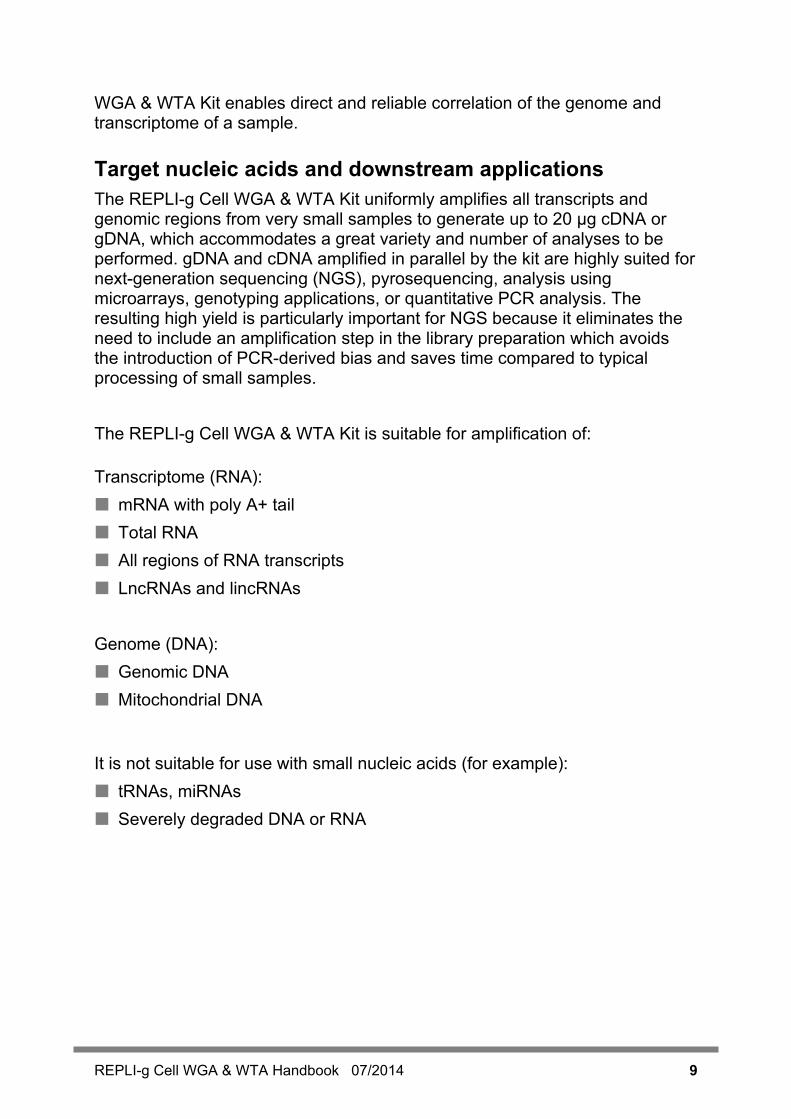
Target nucleic acids and downstream applications The REPLI-g Cell WGA & WTA Kit uniformly amplifies all transcripts and genomic regions from very small samples to generate up to 20 µg cDNA or gDNA, which accommodates a great variety and number of analyses to be performed. gDNA and cDNA amplified in parallel by the kit are highly suited for next-generation sequencing (NGS), pyrosequencing, analysis using microarrays, genotyping applications, or quantitative PCR analysis. The resulting high yield is particularly important for NGS because it eliminates the need to include an amplification step in the library preparation which avoids the introduction of PCR-derived bias and saves time compared to typical processing of small samples. The REPLI-g Cell WGA & WTA Kit is suitable for amplification of: Transcriptome (RNA): mRNA with poly A+ tail Total RNA All regions of RNA transcripts LncRNAs and lincRNAs Genome (DNA): Genomic DNA Mitochondrial DNA
It is not suitable for use with small nucleic acids (for example): tRNAs, miRNAs Severely degraded DNA or RNA
10 REPLI-g Cell WGA & WTA Handbook 07/2014
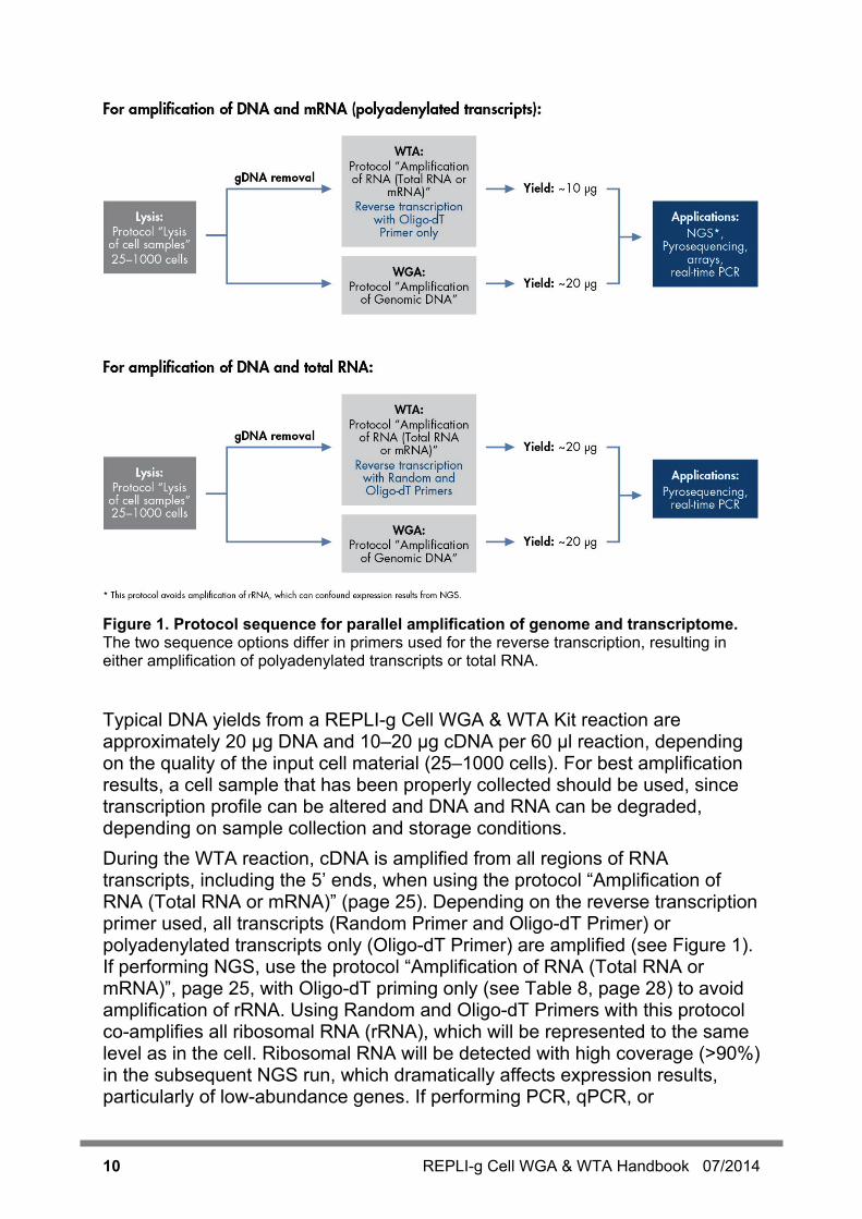
Figure 1. Protocol sequence for parallel amplification of genome and transcriptome. The two sequence options differ in primers used for the reverse transcription, resulting in either amplification of polyadenylated transcripts or total RNA.
Typical DNA yields from a REPLI-g Cell WGA & WTA Kit reaction are approximately 20 µg DNA and 10–20 µg cDNA per 60 µl reaction, depending on the quality of the input cell material (25–1000 cells). For best amplification results, a cell sample that has been properly collected should be used, since transcription profile can be altered and DNA and RNA can be degraded, depending on sample collection and storage conditions. During the WTA reaction, cDNA is amplified from all regions of RNA transcripts, including the 5’ ends, when using the protocol “Amplification of RNA (Total RNA or mRNA)” (page 25). Depending on the reverse transcription primer used, all transcripts (Random Primer and Oligo-dT Primer) or polyadenylated transcripts only (Oligo-dT Primer) are amplified (see Figure 1). If performing NGS, use the protocol “Amplification of RNA (Total RNA or mRNA)”, page 25, with Oligo-dT priming only (see Table 8, page 28) to avoid amplification of rRNA. Using Random and Oligo-dT Primers with this protocol co-amplifies all ribosomal RNA (rRNA), which will be represented to the same level as in the cell. Ribosomal RNA will be detected with high coverage (>90%) in the subsequent NGS run, which dramatically affects expression results, particularly of low-abundance genes. If performing PCR, qPCR, or

REPLI-g Cell WGA & WTA Handbook 07/2014 11
Pyrosequencing, use both Random and Oligo-dT Primers as non-polyA mRNAs and Incs are also be represented.
Kit concept and use Because MDA technology makes use of large DNA molecules for amplification, the ligation reaction of cDNA is necessary to obtain high-molecular-weight DNA for WTA. In order to minimize amplification variation between WTA and WGA, all enzymatic steps are aligned for both reactions wherever possible. That means that reagents, buffers and reaction steps for WGA and WTA are coordinated as tightly as possible in the REPLI-g Cell WGA & WTA Kit: The WGA ready reaction uses the same buffer and primers as the reverse
transcription and the reaction results also in DNA fragments of size similar to cDNA.
Although genomic DNA is amplified during WGA, the gDNA Wipeout Buffer must be used. Specific components of the gDNA Wipeout Buffer are required for the WGA ready reaction. However, the gDNA wipeout function is inactivated prior to adding DNA template and thus, gDNA removal does not take place.
Ligation and amplification are also identical for amplification of gDNA and of cDNA to ensure that method-derived bias between WGA and WTA is held very low and if residually present, bias is comparably brought into the same sequences derived from the genome or transcriptome.
Due to the increased use of highly parallel analysis methods like NGS, the ability to answer more questions in a single experiment has enabled researchers to more easily investigate genomic status and its influence on transcriptional status. Researchers investigating, for example, functional genomics or using a systems biology approach are asking for easy kit concepts that support these complex studies. Applying different methods for WGA and WTA may confound data interpretation as it is uncertain if observed differences are artificial or due to real biology.
The REPLI-g Cell WGA & WTA Kit provides uniform amplification across the entire genome and transcriptome, with negligible sequence bias (2). The amplification method is based on accurate MDA technology, which carries out isothermal genome amplification utilizing a highly processive DNA polymerase capable of replicating up to 70 kb without dissociating from the genomic DNA template. In contrast to PCR-based methods, REPLI-g SensiPhi DNA Polymerase has a 3'5' exonuclease proofreading activity, resulting in 1000-fold higher fidelity than Taq Polymerase during replication. The resulting amplified DNA and cDNA are stable during long-term storage for up to several years with no structural changes or degradation effects, enabling later analysis of sample material.

12 REPLI-g Cell WGA & WTA Handbook 07/2014
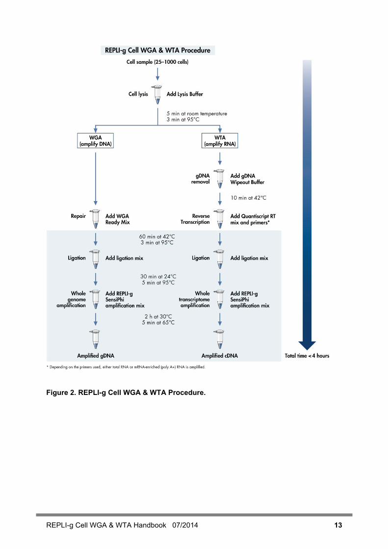
Principle and procedure The REPLI-g Cell WGA & WTA Kit contains reagents for the following sequential reactions (refer to flowchart on page 13): Lysis of cells: a sample (containing 25–1000 cells, see table 1) is lysed
efficiently within 5 minutes, with no effect on RNA or DNA integrity. Lysed cells are divided into two aliquots of equal volume. The first aliquot is used for WGA, while the second aliquot is used for WTA of total RNA, or alternatively, mRNA-enriched (poly A+) RNA.
Whole genome amplification: following cell lysis, DNA from one of the aliquots is enzymatically polished for high efficiency ligation and amplification. Following ligation, amplification using MDA technology takes place using the novel, high-affinity REPLI-g SensiPhi DNA Polymerase. Wherever possible, steps in the WGA procedure are kept identical to the steps in the WTA procedure.
Whole transcriptome amplification: following cell lysis, gDNA is removed from the second aliquot prior to starting the WTA process, since accurate measurement of transcript levels depends on the elimination of false-positive results caused by gDNA contamination. cDNA is synthesized for high efficiency ligation and amplification. Depending on the primer chosen during the reverse transcription reaction, total RNA or only polyadenylated transcripts are amplified (see Table 8, page 28). Wherever possible, steps in the WTA procedure are kept identical to the steps in the WGA procedure.
Due to the nature of the ligation reaction, DNA fragments might not be assembled in the order in which they originally existed in the organism. However, kit chemistry is designed to make these events rare and thus, detection of nucleic acid sequences is not affected (e.g., polymorphisms) in downstream applications, such as NGS, array analysis, or qPCR. Error rates (e.g., chimeras, etc.) observed during kit optimization were in normal range.
REPLI-g Cell WGA & WTA Handbook 07/2014 13
Figure 2. REPLI-g Cell WGA & WTA Procedure.
14 REPLI-g Cell WGA & WTA Handbook 07/2014
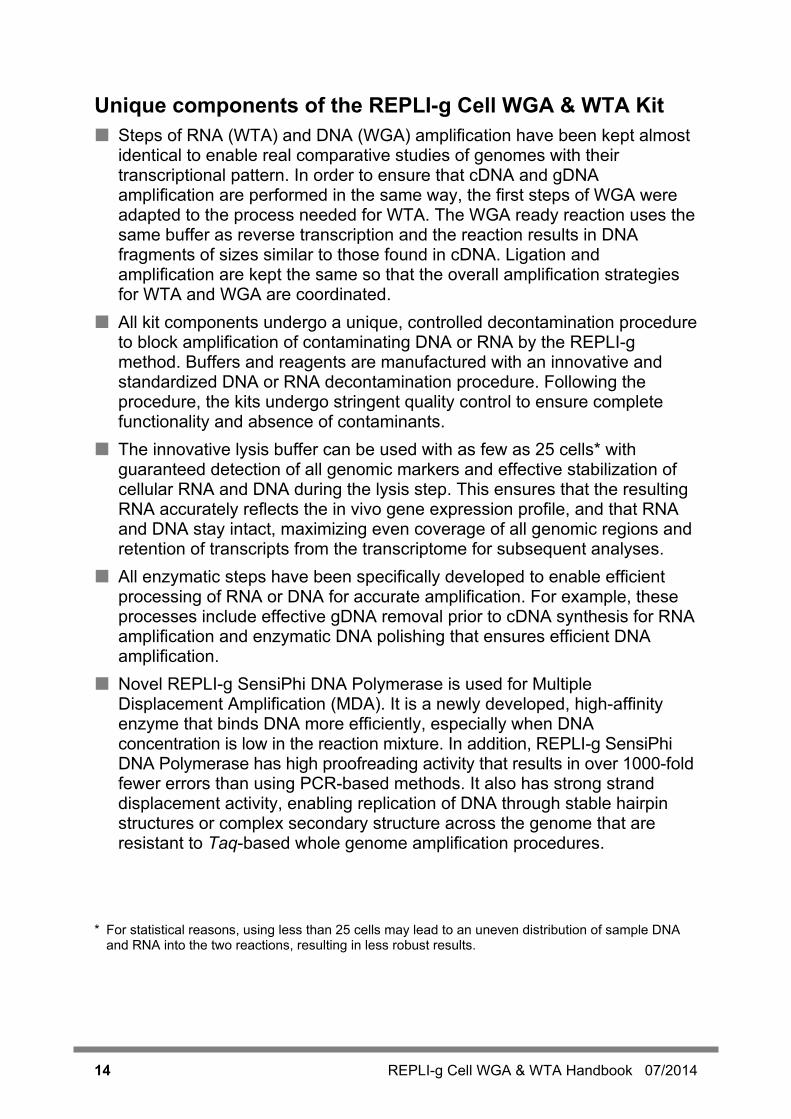
Unique components of the REPLI-g Cell WGA & WTA Kit Steps of RNA (WTA) and DNA (WGA) amplification have been kept almost
identical to enable real comparative studies of genomes with their transcriptional pattern. In order to ensure that cDNA and gDNA amplification are performed in the same way, the first steps of WGA were adapted to the process needed for WTA. The WGA ready reaction uses the same buffer as reverse transcription and the reaction results in DNA fragments of sizes similar to those found in cDNA. Ligation and amplification are kept the same so that the overall amplification strategies for WTA and WGA are coordinated.
All kit components undergo a unique, controlled decontamination procedure to block amplification of contaminating DNA or RNA by the REPLI-g method. Buffers and reagents are manufactured with an innovative and standardized DNA or RNA decontamination procedure. Following the procedure, the kits undergo stringent quality control to ensure complete functionality and absence of contaminants.
The innovative lysis buffer can be used with as few as 25 cells* with guaranteed detection of all genomic markers and effective stabilization of cellular RNA and DNA during the lysis step. This ensures that the resulting RNA accurately reflects the in vivo gene expression profile, and that RNA and DNA stay intact, maximizing even coverage of all genomic regions and retention of transcripts from the transcriptome for subsequent analyses.
All enzymatic steps have been specifically developed to enable efficient processing of RNA or DNA for accurate amplification. For example, these processes include effective gDNA removal prior to cDNA synthesis for RNA amplification and enzymatic DNA polishing that ensures efficient DNA amplification.
Novel REPLI-g SensiPhi DNA Polymerase is used for Multiple Displacement Amplification (MDA). It is a newly developed, high-affinity enzyme that binds DNA more efficiently, especially when DNA concentration is low in the reaction mixture. In addition, REPLI-g SensiPhi DNA Polymerase has high proofreading activity that results in over 1000-fold fewer errors than using PCR-based methods. It also has strong strand displacement activity, enabling replication of DNA through stable hairpin structures or complex secondary structure across the genome that are resistant to Taq-based whole genome amplification procedures.
* For statistical reasons, using less than 25 cells may lead to an uneven distribution of sample DNA
and RNA into the two reactions, resulting in less robust results.
REPLI-g Cell WGA & WTA Handbook 07/2014 15
Description of protocols The protocols in this handbook provide detailed instructions for use of the REPLI-g Cell WGA & WTA Kit for parallel whole genome and whole transcriptome amplification. Although the kit was designed to amplify both DNA and RNA from the same sample, the REPLI-g Cell WGA & WTA Kit contains sufficient buffers and reagents to perform either 24 WGA or 24 WTA reactions from separate samples consisting of more than 25 cells, if desired (see Figure 2 for details about amplicons and yields for each reaction). The protocol “Lysis of Cell Samples”, page 18, is optimized for cell material from all species without a cell wall, for example, cells from vertebrates, sorted cells, tissue culture cells, and cells or tissue from biopsies. Cells with cell walls, such as bacteria (gram positive and gram negative) or plant cells cannot be used. Once cell lysates are prepared, they can be used directly in the subsequent protocols: “Amplification of Genomic DNA”, page 20, which describes efficient enzymatic preparation and accurate amplification of the complete genomic DNA; and “Amplification of RNA (Total RNA or mRNA)”, page 25, which describes sensitive reverse transcription, efficient enzymatic preparation, and accurate amplification of total RNA or poly A+ RNA. Note that rRNA is also amplified when using Random and Oligo-dT Primers, and will represent more than 90% of all sequences after amplification. If working with sequence-specific probes, such as with qPCR, the amplified rRNA will not affect downstream application results. If using the amplified cDNA for RNAseq, be aware that more than 90% of reads are derived from rRNA and therefore, sufficient reads must be obtained if performing whole transcriptome sequencing. Amplification of rRNA does not occur when amplifying mRNA-enriched (poly A+) RNA, which can be easily obtained by omitting the random primer. See Figure 3 for a suggested experimental workflow (page 16).

16 REPLI-g Cell WGA & WTA Handbook 07/2014
The workflow outlined below saves time and handling steps for the efficient parallel performance of the WGA and WTA reactions.
Figure 3. Suggested experimental workflow for parallel WTA and WGA.
REPLI-g Cell WGA & WTA Handbook 07/2014 17
Equipment and Reagents to Be Supplied by User When working with chemicals, always wear a suitable lab coat, disposable gloves, and protective goggles. For more information, consult the appropriate safety data sheets (SDSs), available from the product supplier. Microcentrifuge tubes or PCR-strips Thermal cycler Microcentrifuge Vortexer Pipets and pipet tips Wet ice Nuclease-free water or TE buffer (10 mM Tris·Cl; 1 mM EDTA, pH 8.0)

18 REPLI-g Cell WGA & WTA Handbook 07/2014
Protocol: Lysis of Cell Samples This protocol is for lysis of cell samples and must be done prior to the protocols “Amplification of Genomic DNA”, page 20 and “Amplification of RNA (Total RNA or mRNA)”, page 25.
Important points before starting This protocol is optimized for use with cells from all vertebrate species
(e.g., human, mouse rat, sorted cells, cultured cells, or cells collected under the microscope or via laser microdissection). The protocol cannot be used for bacterial cells. Plant cells or other cells that include cell walls are also not suitable. The protocol cannot be used for cells that have been fixed using formalin or other cross-linking agents (e.g., single cell samples obtained by laser microdissection from formalin-fixed, paraffin-embedded [FFPE] tissues).
Samples containing 25–1000 cells are optimal for whole transcriptome amplification and whole genome amplification reactions using the REPLI-g Cell WGA & WTA Kit.
Avoid DNA or RNA contamination of reagents by using dedicated laboratory equipment (e.g., pipets, filter pipet tips, reaction vials, etc.). Set up the REPLI-g Cell WGA & WTA Kit reaction in a location free of nucleic acids.
Vortex all buffers and reagents before use to ensure thorough mixing.
Procedure 1. Place 13 μl cell material (suspended in PBS) into a microcentrifuge
tube. If using less than 13 μl cell material, add H20 sc to bring the volume up to 13 μl.
2. Add 8 µl Lysis Buffer. Mix carefully by flicking the tube and centrifuge briefly. Note: Ensure that the cell material does not stick to the wall of the tube above the meniscus.
3. Incubate at 24°C for 5 min, followed by 95°C for 3 min. Cool down to 4°C.
4. Transfer a 10 µl aliquot to a fresh reaction tube and immediately perform WGA (see protocol “Amplification of Genomic DNA”, page 20). The tube should be kept on ice.
REPLI-g Cell WGA & WTA Handbook 07/2014 19
5. Transfer a second 10 µl aliquot to a second fresh reaction tube and immediately perform WTA (see protocol “Amplification of RNA (Total RNA or mRNA)”, page 25). The tube should be kept on ice. Note: Whole genome amplification with the protocol “Amplification of Genomic DNA”, page 20 and whole transcriptome amplification using the protocol “Amplification of RNA (Total RNA or mRNA)”, page 25 can be easily processed in parallel. The protocols are identical from the preparation of ligation mix, through to the end of each procedure (step 3 of the protocol “Amplification of Genomic DNA” and step 4 of the protocol “Amplification of RNA (Total RNA or mRNA)”). See Figure 3 (page 16) for a suggested experimental workflow. IMPORTANT: Cell lysates cannot be stored at this point. They must be processed immediately using the protocols “Amplification of Genomic DNA”, page 20 and “Amplification of RNA (Total RNA or mRNA)”, page 25.
20 REPLI-g Cell WGA & WTA Handbook 07/2014
Protocol: Amplification of Genomic DNA This protocol is for whole genome amplification of genomic DNA. For whole transcriptome amplification, refer to the protocol “Amplification of RNA (Total RNA or mRNA)”, page 25. Important points before starting This whole genome amplification protocol is optimized for cells that have
been lysed using the protocol “Lysis of Cell Samples”, page 18. Appropriate lysis of samples must be performed before WGA and/or WTA reactions.
A ligation step has been implemented to eliminate experimental variation between the gDNA and RNA amplification processes in as many steps as possible and thus, achieve consistent and comparable results.
Although genomic DNA is amplified by this process, the gDNA Wipeout Buffer must to be used for the WGA ready reaction. Specific components of the gDNA Wipeout Buffer are required for the WGA ready reaction. However, the gDNA wipeout function is inactivated in the WGA ready mix upon mixing with RT/Polymerase buffer. Thus, gDNA removal does not occur.
Avoid any DNA or RNA contamination of reagents by using dedicated laboratory equipment (e.g., pipets, filter pipet tips, reaction vials, etc.). Set up the reaction in a location free of nucleic acids.
The high-molecular-weight DNA generated by random extension of primers (primer-multimer formation) in no-template controls (NTC) does not contain genetic information and will not affect the quality of downstream applications. These products are outcompeted by DNA of viable cells present during WGA.
If performing PCR/real-time PCR as downstream application, the amplified cDNA must be diluted 1:100. Add 2–3 μl diluted DNA to a 20 μl real-time PCR reaction volume.
If performing microarray as downstream application, the amplified cDNA and DNA must be purified prior to labeling. Please note that the amplified cDNA and gDNA are double-stranded. Follow the instructions of the microarray manufacturer and choose labeling methods that are compatible with the microarray application.
Things to do before starting The REPLI-g Cell WGA & WTA Kit is designed to process protocols
“Amplification of Genomic DNA” and “Amplification of RNA (Total RNA or mRNA)” in parallel until the ligation step for each protocol. See Figure 3 on page 16 for a suggested experimental workflow.
WGA Ready Enzyme, Ligase Mix, and REPLI-g SensiPhi DNA Polymerase should be thawed on ice. All other components can be thawed at room temperature (15–25°C).

REPLI-g Cell WGA & WTA Handbook 07/2014 21
All buffers and reagents should be vortexed before use to ensure thorough mixing.
The WGA ready mix, ligation mix, and REPLI-g SensiPhi amplification mix described in the protocol must always be prepared fresh. They cannot be stored for later use.
For increased speed and convenience, all incubation steps of the protocol can be preprogrammed on a thermal cycler (Table 2).
Table 2. Thermal cycling parameters
Step Time Temperature Additional comments Set the heating lid to 50ºC for all steps
WGA ready reaction
60 min
3 min ∞
42ºC
95ºC
4°C
Polish genomic DNA for ligation*. Add WGA ready mix prior to incubation (step 2) Stops enzymatic polishing of gDNA. Hold
Ligation 30 min 24ºC Add ligation mix prior to incubation (step 4)
5 min ∞
95°C 4°C
Stops ligation Hold
Whole genome amplification
2 h 30ºC Add REPLI-g SensiPhi amplification mix prior to incubation (step 6)
5 min 65ºC Inactivates all enzymes
∞ 4ºC Cools amplified DNA
* This step has been introduced into the protocol to maintain method consistency between the WGA and WTA procedures.
Procedure 1. Prepare the WGA ready mix (Table 3, page 22). 2. Add 10 µl WGA ready mix to the first aliquot of the lysed cell sample
(step 4 from the protocol “Lysis of Cell Samples”, page 18). Mix by vortexing and centrifuge briefly. Note: The WGA ready mix must be prepared fresh.

22 REPLI-g Cell WGA & WTA Handbook 07/2014
3. Incubate at 42ºC for 60 min. Stop the reaction by incubating at 95ºC for 3 min, then cool on ice. IMPORTANT: The following protocol steps are identical to the protocol “Amplification of RNA (Total RNA or mRNA)”, page 25, starting at step 5, and can be processed in parallel. If processing both reactions in parallel, make sure to set up the correct amount of ligation mix for both reactions.
Table 3. Preparing WGA ready mix*
Component Volume/reaction RT/Polymerase Buffer 4 µl
gDNA Wipeout Buffer† 2 µl
H2O sc 1 µl
Random Primer 1 µl
Oligo-dT Primer‡ 1 µl
WGA Ready Enzyme 1 µl
Total volume§ 10 µl
* Scale up accordingly if performing more than one reaction at one time. † Some components of the gDNA Wipeout Buffer are required for the WGA ready reaction.
The gDNA wipeout function is inactivated upon addition to the WGA ready mix. Thus, gDNA removal does not take place.
‡ Oligo-dT primer is included because they are able to also amplify T rich and other regions of gDNA, giving a complete representation of all genomic markers.
§ Mix by vortexing and centrifuge briefly. 4. Prepare the ligation mix (Table 4). Add 10 µl ligation mix to each WGA
ready reaction from step 3 and to each Quantiscript RT reaction from step 4 of the protocol “Amplification of RNA (Total RNA or mRNA)”. Mix by vortexing and centrifuge briefly. IMPORTANT: When preparing the ligation mix, add the components in the order shown in Table 4, page 23. Note: The ligation mix must be prepared fresh.
REPLI-g Cell WGA & WTA Handbook 07/2014 23
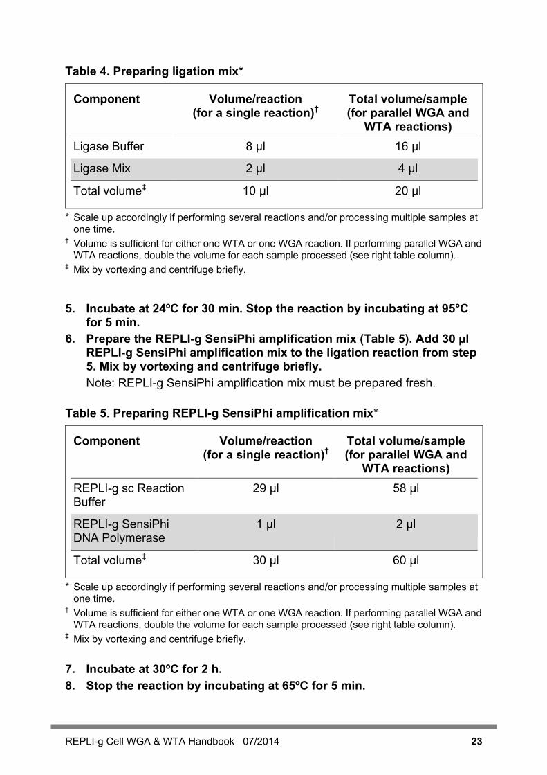
Table 4. Preparing ligation mix*
Component Volume/reaction (for a single reaction)†
Total volume/sample (for parallel WGA and
WTA reactions) Ligase Buffer 8 µl 16 µl
Ligase Mix 2 µl 4 µl
Total volume‡ 10 µl 20 µl
* Scale up accordingly if performing several reactions and/or processing multiple samples at one time.
† Volume is sufficient for either one WTA or one WGA reaction. If performing parallel WGA and WTA reactions, double the volume for each sample processed (see right table column).
‡ Mix by vortexing and centrifuge briefly. 5. Incubate at 24ºC for 30 min. Stop the reaction by incubating at 95°C
for 5 min. 6. Prepare the REPLI-g SensiPhi amplification mix (Table 5). Add 30 µl
REPLI-g SensiPhi amplification mix to the ligation reaction from step 5. Mix by vortexing and centrifuge briefly. Note: REPLI-g SensiPhi amplification mix must be prepared fresh.
Table 5. Preparing REPLI-g SensiPhi amplification mix*
Component Volume/reaction (for a single reaction)†
Total volume/sample (for parallel WGA and
WTA reactions) REPLI-g sc Reaction Buffer
29 µl 58 µl
REPLI-g SensiPhi DNA Polymerase
1 µl 2 µl
Total volume‡ 30 µl 60 µl
* Scale up accordingly if performing several reactions and/or processing multiple samples at one time.
† Volume is sufficient for either one WTA or one WGA reaction. If performing parallel WGA and WTA reactions, double the volume for each sample processed (see right table column).
‡ Mix by vortexing and centrifuge briefly. 7. Incubate at 30ºC for 2 h. 8. Stop the reaction by incubating at 65ºC for 5 min.
24 REPLI-g Cell WGA & WTA Handbook 07/2014
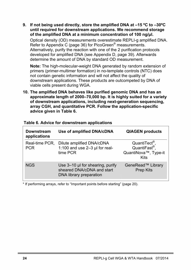
9. If not being used directly, store the amplified DNA at –15 ºC to –30ºC until required for downstream applications. We recommend storage of the amplified DNA at a minimum concentration of 100 ng/μl. Optical density (OD) measurements overestimate REPLI-g amplified DNA. Refer to Appendix C (page 36) for PicoGreen® measurements. Alternatively, purify the reaction with one of the 2 purification protocols developed for amplified DNA (see Appendix D, page 39). Afterwards determine the amount of DNA by standard OD measurement. Note: The high-molecular-weight DNA generated by random extension of primers (primer-multimer formation) in no-template controls (NTC) does not contain genetic information and will not affect the quality of downstream applications. These products are outcompeted by DNA of viable cells present during WGA.
10. The amplified DNA behaves like purified genomic DNA and has an approximate length of 2000–70,000 bp. It is highly suited for a variety of downstream applications, including next-generation sequencing, array CGH, and quantitative PCR. Follow the application-specific advice given in Table 6.
Table 6. Advice for downstream applications
Downstream applications
Use of amplified DNA/cDNA QIAGEN products
Real-time PCR, PCR
Dilute amplified DNA/cDNA 1:100 and use 2–3 µl for real-time PCR
QuantiTect®, QuantiFast®,
QuantiNova™, Type-it Kits
NGS Use 3–10 µl for shearing, purify sheared DNA/cDNA and start DNA library preparation
GeneRead™ Library Prep Kits
* If performing arrays, refer to “Important points before starting” (page 20).
REPLI-g Cell WGA & WTA Handbook 07/2014 25
Protocol: Amplification of RNA (Total RNA or mRNA) This protocol is for whole transcriptome amplification of total RNA, or, optionally, for amplification of mRNA-enriched RNA. For whole genome amplification, refer to the protocol “Amplification of Genomic DNA”, page 20. Note that rRNA is also amplified and will represent more than 90% of all sequences after amplification. If working with sequence-specific probes, such as with qPCR, the amplified rRNA will not affect downstream application results. Amplification of rRNA does not occur when amplifying mRNA-enriched (poly A+) RNA by omitting the Random Primer (refer to step 3).
Important points before starting This whole transcriptome amplification protocol is optimized for cells that
have been lysed using the protocol “Lysis of Cell Samples”, page 18. Appropriate lysis of samples must be performed before WGA and/or WTA reactions.
If performing NGS or microarray analysis, use this protocol with Oligo-dT Primer only.
If performing PCR/real-time PCR, the amplified cDNA must be diluted 1:100. Add 2–3 µl diluted DNA to a 20 µl real-time PCR reaction volume.
Purification of amplified cDNA is only necessary for labeling of cDNA, for example, to be used in microarrays. All other downstream applications, such as NGS or PCR, are not affected by remaining nucleotides.
The high-molecular-weight DNA generated by random extension of primers (primer-multimer formation) in no-template controls (NTC) does not contain genetic information and will not affect the quality of downstream applications. These products are outcompeted by DNA of viable cells present during WGA.
Avoid any DNA or RNA contamination of reagents by using dedicated laboratory equipment (e.g., pipets, filter pipet tips, reaction vials, etc.). Set up the REPLI-g Cell WGA & WTA Kit reaction in a location free of nucleic acids.
Because the REPLI-g Cell WGA & WTA Kit is intended to generate amplified cDNA from a minimal amount of starting RNA, take appropriate measures to avoid inadvertently introducing RNase contamination. Create and maintain an RNase-free environment when working with RNA by following proper microbiological and aseptic technique. The use of disposable plastic tubes and pipet tips from freshly opened boxes or bags is strongly recommended.
The reagents for whole transcriptome amplification are not suitable for the amplification of small RNA molecules, such as tRNAs or miRNAs.
26 REPLI-g Cell WGA & WTA Handbook 07/2014
Although all sequences are well represented, the amplified cDNA does not contain full-length cDNAs. The amplification process is started by random-primed cDNA synthesis. Consequently, transcript sequences are amplified in pieces. Due to the nature of the ligation reaction, DNA fragments might not be assembled in the order in which they originally existed in the organism. However, kit chemistry is designed to make these events rare and thus, detection of nucleic acid sequences is not affected (e.g., polymorphisms) in downstream applications, such as NGS, array analysis, or qPCR. Error rates (e.g., chimeras, etc.) observed during kit optimization were in normal range.
Things to do before starting The REPLI-g Cell WGA & WTA Kit is designed to process protocols
“Amplification of Genomic DNA” and “Amplification of RNA (Total RNA or mRNA)” in parallel until the ligation step for each protocol. See Figure 3 on page 16 for a suggested experimental workflow.
The Quantiscript RT mix, ligation mix, and REPLI-g SensiPhi amplification mix described in the protocol must always be prepared fresh. They cannot be stored for later use.
All buffers and reagents should be vortexed before use to ensure thorough mixing.
Quantiscript RT Enzyme Mix, Ligase Mix, and REPLI-g SensiPhi DNA Polymerase should be thawed on ice. All other components can be thawed at room temperature (15–25°C).
For increased speed and convenience, all incubation steps of the protocol can be preprogrammed on a thermal cycler (Table 7, page 27).
If performing NGS or microarray analysis, use only the Oligo-dT Primer for the reverse transcription to avoid amplifying rRNA.

REPLI-g Cell WGA & WTA Handbook 07/2014 27
Table 7. Thermal cycling parameters
Step Time Temperature Additional comments Set the heating lid to 50ºC for all steps
gDNA removal 10 min 42°C Add gDNA Wipeout Buffer prior to incubation (step 1)
Reverse transcription
60 min 42ºC
Add Quantiscript RT mix prior to incubation (step 3)
3 min ∞
95°C 4°C
Stops reverse transcription Hold
Ligation 30 min 24ºC Add ligation mix prior to incubation (step 5)
5 min ∞
95°C 4°C
Stops ligation Hold
Whole transcriptome amplification
2 h
30ºC
Add REPLI-g SensiPhi amplification mix prior to incubation (step 6)
5 min 65ºC Inactivates all enzymes.
∞ 4ºC Cools amplified cDNA.
Procedure 1. Add 2 µl gDNA Wipeout Buffer to the second aliquot of the lysed cell
sample (step 5 from Protocol: Lysis of Cell Samples, page 18), mix by vortexing, and centrifuge briefly.
2. Incubate at 42°C for 10 min. 3. Prepare Quantiscript RT mix (Table 8). Add 8 µl Quantiscript RT mix
to the lysed cell sample, mix by vortexing, and centrifuge briefly. Note: Quantiscript RT mix must be prepared fresh. IMPORTANT: To enrich the WTA amplification product for mRNA poly A+ sequences, omit Random Primer from the Quantiscript RT mix (see Table 8, page 28). IMPORTANT: If performing NGS, be aware that when amplifying total RNA (i.e., without enriching for mRNA poly A+), ribosomal RNA (rRNA) will be represented to the same level as in the cell, and will be detected with high coverage in the subsequent NGS run. Amplification of rRNA does not occur when amplifying mRNA-enriched (poly A+) RNA (by omitting the Random Primer).
28 REPLI-g Cell WGA & WTA Handbook 07/2014
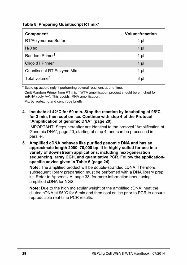
Table 8. Preparing Quantiscript RT mix*
Component Volume/reaction RT/Polymerase Buffer 4 µl
H20 sc 1 µl
Random Primer† 1 µl
Oligo dT Primer 1 µl
Quantiscript RT Enzyme Mix 1 µl
Total volume‡ 8 µl
* Scale up accordingly if performing several reactions at one time. † Omit Random Primer from RT mix if WTA amplification product should be enriched for
mRNA (poly A+). This avoids rRNA amplification. ‡ Mix by vortexing and centrifuge briefly. 4. Incubate at 42ºC for 60 min. Stop the reaction by incubating at 95ºC
for 3 min, then cool on ice. Continue with step 4 of the Protocol “Amplification of genomic DNA” (page 20). IMPORTANT: Steps hereafter are identical to the protocol “Amplification of Genomic DNA”, page 20, starting at step 4, and can be processed in parallel.
5. Amplified cDNA behaves like purified genomic DNA and has an approximate length 2000–70,000 bp. It is highly suited for use in a variety of downstream applications, including next-generation sequencing, array CGH, and quantitative PCR. Follow the application-specific advice given in Table 6 (page 24). Note: The amplified product will be double-stranded cDNA. Therefore, subsequent library preparation must be performed with a DNA library prep kit. Refer to Appendix A, page 33, for more information about using amplified cDNA for NGS. Note: Due to the high molecular weight of the amplified cDNA, heat the diluted cDNA at 95°C for 5 min and then cool on ice prior to PCR to ensure reproducible real-time PCR results.
REPLI-g Cell WGA & WTA Handbook 07/2014 29
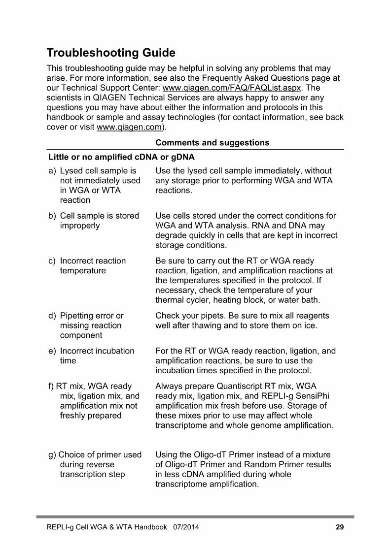
Troubleshooting Guide This troubleshooting guide may be helpful in solving any problems that may arise. For more information, see also the Frequently Asked Questions page at our Technical Support Center: www.qiagen.com/FAQ/FAQList.aspx. The scientists in QIAGEN Technical Services are always happy to answer any questions you may have about either the information and protocols in this handbook or sample and assay technologies (for contact information, see back cover or visit www.qiagen.com).
Comments and suggestions Little or no amplified cDNA or gDNA a) Lysed cell sample is
not immediately used in WGA or WTA reaction
Use the lysed cell sample immediately, without any storage prior to performing WGA and WTA reactions.
b) Cell sample is stored improperly
Use cells stored under the correct conditions for WGA and WTA analysis. RNA and DNA may degrade quickly in cells that are kept in incorrect storage conditions.
c) Incorrect reaction temperature
Be sure to carry out the RT or WGA ready reaction, ligation, and amplification reactions at the temperatures specified in the protocol. If necessary, check the temperature of your thermal cycler, heating block, or water bath.
d) Pipetting error or missing reaction component
Check your pipets. Be sure to mix all reagents well after thawing and to store them on ice.
e) Incorrect incubation time
For the RT or WGA ready reaction, ligation, and amplification reactions, be sure to use the incubation times specified in the protocol.
f) RT mix, WGA ready mix, ligation mix, and amplification mix not freshly prepared
Always prepare Quantiscript RT mix, WGA ready mix, ligation mix, and REPLI-g SensiPhi amplification mix fresh before use. Storage of these mixes prior to use may affect whole transcriptome and whole genome amplification.
g) Choice of primer used during reverse transcription step
Using the Oligo-dT Primer instead of a mixture of Oligo-dT Primer and Random Primer results in less cDNA amplified during whole transcriptome amplification.

30 REPLI-g Cell WGA & WTA Handbook 07/2014
Comments and suggestions DNA yields of approximately 10 µg in negative (no-template) controls, but no positive result in downstream assay (e.g., PCR) DNA is generated
during REPLI-g reaction by random extension of primer-multimers
As long as DNA from viable cells is present in the WGA or WTA reaction, the high-molecular-weight products from the random extension of primer-multimers will be outcompeted. These products do not affect the quality of the samples nor do they impact specific downstream genetic assays.
DNA yields of approximately 10 µg in negative (no-template) controls and positive result in downstream assay (e.g., PCR) DNA is generated
during REPLI-g reaction by contaminating RNA or DNA templates
Decontaminate all laboratory equipment, and take all necessary precautions to avoid contamination of reagents and samples with extraneous DNA or RNA. If possible, work in a laminar-flow hood. Use sterile equipment and barrier pipet tips only, and keep amplification chemistry and templates in separate storage locations.
Little or no target sequence detected in real-time PCR analysis, but DNA yield is approximately 20 µg a) Sample does not
contain cells Dilutions of cells often contain volumes with a lower number of cells than recommended due to Poisson distribution.
b) Cells are not intact Use viable cells for REPLI-g Cell WGA & WTA Kit reactions
c) Low-abundance transcript analyzed
Due to Poisson distribution, the REPLI-g Cell WGA & WTA Kit may provide variable amplification of transcripts that have fewer than 10 copies per reaction.
d) Small transcripts analyzed
Small transcripts, such as tRNA or miRNAs, cannot be amplified by the REPLI-g Cell WGA & WTA Kit. Only RNA transcripts longer than 500 nt can be amplified.
e) Sequences larger than 1.5 kb are analyzed
Due to random priming, amplification of large, continuous sequences is not possible. We recommend analyzing smaller sequences from your target sequence.

REPLI-g Cell WGA & WTA Handbook 07/2014 31
Comments and suggestions f) 5´ regions analyzed
when following protocol “Amplification of RNA (Total RNA or mRNA)”
In the protocol “Amplification of RNA (Total RNA or mRNA)”, 3´ regions of polyadenylated transcripts are amplified preferentially. 5´ regions are underrepresented.
Downstream application results not optimal Sensitive downstream applications may require DNA cleanup after the REPLI-g reaction
Contact QIAGEN Technical Services for DNA cleanup recommendations suitable for your application.
32 REPLI-g Cell WGA & WTA Handbook 07/2014
References 1. Dean, F.B. et al (2002) Comprehensive human genome amplification using
multiple displacement amplification. Proc. Natl. Acad. Sci. USA 99, 5261. 2. Hosono, S. et al (2003) Unbiased whole-genome amplification directly from
clinical samples. Genome Res. 13, 954.
REPLI-g Cell WGA & WTA Handbook 07/2014 33
Appendix A: Use of Amplified cDNA or gDNA for Next-Generation Sequencing Next-generation sequencing (NGS) is a driving force for numerous applications, including cancer research, stem cell research, metagenomics, population genetics, and medical research. Library preparation can be done either with intact DNA, using tagmentation methods like Nextera DNA library prep,or the amplified DNA can be fragmented by mechanical shearing prior to library construction (e.g., Covaris® instrument). If using the Nextera library prep, please check the supplementary protocols available at www.qiagen.com. The efficient QIAGEN GeneRead Library Prep Kits are based on a streamlined, optimized, one-tube protocol that does not require sample cleanup between each step, which saves time and prevents handling errors and loss of valuable samples. The following describes use of these kits. The GeneRead library prep procedure includes an optional, high-fidelity amplification step to ensure high yields of DNA library that are reproducibly generated with minimal sequence bias and low error rates. This step can be omitted because the amount of DNA derived from the REPLI-g amplification is sufficient for several library preps without further amplification.
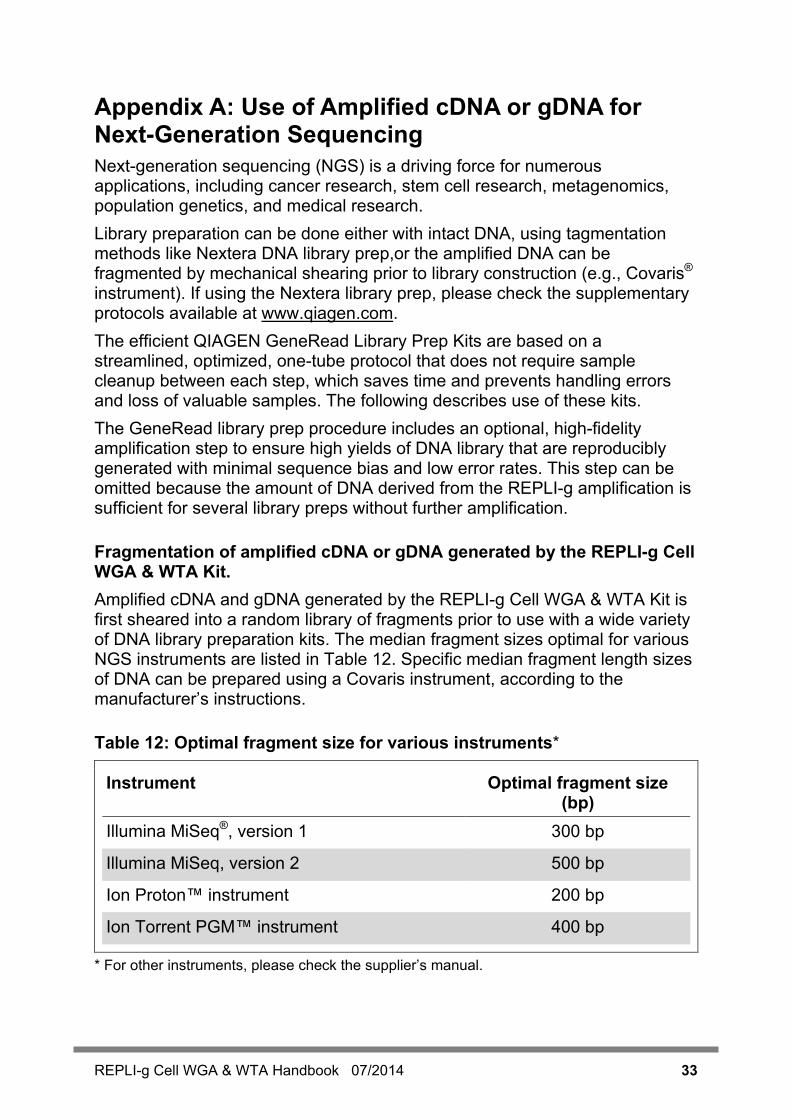
Fragmentation of amplified cDNA or gDNA generated by the REPLI-g Cell WGA & WTA Kit. Amplified cDNA and gDNA generated by the REPLI-g Cell WGA & WTA Kit is first sheared into a random library of fragments prior to use with a wide variety of DNA library preparation kits. The median fragment sizes optimal for various NGS instruments are listed in Table 12. Specific median fragment length sizes of DNA can be prepared using a Covaris instrument, according to the manufacturer’s instructions.
Table 12: Optimal fragment size for various instruments*
Instrument Optimal fragment size (bp)
Illumina MiSeq®, version 1 300 bp
Illumina MiSeq, version 2 500 bp
Ion Proton™ instrument 200 bp
Ion Torrent PGM™ instrument 400 bp
* For other instruments, please check the supplier’s manual.

34 REPLI-g Cell WGA & WTA Handbook 07/2014
Use of fragmented gDNA and cDNA with GeneRead Library Prep Kits A1. Use 2–5 µg of amplified cDNA and/or gDNA generated with the
REPLI-g Cell WGA & WTA Kit. Note: Follow instructions in the Covaris manual to determine the correct instrument settings to achieve the optimal fragment size. Using too much DNA in a Covaris instrument may, for example, lead to incomplete shearing of the DNA. Check the fragmented DNA for the correct size distribution using an agarose gel, QIAxcel® Advanced, or Agilent® Bioanalyzer®.
A2. Purify fragmented cDNA and/or gDNA using the QIAquick PCR Purification Kit (cat. no. 28104)
Note: Follow “QIAquick PCR Purification Kit Protocol, using a microcentrifuge” in the QIAquick Spin Handbook.
A3. Prepare GeneRead Library following the protocols in the kit handbooks.
Note: Choose the correct kit for your application and NGS instrument (Table 13).
Table 13: GeneRead Library Preparation
Instrument GeneRead Library Cat. no. Illumina MiSeq, Illumina HiSeq® 2000 or 2500 Illumina Genome Analyzer
GeneRead DNA Library I Core Kit 180432
GeneRead Adapter I Set 1-plex 180912
GeneRead Adapter I Set 12-plex 180984
GeneRead DNA I Amp Kit 180455
Ion Proton
instrument Ion Torrent PGM instrument
GeneRead DNA Library L Core Kit 180462
GeneRead Adapter L Set 1-plex 180922
GeneRead Adapter L Set 12-plex 180994
GeneRead DNA L Amp Kit 180485

REPLI-g Cell WGA & WTA Handbook 07/2014 35
Appendix B: Determining DNA Concentration and Yield Using the REPLI-g Cell WGA & WTA Kit, a 60 µl reaction typically yields up to 20 µg of DNA, depending on the quality of RNA or DNA within the cell sample. For accurate quantification of the amplification product, it is important to use a DNA quantification method that is specific for double-stranded DNA, since the DNA sample contains unused reaction primers. Alternatively, purify the reaction according to instructions in Appendix D (page 39). Avoid using any other purification method as it will result in reduced yields. Following purification, determine the amount of DNA using a standard OD measurement. PicoGreen reagent displays enhanced binding to double-stranded DNA and may be used, in conjunction with a fluorometer, to quantify the amount of amplified DNA. A protocol for the quantification of amplified DNA is provided in Appendix C.
36 REPLI-g Cell WGA & WTA Handbook 07/2014
Appendix C: PicoGreen Quantification of Amplified DNA This protocol is designed for quantification of double-stranded amplified DNA using PicoGreen reagent. Note: Degraded or old PicoGreen reagent may result in inaccurate DNA quantification. DNA yields in excess of 80 µg should be ignored and, if necessary, quantification should be repeated using fresh PicoGreen reagent. IMPORTANT: When working with hazardous chemicals, always wear a suitable lab coat, disposable gloves, and protective goggles. For more information, please consult the appropriate safety data sheets (SDSs), available from the product supplier.
Equipment and reagents to be supplied by user Quant-iT™ PicoGreen dsDNA reagent (Invitrogen, cat. no. P7581) TE buffer (10 mM Tris·Cl; 1 mM EDTA, pH 8.0) Human genomic DNA (e.g., REPLI-g Control DNA, QIAGEN cat. no.
150090) 2 ml microcentrifuge tube 96-well plates (suitable for use in a fluorescence microplate reader) Fluorescence microplate reader (e.g., TECAN® Ultra)
Procedure
Setup and reading of microplate C1. In a 2 ml microcentrifuge tube, make a 1:150 dilution of PicoGreen
stock solution in TE buffer. Each quantification reaction requires 20 µl. Cover the microcentrifuge tube in aluminum foil or place it in the dark to avoid photodegradation of the PicoGreen reagent. For example, to prepare enough PicoGreen working solution for 100 samples, add 13.3 µl PicoGreen to 1986.7 µl TE buffer. IMPORTANT: Prepare the PicoGreen/TE solution in a plastic container, as the PicoGreen reagent may adsorb to glass surfaces.
C2. Prepare a 16 µg/ml stock solution of genomic DNA in TE buffer. DNA standards will be prepared from this stock solution.
C3. Make 200 µl of 1.6, 0.8, 0.4, 0.2, and 0.1 µg/ml DNA standards by further diluting the 16 µg/ml genomic DNA with TE buffer.
REPLI-g Cell WGA & WTA Handbook 07/2014 37
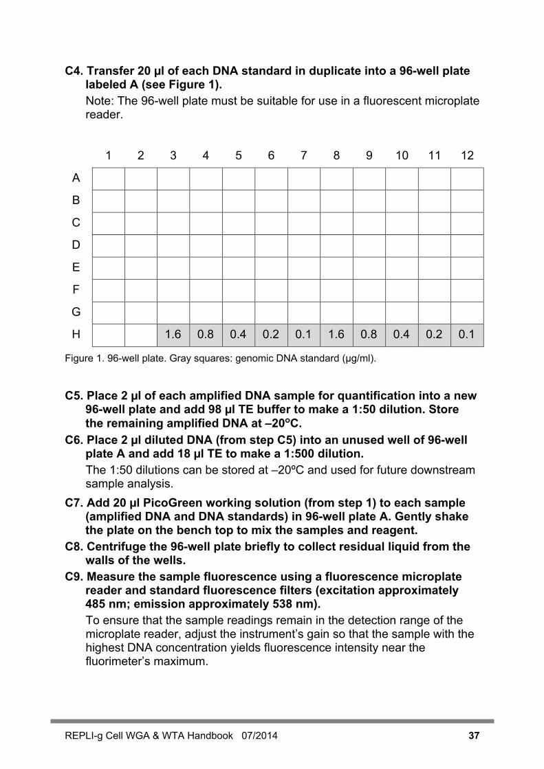
C4. Transfer 20 µl of each DNA standard in duplicate into a 96-well plate labeled A (see Figure 1). Note: The 96-well plate must be suitable for use in a fluorescent microplate reader.
1 2 3 4 5 6 7 8 9 10 11 12
A
B
C
D
E
F
G
H 1.6 0.8 0.4 0.2 0.1 1.6 0.8 0.4 0.2 0.1
Figure 1. 96-well plate. Gray squares: genomic DNA standard (µg/ml).
C5. Place 2 µl of each amplified DNA sample for quantification into a new
96-well plate and add 98 µl TE buffer to make a 1:50 dilution. Store the remaining amplified DNA at –20ºC.
C6. Place 2 µl diluted DNA (from step C5) into an unused well of 96-well plate A and add 18 µl TE to make a 1:500 dilution. The 1:50 dilutions can be stored at –20ºC and used for future downstream sample analysis.
C7. Add 20 µl PicoGreen working solution (from step 1) to each sample (amplified DNA and DNA standards) in 96-well plate A. Gently shake the plate on the bench top to mix the samples and reagent.
C8. Centrifuge the 96-well plate briefly to collect residual liquid from the walls of the wells.
C9. Measure the sample fluorescence using a fluorescence microplate reader and standard fluorescence filters (excitation approximately 485 nm; emission approximately 538 nm). To ensure that the sample readings remain in the detection range of the microplate reader, adjust the instrument’s gain so that the sample with the highest DNA concentration yields fluorescence intensity near the fluorimeter’s maximum.
38 REPLI-g Cell WGA & WTA Handbook 07/2014
Calculation of DNA concentration and yield C10. Generate a standard curve by plotting the concentration of DNA
standards (µg/ml) (x-axis) against the fluorescence reading generated by the microplate reader (y-axis). Plot an average of the fluorescence recorded for each DNA standard of the same concentration.
C11. Use the standard curve to determine the concentration (µg/ml) of the diluted amplified DNA sample. This is achieved by plotting the fluorescence reading of the sample against the standard curve and reading the DNA concentration on the x-axis. Note: The calculation of DNA concentration depends on the standard curve and the determination of the slope. For accurate results, the standard curve should be a straight line. Any deviation from this may cause inaccuracies in the measurement of amplified DNA concentrations.
C12. Multiply the value determined in step 11 by 500 to show the concentration of undiluted sample DNA (as the sample DNA measured by PicoGreen fluorescence had been diluted 1 in 500).
C13. To determine the total amount of DNA in your sample, multiply the concentration of undiluted sample DNA (µg/ml) from step C12 by the reaction volume in milliliters (i.e., for a 50 µl reaction, multiply by 0.05).
REPLI-g Cell WGA & WTA Handbook 07/2014 39
Appendix D: Purification of Amplified cDNA or gDNA for use in labeling reactions or OD measurement Since the amplified cDNA or DNA is particularly long (2–100 kb), standard purification methods, like ethanol precipitation or use of QIAamp® kits, cannot be applied as they will result in low recovery of < 20%. Two protocols have been developed for purification of long DNA with good recovery and yield based on either LiCl/EtOH or the use of AmPure beads. Both are available as supplementary protocols on the REPLI-g Cell WGA & WTA Kit product page at www.qiagen.com.
40 REPLI-g Cell WGA & WTA Handbook 07/2014
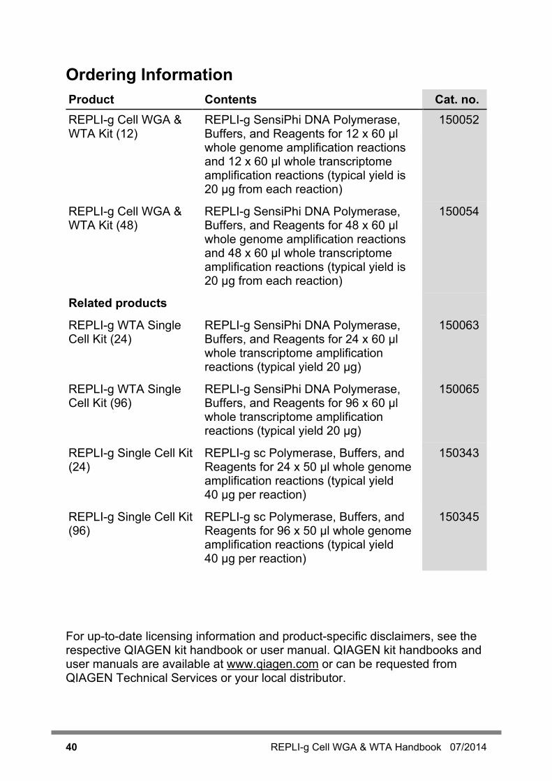
Ordering Information Product Contents Cat. no. REPLI-g Cell WGA & WTA Kit (12)
REPLI-g SensiPhi DNA Polymerase, Buffers, and Reagents for 12 x 60 µl whole genome amplification reactions and 12 x 60 µl whole transcriptome amplification reactions (typical yield is 20 µg from each reaction)
150052
REPLI-g Cell WGA & WTA Kit (48)
REPLI-g SensiPhi DNA Polymerase, Buffers, and Reagents for 48 x 60 µl whole genome amplification reactions and 48 x 60 µl whole transcriptome amplification reactions (typical yield is 20 µg from each reaction)
150054
Related products
REPLI-g WTA Single Cell Kit (24)
REPLI-g SensiPhi DNA Polymerase, Buffers, and Reagents for 24 x 60 µl whole transcriptome amplification reactions (typical yield 20 µg)
150063
REPLI-g WTA Single Cell Kit (96)
REPLI-g SensiPhi DNA Polymerase, Buffers, and Reagents for 96 x 60 µl whole transcriptome amplification reactions (typical yield 20 µg)
150065
REPLI-g Single Cell Kit (24)
REPLI-g sc Polymerase, Buffers, and Reagents for 24 x 50 µl whole genome amplification reactions (typical yield 40 µg per reaction)
150343
REPLI-g Single Cell Kit (96)
REPLI-g sc Polymerase, Buffers, and Reagents for 96 x 50 µl whole genome amplification reactions (typical yield 40 µg per reaction)
150345
For up-to-date licensing information and product-specific disclaimers, see the respective QIAGEN kit handbook or user manual. QIAGEN kit handbooks and user manuals are available at www.qiagen.com or can be requested from QIAGEN Technical Services or your local distributor.
REPLI-g Cell WGA & WTA Handbook 07/2014 41
Notes

42 REPLI-g Cell WGA & WTA Handbook 07/2014
Notes
Trademarks: QIAGEN®, SensiPhi®, QIAamp®, QIAxcel®, GeneRead™, QuantiFast®, Quantiscript®, QuantiTect®, QuantiNova™, REPLI-g® (QIAGEN Group); Affymetrix® (Affymetrix, Inc.); Agilent®, Bioanalyzer® (Agilent Technologies, Inc.); Covaris® (Covaris, Inc.); HiSeq®, MiSeq® (Illumina, Inc.); Ion Proton™, Ion Torrent PGM ™, Quant-iT™ (Life Technologies, Corporation); PicoGreen® (Molecular Probes, Inc.); TECAN® (TECAN Group AG).
Limited License Agreement for the REPLI-g Cell WTA & WGA Kit
Use of this product signifies the agreement of any purchaser or user of the product to the following terms:
1. The product may be used solely in accordance with the protocols provided with the product and this handbook and for use with components contained in the kit only. QIAGEN grants no license under any of its intellectual property to use or incorporate the enclosed components of this kit with any components not included within this kit except as described in the protocols provided with the product, this handbook, and additional protocols available at www.qiagen.com. Some of these additional protocols have been provided by QIAGEN users for QIAGEN users. These protocols have not been thoroughly tested or optimized by QIAGEN. QIAGEN neither guarantees them nor warrants that they do not infringe the rights of third-parties.
2. Other than expressly stated licenses, QIAGEN makes no warranty that this kit and/or its use(s) do not infringe the rights of third-parties.
3. This kit and its components are licensed for one-time use and may not be reused, refurbished, or resold.
4. QIAGEN specifically disclaims any other licenses, expressed or implied other than those expressly stated.
5. The purchaser and user of the kit agree not to take or permit anyone else to take any steps that could lead to or facilitate any acts prohibited above. QIAGEN may enforce the prohibitions of this Limited License Agreement in any Court, and shall recover all its investigative and Court costs, including attorney fees, in any action to enforce this Limited License Agreement or any of its intellectual property rights relating to the kit and/or its components.
For updated license terms, see www.qiagen.com.
© 2014 QIAGEN, all rights reserved.
1082217 07/2014 Sample & Assay
www.qiagen.com
Australia [email protected]
Austria [email protected]
Belgium [email protected]
Brazil [email protected]
Canada [email protected]
China [email protected]
Denmark [email protected]
Finland [email protected]
France [email protected]
Germany [email protected]
Hong Kong [email protected]
India [email protected]
Ireland [email protected]
Italy [email protected]
Japan [email protected]
Korea (South) [email protected]
Luxembourg [email protected]
Mexico [email protected]
The Netherlands [email protected]
Norway [email protected]
Singapore [email protected]
Sweden [email protected]
Switzerland [email protected]