renal pathology ii. - semmelweis
TRANSCRIPT

Renal pathology II.
Áron Somorácz MD PhD

Urologic renal diseases
I. Congenital abnormalities
II. Cystic diseases of the kidney
III. Urolithiasis
IV. Obstructive uropathy
V. Pyelonephritis
VI. Tumors of the kidney
I. Congenital abnormalities
1. Agenesis of the kidney
In utero detected bilateral agenesis indicates abortion
2. Hypoplasia of the kidney
Smaller kidney with contralateral compensatory hypertrophy
3. Oligomeganephronia
Reduced number of nephrons leading to end stage renal disease (ESRD) by the
time of adolescence
4. Horseshoe kidney
The most common congenital anomaly (1/500-1000) resulting from the fusion of
lower (90%) or upper (10%) poles
5. Ectopic kidneys
The kidney lies at the pelvic brim or within the pelvis

I. Congenital abnormalities
6. Duplication of the renal pelvis and the ureter
7. Ureteropelvic junction stenosis
Usually unilateral, leads to hydronephrosis, early operation can save the kidney
8. Accessory renal artery
9. Multicystic renal dysplasia
Developmental anomaly, NOT neoplastic process!
Abnormal tissue elements (cartilage, undifferentiated mesenchyme)
Cysts
Unilateral or bilateral, complete or segmental
Enlarged kidney with insufficent function

Horseshoe kidney Ureteral duplication

Ureteropelvic junction stenosis

Multicystic renal dysplasia

II. Cystic diseases of the kidney
1. Polycystic kidney disease
A) Autosomal-dominant (ADPKD)
Adulthood
PKD1 and PKD2 genes (Polycystin-1 and -2)
Prevalence 1/400-1000
Bilateral and marked enlargement
Grape-like appearance
Symptomes begin in early adulthood
It is responsible for 5-10% of chronic renal failures
Liver and pancreatic cysts, mitral valve prolapse, intracranial berry aneurysms

Autosomal-dominant polycystic kidney disease

II. Cystic diseases of the kidney
1. Polycystic kidney disease
B) Autosomal-recessive (ARPKD)
Perinatal, neonatal, infantile, juvenile subcategories
PKDH1 gene (fibrocystin)
Markedly enlarged kidneys
Lung hypoplasia, oligohydramnion
Medullary and cortical elongated cysts, sponge-like appearance
Usually leads to death within the first month of life

Autosomal-recessive polycystic kidney disease

II. Cystic diseases of the kidney
2. Cystic diseases of the renal medulla
A) Medullary sponge kidney
1-3 mm medullary cysts of the collecting ducts
Does not lead to renal failure
B) Nephronophtysis
Sporadic or familial
Atrophic kidneys, cysts at the corticomedullary junction
Leads to ESRD
3. Simple cysts
Common finding, single or multiple, do not influence the renal function

II. Cystic diseases of the kidney
4. Acquired cystic disease of the kidney
In case of long-standing dialysis
5. Glomerulocystic kidney disease
6. Cysts in hereditary syndromes
von Hippel-Lindau syndrome
Tuberous sclerosis

III. Urolithiasis
Three factors:
Salts that are capable of crystallization
Core that triggers crystallizaton (cell debris, urinary cast)
Lack of inhibitors of crystallization
1. Calcium stones
60-70%
Calcium oxalate/calcium phosphate
Hypercalciuria (with or without hypercalcemia), hyperoxaluria
Brown-black, 1-2 cm, visible by X-ray

III. Urolithiasis
2. Struvite stones
15%
Magnesium ammonium phosphate
After infection (e.g., Proteus)
Grey-yellow, staghorn calculi
3. Uric acid stones
15%
Hyperuricemia (gout, rapid cell turnover e.g., leukemias)
White or orange, radiolucent
4. Cystine stones
1-2%
Cystinuria

Calcium stone Staghorn (struvite) stone

III. Urolithiasis
Clinical features
Uni- (80%) or bilateral
Kidney stone attack: agonizing intense pain in the lower back (lumbal area)
extending into the groin area
Nausea, vomiting
Smaller stones are more hazardous
Hematuria
Might be without symptomes
Predisposes for infections

IV. Obstructive uropathy
Obstruction predisposes for infections and stone formation
Unrelieved obstruction leads to hydronephrosis
Hydronephrosis:
Dilation of the renal pelvis and calyces
Progressive atrophy of the kidney

IV. Obstructive uropathy
Causes:
1. Congenital anomalies
2. Calculi
3. Prostatic hyperplasia
4. Tumors
5. Lower urinary tract inflammations
6. Pregnancy
7. Uterine prolapse
8. Functional disorders

IV. Obstructive uropathy
Clinical features:
Acute obstruction usually provokes pain
Partial obstruction may remain silent
Partial bilateral obstruction leads to inabilty to concentrate the urine resulting in
polyuria followed by chronic tubulointerstitial nephritis
Complete bilateral obstruction leads oliguria or anuria

V. Pyelonephritis
1. Acute pyelonephritis
Inflammation of the tubules, the interstitium, the calyces and the renal pelvis
Caused by bacterias (E. coli, Proteus mirabilis, Klebsiella, Enterococcus)
Usually consequence of an ascending urinary tract infection
Less commonly result of a hematogenous spread
In normal kidneys, or as a complication of urinary tract disorders (e.g. VUR)
Predisposing factors: catheter, diabetes, pregnancy, lower urinary tract
obstruction, immunsuppression

V. Pyelonephritis
1. Acute pyelonephritis
Morphology:
Sligthly enlarged kidney(s)
1-3 mm yellowish abscesses on the surface and in the parenchyme
(pyelonpehritis apostematosa)
The calyces and the renal pelvis are reddish
Patchy interstitial suppurative inflammation, aggregates of neutrophils in the
tubules, tubular necrosis
Glomeruli are also affected in case of hematogenous origin
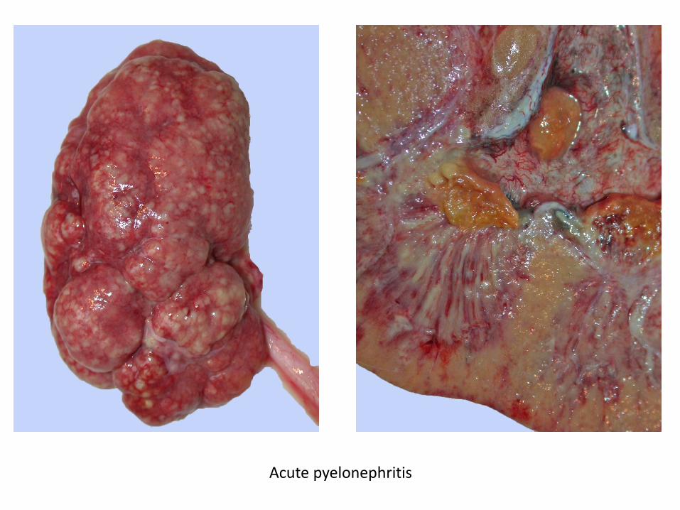
Acute pyelonephritis

Acute pyelonephritis

V. Pyelonephritis
1. Acute pyelonephritis
Clinical features:
Uni- or bilateral
Sudden onset with high fever
Pain at the costovertebral angle
Leukocytosis, high sedimentation rate
Pyuria, bacteruria
Usually follows a benign course (with appropriate antibiotic therapy)
Complicated cases can be fatal

V. Pyelonephritis
2. Chronic pyelonephritis
Chronic injury of the interstitium and the tubules resulting in scar formation
The renal pelvis and the calyces are also affected
Causes: reflux nephropathy, chronic obstruction
Recurrent infections
Morphology:
Kidneys are irregularly scarred, corticomedullary scars overlying dilated calyces,
flattening of the papillae
The calyces are dilated and their mucosa is thickened
Focal interstitial fibrosis, atrophic tubules, tubular casts (thyroidization), lymphoid
infiltration
The mucosa of the calyces and pelvis is fibrotic and contains cronic inflammation

Chronic pyelonephritis

Chronic pyelonephritis

V. Pyelonephritis
2. Chronic pyelonephritis
Clinical features:
Uni- or bilateral
Episodes of acute pyelonephritis, or silent clinical course leading to destruction
Bilateral chronic pyelonephritis can result in hypertension and renal insufficiency
10% of patients on dialysis therapy have chronic pyelonephritis

V. Pyelonephritis
3. Xanthogranulomatous pyelonephritis
Middle-aged women with diabetes
Usually unilateral
Proteus mirabilis infection
Tumor-like lesion
Yellowish areas, extracapsular spread, infiltrative pattern
Foamy histiocytes, giant cells, lymphocytes, plasma cells, neutrophils
Indicates nephrectomy

Xanthogranulomatous pyelonephritis

VI. Tumors of the kidney
Tumor types
Benign tumors
Malignant tumors
Pediatric tumors
Genetic background
Prognostic factors
Clinical features

VI. Tumors of the kidney
Histogenesis of renal tumors
Tubular epithelium
Papillary adenoma
Oncocytoma
Clear cell renal cell carcinoma
(renal cell carcinoma: RCC)
Multilocular cystic neoplasm
Papillary RCC
Chromophobe RCC
Clear cell papillary RCC
Mesenchyme
Angiomyolipoma
Metanephrogenic elements
Metanephric adenoma
Nephroblastoma (Wilms tumor)

VI. Tumors of the kidney
Benign tumors
1. Papillary adenoma
Papillary tumor with low-grade tumors cells, ≤ 15 mm
Grey-white, round nodule, can be multifocal
Commonly incidental finding
Cuboidal, monomorphic tumor cells, papillary architecture, psammoma bodies

VI. Tumors of the kidney
Benign tumors
2. Oncocytoma
5% of renal tumors
Mahogany brown, characteristic central scar
Nested, trabecular architecture, degenerative signs
Oncocytic, bland looking cells (large, granular eosinophilic cytoplasm), however, bizarre
nuclei, extracapsular infiltration, vascular invasion might be encountered


VI. Tumors of the kidney
Benign tumors
3. Angiomyolipoma
PEComa: perivascular epithelioid cell tumor
1% of the resected renal tumors, but it is more common (by US detected tumors
that are not operated)
Well-circumscribed, usually fatty appearance
Adipose tissue, thick-walled vessels, smooth muscle
HMB45+, Melan A+ (melanocytic markers!!)


VI. Tumors of the kidney
Benign tumors
4. Metanephric adenoma
More common in females
Average size is 5 cm
Grey-brown, solid, cystic degeneration may be present
Well-circumscribed but unencapsulated
Tubulary, solid, or papillary architecture
No mitoses
WT1+, CD57+, CK7-, EMA-


VI. Tumors of the kidney
Malignant tumors
1. Clear cell RCC
Most common type (approx. 80%)
Characteristic golden yellow color
Variable architecture: solid, tubular, papillary, microcystic, cystic
Haemorrhages and necroses are usual
Genetic/epigenetic alteration: 3p deletion, VHL mutation, VHL hypermetilation


VI. Tumors of the kidney
Malignant tumors
2. Multilocular cystic renal neoplasm of low malignant potential
2-3% of RCCs
Middle-aged patients, usually incidentally detected
Complex cystic lesion by radiological examination
Composed exclusively of thin-walled cysts
Clear cyst content
Low-grade tumor cells: internal surface of cysts, small groups in the septums
Excellent prognosis


VI. Tumors of the kidney
Malignant tumors
3. Papillary RCC
Second most common (approx. 12-15%)
More commonly multifocal
Type 1
Grey-white, well-circumscribed, encapsulated, haemorrhages
Papillary, tubular, or solid architecture
Cuboidal cells, foamy macrophages, psammoma bodies
Type 2
Variable appearance, necroses, haemorrhages
Larger cells, higher grade
Characteristic genetic alteration: trisomies (7, 17, 12, 16 20), loss of Y, c-Met
mutation


Type 1 Type 2

VI. Tumors of the kidney
Malignant tumors
4. Chromophobe RCC
Approx. 4-5%
Well-circumscribed, grey-white
Clear or eosinophilic cytoplasm, distinct cell borders, binucleated figures,
rasinoid nuclei, perinuclear halo
Widespread chromosomal losses
Better prognosis
DD: oncocytoma


VI. Tumors of the kidney
Malignant tumors
5. Clear cell papillary RCC
Originally descriped being associated with end-stage kidney disease
1-1,5% of renal cell tumors
Well-circumscribed, encapsulated, grey-brown
Solid and cystic areas
Mostly branching tubular, less commonly papillary architecture
Clear cells with low-grade morphology
Subnuclear vacuolization
Indolent behavior (no metastatic case has been published to date)


VI. Tumors of the kidney
Malignant tumors
Rare types
Collecting duct (Bellini) carcinoma
Hereditary leiomyomatosis and renal cell carcinoma-associated RCC
MiT gene family translocation RCC
Succinate dehydrogenase-deficient RCC
Tubulocystic RCC
Acquired cystic disease-associated RCC
Mucinous tubular and spindle cell RCC
Medullary RCC

VI. Tumors of the kidney
Pediatric tumors
1. Nephroblastoma (Wilms tumor)
Malignant tumor that derives from nephrogenic blastema cells
10% associated with congenital malformations (e.g. WAGR)
98% diagnosed prior to age 10 years, very rarely detected in adults
Palpable abdominal mass, abdominal pain, haematuria
10% bilateral, greyish tumor, cystic areas
Triphasic appearance: blastema cells, epithelial elements, stroma
WT1 deletion in 1/3, as well as WT1 mutation in 1/10 of sporadic cases
Usually good prognosis


VI. Tumors of the kidney
Pediatric tumors
2. Mesoblastic nephroma
Derives from nephrogenic mesenchyme
Congenital tumor
Indolent behavior
3. Clear cell sarcoma of the kidney
Derives from nephrogenic mesenchyme
Mean age of presentation is 2 years
Bone metastases
Poor outcome
4. Rhabdoid tumor of the kidney
Large eosinophilic cytoplasma, excentric nuclei
Prior to age 2 years
Poor prognosis

VI. Tumors of the kidney
Genetic background of RCCs
VHL gene
von Hippel-Lindau syndrome
sporadic clear cell RCCs
Degradation of HIF1 (hypoxia-inducible factor, HIF1α, HIF2 α)
HIF target genes: MAPK pathway, mTOR pathway, c-Myc

Kidney Int. 2009 Nov; 76(9): 939–945.

VI. Tumors of the kidney
Genetic background of RCCs
MET gene
Product: c-Met/HGF receptor
Hereditary papillary RCC
Mutation of MET can be detected in a subset of type 1 papillary RCCs
c-Met inhibitor: foretinib
TSC1/TSC2
Tuberous sclerosis complex
Renal manifestation of tuberous sclerosis: angiomyolipoma, clear cell RCC
mTOR pathway

VI. Tumors of the kidney
Genetic background of RCCs
Folliculin
Birt-Hogg-Dubé syndrome
Fumarate hydratase
Hereditary leiomyomatosis and renal cell carcinoma
HIF1 accumulation
Succinate dehydrogenase
Paraganglioma, pheochromocytoma, RCC

VI. Tumors of the kidney
Prognostic factors
Tumor type
Kuthi L et al. Pathol Oncol Res. 2017 Jul;23(3):689-698.

VI. Tumors of the kidney
Prognostic factors
Stage
pT:
pT1 – tumor ≤7 cm in greatest dimension, limited to the kidney (pT1a ≤4 cm, pT1b >4 cm)
pT2 – tumor >7 cm in greatest dimension, limited to the kidney (pT2a ≤10 cm, pT2b >10
cm)
pT3 – tumor extends into major veins (renal vein, VCI) or perinephric tissues (ERE)
(pT3a renal vein invasion and/or ERE, pT3b VCI invasion below diaphragm, pT3c VCI
invasion above diaphragm)
pT4 – tumor invades beyond the Gerota fascia (including contiguous extension into the
ipsilateral adrenal gland)

VI. Tumors of the kidney
Prognostic factors
Grade:
Fuhrman grade/ISUP grade
Clear cell RCC, papillary RCC

Kuthi L et al. Pathol Oncol Res. 2017 Jul;23(3):689-698.
VI. Tumors of the kidney
Prognostic factors
Grade:
Fuhrman grade/ISUP grade

VI. Tumors of the kidney
Prognostic factors
Kuthi L et al. Pathol Oncol Res. 2017 Jul;23(3):689-698.
VI. Tumors of the kidney
Clinical features
1. Symptomes
Costovertebral pain
Hematuria
Palpable mass
Fever (FUO)
Weight loss
Malaise
Paraneoplastic syndromes (polycythemia, hypercalcemia, hypertension, hepatic
dysfunction, feminization, masculinization, Cushing syndrome, eosinophilia,
leukemoid reaction, amyloidosis)
Symptomes of metastases (any organ can be affected)

VI. Tumors of the kidney
Clinical features
2. Diagnostics
Majority of renal tumors are discovered incidentally by abdominal imaging
US, CT, MR
Preoperative diagnostics: FNAB, core biopsy (US- or CT-guided)

VI. Tumors of the kidney
Clinical features
3. Treatment
Partial (pT1 tumors) or radical nephrectomy (open or laparoscopic surgery)
Targeted therapy (metastatic cases): sunitinib,

Acknowledgement
Eszter Székely
For a subset of the pictures