renal disease pathophysiology and treatment: contributions ... · renal disease pathophysiology and...
TRANSCRIPT

REVIEW SPECIAL COLLECTION: TRANSLATIONAL IMPACT OF RAT
Renal disease pathophysiology and treatment: contributions fromthe ratLinda J. Mullins*, Bryan R. Conway, Robert I. Menzies, Laura Denby and John J. Mullins
ABSTRACTThe rat has classically been the species of choice forpharmacological studies and disease modeling, providing a sourceof high-quality physiological data on cardiovascular and renalpathophysiology over many decades. Recent developments ingenome engineering now allow us to capitalize on the wealth ofknowledge acquired over the last century. Here, we review rat modelsof hypertension, diabetic nephropathy, and acute and chronic kidneydisease. These models have made important contributions to ourunderstanding of renal diseases and have revealed key genes, suchas Ace and P2rx7, involved in renal pathogenic processes. Bytargeting these genes of interest, researchers are gaining a betterunderstanding of the etiology of renal pathologies, with the promisedpotential of slowing disease progression or even reversing thedamage caused. Some, but not all, of these target genes have provedto be of clinical relevance. However, it is now possible to generatemore sophisticated and appropriate disease models in the rat, whichcan recapitulate key aspects of human renal pathology. Theseadvances will ultimately be used to identify new treatments andtherapeutic targets of much greater clinical relevance.
KEY WORDS: Rat, Chronic kidney disease, Diabetic nephropathy,Genetically modified rats, End-organ damage, Renal transplantation
IntroductionThe prevalence of chronic kidney disease (CKD) is estimated to be8-16% worldwide (Jha et al., 2013; Stevens et al., 2007). With anaging population, and rising levels of hypertension, diabetes andobesity, renal diseases pose an increasing burden on publichealthcare. Two million people worldwide are currently on renalreplacement therapy (RRT), dialysis or have a renal transplant.However, this figure makes up only ∼10% of all individuals whoactually need RRT, with a greater number dying due to theinadequate availability of therapies (https://www.kidney.org/kidneydisease/global-facts-about-kidney-disease#_ENREF_3) andskewed treatment towards affluent countries with access tohealthcare (Jha et al., 2013). Furthermore, kidney diseaserepresents an independent risk factor for cardiovascular mortality(Tonelli et al., 2006). Individuals often present with complex renalpathologies resulting from numerous insults, both genetic andenvironmental. The interactions of combined metabolic and
cardiovascular factors make it difficult to identify individuals whowill benefit most from available treatments to slow or preventdisease progression (Jha et al., 2013). It is therefore imperative thatwe develop new strategies to identify those at high risk ofprogressive kidney disease and to discover new therapies to slowthe rate of disease progression in these individuals. Animal modelscan provide insight into the pathophysiology of kidney disease andcan be used to test novel therapies. However, their utility is limitedby how well they recapitulate the key features and mechanisms ofprogressive human disease. Although it can be argued that rodentsare poor replacements for humans in studies of kidney disease(Becker and Hewitson, 2013), much valuable information about theunderlying etiology of renal disease has been revealed by studyingrat models.
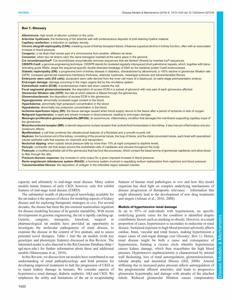
The functional unit of the kidney is the nephron (see Glossary,Box 1), which is closely integrated with the renal blood supply(Fig. 1). The human kidney filters 180 liters of plasma through itsglomeruli, and produces 1 to 2 liters of urine daily. Approximately99% of filtered sodium is retrieved as it passes through varioussections of the nephron before reaching the collecting duct.
Acute kidney injury (AKI) occurs when there is a rapid decline inglomerular filtration rate (GFR; see Glossary, Box 1), usuallyaccompanied by impaired microcirculation, inflammation and/ortubular injury or necrosis and reduced renal blood flow (Basile et al.,2012). AKI is initiated by various clinical insults, includinghypotensive shock, sepsis, surgery or the administration ofnephrotoxic agents such as cisplatin (Tanaka et al., 2005) andcontrast agents (commonly used for medical imaging) (Mehran andNikolsky, 2006). Following mild kidney injury, an adaptive repairresponse might ensue, leading to kidney regeneration. However,with more severe injury, regeneration is incomplete and nephronmass can be replaced by scar tissue, leading to CKD (Bucaloiu et al.,2012; Chawla et al., 2011). There are limited treatment optionsavailable for AKI, and its associated mortality remains high(Ferenbach and Bonventre, 2015). AKI can be induced in rats byperforming ischemia-reperfusion surgery or by administering toxinssuch as cisplatin. However, these single insults are unlikely to fullyrecapitulate the multiple injurious processes that have typicallyoccurred in individuals with AKI.
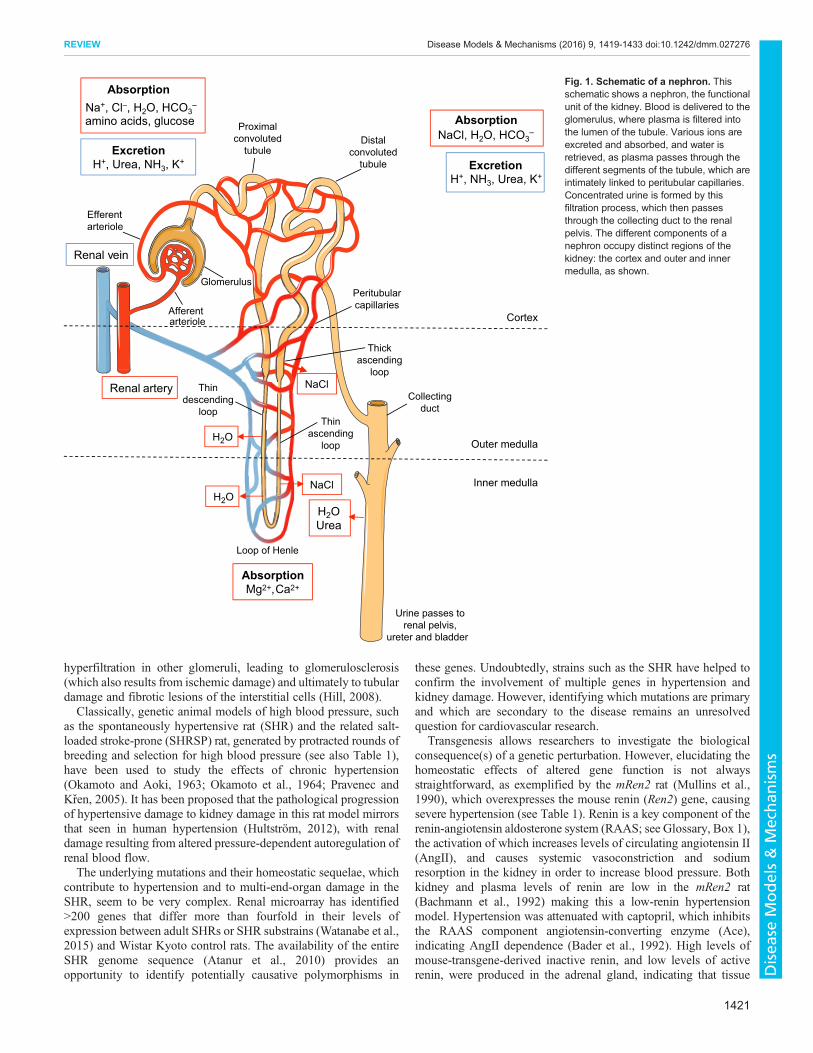
CKD is an umbrella term for any renal disease that results in theprogressive loss of kidney function over time. The kidney possessesonly a limited capacity for regeneration, and repeated or sustainedinjury to the kidney results in maladaptive responses (Ferenbach andBonventre, 2015), including the deposition of excess extracellularmatrix (ECM; see Glossary, Box 1), particularly collagen, inthe glomerulus and tubulointerstitium of the kidney (Fig. 2).The pathological changes associated with CKD includeglomerulosclerosis and tubulointerstitial fibrosis (see Glossary,Box 1), which result in the loss of normal renal architecture,microvascular capillary rarefaction (see Glossary, Box 1), hypoxiaand tubular atrophy. These changes lead to the loss of renal filtrative
University of Edinburgh/British Heart Foundation Centre for CardiovascularScience, Queen’s Medical Research Institute, 47 Little France Crescent, EdinburghEH16 4TJ, UK.
*Author for correspondence ([email protected])
L.J.M., 0000-0002-6743-8707; J.J.M., 0000-0001-5745-5258
This is an Open Access article distributed under the terms of the Creative Commons AttributionLicense (http://creativecommons.org/licenses/by/3.0), which permits unrestricted use,distribution and reproduction in any medium provided that the original work is properly attributed.
1419
© 2016. Published by The Company of Biologists Ltd | Disease Models & Mechanisms (2016) 9, 1419-1433 doi:10.1242/dmm.027276
Disea
seModels&Mechan
isms
capacity and ultimately to end-stage renal disease. Many rodentmodels mimic features of early CKD; however, only few exhibitfeatures of end-stage renal disease (ESRD).The substantial wealth of physiological knowledge available for
the rat makes it the species of choice for modeling aspects of kidneydisease and for exploring therapeutic strategies in vivo. For severaldecades, the mouse has been the pre-eminent mammalian organismfor disease modeling because of its genetic tractability. With recentdevelopments in genome engineering, the rat is rapidly catching up.Genetic, congenic, transgenic, knockout, surgical orpharmacological rat models have provided an opportunity toinvestigate the molecular pathogenesis of renal disease, toexamine the disease in the context of live animals, and to assesspotential novel therapies. Table 1 lists the rat models (with keygenotypic and phenotypic features) discussed in this Review. Theinterested reader is also directed to the Rat Genome Database (http://rgd.mcw.edu/) for further information about these and additionalmodels (Shimoyama et al., 2016).In this Review, we discuss how rat models have contributed to our
understanding of renal pathophysiology and hold promise fordeveloping improved treatments to halt the progression of CKD orto repair kidney damage in humans. We consider aspects ofhypertensive renal damage, diabetic nephritis, AKI and CKD. Weemphasize the utility and limitations of the rat in recapitulating
features of human renal pathologies in vivo and how this modelorganism has shed light on complex underlying mechanisms ofdisease progression of therapeutic relevance – information thatmight ultimately lead to the development of new drug treatmentsand targets (Aitman et al., 2016, 2008).
Models of hypertensive renal damageIn up to 95% of individuals with hypertension, no specificunderlying genetic cause for the condition is identified despitecontributory factors such as smoking or obesity. However, in a smallproportion of cases, hypertension is secondary to endocrine or renaldisease. Sustained exposure to high blood pressure adversely affectscardiac, brain, vascular and renal tissues, making hypertension amajor cause of end-organ damage (see Glossary; Box 1). Hence,renal disease might be both a cause and consequence ofhypertension, forming a vicious circle whereby hypertensioncauses kidney damage, which then exacerbates the high bloodpressure. Hypertensive nephrosclerosis is characterized by arterialwall thickening, loss of renal autoregulation, glomerulosclerosis,tubular atrophy and interstitial fibrosis (Hill, 2008). Arterialstiffening due to increased pulse pressure affects autoregulation ofthe preglomerular afferent arterioles, and leads to progressiveglomerular hypertrophy and damage with atrophy of the attachedtubule. Reduced glomerular filtration causes compensatory
Box 1. Glossary
Albuminuria: high levels of albumin (protein) in the urine.Arteriolar hyalinosis: the thickening of the arteriole wall with proteinaceous deposits of pink-staining hyaline material.Capillary rarefaction: a reduction in capillary density.Chronic allograft nephropathy (CAN): a leading cause of kidney transplant failure; it features a gradual decline in kidney function, often with an associatedincrease in blood pressure.Congenic: a rat strain that carries part of a chromosome from another, different rat strain.Consomic: when two rat strains carry the same transgene inserted at the same place in the genome.Cre recombinase/loxP: Cre recombinase enzymatically removes sequences that are flanked (floxed) by inserted loxP sequences.CRISPR-Cas9: a genome-engineering technique. CRISPR stands for clustered regularly interspaced short palindromic repeats, which, together with trans-activating guide RNAs, target the sequence-specific double-stranded breakage of DNA by the bacterial protein Cas9 endonuclease.Diabetic nephropathy (DN): a progressive form of kidney disease in diabetics, characterized by albuminuria, a >50% decline in glomerular filtration rate(GFR), increased glomerular basement-membrane thickness, arteriolar hyalinosis, mesangial sclerosis and tubulointerstitial fibrosis.Embryonic stem cells (ES cells): pluripotent stem cells derived from the inner cell mass of a blastocyst, an early-stage preimplantation embryo.End-organ damage: damage occurring in the major organs fed by the circulatory system.Extracellular matrix (ECM): a proteinaceous matrix laid down outside the cell.Focal segmental glomerulosclerosis: the deposition of excess ECM in a subset of glomeruli with only part of each glomerulus affected.Glomerular filtration rate (GFR): the rate at which plasma is filtered through the glomerulus.Glomerulosclerosis: the deposition of excess ECM in the glomerulus.Hyperglycemia: abnormally increased sugar content in the blood.Hyperkalemia: abnormally high potassium concentration in the blood.Hypokalemia: abnormally low potassium concentration in the blood.Ischemia-reperfusion injury (IRI): the tissue damage caused when blood supply returns to the tissue after a period of ischemia or lack of oxygen.Malignant hypertension: a rapid and severe increase in blood pressure, leading to end-organ damage.Mesangio-proliferative glomerulonephritis (MPGN): an autoimmune, inflammatory condition that damages the membrane supporting capillary loops ofthe glomerulus.Mineralocorticoid receptor (MR): a steroid-responsive nuclear receptor that controls fluid homeostasis in the kidney; it also has pro-inflammatory and pro-proteinuric effects.Myofibroblast: a cell that combines the ultrastructural features of a fibroblast and a smooth-muscle cell.Nephron: the functional unit of the kidney, consisting of the proximal tubule, the loop of Henle, and the distal convoluted tubule, each lined with specializedtubular epithelial cells that express ion channels and transporters.Nocturnal dipping: when systolic blood pressure falls by more than 10% at night compared to daytime levels.Pericyte: contractile cell that wraps around the endothelial cells of capillaries and venules throughout the body.Podocyte: a modified epithelial cell of the glomerulus that has foot-like processes, which contact the basal lamina of glomerular capillaries and allow bloodto filter through the slits.Pressure-diuresis response: the increase in urine output for a given imposed increase in blood pressure.Renin-angiotensin aldosterone system (RAAS): a hormone system involved in regulating sodium reabsorption from nephrons and blood pressure.Tubulointerstitial fibrosis: the deposition of collagen in the interstitial region between tubules.
1420
REVIEW Disease Models & Mechanisms (2016) 9, 1419-1433 doi:10.1242/dmm.027276
Disea
seModels&Mechan
isms
hyperfiltration in other glomeruli, leading to glomerulosclerosis(which also results from ischemic damage) and ultimately to tubulardamage and fibrotic lesions of the interstitial cells (Hill, 2008).Classically, genetic animal models of high blood pressure, such
as the spontaneously hypertensive rat (SHR) and the related salt-loaded stroke-prone (SHRSP) rat, generated by protracted rounds ofbreeding and selection for high blood pressure (see also Table 1),have been used to study the effects of chronic hypertension(Okamoto and Aoki, 1963; Okamoto et al., 1964; Pravenec andKren, 2005). It has been proposed that the pathological progressionof hypertensive damage to kidney damage in this rat model mirrorsthat seen in human hypertension (Hultström, 2012), with renaldamage resulting from altered pressure-dependent autoregulation ofrenal blood flow.The underlying mutations and their homeostatic sequelae, which
contribute to hypertension and to multi-end-organ damage in theSHR, seem to be very complex. Renal microarray has identified>200 genes that differ more than fourfold in their levels ofexpression between adult SHRs or SHR substrains (Watanabe et al.,2015) and Wistar Kyoto control rats. The availability of the entireSHR genome sequence (Atanur et al., 2010) provides anopportunity to identify potentially causative polymorphisms in
these genes. Undoubtedly, strains such as the SHR have helped toconfirm the involvement of multiple genes in hypertension andkidney damage. However, identifying which mutations are primaryand which are secondary to the disease remains an unresolvedquestion for cardiovascular research.
Transgenesis allows researchers to investigate the biologicalconsequence(s) of a genetic perturbation. However, elucidating thehomeostatic effects of altered gene function is not alwaysstraightforward, as exemplified by the mRen2 rat (Mullins et al.,1990), which overexpresses the mouse renin (Ren2) gene, causingsevere hypertension (see Table 1). Renin is a key component of therenin-angiotensin aldosterone system (RAAS; see Glossary, Box 1),the activation of which increases levels of circulating angiotensin II(AngII), and causes systemic vasoconstriction and sodiumresorption in the kidney in order to increase blood pressure. Bothkidney and plasma levels of renin are low in the mRen2 rat(Bachmann et al., 1992) making this a low-renin hypertensionmodel. Hypertension was attenuated with captopril, which inhibitsthe RAAS component angiotensin-converting enzyme (Ace),indicating AngII dependence (Bader et al., 1992). High levels ofmouse-transgene-derived inactive renin, and low levels of activerenin, were produced in the adrenal gland, indicating that tissue
Proximalconvoluted
tubule
Thin ascending loop
Afferent arteriole
Efferentarteriole
Thindescending
loop
Loop of Henle
Thinascending
loop
Thickascending
loop
Distalconvoluted
tubule
Collectingduct
Cortex
Outer medulla
Inner medulla
H2O
H2OUrea
Absorption Na+, Cl–, H2O, HCO3
– amino acids, glucose
glucose
Peritubularcapillaries
Excretion H+, Urea, NH3, K+
Absorption NaCl, H2O, HCO3
–
Excretion H+, NH3, Urea, K+
Urine passes torenal pelvis,
ureter and bladder
NaCl
NaCl H2O
Absorption Mg2+, Ca2+
Renal vein
Renal artery
Glomerulus
Fig. 1. Schematic of a nephron. Thisschematic shows a nephron, the functionalunit of the kidney. Blood is delivered to theglomerulus, where plasma is filtered intothe lumen of the tubule. Various ions areexcreted and absorbed, and water isretrieved, as plasma passes through thedifferent segments of the tubule, which areintimately linked to peritubular capillaries.Concentrated urine is formed by thisfiltration process, which then passesthrough the collecting duct to the renalpelvis. The different components of anephron occupy distinct regions of thekidney: the cortex and outer and innermedulla, as shown.
1421
REVIEW Disease Models & Mechanisms (2016) 9, 1419-1433 doi:10.1242/dmm.027276
Disea
seModels&Mechan
isms
RAAS is responsible for hypertension in this model (Peters et al.,1993). The crossing of the renin transgene onto a closely relatedoutbred Sprague Dawley strain generated animals that developedmalignant hypertension and end-organ damage by 8 weeks of age(see Glossary, Box 1) (Whitworth et al., 1994). In particular, thekidney exhibited glomerulosclerosis and interstitial fibrotic lesions.When the mRen2 transgene was crossed onto the inbred Fischer(F344) and Lewis rat strains, the resulting consomic strains (seeGlossary, Box 1) were susceptible and resistant to malignanthypertension, respectively. Genome-wide screening andquantitative trait analysis identified two modifier loci onchromosomes 10 and 17, which contributed to malignanthypertension susceptibility (Kantachuvesiri et al., 1999). ThemRen2 rat strains have been studied extensively for over 25 years,under both hypertensive and hyperglycemic conditions.In a more refined model, the Cyp1a1Ren2 rat (Kantachuvesiri
et al., 2001), expression of themRen2 gene is under the control of aninducible promoter in the inbred Fischer strain. This allows the
researcher to control the degree of AngII-dependent hypertensionand consequent end-organ damage, its speed of attainment and,also, to look at repair processes, once the inducer (indole-3-carbinol; I-3-C) is withdrawn (see ‘Models of diabetic nephropathy’below). The earliest hypertension-induced renal injury identified inthe Cyp1a1Ren2.Fischer strain is limited to the preglomerularvasculature (Ashek et al., 2012). The later-onset hypertensivekidney damage includes arterial wall thickening,glomerulosclerosis, interstitial fibrosis and tubular injury(Kantachuvesiri et al., 2001) similar to the renal damage causedby hypertension in humans. Increases in urinary albumin andangiotensinogen were observed with malignant hypertension(Milani et al., 2010), although the latter did not reflect changes inangiotensinogen gene expression in the kidney cortex (Prieto et al.,2011). Proteinuria was alleviated in this model by antagonism ofthe mineralocorticoid receptor (MR; see Glossary, Box 1) withspironolactone (Ortiz et al., 2007). After the transient inductionof hypertension, Cyp1a1Ren2 rats developed salt-sensitive
Reduced glomerular filtration Reduced renal perfusion Loss of podocytes Perivascular fibrosis Glomerulosclerosis#
Hypertension Diabetes Glomerulonephritis
Normal healthy cortical tubular
epithelium
Basement membrane
Peritubular capillary
Hypoxia Pro-fibrotic signals, e.g. TGFβ Pro-inflammatory signals, e.g. IL-6
Chronic injury
Flattened tubular epithelium. Some cell- cycle-arrested cells. Atrophy of tubules
Fibroblast activation and recruitment
ECM production Inflammatory cell infiltrate
Tubulointerstitial fibrosis*
Injured activated endothelium Increased apoptosis Eventual capillary rarefaction Increased hypoxia
*
#
A
B
C D
Fig. 2. The pathophysiological processes linked to kidney disease. (A) A normal, healthy kidney (left), and amagnified view of the structure of a tubule and itsassociated vasculature (right). (B) A chronically diseased kidney, showing the processes that lead to tubulointerstitial fibrosis. (C,D) Histological sections ofan adult rat kidney, stained with Masson’s trichrome (20× magnification; scale bars: 50 µm). (C) The glomerular and tubular architecture of a normal adult ratkidney, and (D) glomerulosclerosis (#) and tubulointerstitial fibrosis (*) in a 12-month-old hydroxysteroid dehydrogenase 2 (Hsd11b2)-knockout rat exhibitingend-stage renal disease.
1422
REVIEW Disease Models & Mechanisms (2016) 9, 1419-1433 doi:10.1242/dmm.027276
Disea
seModels&Mechan
isms
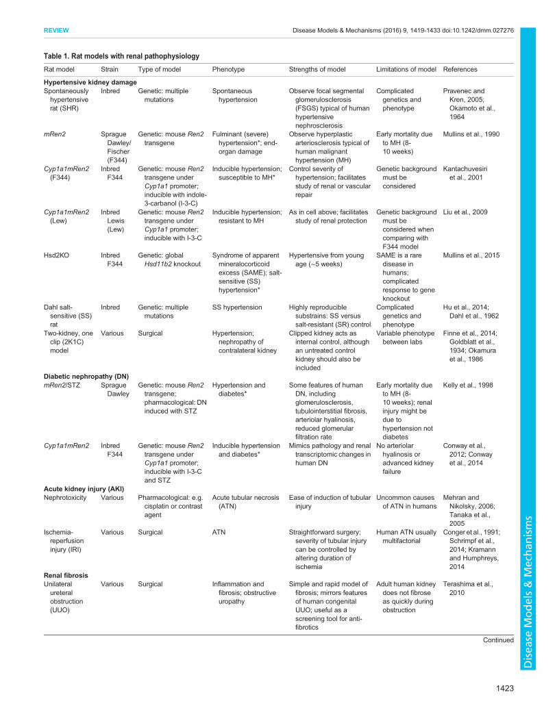
Table 1. Rat models with renal pathophysiology
Rat model Strain Type of model Phenotype Strengths of model Limitations of model References
Hypertensive kidney damageSpontaneouslyhypertensiverat (SHR)
Inbred Genetic: multiplemutations
Spontaneoushypertension
Observe focal segmentalglomerulosclerosis(FSGS) typical of humanhypertensivenephrosclerosis
Complicatedgenetics andphenotype
Pravenec andKren, 2005;Okamoto et al.,1964
mRen2 SpragueDawley/Fischer(F344)
Genetic: mouse Ren2transgene
Fulminant (severe)hypertension*; end-organ damage
Observe hyperplasticarteriosclerosis typical ofhuman malignanthypertension (MH)
Early mortality dueto MH (8-10 weeks)
Mullins et al., 1990
Cyp1a1mRen2(F344)
InbredF344
Genetic: mouse Ren2transgene underCyp1a1 promoter;inducible with indole-3-carbanol (I-3-C)
Inducible hypertension;susceptible to MH*
Control severity ofhypertension; facilitatesstudy of renal or vascularrepair
Genetic backgroundmust beconsidered
Kantachuvesiriet al., 2001
Cyp1a1mRen2(Lew)
InbredLewis(Lew)
Genetic: mouse Ren2transgene underCyp1a1 promoter;inducible with I-3-C
Inducible hypertension;resistant to MH
As in cell above; facilitatesstudy of renal protection
Genetic backgroundmust beconsidered whencomparing withF344 model
Liu et al., 2009
Hsd2KO InbredF344
Genetic: globalHsd11b2 knockout
Syndrome of apparentmineralocorticoidexcess (SAME); salt-sensitive (SS)hypertension*
Hypertensive from youngage (∼5 weeks)
SAME is a raredisease inhumans;complicatedresponse to geneknockout
Mullins et al., 2015
Dahl salt-sensitive (SS)rat
Inbred Genetic: multiplemutations
SS hypertension Highly reproduciblesubstrains: SS versussalt-resistant (SR) control
Complicatedgenetics andphenotype
Hu et al., 2014;Dahl et al., 1962
Two-kidney, oneclip (2K1C)model
Various Surgical Hypertension;nephropathy ofcontralateral kidney
Clipped kidney acts asinternal control, althoughan untreated controlkidney should also beincluded
Variable phenotypebetween labs
Finne et al., 2014;Goldblatt et al.,1934; Okamuraet al., 1986
Diabetic nephropathy (DN)mRen2/STZ Sprague
DawleyGenetic: mouse Ren2transgene;pharmacological: DNinduced with STZ
Hypertension anddiabetes*
Some features of humanDN, includingglomerulosclerosis,tubulointerstitial fibrosis,arteriolar hyalinosis,reduced glomerularfiltration rate
Early mortality dueto MH (8-10 weeks); renalinjury might bedue tohypertension notdiabetes
Kelly et al., 1998
Cyp1a1mRen2 InbredF344
Genetic: mouse Ren2transgene underCyp1a1 promoter;inducible with I-3-Cand STZ
Inducible hypertensionand diabetes*
Mimics pathology and renaltranscriptomic changes inhuman DN
No arteriolarhyalinosis oradvanced kidneyfailure
Conway et al.,2012; Conwayet al., 2014
Acute kidney injury (AKI)Nephrotoxicity Various Pharmacological: e.g.
cisplatin or contrastagent
Acute tubular necrosis(ATN)
Ease of induction of tubularinjury
Uncommon causesof ATN in humans
Mehran andNikolsky, 2006;Tanaka et al.,2005
Ischemia-reperfusioninjury (IRI)
Various Surgical ATN Straightforward surgery;severity of tubular injurycan be controlled byaltering duration ofischemia
Human ATN usuallymultifactorial
Conger et al., 1991;Schrimpf et al.,2014; Kramannand Humphreys,2014
Renal fibrosisUnilateralureteralobstruction(UUO)
Various Surgical Inflammation andfibrosis; obstructiveuropathy
Simple and rapid model offibrosis; mirrors featuresof human congenitalUUO; useful as ascreening tool for anti-fibrotics
Adult human kidneydoes not fibroseas quickly duringobstruction
Terashima et al.,2010
Continued
1423
REVIEW Disease Models & Mechanisms (2016) 9, 1419-1433 doi:10.1242/dmm.027276
Disea
seModels&Mechan
isms

hypertension, which could be attenuated by the superoxide dismutasemimetic tempol, implicating the superoxide anion in the developmentof salt-sensitive hypertension (Howard et al., 2005).The Cyp1a1Ren2 transgene is carried on the Y chromosome and,
by crossing the inducible Fischer male to a Lewis female, followedby selective backcrossing of the F1 progeny to Lewis or Fischeranimals, congenic lines (see Glossary, Box 1) were derived. Theselines retain the transgene and either susceptibility or resistance toend-organ damage, on an otherwise resistant or susceptiblebackground (Kantachuvesiri et al., 1999). Whole-renal,microarray-based, gene-expression profiling studies of theparental and congenic strains revealed genes in the congenicregion that were differentially expressed between the parental andcongenic strains (Liu et al., 2009). This strategy identifiedangiotensin-converting enzyme Ace as a principal modifier ofhypertension-induced microvascular renal injury in theCyp1a1Ren2 rat model (Liu et al., 2009). The C-domain of Ace isthought to mediate blood pressure control through its action onangiotensin I. However, it is now recognized that Ace has othereffects, such as cleavage of the naturally occurring tetra-peptideacetyl-N-Ser-Asp-Lys-Pro (AcSDKP) by the N-terminal domain ofAce (Bernstein et al., 2011). AcSDKP has been shown to reverseinflammation, cell proliferation and fibrosis in rat models ofhypertension (Liu et al., 2009; Zuo et al., 2013). As predicted,AcSDKP was present at significantly lower levels in the kidneys ofthe injury-susceptible Fischer rat than in the kidneys of the moreprotected Lewis rat (Liu et al., 2009).Microarray-based gene-expression profiling of the congenic
Fischer and Lewis kidneys was further used to identify previouslyunknown candidate genes that might associate with a susceptibility
to kidney injury (Menzies et al., 2013). A bioinformatic enrichmentanalysis identified multiple candidate genes in addition to Ace. Thesecond- and third-ranked susceptibility genes were the purinereceptors P2X7 and P2X4 (Menzies et al., 2013). There are sevenP2X receptors in the rat, as in humans. These adenosine-5′-triphosphate-activated cation channels are part of the largermammalian purine receptor family, which includes G-proteincoupled P2Y receptors and adenosine P1 receptors (Ralevic andBurnstock, 1998). Both P2X and P2Y purine receptors have beenimplicated in preclinical rodent models of hypertension (Menzieset al., 2015b) and kidney disease (Menzies et al., 2016; Ralevic andBurnstock, 1998). In humans, genetic variation that causes thefunctional impairment of P2X7 is associated with a reduced risk ofstroke (Gidlöf et al., 2012). Conversely, P2X4 loss of function isassociated with increased pulse pressure (Stokes et al., 2011). Therenal pressure-diuresis response (see Glossary, Box 1) of Fischer,but not of Lewis, rats was improved with combined P2X7 and P2X4receptor antagonism using the dye, Brilliant Blue G (BBG)(Menzies et al., 2013). Renal vascular resistance was unaffectedby BBG in Lewis rats, but both blood pressure and vascularresistance decreased in Fischer rats, suggesting that P2X7 mightsupport tonic vasoconstriction in the susceptible strain. SpecificP2X7 receptor antagonism using the compound AZ11657312caused rapid vasodilation. Acute antagonism of the receptor P2X7in Fischer rats, chronically infused with AngII, significantlyimproved renal perfusion and tissue oxygenation (Menzies et al.,2015a). Recently, P2X7 receptor antagonism has also been shownto attenuate renal injury in Dahl salt-sensitive rats (Ji et al., 2012).
P2X7 has been implicated in a wide range of neurological,inflammatory and musculoskeletal disorders, in addition to its role
Table 1. Continued
Rat model Strain Type of model Phenotype Strengths of model Limitations of model References
Chronic kidney disease (CKD)Humandiphtheria toxinreceptor(hDTR)
InbredF344
Genetic: humandiphtheria toxintransgene
Podocyte loss; focalsegmentalglomerulosclerosis(FSGS)
Develops nephrotic rangeproteinuria, podocyteloss, FSGS
Artificial mechanismof injury: podocyteloss rapid andsimultaneous
Wharram et al.,2005
AA-4E-BP1 InbredF344
Genetic: AA-4E-BP1‡
transgene driven bypodicin promoter
Mechanical failure ofpodocytes;proteinuria; FSGS
– Artificial mechanismof injury
Fukuda et al.,2012a
PassiveHeymannnephritis(PHN)
SpragueDawley
Pharmacological: anti-Fx1A antibody
PHN; membraneousnephropathy
Develop immune depositsand proteinuria
Antibody in humandisease is directedagainstphospholipase A2receptor
Salant et al., 1979
Anti-Thy 1.1 Various Pharmacological: IgAnephropathy
Mesangio-proliferativeglomerulonephritis(MPGN)
Has several features of thehuman clinical pathology,e.g. mesangialproliferation, glomerularECM deposition
Self-limiting diseasecourse in rat,limited tubularinvolvement andminimal renalfunctional change
Nazeer et al., 2009;Denby et al.,2011
5/6thnephrectomy
Various Surgical Reduced nephronnumber; reducedglomerular filtrationrate (GFR)
Can exhibit progressivedecline in renal function(strain specific) andincrease in bloodpressure
Difficult surgery;high mortality
Gilbert et al., 2012
Nephrotoxicnephritis (NTN)
Various Pharmacological:nephrotoxic globulin
Immune-complex-mediated glomerularnephritis; proteinuria;P2RX7 increase
Develops proteinuria andsome histopathologicalchanges that areobserved in humandisease
Batch-to-batchvariation indisease severity
Turner et al., 2007;Taylor et al.,2009
*UK Home Office regulations for animal research do not allow end-stage renal failure (ESRF) or malignant hypertension (MH) as end point of experiment.‡AA-4E-BP1, eukaryotic translation initiation factor binding protein 1 (EIF4EBP1), a member of the mammalian target of rapamycin complex 1 pathway.STZ, streptozotocin.
1424
REVIEW Disease Models & Mechanisms (2016) 9, 1419-1433 doi:10.1242/dmm.027276
Disea
seModels&Mechan
isms
in hypertension and renal disease. Clinical trials of P2X7antagonists in the treatment of inflammatory diseases have shownlimited therapeutic benefit to date (Bartlett et al., 2014). Given thelarge number of splice variants (Cheewatrakoolpong et al., 2005)and disease-related single-nucleotide polymorphisms (SNPs) (Jianget al., 2013) in the human P2RX7 gene, a productive future researchstrategy could be the selective humanization of rats to developtissue-specific or disease-relevant therapeutic strategies.In the two-kidney, one clip (2K1C) hypertensive system
(Goldblatt et al., 1934), which has been implemented in rats, aclip on the left renal artery activates the RAAS system. Althoughboth kidneys are exposed to an equivalent increase in AngII, onlythe non-clipped rat kidney shows hypertensive damage (Cervenkaet al., 1999). Recently, the non-clipped kidney was found to haveincreased mRNA, protein and urinary levels of angiotensinogen,suggesting that kidney damage occurs through increased AngII, andthat angiotensinogen could be used as an early biomarker of kidneydamage (Shao et al., 2016). Exposure of the non-clipped kidney toincreased AngII was ameliorated by nitric oxide (NO) release,suggesting that this is a protective mechanism (Helle et al., 2008).Additional early hypertension-induced changes in the renal tubuleswere identified by micro-dissection of visibly undamagedtubulointerstitial tissue from the non-clipped kidney. Proteomicanalysis using mass spectrometry revealed the differentialexpression of over 300 proteins compared to control samples,with profibrotic Rho-signaling proteins being the most highlyoverrepresented (Finne et al., 2016). Such studies should help toidentify additional biomarkers of early tubule damage, which intime could be used diagnostically. It should be noted, however, thatthe clipped kidney is not physiologically equivalent to an untreated(sham) control kidney; thus, the latter should always be included asa control when comparing clipped and non-clipped kidneys (Palmet al., 2008, 2010).Despite complexities of the SHR, SHRSP and 2K1C hypertension
models, a recent gene-expression profiling study revealed a commonprogression in hypertensive renal damage (Skogstrand et al., 2015).Of the 88 genes similarly regulated in all three models, 40 were alsoidentified in gene-expression profiles from human fibrotic kidneys.This suggests that pathogenic pathways underlying kidney damageare conserved between rats and humans.
Hypertensive models generated by genetic modificationGene-knockout technology has only recently become available forthe rat with the isolation of rat embryonic stem (ES) cells (seeGlossary, Box 1) (Buehr et al., 2008; Li et al., 2008), which can beused as a tool for gene modification. The genetic tractability of therat has also been greatly facilitated by genome-engineeringtechnologies, such as zinc-finger nucleases (ZFNs) (Geurts et al.,2009), transcription activator-like effector nucleases (TALENs)(Tesson et al., 2011) and the CRISPR-Cas9 system (see Glossary,Box 1) (Li et al., 2013). Genome endonuclease technologiesgenerate a sequence-specific DNA double-strand break, which isrepaired by error-prone, non-homologous end-joining. Anyinsertions or deletions introduced at the target site cause missenseor nonsense mutations. The PhysGen knockout program (http://pga.mcw.edu/) has utilized these technologies to generate a wide varietyof knockout rat models in genes associated with cardiovascular orrenal disease. One of the earliest ZFN-knockout rat modelsgenerated with a clear renal phenotype was the hypotensive renin-knockout rat (Moreno et al., 2011). Disruption of the renin genecaused profound disruption to normal kidney development. Theinner renal medulla was morphologically rudimentary and there
were signs of cortical interstitial fibrosis. These changes could berelated to the concomitant reduction in AngII production, andsupport the assertion that the RAAS is essential for normal kidneydevelopment in mammals (Guron and Friberg, 2000).
Another rat knockout model that exhibits reduced renin levels isthe Hsd2KO rat (Mullins et al., 2015). The enzyme 11-β-hydroxysteroid dehydrogenase type 2 (Hsd11b2) protects the MRfrom inappropriate activation by cortisol (corticosterone), in thekidney principal cell, by inactivating it to cortisone (11-dehydrocorticosterone). In this model, ZFN-induced knockout ofthe Hsd11b2 gene causes inappropriate activation of the MR,leading to salt-sensitive hypertension, suppression of reninsecretion, and hypokalemia (see Glossary, Box 1). Thisphenotype closely models the human syndrome of apparentmineralocorticoid excess (SAME). The rats exhibit severe renalinjury, including protein casts and atrophic tubules, segmentalglomerulosclerosis, tubule-interstitial fibrosis and proteinuria(Mullins et al., 2015). These are all features associated withchronic exposure to hypertension and with MR activation seen inhuman kidney disease (Ueda and Nagase, 2014). Interestingly, theHsd2KO rat model demonstrates metabolic protection, includingincreased insulin sensitivity and reduced mesenteric fataccumulation, due to the depletion of the substrate for Hsd11b1 inadipose tissue. This suggests that treatment with MR inhibitorsmight reverse the adverse cardiovascular effects of SAME (whichinclude hypokalemia, hypertension, proteinuria and end-organdamage), while promoting the beneficial metabolic effects ofHsd11b2 inactivation (Mullins et al., 2015).
Salt-sensitive hypertension involves a complex feedback loop ofsalt appetite and sodium retention. Hsd11b2 in the murine braintriggers a central drive to consume salt (Evans et al., 2016). The ratHsd2KO model offers a more robust platform to investigate thephysiological mechanisms of central versus renal-centric saltsensitivity than is feasible in the mouse. Decreasing dietary saltconsumption might reduce the burden of CKD in humans(McMahon et al., 2013). Intriguingly, an alternative, albeit moreinvasive, strategy to ameliorate salt-sensitive hypertension has beenrecently demonstrated. Renal medullary dysfunction in salt-sensitive Dahl rats (Dahl et al., 1962) was found to reflect areduction in adult (CD133+) mesenchymal stem cells (MSCs) in themedulla. Injection of MSCs, but not of renal medullary interstitialcells, into the renal medulla attenuated immune-cell infiltration andsodium retention, and reduced systemic blood pressure (Hu et al.,2014). The rationale for using MSCs stems from numerous animalstudies, which have demonstrated that these cells have protectiveeffects in acute and chronic kidney injury models (Fleig andHumphreys, 2014; Wang et al., 2013).
The co-injection of single-strand oligonucleotides with ZFNs,TALENs or CRISPR-Cas9 components can be used to introducetargeted SNPs or to repair mutations, through homology-drivenrepair (HDR). Rapid improvements in CRISPR-Cas9 technology,using donor plasmids as HDR templates, have included theintroduction of fluorescent reporters (Ma et al., 2014a), the one-step generation of a floxed allele (loxP sites flanking an exon) (Maet al., 2014b) and conditional knockout using Cre-recombinase ratstrains (see Glossary, Box 1) (Ma et al., 2014a). Recently, Wistar-Kyoto rats and SHRs that ubiquitously express GFP have beenproduced, using the Sleeping Beauty transposon system. Thesestrains will prove useful for investigating cell fate andtransplantation in the hypertensive kidney (Garcia Diaz et al., 2016).
The identification of genes such as Ace, P2rx7 and Hsd11b2, orspecific genetic variants or splice variants of genes, that seem to
1425
REVIEW Disease Models & Mechanisms (2016) 9, 1419-1433 doi:10.1242/dmm.027276
Disea
seModels&Mechan
isms
play key roles in moderating hypertensive damage, renal pathologyand salt-sensitivity has the potential to enable future identificationof individuals at risk of hypertensive kidney damage based on theirgenetic profile. With the availability of humanized transgenicmodels, Cre-loxP technology, reporter strains, gene knockouts andknock-ins, and the ability to correct candidate genes in mutant ratstrains, many of the tools available to the mouse community are nowavailable in the rat. Although the inherent problem of off-targetevents remain for genome-engineering technologies, targeting in ratES cells and screening for clones free of off-target events remains apossibility. Thus, many more-refined and increasingly sophisticatedrat models, which more closely recapitulate human renal pathologycaused by hypertensive damage, can be expected in the future, andmight help to predict targeted therapeutic response more faithfully.
Models of diabetic nephropathyDiabetic nephropathy (DN; see Glossary, Box 1) is the single mostcommon cause of end-stage kidney disease in the western world(Saran et al., 2015). The use of reliable animal models of DN couldgreatly facilitate research by providing mechanistic insights into thisdisease to help identify novel therapeutic targets. These in turncould provide a platform for preclinical testing of such noveltherapies. Unfortunately, one of the roadblocks to DN research is thelack of preclinical models that recapitulate important functional,structural and molecular pathological features of progressive humandiabetic kidney disease. Although several rodent models of type 1diabetes [streptozotocin (STZ)-induced (Cooper et al., 1988)] andtype 2 diabetes [Zucker, Goto Kakizaki (Janssen et al., 2003)] havebeen employed to study DN (see Glossary, Box 1), these models failto recapitulate all of the hallmarks of this disease as defined bythe Diabetic Complications Consortium (DiaComp; https://www.diacomp.org/shared/validationcriteria.aspx). The inability of animalmodels to fully replicate human DN might explain why manytherapies that have been beneficial in preclinical models of thisdisease have proven to be ineffective in clinical trials. For example,direct renin inhibitors were beneficial in reducing proteinuria inrodent models (Kelly et al., 2007). However, the absence ofprogressive renal failure in these models meant that the efficacy ofthese inhibitors in reducing renal function could not be tested.Human studies confirmed a beneficial effect of direct renininhibitors on reducing proteinuria (Parving et al., 2008) but,importantly, they did not slow the rate of renal-function decline(Parving et al., 2012). Furthermore, the increased risk ofhyperkalemia (see Glossary, Box 1) resulting from treatment withdirect renin inhibitors in patients with impaired renal function(Parving et al., 2012) was not highlighted in the rodent models,where blood potassium levels remained normal.Although hyperglycemia (see Glossary, Box 1) is a pre-requisite
for the development of DN, hemodynamic factors play a substantialrole in the progression of this disease. Individuals with advancedDN invariably have hypertension, and tight control of bloodpressure is as important as glycemic control in slowing diseaseprogression (Mogensen, 1998). Hypertension might not only be aconsequence of nephropathy but a key driver of kidney disease indiabetes. Indeed, subtle abnormalities in blood pressure, such asloss of nocturnal dipping (see Glossary, Box 1), precede the onset ofalbuminuria (see Glossary, Box 1) in adolescents with type 1diabetes (Lurbe et al., 2002). Furthermore, there are two case reportsregarding individuals with longstanding diabetes, hypertension andunilateral renal artery stenosis (Berkman and Rifkin, 1973;Béroniade et al., 1987) whose conditions mimic the 2K1C ratmodel of hypertension. Autopsy findings in both cases revealed no
pathological evidence of nephropathy in the kidney downstream ofthe arterial stenosis, despite severe nephropathy in the contralateralkidney. The implications of these findings are that unilateral renalartery stenosis might prevent the transmission of systemichypertension to the kidney parenchyma and the subsequentdevelopment of nephropathy, even though both kidneys have beenexposed to an equivalent degree of hyperglycemia and to increasedAngII exposure. Thus, hyperglycemia or elevated angiotensin levelsalone are insufficient to promote advanced DN; the development ofhypertension is a prerequisite for disease progression. Howhypertension interacts with hyperglycemia to promotenephropathy is unclear, but the application of cyclical stretch tomesangial cells cultured in high-glucose media increases theexpression of pro-fibrotic genes, suggesting a role for increasedmechanical strain (Gruden et al., 2000). In rat mesangial cells grownin high-glucose media, ATP and a P2X7 agonist dose-dependentlyincreased ECM deposition and levels of transforming growth factorbeta (TGFβ; a pro-fibrotic cytokine), whereas P2X7 inhibitionattenuated the response (Solini et al., 2005), indicating theinvolvement of purinergic receptors.
Several approaches have been taken to recapitulate theseimportant hemodynamic factors in rodent models of DN. In the1980s, the Brenner group determined that a high-protein dietincreased intra-glomerular pressure and promoted glomerular injuryin diabetic rats and that these features could be successfullyprevented by Ace inhibition (Zatz et al., 1986, 1985). These seminalstudies led directly to clinical trials of ACE inhibitors in patientswith DN, and they represent one of the best examples of how rodentmodels can be utilized to provide important mechanistic insightsthat subsequently lead to therapeutic advances. Indeed, ACEinhibitors have since become the mainstay of preventing theprogression of renal disease in individuals with DN (Lewis et al.,1993). Conversely, many therapies that have been effective inanimal models of DN that targeted hyperglycemia alone haveproven unsuccessful in clinical trials (B.R.C., personalobservation).
Rat models of DNGenetic models of hypertension have also been utilized to modelprogressive DN. The induction of diabetes with STZ leads to higherlevels of albuminuria in SHRs than in rat strains with diabetes orhypertension alone (Cooper et al., 1988). Treatment with Aceinhibitors abrogates the increase in albuminuria in SHR strains.Activation of the RAAS plays a pre-eminent role in clinical DN.Therefore, a logical approach was to induce diabetes in mRen2 rats(Kelly et al., 1998). The renin-dependent hypertension in mRen2rats accelerates the development of nephropathy, and this model hasbeen used to study not only the role of the RAAS in DN, but alsothat of other pathways, including oxidative stress (Advani et al.,2009). It has been shown that sustained hyperglycemia causesincreased tubular oxygen consumption due to mitochondrialdysfunction and reduced electrolyte transport efficiency (reviewedin Hansell et al., 2013). The onset of malignant hypertension in themRen2 model results in accelerated renal injury and in earlymortality, which is atypical of the slowly progressive courseobserved in human diabetic kidney disease (Hartner et al., 2007).This problem was overcome by using Cyp1a1mRen2 rats, whereadjustment of I-3-C concentration in the diet controls the timing andseverity of hypertension. Following induction of diabetes usingSTZ, the addition of 0.125% I-3-C resulted in a gradual increase inblood pressure, mimicking the evolution of hypertension in humanDN (Conway et al., 2012). The hyperglycemia and hypertension
1426
REVIEW Disease Models & Mechanisms (2016) 9, 1419-1433 doi:10.1242/dmm.027276
Disea
seModels&Mechan
isms
synergized to promote a 500-fold increase in albuminuria, andcaused moderate glomerulosclerosis and tubulointerstitial fibrosis –all features of moderately advanced human DN. However, there wasno significant decline in renal function in this model, and some keypathological features of DN, such as arteriolar hyalinosis (seeGlossary, Box 1), were not observed.Microarray and RNA-sequencing technologies provide a non-
biased view of gene expression changes. Thus, comparingtranscriptomic changes in DN patients with rat models of thedisease might reveal common disease mechanisms, identifyrelevant biomarkers and therapeutic targets, and enable therational selection of the rodent model that most closelyrecapitulates changes seen in DN kidneys. Up to 50% of genesthat were differentially expressed in the tubulointerstitialcompartment of the kidney in human DN (Lindenmeyer et al.,2007) were also similarly up- or downregulated in the renal cortex ofhyperglycemic and hypertensive Cyp1a1mRen2 rats (Conway et al.,2012). For example, one downregulated gene in both the rat modeland in the kidneys of individuals with DN was epidermal growthfactor (EGF). Urinary EGF levels reflect renal EGF expression, andsubsequent studies confirmed that low levels of urinary EGFexcretion predict a poor renal outcome in individuals with DN andwith other CKDs (Betz et al., 2016; Ju et al., 2015). Hence, non-biased transcriptomic approaches could be used to identify as-yet-unknown prognostic biomarkers for therapeutic targets or to recruithigh-risk individuals for clinical trials. Such transcriptomic datasetsshould be made freely available on databases such as Geodataset(http://www.ncbi.nlm.nih.gov/gds/) or Nephroseq (https://www.nephroseq.org), as this will enable researchers to select the modelin which their pathway of interest is differentially activated in asimilar manner to human disease. Such ‘precision modeling’ couldimprove the chances of translating findings made in rodent modelsto the clinic.Although the natural history of DN is one of inexorable
progression towards end-stage kidney disease, the tight control ofblood glucose and blood pressure can lead to the regression ofalbuminuria in up to 50% of individuals with DN (Perkins et al.,2003). More remarkably, regression of establishedglomerulosclerosis and tubulointerstitial fibrosis has been observedin individuals with moderately advanced DN who achieve sustainednormoglycemia after receiving a pancreas transplant (Fioretto et al.,1998, 2006), although this takes up to 10 years to become evident.The pathways that promote regression remain poorly understood,largely because serial biopsies are rarely performed in individualswho are responding to treatment.Rodent models provide insights into mechanisms of injury,
regeneration and repair. The Cyp1a1mRen2 rat model of DN isparticularly useful in this regard because hypertension can beinduced and then blood pressure normalized by adding and thenremoving dietary I-3-C; inserting subcutaneous insulin implants canalso control STZ-induced hyperglycemia. In one study, 28 weeks ofhyperglycemia and hypertension (the injury phase) were followedby tight glycemic and blood pressure control for an additional 8weeks (the reversal phase), resulting in the partial regression ofalbuminuria (Conway et al., 2014). Microarray analysis of the renaltranscriptome during both the injury and reversal phases revealed∼650 genes that were upregulated during injury, almost 100 ofwhich reverted to control levels following reversal ofhyperglycemia and hypertension. This gene set was enriched forgenes that encoded ECM proteins, fibroblast markers and acute-phase reactants, indicating that the tight control of glucose andblood pressure might suffice to switch off the formation of new scar
tissue. This was supported by the finding that there was no furtherincrease in the severity of glomerulosclerosis or tubulointerstitialfibrosis during the 8-week reversal phase. In addition, many genesof unknown function, which reverted to control levels during repair,might be implicated in the fibrotic- or acute-phase response andhence they merit further investigation. Conversely, almost 400genes remained significantly upregulated despite the normalizationof blood glucose and blood pressure. This gene set was enriched forgenes that encoded proteins implicated in innate and adaptiveimmunity, in particular pro-resolution macrophages and regulatoryT cells, suggesting that attempts at repair have been initiated.Although glomerulosclerosis and tubulointerstitial fibrosis did notreduce during the reversal phase, this was to be expected given theprotracted period required for regression of fibrosis followingpancreas transplantation in humans (Fioretto et al., 2006).Permanent or long-term upregulation of some of these genesmight be responsible for the salt sensitivity observed in I-3-C-induced rats (Howard et al., 2005).
Bilateral renal denervation has emerged as a potential treatmentfor multiple-drug-resistant hypertension in individuals with bilateralsingle renal arteries, but results from recent clinical trials havequestioned its efficacy for individuals with secondary (or accessory)renal arteries (Bhatt et al., 2014; Hering et al., 2016; Khan et al.,2014). When bilateral renal denervation was performed in themRen2/STZ rat model, it reduced signs of renal pathology,albuminuria and the expression of fibrotic markers. This suggeststhat renal denervation might attenuate renal injury in DN (Yao et al.,2014), presumably with similar caveats regarding efficacy.
In summary, rat studies can mimic many of the features of humanDN, including progressive proteinuria, key pathological featuressuch as glomerulosclerosis and tubulointerstitial fibrosis, and theactivation of many pathways that are implicated in human DN.However, none fully recapitulate human DN, with few exhibitingarteriolar hyalinosis and a progressive decline in renal function. Ratmodels have highlighted the benefits of Ace inhibitors and theprognostic value of EGF in the treatment of DN. A comparison ofthe results from microarray and RNA-sequencing technologies inrodent models and human DN will continue to identify newcandidates for therapeutic interventions to prevent kidney damageor to enhance repair and regeneration.
Models of acute and chronic kidney diseaseAKI affects multiple cell types in the kidney, including endothelialand tubular cells, which are adversely affected by hypoxia. It is notclear whether hypoxia (the reduction of tissue oxygen supply tobelow physiological levels) or re-oxygenation (increased exposureto oxygen, as seen with reperfusion following ischemia) causesAKI, but it is associated with altered intra-renal microcirculationand oxygenation (Rosenberger et al., 2006). Ischemia-reperfusioninjury (IRI; see Glossary, Box 1) is extensively used as a model ofAKI, but hypoxic damage predominantly affects proximal tubulesegments in the outer stripe of the outer medulla and might notrecapitulate human AKI, which often includes medullary oxygeninsufficiency. Damage to the thick ascending limb is attenuatedfollowing IRI, probably because the reduced solute transport leadsto improved oxygenation of the distal tubule (Rosenberger et al.,2006). Following acute IRI, the vascular function of rats remainsimpaired for several days (Conger et al., 1991). The pericyte (seeGlossary, Box 1) detaches from the endothelium under pathologicalconditions, leading to microvascular rarefaction and hypoxia(Schrimpf et al., 2014). Pericytes might contribute to the pool ofscar-forming myofibroblasts (see Glossary, Box 1) (Kramann and
1427
REVIEW Disease Models & Mechanisms (2016) 9, 1419-1433 doi:10.1242/dmm.027276
Disea
seModels&Mechan
isms

Humphreys, 2014), making them key to both regeneration and thedevelopment of fibrosis (Schrimpf and Duffield, 2011), althoughmyofibroblasts can also arise from other sources (Falke et al., 2015;Micallef et al., 2012).Agents affecting both cortical and medullary blood flow and
oxygen tension include radio-contrast agents (Heyman et al., 1991),endotoxin [sepsis (Heyman et al., 2000)] and NO inhibitors (Breziset al., 1991). Together with non-steroidal anti-inflammatory drugs,which cause a selective reduction in medullary blood flow andtissue oxygenation, these could provide better models of AKI andcould enable investigation of hypoxia-inducible factors, adaptiveresponses and potential therapies (Rosenberger et al., 2006). Thedevelopment of rat models should enhance our understanding ofAKI and help to design therapeutic strategies to block maladaptiveresponses.Pre-existing CKD affects the severity of AKI in humans and their
recovery (Liangos et al., 2006). This has been experimentallymodeled in rats using the renal-mass-reduction model of CKD withan additional induced IRI. CKD develops in the 5/6th nephrectomyrat model (in which the 5/6th of renal mass is surgically ablated; seeTable 1). When AKI is induced in this model via IRI, adisproportionate number of regenerating tubules fail to re-differentiate. This is associated with significant loss of tubularVEGF expression and with substantial capillary rarefaction.Defective tubules also have pro-fibrotic properties that increasetubulointerstitial fibrosis (Polichnowski et al., 2014). Furtherinvestigation of this model will provide a greater understanding atthe molecular level of the AKI to CKD transition seen in humans.Reporter rats should prove invaluable for mechanistic studies and
for the identification of the molecular pathways and cell lineagesinvolved in kidney disease (Garcia Diaz et al., 2016). The creationof reporter transgenic rats has allowed the mapping of cells thatcontribute to renal fibrosis and the testing of novel anti-fibroticagents on key pro-fibrotic pathways (Terashima et al., 2010). Usingtransgenic rats carrying a luciferase reporter gene under the controlof rat α1(I) collagen and rat α2(II) collagen, the anti-fibrotic effectsof inhibiting TGFβ signaling (using a TGFβR1 inhibitor) and AngIIsignaling [using an AngII-receptor blocker (ARB), olmesartan]were examined (Terashima et al., 2010). This study revealed thatARBs had an anti-fibrotic effect, independent of hemodynamiceffects, in the unilateral ureteral obstruction (UUO) model of rapidrenal fibrosis (see Table 1), which induces a marked change in renalperfusion.Rat models of AKI and CKD have been used as a platform to test
potential new therapies, including novel anti-fibrotic agents. FT011is a derivative of the anti-allergy drug Tranilast (Miyazawa et al.,1995), and it inhibits the proliferative actions of TGFβ and platelet-derived growth factor (PDGF). FT011 stemmed the decline in GFRin the 5/6th nephrectomy model of progressive CKD (see Table 1)and reduced proteinuria and structural injury (Gilbert et al., 2012).In the diabetic, hypertensive mRen2/STZ model, FT011 markedlyattenuated the development of proteinuria, as well as reducingfibrosis in both the glomerulus and tubulointerstitium, andinterstitial macrophage infiltration, but GFR was unaffected(Gilbert et al., 2012).In a rat model of aristolochic-acid-induced nephropathy, the
neutralization of TGFβ with anti-TGFβ antibody improved renalfunction and reduced acute tubular necrosis, interstitialinflammation, vascular rarefaction and myofibroblastaccumulation (Pozdzik et al., 2016). The disruption of proximaltubule organelle ultrastructure was also prevented. However, thesefindings have not translated to the clinic; agents that block TGFβ and
retard CKD have failed to improve renal function despite thepromising preclinical results (Lee et al., 2015). These findings againsupport the observation that animal models typically recapitulateonly part of the human condition – particularly CKD and itsprogression to ESRD. Animal models such as the UUO rat, used as amodel of renal fibrosis, can be studied for a few weeks at most,whereas, in humans, these conditions usually develop over manyyears. Pathways that are important initially might not be as importantin the pathophysiology of later disease and could explain the lack oftranslation of successful preclinical compounds.
Studies performed in various transgenic rat models have led tonew insights into glomerulosclerosis, and in particular into the roleof the podocyte (see Glossary, Box 1). A direct causativerelationship exists between the degree of podocyte depletion andthe development of proteinuria and glomerulosclerosis (Kim et al.,2001; Wharram et al., 2005). However, the mechanisms by whichpodocyte depletion can lead CKD to progress to end-stage kidneydisease are poorly understood.
To examine the effect of podocyte depletion, the humandiphtheria toxin receptor (hDTR) was specifically expressed inpodocytes, generating the hDTR Fischer rat model (see Table 1),which has histopathological features commonly seen in the humandisease focal segmental glomerulosclerosis (FSGS; see Glossary,Box 1), including mesangial expansion, segmental and globalsclerosis (Wharram et al., 2005). These features occur in proportionto the degree of podocyte depletion. Although a return to normalglomerular architecture over time did not occur, once theglomerulus was destabilized by a critical degree of podocyte loss,the continuous infusion of an ACE inhibitor (enalapril) and ARB(losartan) was found sufficient to stabilize the glomeruli. The reno-protective effect of ARBs is not through blood pressure reductionalone and seems to be due to a direct effect on the podocyte (Fukudaet al., 2012b; Wharram et al., 2005).
Another transgenic Fischer rat model, this time expressing adominant-negative phosphorylation site mutant of AA-4E-BP1, theeukaryotic translation initiation factor binding protein 1 (EIF4EBP1)transgene (see Table 1), has been used to examine the effect of growthon podocyte failure (Fukuda et al., 2012a). Driven by the podocinpromoter, the EIF4EBP1 transgene encodes a member of themammalian target of rapamycin complex 1 (mTORC1) pathway,which is a key determinant of the cellular hypertrophic response,driven by the podocin promoter. Transgenic AA-4E-BP1 rats havenormal kidney histologywith no proteinuria below 100 g bodyweight,but develop end-stage renal disease by 12 months. The observedproteinuria and glomerulosclerosis were linearly related to bodyweight increases and transgene dose. Histological observationsrevealed bare areas of glomerular basement membrane, wherepodocyte foot processes had pulled apart, and consequent adhesionto the Bowman capsule. In the AA-4E-BP1 model, it seems thatproteinuria develops through mechanical failure of the podocyteepithelial layer. This mechanism of podocyte depletion is differentfrom direct podocyte damage and death. It also provides a mechanisticexplanation for a separate group of diseases that lead to globalglomerulosclerosis or focal segmental glomerulosclerosis (seeGlossary, Box 1) in childhood and obesity (Fukuda et al., 2012a),suggesting that limiting calorie intake could be beneficial in reducingthe severity of the human condition. With additional developments,such as intravital imaging (Peti-Peterdi et al., 2016) and visualizationof calcium dynamics (Szebenyi et al., 2015) to observe podocytefunction/glomerular injury processes in real time, a deeperunderstanding of the mechanisms that lead to the development ofrenal pathology should identify novel therapeutic targets.
1428
REVIEW Disease Models & Mechanisms (2016) 9, 1419-1433 doi:10.1242/dmm.027276
Disea
seModels&Mechan
isms
Novel monogenic rat models of glomerulosclerosis have also beengenerated, such as the TGR(hET-2)37 rat model, which expresses highlevels of human endothelin-2 (ET2) in the kidney (Hocher et al., 1996).These rats develop blood-pressure-independent glomerulosclerosis,which demonstrates that the human ET2 gene can have a blood-pressure-independent, growth-promoting effect on the rat glomerulus.Apoptosis is a key feature of the progression of CKD. Recently,
ouabain, which is a cardiotonic steroid, has been found to have anti-apoptotic actions. Chronic ouabain treatment of rats with passiveHeymann nephritis [PHN; a model of human membranousnephropathy, a slow progressive proteinuric kidney disease(Salant et al., 1979)] prevented the loss of podocytes, reduced thelevel of apoptotic proximal tubule cells and reduced renal fibrosis(Burlaka et al., 2016). Ouabain might represent a novel therapy thatcould potentially protect against apoptosis and prevent the loss offunctional tissue in chronic proteinuric kidney disease.The anti-Thy1.1 model of glomerulonephritis is an experimental
rat model that mimics human antigen-triggered, immune-inducedmesangio-proliferative glomerulonephritis (MPGN; see Glossary,Box 1), such as IgA nephropathy. This well-characterized model ofglomerular injury has been used to investigate molecularmechanisms of mesangial proliferation. Proteomic studies haverevealed several proteins that show altered expression in this model(Nazeer et al., 2009), particularly the four and a half LIM domainprotein 2 (FHL2), which increases mesangial cell proliferation invitro (Lu et al., 2012) and could represent a new target for treatingMPGN. This model has proven to be useful in identifying key stress-induced microRNAs, such as miR-21 and miR-214 (Denby et al.,2011), which are upregulated during renal injury. ThesemicroRNAs have since been found to be differentially expressedin human biopsies of individuals with IgA nephropathy, and theirupregulation correlates linearly with renal fibrosis (Hennino et al.,2016), demonstrating the translational relevance of this model.Other rat models of glomerulonephritis include the nephrotoxic
nephritis (NTN) model (see Table 1), which established that levelsof the P2X7 receptor protein are increased in the glomerulus. Thiscorrelates with increased glomerular P2X7 in human biopsysamples from patients with nephritis due to lupus (Turner et al.,2007). In the rat NTN model, the P2X7 antagonist A-438079prevented antibody-mediated glomerulonephritis through reducedinflammatory damage due to a reduction in macrophage infiltrationinto the glomerulus (Taylor et al., 2009).Rat models have proved to be invaluable in the field of
regenerative cell therapy for renal disease. The potential of bone-marrow-derived MSCs to accelerate healing has been demonstratedin several rat models of hypertension (as discussed above) and ofrenal disease, including in the anti-Thy1.1 model (Li et al., 2006),the 5/6th nephrectomy model of progressive CKD (Cavaglieri et al.,2009; Choi et al., 2009) and in an AKI model induced by cisplatin(Urt-Filho et al., 2016). MSCs might reverse AKI by a paracrinemechanism rather than by MSC transdifferentiation. Intravenousinjection of microvesicles, released from cultured human MSCs,inhibited tubular apoptosis and stimulated regeneration (Gatti et al.,2011). The renoprotective effect was lost if microvesicles were pre-treated with RNAse, or if the pro-angiogenic microRNAs, miR-126and miR-296, were depleted. This suggests that the miRNAs,delivered by microvesicles, are able to reprogram hypoxic residentrenal cells (Cantaluppi et al., 2012). Importantly, MSCs taken fromeither the 5/6th nephrectomy model or the adenine-inducednephropathy model and transplanted into the anti-Thy1.1 modelfailed to induce healing. Both CKD and uremia adversely affectedtransplanted MSCs, which exhibited cellular senescence
(Klinkhammer et al., 2014). This result brings into question theuse of autologous MSCs for the treatment of CKD.
In summary, AKI and CKD share a spectrum of renal pathologies.The identification of early biomarkers could allow the practitioner toharness adaptive repair and regenerative mechanisms, and prevent themaladaptive profibrotic pathways. A better understanding of the rolesof, and of the potential cross-talk between, pericytes, myofibroblasts,tubular epithelium and podocytes is key to developing new therapies,and the rat is well placed to deliver such advances.
Renal transplantationRenal transplantation was first performed in the rat over 50 yearsago. Although the microsurgical techniques involved remainchallenging, they are more readily mastered in rats than in mice.Several different combinations of inbred and outbred rat strains canbe used to model various complications of renal transplantation,including IRI, acute rejection and chronic allograft nephropathy(CAN; see Glossary, Box 1) (Shrestha and Haylor, 2014). Renaltransplantation from a Fischer donor to a Lewis recipient is the mostcommon model of CAN in rats (White et al., 1969). Fisher andLewis rat strains differ partially at the major histocompatibility loci(MHC) I and II, and this weak histocompatible combination resultsin CAN in the absence of immunosuppression (Hancock et al.,1992; Paul et al., 1998). Ace inhibition can limit kidney damage inthis transplant model (Noris et al., 2003), which has also been usedto assess the development of alloimmunity (de Heer et al., 1994), theefficacy of immunosuppressants (Chandraker et al., 1998), non-immune therapies (Magee et al., 1999) and the development offibrosis in the graft (Jain et al., 2000). The small molecule BB3 is ahepatocyte growth-factor mimetic, and studies in an IRI-induced ratmodel of AKI revealed that BB3 protected the kidney from tubularapoptosis and necrosis (Narayan et al., 2016). These data form thebasis of a clinical trial using BB3 in kidney-transplant recipientswho present with delayed graft function.
Allograft and isograft renal transplantation can also be used todetermine the relative importance of intrinsic renal cells versusbone-marrow-derived cells in the pathogenesis of a wide range ofrenal diseases. Ex vivo injection of MSCs into the kidney prior totransplantation proved beneficial, whereas systemic injection ofMSCs failed to improve recipient survival (Iwai et al., 2014). Recentimprovements in the ability to genetically manipulate rats open upan exciting new area of research for renal transplantation studies(Doorschodt et al., 2014).
Conclusions and future perspectivesDisparities between animal models and human disease might haveresulted in promising preclinical therapies failing to be effective inclinical trials. Recent developments in genome engineering andtranscriptomic profiling now allow the researcher to design andrefine models, to more closely interrogate specific aspects of renaldisease. The rat has and will continue to play a major role in theidentification of key genes that increase disease susceptibility, ofearly biomarkers that highlight disease progression, and of genes,pathways and cells that are fundamentally involved in kidneyregeneration or damage.
As highlighted in this Review, hypoxia, AngII, ACE and P2X7play key roles in many aspects of kidney damage, placing them atthe forefront of therapeutic targets to be explored using rat models.Given the complex nature of, for example, human P2X7 transcripts,humanization of the rat could help to identify which isoforms aredisease-promoting, and could aid in the development of noveltreatment strategies.
1429
REVIEW Disease Models & Mechanisms (2016) 9, 1419-1433 doi:10.1242/dmm.027276
Disea
seModels&Mechan
isms

Of particular interest is the application of MSC technology to thetreatment of AKI, CKD and renal transplantation. A number ofMSC-based clinical trials have been set up, despite safety concernsraised by animal studies (Kunter et al., 2007). In a rat model ofglomerulonephritis, MSCs produced a short-term improvement, butultimately differentiated into intraglomerular adipocytes, resultingin glomerulosclerosis (Kunter et al., 2007). Enhanced recruitment ofendogenous MSCs or the use of cell-free cocktails of secretedfactors might be preferable approaches (Kunter et al., 2011).It is important to note that the ‘treatment’ of kidney disease might
not lead to repair of all aspects of organ damage. However, thecomplexity of renal pathologies means that better design and use ofrat models as a resource could ultimately result in stratification ofdiagnosis and tailored therapy.
This article is part of a special subject collection ‘Spotlight on Rat: Translational Impact’,guest edited by Tim Aitman and Aron Geurts. See related articles in this collection athttp://dmm.biologists.org/collection/rat-disease-model.
AcknowledgementsFigures were adapted using the Servier Powerpoint image bank (http://www.servier.com/Powerpoint-image-bank).
Competing interestsThe authors declare no competing or financial interests.
FundingThe authors acknowledge support from the British Heart Foundation (BHF) Centre ofResearch Excellence (RE/08/001/23904) and Kidney Research UK. R.I.M. is a BHFImmediate Postdoctoral Basic Science Fellow (award number FS/15/60/31510).L.D. is a Kidney Research UK Fellow (award number PD6/2012).
ReferencesAdvani, A., Gilbert, R. E., Thai, K., Gow, R. M., Langham, R. G., Cox, A. J.,Connelly, K. A., Zhang, Y., Herzenberg, A. M., Christensen, P. K. et al. (2009).Expression, localization, and function of the thioredoxin system in diabeticnephropathy. J. Am. Soc. Nephrol. 20, 730-741.
Aitman, T. J., Critser, J. K., Cuppen, E., Dominiczak, A., Fernandez-Suarez,X. M., Flint, J., Gauguier, D., Geurts, A. M., Gould, M., Harris, P. C. et al. (2008).Progress and prospects in rat genetics: a community view. Nat. Genet. 40,516-522.
Aitman, T., Dhillon, P. and Geurts, A. M. (2016). A RATional choice fortranslational research? Dis. Model. Mech. 9, 1069-1072.
Ashek, A., Menzies, R. I., Mullins, L. J., Bellamy, C. O. C., Harmar, A. J., Kenyon,C. J., Flatman, P. W., Mullins, J. J. and Bailey, M. A. (2012). Activation ofthiazide-sensitive co-transport by angiotensin II in the cyp1a1-Ren2 hypertensiverat. PLoS ONE 7, e36311.
Atanur, S. S., Birol, I., Guryev, V., Hirst, M., Hummel, O., Morrissey, C.,Behmoaras, J., Fernandez-Suarez, X. M., Johnson, M. D., McLaren, W. M.et al. (2010). The genome sequence of the spontaneously hypertensive rat:analysis and functional significance. Genome Res. 20, 791-803.
Bachmann, S., Peters, J., Engler, E., Ganten, D. and Mullins, J. (1992).Transgenic rats carrying the mouse renin gene—morphological characterizationof a low-renin hypertension model. Kidney Int. 41, 24-36.
Bader, M., Zhao, Y., Sander, M., Lee, M. A., Bachmann, J., Bohm, M., Djavidani,B., Peters, J., Mullins, J. J. and Ganten, D. (1992). Role of tissue renin in thepathophysiology of hypertension in TGR(mREN2)27 rats. Hypertension 19,681-686.
Bartlett, R., Stokes, L. and Sluyter, R. (2014). The P2X7 receptor channel: recentdevelopments and the use of P2X7 antagonists in models of disease. Pharmacol.Rev. 66, 638-675.
Basile, D. P., Anderson, M. D. and Sutton, T. A. (2012). Pathophysiology of acutekidney injury. Compr. Physiol. 2, 1303-1353.
Becker, G. J. andHewitson, T. D. (2013). Animal models of chronic kidney disease:useful but not perfect. Nephrol. Dial. Transplant. 28, 2432-2438.
Berkman, J. and Rifkin, H. (1973). Unilateral nodular diabetic glomerulosclerosis(Kimmelstiel-Wilson): report of a case. Metabolism 22, 715-722.
Bernstein, K. E., Shen, X. Z., Gonzalez-Villalobos, R. A., Billet, S., Okwan-Duodu, D., Ong, F. S. and Fuchs, S. (2011). Different in vivo functions of the twocatalytic domains of angiotensin-converting enzyme (ACE). Curr. Opin.Pharmacol. 11, 105-111.
Beroniade, V. C., Lefebvre, R. and Falardeau, P. (1987). Unilateral nodulardiabetic glomerulosclerosis: recurrence of an experiment of nature.Am. J. Nephrol. 7, 55-59.
Betz, B. B., Jenks, S. J., Cronshaw, A. D., Lamont, D. J., Cairns, C., Manning,J. R., Goddard, J., Webb, D. J., Mullins, J. J., Hughes, J. et al. (2016). Urinarypeptidomics in a rodent model of diabetic nephropathy highlights epidermalgrowth factor as a biomarker for renal deterioration in patients with type 2 diabetes.Kidney Int. 89, 1125-1135.
Bhatt, D. L., Kandzari, D. E., O’Neill, W. W., D’Agostino, R., Flack, J. M., Katzen,B. T., Leon, M. B., Liu, M., Mauri, L., Negoita, M. et al. (2014). A controlled trial ofrenal denervation for resistant hypertension. N. Engl. J. Med. 370, 1393-1401.
Brezis, M., Heyman, S. N., Dinour, D., Epstein, F. H. andRosen, S. (1991). Role ofnitric oxide in renal medullary oxygenation. Studies in isolated and intact ratkidneys. J. Clin. Invest. 88, 390-395.
Bucaloiu, I. D., Kirchner, H. L., Norfolk, E. R., Hartle, J. E., II and Perkins, R. M.(2012). Increased risk of death and de novo chronic kidney disease followingreversible acute kidney injury. Kidney Int. 81, 477-485.
Buehr, M., Meek, S., Blair, K., Yang, J., Ure, J., Silva, J., McLay, R., Hall, J., Ying,Q.-L. and Smith, A. (2008). Capture of authentic embryonic stem cells from ratblastocysts. Cell 135, 1287-1298.
Burlaka, I., Nilsson, L. M., Scott, L., Holtback, U., Eklof, A.-C., Fogo, A. B.,Brismar, H. and Aperia, A. (2016). Prevention of apoptosis averts glomerulartubular disconnection and podocyte loss in proteinuric kidney disease. Kidney Int.90, 135-148.
Cantaluppi, V., Gatti, S., Medica, D., Figliolini, F., Bruno, S., Deregibus, M. C.,Sordi, A., Biancone, L., Tetta, C. and Camussi, G. (2012). Microvesiclesderived from endothelial progenitor cells protect the kidney from ischemia-reperfusion injury by microRNA-dependent reprogramming of resident renal cells.Kidney Int. 82, 412-427.
Cavaglieri, R. C., Martini, D., Sogayar, M. C. and Noronha, I. L. (2009).Mesenchymal stem cells delivered at the subcapsule of the kidney amelioraterenal disease in the rat remnant kidney model. Transplant. Proc. 41, 947-951.
Cervenka, L., Wang, C.-T., Mitchell, K. D. and Navar, L. G. (1999). Proximaltubular angiotensin II levels and renal functional responses to AT1 receptorblockade in nonclipped kidneys of Goldblatt hypertensive rats. Hypertension 33,102-107.
Chandraker, A., Azuma, H., Nadeau, K., Carpenter, C. B., Tilney, N. L., Hancock,W. W. and Sayegh, M. H. (1998). Late blockade of T cell costimulation interruptsprogression of experimental chronic allograft rejection. J. Clin. Invest. 101,2309-2318.
Chawla, L. S., Amdur, R. L., Amodeo, S., Kimmel, P. L. and Palant, C. E. (2011).The severity of acute kidney injury predicts progression to chronic kidney disease.Kidney Int. 79, 1361-1369.
Cheewatrakoolpong, B., Gilchrest, H., Anthes, J. C. and Greenfeder, S. (2005).Identification and characterization of splice variants of the human P2X7 ATPchannel. Biochem. Biophys. Res. Commun. 332, 17-27.
Choi, S., Park, M., Kim, J., Hwang, S., Park, S. and Lee, Y. (2009). The role ofmesenchymal stem cells in the functional improvement of chronic renal failure.Stem Cells Dev. 18, 521-529.
Conger, J. D., Robinette, J. B. and Hammond, W. S. (1991). Differences invascular reactivity in models of ischemic acute renal failure. Kidney Int. 39,1087-1097.
Conway, B. R., Rennie, J., Bailey, M. A., Dunbar, D. R., Manning, J. R., Bellamy,C. O., Hughes, J. and Mullins, J. J. (2012). Hyperglycemia and renin-dependenthypertension synergize to model diabetic nephropathy. J. Am. Soc. Nephrol. 23,405-411.
Conway, B. R., Betz, B., Sheldrake, T. A., Manning, J. R., Dunbar, D. R., Dobyns,A., Hughes, J. and Mullins, J. J. (2014). Tight blood glycaemic and bloodpressure control in experimental diabetic nephropathy reduces extracellularmatrix production without regression of fibrosis. Nephrology 19, 802-813.
Cooper, M. E., Allen, T. J., Macmillan, P., Bach, L., Jerums, G. and Doyle, A. E.(1988). Genetic hypertension accelerates nephropathy in the streptozotocindiabetic rat. Am. J. Hypertens. 1, 5-10.
Dahl, L. K., Heine, M. and Tassinari, L. (1962). Role of genetic factors insusceptibility to experimental hypertension due to chronic excess salt ingestion.Nature 194, 480-482.
de Heer, E., Davidoff, A., van der Wal, A., van Geest, M. and Paul, L. C. (1994).Chronic renal allograft rejection in the rat. Transplantation-induced antibodiesagainst basement membrane antigens. Lab. Invest. 70, 494-502.
Denby, L., Ramdas, V., McBride, M. W., Wang, J., Robinson, H., McClure, J.,Crawford, W., Lu, R., Hillyard, D. Z., Khanin, R. et al. (2011). miR-21 and miR-214 are consistently modulated during renal injury in rodent models.Am. J. Pathol.179, 661-672.
Doorschodt, B. M., Teubner, A., Kobayashi, E. and Tolba, R. H. (2014).Promising future for the transgenic rat in transplantation research. Transplant.Rev. 28, 155-162.
Evans, L. C., Ivy, J. R., Wyrwoll, C., McNairn, J. A., Menzies, R. I., Christensen,T. H., Al-Dujaili, E. A. S., Kenyon, C. J., Mullins, J. J., Seckl, J. R. et al. (2016).Conditional deletion of Hsd11b2 in the brain causes salt appetite andhypertension. Circulation 133, 1360-1370.
Falke, L. L., Gholizadeh, S., Goldschmeding, R., Kok, R. J. and Nguyen, T. Q.(2015). Diverse origins of the myofibroblast-implications for kidney fibrosis. Nat.Rev. Nephrol. 11, 233-244.
1430
REVIEW Disease Models & Mechanisms (2016) 9, 1419-1433 doi:10.1242/dmm.027276
Disea
seModels&Mechan
isms

Ferenbach, D. A. and Bonventre, J. V. (2015). Mechanisms of maladaptive repairafter AKI leading to accelerated kidney ageing and CKD. Nat. Rev. Nephrol. 11,264-276.
Finne, K., Vethe, H., Skogstrand, T., Leh, S., Dahl, T. D., Tenstad, O., Berven,F. S., Reed, R. K. and Vikse, B. E. (2014). Proteomic analysis of formalin-fixedparaffin-embedded glomeruli suggests depletion of glomerular filtration barrierproteins in two-kidney, one-clip hypertensive rats. Nephrol. Dial. Transplant. 29,2217-2227.
Finne, K., Marti, H.-P., Leh, S., Skogstrand, T., Vethe, H., Tenstad, O., Berven,F. S., Scherer, A. and Vikse, B. E. (2016). Proteomic analysis of minimallydamaged renal tubular tissue from two-kidney-one-clip hypertensive ratsdemonstrates extensive changes compared to tissue from controls. Nephron132, 70-80.
Fioretto, P., Steffes, M. W., Sutherland, D. E. R., Goetz, F. C. and Mauer, M.(1998). Reversal of lesions of diabetic nephropathy after pancreas transplantation.N. Engl. J. Med. 339, 69-75.
Fioretto, P., Sutherland, D. E. R., Najafian, B. and Mauer, M. (2006). Remodelingof renal interstitial and tubular lesions in pancreas transplant recipients.Kidney Int.69, 907-912.
Fleig, S. V. and Humphreys, B. D. (2014). Rationale of mesenchymal stem celltherapy in kidney injury. Nephron Clin. Pract. 127, 75-80.
Fukuda, A., Chowdhury, M. A., Venkatareddy, M. P., Wang, S. Q., Nishizono, R.,Suzuki, T., Wickman, L. T., Wiggins, J. E., Muchayi, T., Fingar, D. et al.(2012a). Growth-dependent podocyte failure causes glomerulosclerosis. J. Am.Soc. Nephrol. 23, 1351-1363.
Fukuda, A., Wickman, L. T., Venkatareddy, M. P., Sato, Y., Chowdhury, M. A.,Wang, S. Q., Shedden, K. A., Dysko, R. C., Wiggins, J. E. and Wiggins, R. C.(2012b). Angiotensin II-dependent persistent podocyte loss from destabilizedglomeruli causes progression of end stage kidney disease. Kidney Int. 81, 40-55.
Garcia Diaz, A. I., Moyon, B., Coan, P. M., Alfazema, N., Venda, L., Woollard, K.and Aitman, T. (2016). New Wistar Kyoto and spontaneously hypertensive rattransgenic models with ubiquitous expression of green fluorescent protein. Dis.Model. Mech. 9, 463-471.
Gatti, S., Bruno, S., Deregibus, M. C., Sordi, A., Cantaluppi, V., Tetta, C. andCamussi, G. (2011). Microvesicles derived from human adult mesenchymal stemcells protect against ischaemia-reperfusion-induced acute and chronic kidneyinjury. Nephrol. Dial. Transplant. 26, 1474-1483.
Geurts, A. M., Cost, G. J., Freyvert, Y., Zeitler, B., Miller, J. C., Choi, V. M.,Jenkins, S. S., Wood, A., Cui, X., Meng, X. et al. (2009). Knockout rats viaembryo microinjection of zinc-finger nucleases. Science 325, 433.
Gidlof, O., Smith, J. G., Melander, O., Lovkvist, H., Hedblad, B., Engstrom, G.,Nilsson, P., Carlson, J., Berglund, G., Olsson, S. et al. (2012). A commonmissense variant in the ATP receptor P2X7 is associated with reduced risk ofcardiovascular events. PLoS ONE 7, e37491.
Gilbert, R. E., Zhang, Y., Williams, S. J., Zammit, S. C., Stapleton, D. I., Cox,A. J., Krum, H., Langham, R. and Kelly, D. J. (2012). A purpose-synthesisedanti-fibrotic agent attenuates experimental kidney diseases in the rat. PLoS ONE7, e47160.
Goldblatt, H., Lynch, J., Hanzal, R. F. and Summerville, W. W. (1934). Studies onexperimental hypertension: I. The production of persistent elevation of systolicblood pressure by means of renal ischemia. J. Exp. Med. 59, 347-379.
Gruden, G., Zonca, S., Hayward, A., Thomas, S., Maestrini, S., Gnudi, L. andViberti, G. C. (2000). Mechanical stretch-induced fibronectin and transforminggrowth factor-beta1 production in humanmesangial cells is p38mitogen-activatedprotein kinase-dependent. Diabetes 49, 655-661.
Guron, G. and Friberg, P. (2000). An intact renin-angiotensin system is aprerequisite for normal renal development. J. Hypertens. 18, 123-137.
Hancock, W.W., Whitley, W. D., Baldwin, W. M., III and Tilney, N. L. (1992). Cells,cytokines, adhesion molecules, and humoral responses in a rat model of chronicrenal allograft rejection. Transplant Proc. 24, 2315-2316.
Hansell, P., Welch, W. J., Blantz, R. C. and Palm, F. (2013). Determinants ofkidney oxygen consumption and their relationship to tissue oxygen tension indiabetes and hypertension. Clin. Exp. Pharmacol. Physiol. 40, 123-137.
Hartner, A., Cordasic, N., Klanke, B., Wittmann, M., Veelken, R. and Hilgers,K. F. (2007). Renal injury in streptozotocin-diabetic Ren2-transgenic rats is mainlydependent on hypertension, not on diabetes. Am. J. Physiol. Renal. Physiol. 292,F820-F827.
Helle, F., Hultstrom, M., Skogstrand, T., Palm, F. and Iversen, B. M. (2008).Angiotensin II-induced contraction is attenuated by nitric oxide in afferentarterioles from the nonclipped kidney in 2K1C. Am. J. Physiol. Renal Physiol.296, F78-F86.
Hennino, M.-F., Buob, D., Van der Hauwaert, C., Gnemmi, V., Jomaa, Z., Pottier,N., Savary, G., Drumez, E., Noel, C., Cauffiez, C. et al. (2016). miR-21-5p renalexpression is associated with fibrosis and renal survival in patients with IgAnephropathy. Sci. Rep. 6, 27209.
Hering, D., Marusic, P., Walton, A. S., Duval, J., Lee, R., Sata, Y., Krum, H.,Lambert, E., Peter, K., Head, G. et al. (2016). Renal artery anatomy affects theblood pressure response to renal denervation in patients with resistanthypertension. Int. J. Cardiol. 202, 388-393.
Heyman, S. N., Brezis, M., Epstein, F. H., Spokes, K., Silva, P. and Rosen, S.(1991). Early renal medullary hypoxic injury from radiocontrast and indomethacin.Kidney Int. 40, 632-642.
Heyman, S. N., Darmon, D., Goldfarb, M., Bitz, H., Shina, A., Rosen, S. andBrezis, M. (2000). Endotoxin-induced renal failure. I. A role for altered renalmicrocirculation. Exp. Nephrol. 8, 266-274.
Hill, G. S. (2008). Hypertensive nephrosclerosis. Curr. Opin. Nephrol. Hypertens.17, 266-270.
Hocher, B., Liefeldt, L., Thone-Reineke, C., Orzechowski, H.-D., Distler, A.,Bauer, C. and Paul, M. (1996). Characterization of the renal phenotype oftransgenic rats expressing the human endothelin-2 gene. Hypertension 28,196-201.
Howard, L. L., Patterson, M. E., Mullins, J. J. and Mitchell, K. D. (2005). Salt-sensitive hypertension develops after transient induction of ANG II-dependenthypertension in Cyp1a1-Ren2 transgenic rats. Am. J. Physiol. Renal. Physiol.288, F810-F815.
Hu, J., Zhu, Q., Xia, M., Guo, T. L., Wang, Z., Li, P.-L., Han, W.-Q., Yi, F. and Li, N.(2014). Transplantation of mesenchymal stem cells into the renal medullaattenuated salt-sensitive hypertension in Dahl S rat. J. Mol. Med. 92, 1139-1145.
Hultstrom, M. (2012). Development of structural kidney damage in spontaneouslyhypertensive rats. J. Hypertens. 30, 1087-1091.
Iwai, S., Sakonju, I., Okano, S., Teratani, T., Kasahara, N., Yokote, S., Yokoo, T.and Kobayash, E. (2014). Impact of ex vivo administration of mesenchymal stemcells on the function of kidney grafts from cardiac death donors in rat. Transplant.Proc. 46, 1578-1584.
Jain, S., Furness, P. N. and Nicholson, M. L. (2000). The role of transforminggrowth factor beta in chronic renal allograft nephropathy. Transplantation 69,1759-1766.
Janssen, U., Riley, S. G., Vassiliadou, A., Floege, J. and Phillips, A. O. (2003).Hypertension superimposed on type II diabetes in Goto Kakizaki rats inducesprogressive nephropathy. Kidney Int. 63, 2162-2170.
Jha, V., Garcia-Garcia, G., Iseki, K., Li, Z., Naicker, S., Plattner, B., Saran, R.,Wang, A. Y.-M. and Yang, C.-W. (2013). Chronic kidney disease: globaldimension and perspectives. Lancet 382, 260-272.
Ji, X., Naito, Y., Hirokawa, G., Weng, H., Hiura, Y., Takahashi, R. and Iwai, N.(2012). P2X(7) receptor antagonism attenuates the hypertension and renal injuryin Dahl salt-sensitive rats. Hypertens. Res. 35, 173-179.
Jiang, L.-H., Baldwin, J. M., Roger, S. and Baldwin, S. A. (2013). Insights into themolecular mechanisms underlying mammalian P2X7 receptor functions andcontributions in diseases, revealed by structural modeling and single nucleotidepolymorphisms. Front. Pharmacol. 4, 55.
Ju, W., Nair, V., Smith, S., Zhu, L., Shedden, K., Song, P. X. K., Mariani, L. H.,Eichinger, F. H., Berthier, C. C., Randolph, A. et al. (2015). Tissuetranscriptome-driven identification of epidermal growth factor as a chronickidney disease biomarker. Sci. Transl. Med. 7, 316ra193.
Kantachuvesiri, S., Haley, C. S., Fleming, S., Kurian, K., Whitworth, C. E.,Wenham, P., Kotelevtsev, Y. and Mullins, J. J. (1999). Genetic mapping ofmodifier loci affecting malignant hypertension in TGRmRen2 rats. Kidney Int. 56,414-420.
Kantachuvesiri, S., Fleming, S., Peters, J., Peters, B., Brooker, G., Lammie,A. G., McGrath, I., Kotelevtsev, Y. and Mullins, J. J. (2001). Controlledhypertension, a transgenic toggle switch reveals differential mechanismsunderlying vascular disease. J. Biol. Chem. 276, 36727-36733.
Kelly, D. J., Wilkinson-Berka, J. L., Allen, T. J., Cooper, M. E. and Skinner, S. L.(1998). A newmodel of diabetic nephropathy with progressive renal impairment inthe transgenic (mRen-2)27 rat (TGR). Kidney Int. 54, 343-352.
Kelly, D. J., Zhang, Y., Moe, G., Naik, G. and Gilbert, R. E. (2007). Aliskiren, anovel renin inhibitor, is renoprotective in a model of advanced diabeticnephropathy in rats. Diabetologia 50, 2398-2404.
Khan, N. A., Herman, R. J., Quinn, R. R., Rabkin, S. W., Ravani, P., Tobe, S. W.,Feldman, R. D., Wijeysundera, H. C, Padwal, R. S. and CanadianHypertension Education Program. (2014). Renal denervation therapy for thetreatment of resistant hypertension: a position statement by the CanadianHypertension Education Program. Can. J. Cardiol. 30, 16-21.
Kim, Y. H., Goyal, M., Kurnit, D., Wharram, B., Wiggins, J., Holzman, L.,Kershaw, D. andWiggins, R. (2001). Podocyte depletion and glomerulosclerosishave a direct relationship in the PAN-treated rat. Kidney Int. 60, 957-968.
Klinkhammer, B. M., Kramann, R., Mallau, M., Makowska, A., van Roeyen, C. R.,Rong, S., Buecher, E. B., Boor, P., Kovacova, K., Zok, S. et al. (2014).Mesenchymal stem cells from rats with chronic kidney disease exhibit prematuresenescence and loss of regenerative potential. PLoS ONE 9, e92115.
Kramann, R. and Humphreys, B. D. (2014). Kidney pericytes: roles in regenerationand fibrosis. Semin. Nephrol. 34, 374-383.
Kunter, U., Rong, S., Boor, P., Eitner, F., Muller-Newen, G., Djuric, Z., vanRoeyen, C. R., Konieczny, A., Ostendorf, T., Villa, L. et al. (2007).Mesenchymal stem cells prevent progressive experimental renal failure butmaldifferentiate into glomerular adipocytes. J. Am. Soc. Nephrol. 18, 1754-1764.
Kunter, U., Rong, S., Moeller, M. J. and Floege, J. (2011). Mesenchymal stemcells as a therapeutic approach to glomerular diseases: benefits and risks. KidneyInt. Suppl. 1, 68-73.
1431
REVIEW Disease Models & Mechanisms (2016) 9, 1419-1433 doi:10.1242/dmm.027276
Disea
seModels&Mechan
isms

Lee, S.-Y., Kim, S. I. and Choi, M. E. (2015). Therapeutic targets for treating fibrotickidney diseases. Transl. Res. 165, 512-530.
Lewis, E. J., Hunsicker, L. G., Bain, R. P. and Rohde, R. D. (1993). The effect ofangiotensin-converting-enzyme inhibition on diabetic nephropathy. TheCollaborative Study Group. N. Engl. J. Med. 329, 1456-1462.
Li, B., Morioka, T., Uchiyama, M. and Oite, T. (2006). Bone marrow cell infusionameliorates progressive glomerulosclerosis in an experimental rat model. KidneyInt. 69, 323-330.
Li, P., Tong, C., Mehrian-Shai, R., Jia, L.,Wu, N., Yan, Y., Maxson, R. E., Schulze,E. N., Song, H., Hsieh, C.-L. et al. (2008). Germline competent embryonic stemcells derived from rat blastocysts. Cell 135, 1299-1310.
Li, D., Qiu, Z., Shao, Y., Chen, Y., Guan, Y., Liu, M., Li, Y., Gao, N., Wang, L., Lu,X. et al. (2013). Heritable gene targeting in the mouse and rat using a CRISPR-Cas system. Nat. Biotechnol. 31, 681-683.
Liangos, O., Wald, R., O’Bell, J. W., Price, L., Pereira, B. J. and Jaber, B. L.(2006). Epidemiology and outcomes of acute renal failure in hospitalized patients:a national survey. Clin. J. Am. Soc. Nephrol. 1, 43-51.
Lindenmeyer, M. T., Kretzler, M., Boucherot, A., Berra, S., Yasuda, Y., Henger,A., Eichinger, F., Gaiser, S., Schmid, H., Rastaldi, M. P. et al. (2007). Interstitialvascular rarefaction and reduced VEGF-A expression in human diabeticnephropathy. J. Am. Soc. Nephrol. 18, 1765-1776.
Liu, X., Bellamy, C. O. C., Bailey, M. A., Mullins, L. J., Dunbar, D. R., Kenyon,C. J., Brooker, G., Kantachuvesiri, S., Maratou, K., Ashek, A. et al. (2009).Angiotensin-converting enzyme is a modifier of hypertensive end organ damage.J. Biol. Chem. 284, 15564-15572.
Lu, Y., Liu, X., Shi, S., Su, H., Bai, X., Cai, G., Yang, F., Xie, Z., Zhu, Y., Zhang, Y.et al. (2012). Bioinformatics analysis of proteomic profiles during the process ofanti-Thy1 nephritis. Mol. Cell. Proteomics 11, M111 008755.
Lurbe, E., Redon, J., Kesani, A., Pascual, J. M., Tacons, J., Alvarez, V. andBatlle, D. (2002). Increase in nocturnal blood pressure and progression tomicroalbuminuria in type 1 diabetes. N. Engl. J. Med. 347, 797-805.
Ma, Y., Ma, J., Zhang, X., Chen,W., Yu, L., Lu, Y., Bai, L., Shen, B., Huang, X. andZhang, L. (2014a). Generation of eGFP and Cre knockin rats by CRISPR/Cas9.FEBS J. 281, 3779-3790.
Ma, Y., Zhang, X., Shen, B., Lu, Y., Chen, W., Ma, J., Bai, L., Huang, X. andZhang, L. (2014b). Generating rats with conditional alleles using CRISPR/Cas9.Cell Res. 24, 122-125.
Magee, C. C., Azuma, H., Knoflach, A., Denton, M. D., Chandraker, A., Iyer, S.,Buelow, R. and Sayegh, M. (1999). In vitro and in vivo immunomodulatory effectsof RDP1258, a novel synthetic peptide. J. Am. Soc. Nephrol. 10, 1997-2005.
McMahon, E. J., Bauer, J. D., Hawley, C. M., Isbel, N. M., Stowasser, M.,Johnson, D.W. and Campbell, K. L. (2013). A randomized trial of dietary sodiumrestriction in CKD. J. Am. Soc. Nephrol. 24, 2096-2103.
Mehran, R. and Nikolsky, E. (2006). Contrast-induced nephropathy: definition,epidemiology, and patients at risk. Kidney Int., 69, S11-S15.
Menzies, R. I., Unwin, R. J., Dash, R. K., Beard, D. A., Cowley, A.W., Jr, Carlson,B. E., Mullins, J. J. and Bailey, M. A. (2013). Effect of P2X4 and P2X7 receptorantagonism on the pressure diuresis relationship in rats. Front. Physiol. 4, 305.
Menzies, R. I., Howarth, A. R., Unwin, R. J., Tam, F. W., Mullins, J. J. and Bailey,M. A. (2015a). Inhibition of the purinergic P2X7 receptor improves renal perfusionin angiotensin-II-infused rats. Kidney Int. 88, 1079-1087.
Menzies, R. I., Unwin, R. J. and Bailey, M. A. (2015b). Renal P2 receptors andhypertension. Acta Physiol. 213, 232-241.
Menzies, R. I., Tam, F. W., Unwin, R. J. and Bailey, M. A. (2016). Purinergicsignaling in kidney disease. Kidney Int. [Epub ahead of print], doi:10.1016/j.kint.2016.08.029.
Micallef, L., Vedrenne, N., Billet, F., Coulomb, B., Darby, I. A. and Desmouliere,A. (2012). The myofibroblast, multiple origins for major roles in normal andpathological tissue repair. Fibrogenesis Tissue Repair 5, S5.
Milani, C. J., Kobori, H., Mullins, J. J. and Mitchell, K. D. (2010). Enhancedurinary angiotensinogen excretion in Cyp1a1-Ren2 transgenic rats with inducibleANG II-dependent malignant hypertension. Am. J. Med. Sci. 340, 389-394.
Miyazawa, K., Kikuchi, S., Fukuyama, J., Hamano, S. and Ujiie, A. (1995).Inhibition of PDGF- and TGF-beta 1-induced collagen synthesis, migration andproliferation by tranilast in vascular smooth muscle cells from spontaneouslyhypertensive rats. Atherosclerosis 118, 213-221.
Mogensen, C. E. (1998). Combined high blood pressure and glucose in type 2diabetes: double jeopardy. British trial shows clear effects of treatment, especiallyblood pressure reduction. BMJ 317, 693-694.
Moreno, C., Hoffman, M., Stodola, T. J., Didier, D. N., Lazar, J., Geurts, A. M.,North, P. E., Jacob, H. J. and Greene, A. S. (2011). Creation andcharacterization of a renin knockout rat. Hypertension 57, 614-619.
Mullins, J. J., Peters, J. and Ganten, D. (1990). Fulminant hypertension intransgenic rats harbouring the mouse Ren-2 gene. Nature 344, 541-544.
Mullins, L. J., Kenyon, C. J., Bailey, M. A., Conway, B. R., Diaz, M. E. andMullins, J. J. (2015). Mineralocorticoid excess or glucocorticoid insufficiency:renal and metabolic phenotypes in a rat Hsd11b2 knockout model. Hypertension66, 667-673.
Narayan, P., Duan, B., Jiang, K., Li, J., Paka, L., Yamin, M., Friedman, S. L., Weir,M. R. and Goldberg, I. D. (2016). Late intervention with the small molecule BB3
mitigates post-ischemic kidney injury. Am. J. Physiol. Renal Physiol. 311,F352-F361.
Nazeer, K., Janech, M. G., Lin, J. J.-C., Ryan, K. J., Arthur, J. M. andBudisavljevic, M. N. (2009). Changes in protein profiles during course ofexperimental glomerulonephritis. Am. J. Physiol. Renal Physiol. 296, F186-F193.
Noris, M., Mister, M., Pezzotta, A., Azzollini, N., Cassis, P., Benigni, A.,Gagliardini, E., Perico, N. and Remuzzi, G. (2003). ACE inhibition limits chronicinjury of kidney transplant even with treatment started when lesions areestablished. Kidney Int. 64, 2253-2261.
Okamoto, K. and Aoki, K. (1963). Development of a strain of spontaneouslyhypertensive rats. Jpn. Circ. J. 27, 282-293.
Okamoto, K., Aoki, K., Nosaka, S. and Fukushima, M. (1964). Cardiovasculardiseases in the spontaneously hypertensive rat. Jpn. Circ. J. 28, 943-952.
Okamura, T., Miyazaki, M., Inagami, T. and Toda, N. (1986). Vascular renin-angiotensin system in two-kidney, one clip hypertensive rats. Hypertension 8,560-565.
Ortiz, R. M., Graciano, M. L., Mullins, J. J. and Mitchell, K. D. (2007). Aldosteronereceptor antagonism alleviates proteinuria, but not malignant hypertension, inCyp1a1-Ren2 transgenic rats. Am. J. Physiol. Renal Physiol. 293, F1584-F1591.
Palm, F., Connors, S. G., Mendonca, M., Welch, W. J. and Wilcox, C. S. (2008).Angiotensin II type 2 receptors and nitric oxide sustain oxygenation in the clippedkidney of early Goldblatt hypertensive rats. Hypertension 51, 345-351.
Palm, F., Onozato, M., Welch, W. J. and Wilcox, C. S. (2010). Blood pressure,blood flow, and oxygenation in the clipped kidney of chronic 2-kidney, 1-clip rats:effects of tempol and Angiotensin blockade. Hypertension 55, 298-304.
Parving, H.-H., Persson, F., Lewis, J. B., Lewis, E. J., Hollenberg, N. K. andInvestigators, A. S. (2008). Aliskiren combined with losartan in type 2 diabetesand nephropathy. N. Engl. J. Med. 358, 2433-2446.
Parving, H.-H., Brenner, B. M., McMurray, J. J. V., de Zeeuw, D., Haffner, S. M.,Solomon, S. D., Chaturvedi, N., Persson, F., Desai, A. S., Nicolaides, M. et al.(2012). Cardiorenal end points in a trial of aliskiren for type 2 diabetes.N. Engl. J. Med. 367, 2204-2213.
Paul, L. C., Muralidharan, J., Muzaffar, S. A., Manting, E. H., Valentin, J. F., deHeer, E. andKashgarian,M. (1998). Antibodies againstmesangial cells and theirsecretory products in chronic renal allograft rejection in the rat. Am. J. Pathol. 152,1209-1223.
Perkins, B. A., Ficociello, L. H., Silva, K. H., Finkelstein, D. M.,Warram, J. H. andKrolewski, A. S. (2003). Regression of microalbuminuria in type 1 diabetes.N. Engl. J. Med. 348, 2285-2293.
Peters, J., Munter, K., Bader, M., Hackenthal, E., Mullins, J. J. and Ganten, D.(1993). Increased adrenal renin in transgenic hypertensive rats, TGR(mREN2)27,and its regulation by cAMP, angiotensin II, and calcium. J. Clin. Invest. 91,742-747.
Peti-Peterdi, J., Kidokoro, K. and Riquier-Brison, A. (2016). Intravital imaging inthe kidney. Curr. Opin. Nephrol. Hypertens. 25, 168-173.
Polichnowski, A. J., Lan, R., Geng, H., Griffin, K. A., Venkatachalam, M. A. andBidani, A. K. (2014). Severe renal mass reduction impairs recovery and promotesfibrosis after AKI. J. Am. Soc. Nephrol. 25, 1496-1507.
Pozdzik, A. A., Giordano, L., Li, G., Antoine, M.-H., Quellard, N., Godet, J., DePrez, E., Husson, C., Decleves, A.-E., Arlt, V. M. et al. (2016). Blocking TGF-beta signaling pathway preserves mitochondrial proteostasis and reduces earlyactivation of PDGFRbeta+ pericytes in aristolochic acid induced acute kidneyinjury in wistar male rats. PLoS ONE 11, e0157288.
Pravenec, M. and Kren, V. (2005). Genetic analysis of complex cardiovasculartraits in the spontaneously hypertensive rat. Exp. Physiol. 90, 273-276.
Prieto, M. C., Williams, D. E., Liu, L., Kavanagh, K. L., Mullins, J. J. andMitchell,K. D. (2011). Enhancement of renin and prorenin receptor in collecting duct ofCyp1a1-Ren2 rats may contribute to development and progression of malignanthypertension. Am. J. Physiol. Renal. Physiol. 300, F581-F588.
Ralevic, V. and Burnstock, G. (1998). Receptors for purines and pyrimidines.Pharmacol. Rev. 50, 413-492.
Rosenberger, C., Rosen, S. and Heyman, S. N. (2006). Renal parenchymaloxygenation and hypoxia adaptation in acute kidney injury. Clin. Exp. Pharmacol.Physiol. 33, 980-988.
Salant, D. J., Belok, S., Stilmant, M. M., Darby, C. and Couser, W. G. (1979).Determinants of glomerular localization of subepithelial immune deposits: effectsof altered antigen to antibody ratio, steroids, vasoactive amine antagonists, andaminonucleoside of puromycin on passive Heymann nephritis in rats. Lab. Invest.41, 89-99.
Saran, R., Li, Y., Robinson, B., Ayanian, J., Balkrishnan, R., Bragg-Gresham, J.,Chen, J. T. L., Cope, E., Gipson, D., He, K. et al. (2015). US Renal Data System2014 annual data report: epidemiology of kidney disease in the United States.Am. J. Kidney. Dis. 66, Svii, S1-305.
Schrimpf, C. and Duffield, J. S. (2011). Mechanisms of fibrosis: the role of thepericyte. Curr. Opin. Nephrol. Hypertens. 20, 297-305.
Schrimpf, C., Teebken, O. E., Wilhelmi, M. and Duffield, J. S. (2014). The role ofpericyte detachment in vascular rarefaction. J. Vasc. Res. 51, 247-258.
Shao, W., Miyata, K., Katsurada, A., Satou, R., Seth, D. M., Rosales, C. B.,Prieto, M. C., Mitchell, K. D. andNavar, L. G. (2016). Increased angiotensinogenexpression, urinary angiotensinogen excretion, and tissue injury in nonclipped
1432
REVIEW Disease Models & Mechanisms (2016) 9, 1419-1433 doi:10.1242/dmm.027276
Disea
seModels&Mechan
isms

kidneys of two-kidney, one-clip hypertensive rats. Am. J. Physiol. Renal Physiol.311, F278-F290.
Shimoyama, M., Laulederkind, S. J. F., De Pons, J., Nigam, R., Smith, J. R.,Tutaj, M., Petri, V., Hayman, G. T., Wang, S.-J., Ghiasvand, O. et al. (2016).Exploring human disease using the Rat Genome Database. Dis. Model. Mech. 9,1089-1095.
Shrestha, B. and Haylor, J. (2014). Experimental rat models of chronic allograftnephropathy: a review. Int. J. Nephrol. Renovasc. Dis. 2014, 315-322.
Skogstrand, T., Leh, S., McClure, J., Dashti, M., Iversen, B. M., Graham, D.,McBride, M. W. and Hultstrom, M. (2015). Identification of a common molecularpathway in hypertensive renal damage: comparison of rat and human geneexpression profiles. J. Hypertens. 33, 584-596; discussion 596.
Solini, A., Iacobini, C., Ricci, C., Chiozzi, P., Amadio, L., Pricci, F., Di Mario, U.,Di Virgilio, F. and Pugliese, G. (2005). Purinergic modulation of mesangialextracellular matrix production: role in diabetic and other glomerular diseases.Kidney Int. 67, 875-885.
Stevens, P. E., O’Donoghue, D. J., de Lusignan, S., Van Vlymen, J., Klebe, B.,Middleton, R., Hague, N., New, J. and Farmer, C. K. T. (2007). Chronic kidneydisease management in the United Kingdom: NEOERICA project results. KidneyInt. 72, 92-99.
Stokes, L., Scurrah, K., Ellis, J. A., Cromer, B. A., Skarratt, K. K., Gu, B. J.,Harrap, S. B. and Wiley, J. S. (2011). A loss-of-function polymorphism in thehuman P2X4 receptor is associated with increased pulse pressure. Hypertension58, 1086-1092.
Szebenyi, K., Furedi, A., Kolacsek, O., Csohany, R., Prokai, A., Kis-Petik, K.,Szabo, A., Bosze, Z., Bender, B., Tovari, J. et al. (2015). Visualization ofcalcium dynamics in kidney proximal tubules. J. Am. Soc. Nephrol. 26,2731-2740.
Tanaka, T., Kojima, I., Ohse, T., Inagi, R., Miyala, T., Ingelfinger, J. R., Fujita, T.and Nangaku, M. (2005). Hypoxia-inducible factor modulates tubular cell survivalin cisplatin nephrotoxicity. Am. J. Physiol. Renal. Physiol. 289, F1123-F1133.
Taylor, S. R. J., Turner, C. M., Elliott, J. I., McDaid, J., Hewitt, R., Smith, J.,Pickering, M. C., Whitehouse, D. L., Cook, H. T., Burnstock, G. et al. (2009).P2X7 deficiency attenuates renal injury in experimental glomerulonephritis. J. Am.Soc. Nephrol. 20, 1275-1281.
Terashima, H., Kato, M., Yasumo, H., Tsuchida, H., Mizuno, M. and Sada, T.(2010). A sensitive short-term evaluation of antifibrotic effects using newlyestablished type I collagen reporter transgenic rats. Am. J. Physiol. Renal Physiol.299, F792-F801.
Tesson, L., Usal, C., Menoret, S., Leung, E., Niles, B. J., Remy, S., Santiago, Y.,Vincent, A. I., Meng, X., Zhang, L. et al. (2011). Knockout rats generated byembryo microinjection of TALENs. Nat. Biotechnol. 29, 695-696.
Tonelli, M., Wiebe, N., Culleton, B., House, A., Rabbat, C., Fok, M., McAlister, F.and Garg, A. X. (2006). Chronic kidney disease and mortality risk: a systematicreview. J. Am. Soc. Nephrol. 17, 2034-2047.
Turner, C. M., Tam, F. W. K., Lai, P.-C., Tarzi, R. M., Burnstock, G., Pusey, C. D.,Cook, H. T. and Unwin, R. J. (2007). Increased expression of the pro-apoptoticATP-sensitive P2X7 receptor in experimental and human glomerulonephritis.Nephrol. Dial. Transplant. 22, 386-395.
Ueda, K. and Nagase, M. (2014). Mineralocorticoid receptor activation as anetiological factor in kidney diseases. Clin. Exp. Nephrol. 18, 16-23.
Urt-Filho, A. F., Oliveira, R. J., Hermeto, L. C., Pesarini, J. R., David, N., Cantero,W. B., Falcao, G., Marks, G. and Antoniolli-Silva, A. C. (2016). Mesenchymalstem cell therapy promotes the improvement and recovery of renal function in apreclinical model. Genet. Mol. Biol. 39, 290-299.
Wang, Y., He, J., Pei, X. and Zhao,W. (2013). Systematic review andmeta-analysisof mesenchymal stem/stromal cells therapy for impaired renal function in smallanimal models. Nephrology 18, 201-208.
Watanabe, Y., Yoshida, M., Yamanishi, K., Yamamoto, H., Okuzaki, D., Nojima,H., Yasunaga, T., Okamura, H., Matsunaga, H. and Yamanishi, H. (2015).Genetic analysis of genes causing hypertension and stroke in spontaneouslyhypertensive rats: gene expression profiles in the kidneys. Int. J. Mol. Med. 36,712-724.
Wharram, B. L., Goyal, M., Wiggins, J. E., Sanden, S. K., Hussain, S., Filipiak,W. E., Saunders, T. L., Dysko, R. C., Kohno, K., Holzman, L. B. et al. (2005).Podocyte depletion causes glomerulosclerosis: diphtheria toxin-inducedpodocyte depletion in rats expressing human diphtheria toxin receptortransgene. J. Am. Soc. Nephrol. 16, 2941-2952.
White, E., Hildemann, W. H. and Mullen, Y. (1969). Chronic kidney allograftreactions in rats. Transplantation 8, 602-617.
Whitworth, C. E., Fleming, S., Cumming, A. D., Morton, J. J., Burns, N. J. T.,Williams, B. C. and Mullins, J. J. (1994). Spontaneous development ofmalignant phase hypertension in transgenic Ren-2 rats. Kidney Int. 46,1528-1532.
Yao, Y., Fomison-Nurse, I. C., Harrison, J. C., Walker, R. J., Davis, G. andSammut, I. A. (2014). Chronic bilateral renal denervation attenuates renal injury ina transgenic rat model of diabetic nephropathy. Am. J. Physiol. Renal. Physiol.307, F251-F262.
Zatz, R., Meyer, T. W., Rennke, H. G. and Brenner, B. M. (1985). Predominance ofhemodynamic rather than metabolic factors in the pathogenesis of diabeticglomerulopathy. Proc. Natl. Acad. Sci. USA 82, 5963-5967.
Zatz, R., Dunn, B. R., Meyer, T. W., Anderson, S., Rennke, H. G. and Brenner,B. M. (1986). Prevention of diabetic glomerulopathy by pharmacologicalamelioration of glomerular capillary hypertension. J. Clin. Invest. 77, 1925-1930.
Zuo, Y., Chun, B., Potthoff, S. A., Kazi, N., Brolin, T. J., Orhan, D., Yang, H.-C.,Ma, L.-J., Kon, V., Myohanen, T. et al. (2013). Thymosin beta4 and itsdegradation product, Ac-SDKP, are novel reparative factors in renal fibrosis.Kidney Int. 84, 1166-1175.
1433
REVIEW Disease Models & Mechanisms (2016) 9, 1419-1433 doi:10.1242/dmm.027276
Disea
seModels&Mechan
isms