regulation of polyp-to-jellyfish transition in … we show in the moon jelly aurelia aurita that the...
TRANSCRIPT
Please cite this article in press as: Fuchs et al., Regulation of Polyp-to-Jellyfish Transition in Aurelia aurita, Current Biology (2014),http://dx.doi.org/10.1016/j.cub.2013.12.003
Regulation of Polyp-to-Jellyfi
Current Biology 24, 1–11, February 3, 2014 ª2014 Elsevier Ltd All rights reserved http://dx.doi.org/10.1016/j.cub.2013.12.003
Articlesh
Transition in Aurelia aurita
Bjorn Fuchs,1,5,7 Wei Wang,1,7 Simon Graspeuntner,1
Yizhu Li,1 Santiago Insua,1 Eva-Maria Herbst,1
Philipp Dirksen,1 Anna-Marei Bohm,1 Georg Hemmrich,2
Felix Sommer,4 Tomislav Domazet-Lo�so,3
Ulrich C. Klostermeier,2 Friederike Anton-Erxleben,1
Philip Rosenstiel,2 Thomas C.G. Bosch,1
and Konstantin Khalturin1,6,8,*1Zoologisches Institut, Christian-Albrechts-Universitat zu Kiel,Am Botanischen Garten 1–9, 24118 Kiel, Germany2Institut fur Klinische Molekularbiologie, UniversitatsklinikumSchleswig-Holstein, Schittenhelmstrasse 12, 24105 Kiel,Germany3Institut RuCer Bo�skovi�c, Bijeni�cka cesta 54, 10000 Zagreb,Croatia4Wallenberg Laboratory for Cardiovascular and MetabolicResearch, Sahlgrenska University Hospital, University ofGothenburg, 413 45 Gothenburg, Sweden5Leica Microsystems AG, Max Schmidheiny Strasse 201,9435 Heerbrugg, Switzerland6Department of Invertebrate Zoology, Saint Petersburg StateUniversity, Universitetskaya naberezhnaya 7/9, 199034 SaintPetersburg, Russia
Summary
Background: The life cycle of scyphozoan cnidarians alter-nates between sessile asexual polyps and pelagic medusa.Transition from one life form to another is triggered by environ-mental signals, but the molecular cascades involved in thedrastic morphological and physiological changes remainunknown.Results: We show in the moon jelly Aurelia aurita that themolecular machinery controlling transition of the sessile polypinto a free-swimming jellyfish consists of two parts. One isconserved and relies on retinoic acid signaling. The second,novel part is based on secreted proteins that are stronglyupregulated prior to metamorphosis in response to the sea-sonal temperature changes. One of these proteins functionsas a temperature-sensitive ‘‘timer’’ and encodes the precursorof the strobilation hormone of Aurelia.Conclusions: Our findings uncover the molecule frameworkcontrolling the polyp-to-jellyfish transition in a basal metazoanand provide insights into the evolution of complex life cycles inthe animal kingdom.
Introduction
In humans, one genome corresponds to only one body plan.Among metazoans, this is more the exception than the rule.In the majority of animal taxa, one genome encodes severallife forms that follow each other during ontogeny. A life cycle,accordingly, includes one or several drastic transformations of
7These authors contributed equally to this work8Present address: Marine Genomics Unit, Okinawa Institute of Science and
Technology, 1919-1 Tancha, Onna-son, Kunigami-gun, Okinawa 904-0495,
Japan
*Correspondence: [email protected]
a body plan, termed metamorphosis. Classic examples ofbilaterians with complex life cycles are insects and amphib-ians, in which the molecular machinery of metamorphosishas been intensively studied. In both groups, the transitionbetween life stages is tightly regulated by the neuronal andhormonal signals, which are integrated at the level of nuclearhormone receptors (EcR, TR, USP, and RxR) that activatethe metamorphosis-specific genes [1–3]. Outside of arthro-pods and chordates, our knowledge of molecular pathwaysresponsible for metamorphosis remains fragmented. For theprevailing majority of the invertebrate taxa, neither the meta-morphosis hormones nor the molecular cascades responsiblefor life-cycle regulation are known.Cnidarians represent one of the most basal animal groups in
which complex life cycles are present (Figure 1A). Their phylo-genetic position provides an opportunity to explore the ances-tral characteristics of the life-cycle regulation machinery. Thelife cycle of Medusozoa (Hydrozoa, Cubozoa, and Scyphozoa)consists of two morphologically disparate generations withthree well-defined life stages—planula larvae, polyp, and jelly-fish (Figure 1B). Transition from one stage into another istightly regulated by environmental stimuli and depends onseasonal rhythms [4–7]. In corals and sea anemones (Antho-zoa), a life cycle consisting only of planula and polyp stagesis common [8, 9].Cnidarian life-cycle evolution has been debated for more
than 100 years [9, 10], but so far its regulation is only poorly un-derstood at the molecular level. The only knownmolecule withlife-cycle regulatory potential is a neuropeptide, LWamide,that induces larva-to-polyp transition in the hydrozoanHydractinia echinata [11]. Besides that, retinoids have been re-ported to influence the metamorphosis of polyps in Hydracti-nia [12], and a retinoic acid X receptor (jRxR) with a remarkablehomology to vertebrate retinoic X receptors (RxRs) has beenisolated from cubozoan Tripedalia cystophora [13]. Recently,the anti-inflammatory drug indomethacin was reported byKuniyoshi and colleagues to be a potent inducer of polyp-to-jellyfish transition in Aurelia [14]. However, the reason for itsfunction has remained obscure. Thus, despite a long historyof studies and several important findings mentionedabove, genetic regulation of a metagenetic life cycle remainsenigmatic.In order to elucidate the molecular machinery of metamor-
phosis in the basal Metazoa, we investigated the polyp-to-jellyfish transition, termed strobilation, in the moon jellyAurelia aurita (Figure 1B). In nature, strobilation in Aurelia isa seasonal process that starts in winter or early spring [7,15]. Most probably, the winter decrease in water temperatureserves as an environmental cue perceived by the polyps. Inlaboratory conditions, similar to that in nature, metamor-phosis can be induced by lowering the water temperatureby several degrees (Figure 1C) [6, 16]. The characteristics ofthe temperature modulation needed and its duration are strainspecific and depend on the geographical origin of the animalstrain [17].Polyp-to-jellyfish transition can be divided into three suc-
cessive phases: induction of metamorphosis by temperatureshift, strobilation, and jellyfish morphogenesis. Strobilation isa segmentation-like process that starts at the apical part of
A B C
D E F G H I
J
N
K L M
O P Q
R
S
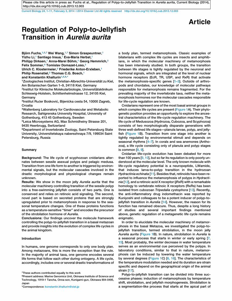
Figure 1. Metamorphosis in Aurelia aurita Is Initiated by Temperature Shift and Is Regulated by a Secreted Strobilation Inducer
(A) Scyphozoan jellyfishes belong to the phylum Cnidaria, which is a sister group to all bilaterian animals.
(B) Life cycle of Aurelia aurita. Each polyp transforms into multiple ephyra during strobilation.
(C) Polyp-to-jellyfish transition is induced by cold temperature. Polyps maintained at constant temperature (+18�C) do not strobilate. DT, temperature shift;
open square, cooling from +20�C to +10�C; closed circles, cooling from +18�C to 10�C; n, total number of animals in three independent experiments.
(D–I) Strobilation in Aurelia. The polyp (D) is divided into multiple segments (E–G) that develop into young jellyfishes (H and I). White arrows, strobila
segments.
(J–M) Metamorphosis induction by transplantation (J). Segmentation starts 72 hr after transplantation (K–M). White arrowheads, transplanted strobila
segments; white arrows, newly developed segments.
(N–Q) Metamorphosis induction by feeding with strobila segments (N). Polyp fed with strobila segments 24 hr (O), 48 hr (P), and 72 hr (Q) after feeding. Black
arrowhead, strobila segments inside of the polyp; white arrows, strobila segments.
(R) Feeding experiments. The segmented part of the strobila (rings) contains the highest amount of the strobilation inducer.
(S) Putative distribution of the strobilation inducer along the body of the strobila.
See also Figure S1.
Current Biology Vol 24 No 32
Please cite this article in press as: Fuchs et al., Regulation of Polyp-to-Jellyfish Transition in Aurelia aurita, Current Biology (2014),http://dx.doi.org/10.1016/j.cub.2013.12.003
the polyp and progresses downward (Figures 1D–1F). As aresult, a stack of disk-shaped segments is generated (Figures1E–1G). Each of those segments subsequently turns into ayoung jellyfish called an ephyra (Figures 1H and 1I). The youngephyra detaches from the strobila (Figures 1H and 1I), startsindependent planktonic life, and within several weeksdevelops into an adult jellyfish (Figure 1B).
By using a combination of classical transplantation ex-periments, transcriptomic analysis, and a loss-of-function
approach, we uncovered the molecules critical in controllingthe polyp-to-jellyfish transition in the life cycle of Aurelia.
Results
Diffusible Molecules Induce Metamorphosis and StrobilaFormation
In Aurelia aurita (strain Roscoff), polyp-to-jellyfish transitioncan be induced by temperature shift from +18�C to +10�C.
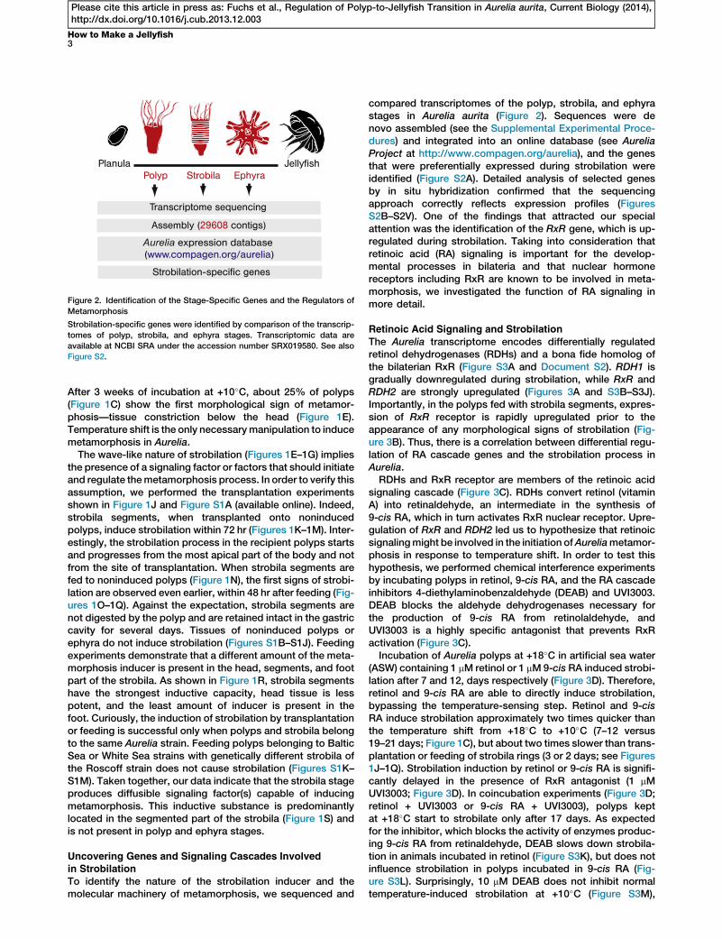
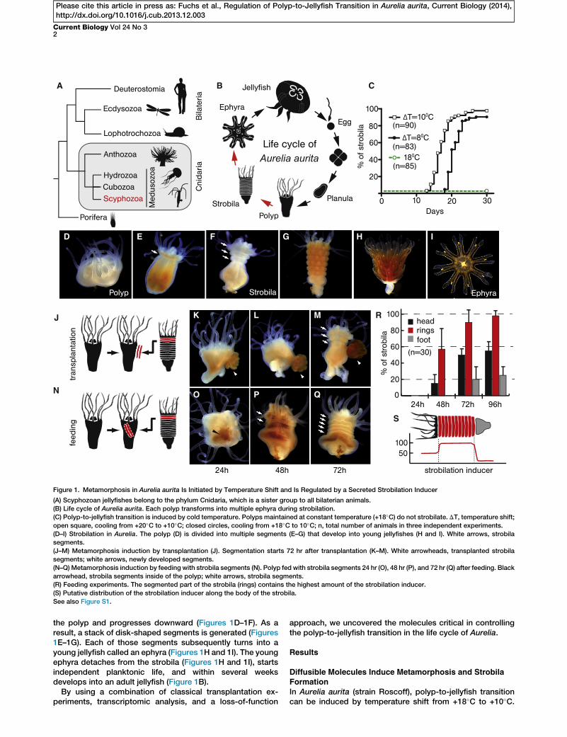
Figure 2. Identification of the Stage-Specific Genes and the Regulators of
Metamorphosis
Strobilation-specific genes were identified by comparison of the transcrip-
tomes of polyp, strobila, and ephyra stages. Transcriptomic data are
available at NCBI SRA under the accession number SRX019580. See also
Figure S2.
How to Make a Jellyfish3
Please cite this article in press as: Fuchs et al., Regulation of Polyp-to-Jellyfish Transition in Aurelia aurita, Current Biology (2014),http://dx.doi.org/10.1016/j.cub.2013.12.003
After 3 weeks of incubation at +10�C, about 25% of polyps(Figure 1C) show the first morphological sign of metamor-phosis—tissue constriction below the head (Figure 1E).Temperature shift is the only necessarymanipulation to inducemetamorphosis in Aurelia.
The wave-like nature of strobilation (Figures 1E–1G) impliesthe presence of a signaling factor or factors that should initiateand regulate themetamorphosis process. In order to verify thisassumption, we performed the transplantation experimentsshown in Figure 1J and Figure S1A (available online). Indeed,strobila segments, when transplanted onto noninducedpolyps, induce strobilation within 72 hr (Figures 1K–1M). Inter-estingly, the strobilation process in the recipient polyps startsand progresses from the most apical part of the body and notfrom the site of transplantation. When strobila segments arefed to noninduced polyps (Figure 1N), the first signs of strobi-lation are observed even earlier, within 48 hr after feeding (Fig-ures 1O–1Q). Against the expectation, strobila segments arenot digested by the polyp and are retained intact in the gastriccavity for several days. Tissues of noninduced polyps orephyra do not induce strobilation (Figures S1B–S1J). Feedingexperiments demonstrate that a different amount of the meta-morphosis inducer is present in the head, segments, and footpart of the strobila. As shown in Figure 1R, strobila segmentshave the strongest inductive capacity, head tissue is lesspotent, and the least amount of inducer is present in thefoot. Curiously, the induction of strobilation by transplantationor feeding is successful only when polyps and strobila belongto the same Aurelia strain. Feeding polyps belonging to BalticSea or White Sea strains with genetically different strobila ofthe Roscoff strain does not cause strobilation (Figures S1K–S1M). Taken together, our data indicate that the strobila stageproduces diffusible signaling factor(s) capable of inducingmetamorphosis. This inductive substance is predominantlylocated in the segmented part of the strobila (Figure 1S) andis not present in polyp and ephyra stages.
Uncovering Genes and Signaling Cascades Involvedin Strobilation
To identify the nature of the strobilation inducer and themolecular machinery of metamorphosis, we sequenced and
compared transcriptomes of the polyp, strobila, and ephyrastages in Aurelia aurita (Figure 2). Sequences were denovo assembled (see the Supplemental Experimental Proce-dures) and integrated into an online database (see AureliaProject at http://www.compagen.org/aurelia), and the genesthat were preferentially expressed during strobilation wereidentified (Figure S2A). Detailed analysis of selected genesby in situ hybridization confirmed that the sequencingapproach correctly reflects expression profiles (FiguresS2B–S2V). One of the findings that attracted our specialattention was the identification of the RxR gene, which is up-regulated during strobilation. Taking into consideration thatretinoic acid (RA) signaling is important for the develop-mental processes in bilateria and that nuclear hormonereceptors including RxR are known to be involved in meta-morphosis, we investigated the function of RA signaling inmore detail.
Retinoic Acid Signaling and Strobilation
The Aurelia transcriptome encodes differentially regulatedretinol dehydrogenases (RDHs) and a bona fide homolog ofthe bilaterian RxR (Figure S3A and Document S2). RDH1 isgradually downregulated during strobilation, while RxR andRDH2 are strongly upregulated (Figures 3A and S3B–S3J).Importantly, in the polyps fed with strobila segments, expres-sion of RxR receptor is rapidly upregulated prior to theappearance of any morphological signs of strobilation (Fig-ure 3B). Thus, there is a correlation between differential regu-lation of RA cascade genes and the strobilation process inAurelia.RDHs and RxR receptor are members of the retinoic acid
signaling cascade (Figure 3C). RDHs convert retinol (vitaminA) into retinaldehyde, an intermediate in the synthesis of9-cis RA, which in turn activates RxR nuclear receptor. Upre-gulation of RxR and RDH2 led us to hypothesize that retinoicsignalingmight be involved in the initiation ofAureliametamor-phosis in response to temperature shift. In order to test thishypothesis, we performed chemical interference experimentsby incubating polyps in retinol, 9-cis RA, and the RA cascadeinhibitors 4-diethylaminobenzaldehyde (DEAB) and UVI3003.DEAB blocks the aldehyde dehydrogenases necessary forthe production of 9-cis RA from retinolaldehyde, andUVI3003 is a highly specific antagonist that prevents RxRactivation (Figure 3C).Incubation of Aurelia polyps at +18�C in artificial sea water
(ASW) containing 1 mM retinol or 1 mM9-cis RA induced strobi-lation after 7 and 12, days respectively (Figure 3D). Therefore,retinol and 9-cis RA are able to directly induce strobilation,bypassing the temperature-sensing step. Retinol and 9-cisRA induce strobilation approximately two times quicker thanthe temperature shift from +18�C to +10�C (7–12 versus19–21 days; Figure 1C), but about two times slower than trans-plantation or feeding of strobila rings (3 or 2 days; see Figures1J–1Q). Strobilation induction by retinol or 9-cis RA is signifi-cantly delayed in the presence of RxR antagonist (1 mMUVI3003; Figure 3D). In coincubation experiments (Figure 3D;retinol + UVI3003 or 9-cis RA + UVI3003), polyps keptat +18�C start to strobilate only after 17 days. As expectedfor the inhibitor, which blocks the activity of enzymes produc-ing 9-cis RA from retinaldehyde, DEAB slows down strobila-tion in animals incubated in retinol (Figure S3K), but does notinfluence strobilation in polyps incubated in 9-cis RA (Fig-ure S3L). Surprisingly, 10 mM DEAB does not inhibit normaltemperature-induced strobilation at +10�C (Figure S3M),
A B
C D
FE
Figure 3. Retinoic Acid Signaling Is Necessary
for the Metamorphosis in Aurelia
(A) Expression of retinoic X receptor (RxR), retinol
dehydrogenase 1 (RDH1), and retinol dehydroge-
nase 2 (RDH2) during strobilation. P, noninduced
polyp; ES, early strobila with one segment; LS,
late strobila with several segments; E, ephyra.
(B) RxR upregulation in feeding experiments.
Expression levels in nonfed animals (0 hr) and
after feeding with strobila and polyp tissue
(12 hr and 24 hr) are shown.
(C) Schematic representation of retinoic acid
signaling cascade.
(D) Strobilation induction by 1 mM solution of
retinol (ROL) or 9-cis RA at +18�C. Coincubationof polyps in ROL or RA and 1 mM UVI3003 slows
down strobilation induction. The total number of
animals in each treatment is n = 30. ***p <
0.0001, log-rank (Mantel-Cox) test.
(E) RxR expression in polyps incubated in 9-cis
RA (1 mM) at +18�C. Expression in nontreated
animals (0) and after 3, 6, and 9 days of incubation
is shown.
(F) Temperature-induced strobilation at +10�C is
repressed by UVI3003 (1 mM, 2.5 mM, and 5 mM).
The total number of animals in each treatment is
n = 30. ***p < 0.0001, log-rank (Mantel-Cox) test.
ASW, artificial sea water.
Expression levels in (A), (B), and (E) were deter-
mined by quantitative RT-PCR. EF1a was used
for equilibration. Mean expression6 SD is shown
(n = 3).
See also Figure S3 and Table S1.
Current Biology Vol 24 No 34
Please cite this article in press as: Fuchs et al., Regulation of Polyp-to-Jellyfish Transition in Aurelia aurita, Current Biology (2014),http://dx.doi.org/10.1016/j.cub.2013.12.003
which is unexpected and implies that additional strobilationregulators might be present.
As shown on Figure 3E, incubation of polyps in 1 mM9-cisRAcauses upregulation ofRxR similar to that in the feeding exper-iments (Figure 3B) or during the temperature induction (Fig-ure 3A). After 6 days of incubation, the first changes in RxRexpression level are detectable, and after 9 days of incubation,its expression is upregulated 3-fold. Importance of the RxR forthe metamorphosis is additionally substantiated by the factthat the temperature-induced strobilation can be inhibited byUVI3003 in a concentration-dependent manner (Figure 3F).As shown in Figure 3F, strobilation is significantly delayed in1 mM solution of UVI3003, and it is completely repressed in5 mM solution.
Thus, RxR receptor seems to be important for the strobila-tion process, and 9-cisRAmight serve as a signaling moleculein Aurelia. We have not directly shown that 9-cis RA is pro-duced in this species, but the upregulation of aRDH2 and theretardation of strobilation by DEAB both speak in favor ofthis possibility. Incubation in retinol or 9-cis RA can function-ally substitute the temperature shift that induces strobilationunder natural conditions. Hence, retinoic acid signaling mightserve as a link between yet unknown temperature-sensing re-ceptor and the downstream genes responsible for the onset ofmetamorphosis. It is also important to mention that 9-cis RAand retinol, even in their highest nontoxic concentrations(5 mM), are not able to induce strobilation within 48 hr as ithappens when polyps are fed with strobila segments. Thisindicates that 9-cis RA is important, but not the only regulatorof strobilation.
Unbiased Identification of Strobilation-Inducing Molecules
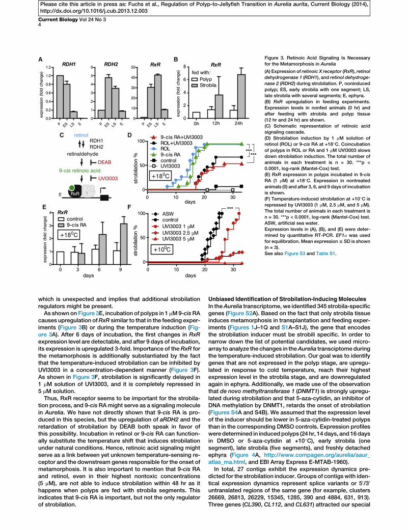
In theAurelia transcriptome, we identified 345 strobila-specificgenes (Figure S2A). Based on the fact that only strobila tissueinduces metamorphosis in transplantation and feeding exper-iments (Figures 1J–1Q and S1A–S1J), the gene that encodesthe strobilation inducer must be strobili specific. In order tonarrow down the list of potential candidates, we used micro-array to analyze the changes in theAurelia transciptomeduringthe temperature-induced strobilation. Our goal was to identifygenes that are not expressed in the polyp stage, are upregu-lated in response to cold temperature, reach their highestexpression level in the strobila stage, and are downregulatedagain in ephyra. Additionally, we made use of the observationthat de novo methyltransferase 1 (DNMT1) is strongly upregu-lated during strobilation and that 5-aza-cytidin, an inhibitor ofDNA methylation by DNMT1, retards the onset of strobilation(Figures S4A and S4B). We assumed that the expression levelof the inducer should be lower in 5-aza-cytidin-treated polypsthan in the corresponding DMSO controls. Expression profilesweredetermined in inducedpolyps (24 hr, 14 days, and 16daysin DMSO or 5-aza-cytidin at +10�C), early strobila (onesegment), late strobila (five segments), and freshly detachedephyra (Figure 4A, http://www.compagen.org/aurelia/aaur_atlas_ma.html, and EBI Array Express E-MTAB-1960).In total, 27 contigs exhibit the expression dynamics pre-
dicted for the strobilation inducer. Groups of contigswith iden-tical expression dynamics represent splice variants or 50/30
untranslated regions of the same gene (for example, clusters26669, 26813, 26229, 15345, 1285, 390 and 4884, 631, 913).Three genes (CL390, CL112, and CL631) attracted our special
How to Make a Jellyfish5
Please cite this article in press as: Fuchs et al., Regulation of Polyp-to-Jellyfish Transition in Aurelia aurita, Current Biology (2014),http://dx.doi.org/10.1016/j.cub.2013.12.003
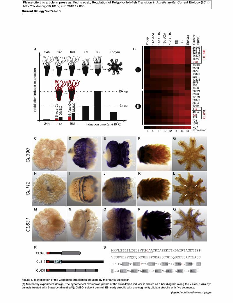
attention due to their drastic upregulation at +10�C. As shownin Figure 4B, the increase in the amount of transcript betweenpolyp and early strobila is more than 35,000-fold for CL390,2,700-fold for CL112, and 3,200-fold for CL631.
Transcript localization of CL390, CL112, and CL631 wasexamined by in situ hybridization in noninduced polyps, ininduced polyps with the first constriction (VES), in early stro-bila (ES), in late strobila (LS), during ephyra morphogenesis,and in ephyra. As shown in Figures 4C–4Q, the candidategenes are not expressed in the polyp, but all of them arestrongly upregulated during strobilation.
All three candidate genes encode proteins lacking similarityto any known proteins from other animals (BLASTP e value <1 3 1025). Thus, they most probably represent Scyphozoa-specific or even Aurelia-specific genes. All three proteinscontain a putative signal peptide sequence at the N terminus(Figure 4R). CL390 protein consists of two qualitativelydifferent regions: an N-terminal portion containing largenumbers of hydrophobic amino acids and a C-terminal portioncontaining highly charged motifs made of arginine-rich re-peats (Figure 4S). CL112 contains a domain consisting of sixcystein residues with low sequence similarity to epidermalgrowth factor (EGF; E = 1 3 1022). Similar to CL390, CL631contains large proportions of hydrophobic amino acids in itsN-terminal region. Three repeats in the C-terminal region ofCL631 are separated by the putative endopeptidase process-ing sites KR and KK.
CL390, CL112, and CL631 encode novel secreted proteins,and due to their strong expression during metamorphosis,they might be considered as potential strobilation inducers.CL390, however, seems to be the most promising candidatesince it is not expressed at the polyp stage, it is strongly upre-gulated in strobila in response to the temperature changes,and its expression is drastically downregulated in the ephyrastage.
Identification of a Temperature Dependent ‘‘Timer’’ for the
Onset of MetamorphosisIn nature, polyps start to strobilate in spring after beingexposed to a prolonged period of cold water temperature.How does the metamorphosis-controlling system distinguishwinter from occasional short-term temperature fluctuations?Theoretically, there must be a minimal duration of the ‘‘cold’’period that causes metamorphosis, and such a system wouldrequire cold-sensitive genes to operate.
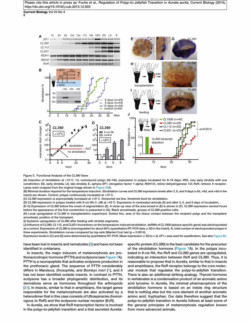
Indeed, CL390, CL112, and CL631 are cold sensitive and anincrease in their transcript levels occurs earlier than the onsetof segmentation (Figure 5A). The most intriguing dynamicscan be observed with CL390. While the upregulation ofCL112 and CL631 is detectable only after 17 and 19 days, theupregulation of CL390 is first detectable already after 9 daysat +10�C.
To determine theminimal duration of the cold period neededto induce strobilation, we placed groups of polyps at +10�Cand returned them back to +18�C after 6, 9, and 12 days ofcold treatment (Figure 5B, inset; Figure S5A). As shown in Fig-ure 5B, none of the polyps incubated at +10�C for just 6 daysmetamorphosed, while incubation for 9 days was sufficientto induce strobilation. Animals kept at +10�C for 12 daysexhibit the same strobilation kinetics as the animals in the con-trol group, which remained at +10�C. Expression of CL390 inthe polyps that were returned back to +18�C after 3, 6, and9 days of cooling is shown in Figures 5B (inset) and S5A. Theexpression level of CL390 shows fluctuations and has a
tendency to decrease at +18�C after the initial upregulationat +10�C.Detailed analysis of CL390 expression by real-time PCR
(Figure 5C) shows that at +10�C the amount of transcript in-creases exponentially, with mean upregulation of 5.53 after6 days, 313 after 9 days, and 110,0003 after 19 days. Theamount of CL390 transcript in a polyp is positively and directlycorrelated with the amount of time spent by Aurelia polyps atlow temperature. Our results indicate that as soon as the upre-gulation exceeds the threshold of about 15-fold (between 6and 9 days at +10�C), the strobilation program is activatedand becomes independent from temperature input (Figure 5C).CL390 therefore represents an example of a novel tempera-ture-dependent ‘‘timer’’ whose expression level allows Aureliapolyps to ‘‘measure’’ the duration of a cold period.As shown in Figure 5A, genes involved in retinoic acid
signaling (RDH1/RDH2 and RxR) are also sensitive to temper-ature changes. The upregulation of RxR, similar to that ofCL390, precedes morphological changes and is detectableafter 6 days at +10�C. Interestingly, CL390 expression can bedirectly activated by incubation of polyps in 9-cis RAat +18�C (Figure 5D). This result indicates a possible linkbetween retinoic acid signaling and the activation of CL390in Aurelia. In support of this view, the 50 flanking region andthe first intron of CL390 contain three putative binding sitesfor RxR nuclear hormone receptor (Figures S5B–S5D). Wedid not perform functional tests for these elements, but theidentical half sites with the sequence AGGTCA from the pro-moters of crystallin genes have been shown previously tobind jRxR of Tripedalia cystophora [13]. Taking into consider-ation the high degree of sequence conservation between DNAbinding domains (DBDs) of RxRs from Aurelia and Tripedalia(Figure S5E), the probability of RxR binding to AGGTCAelements in the promoter of CL390 is relatively high.
CL390 Acts as Strobilation Inducer in AureliaOn the cellular level, cooling activates the expression ofCL390in the ectodermal epithelial cells, which initially form smallpatches (Figures 5E, 5F, and S5F) that increase in numberand expand in size. Several hours before the initiation of thesegmentation process, all ectodermal epithelial cells of apolyp (except the ones in the tentacles and in the foot) becomeCL390 positive (Figure 5G).Similar upregulation and spreading of CL390 expression is
observed in transplantation and feeding experiments. Asdescribed previously (see Figures 1J–1Q), strobilation can beinduced by transplantation or feeding within 72 and 48 hr,respectively. As shown by in situ hybridization in Figure 5H,transplantation of strobila segments induces local upregula-tion of CL390 in the region adjacent to the transplant (dottedline in Figure 5H). A patch ofCL390-expressing cells surroundsthe area where the transplant was located (transplanted stro-bila tissue has been removed from the recipient animal priorto fixation). In the feeding experiments, CL390 is systemicallyand strongly upregulated in all ectodermal epithelial cellsthroughout the polyp body within 24 hr (Figure 5I).In order to directly examine the influence of CL390, CL112,
and CL631 on strobilation, we developed RNAi technologyfor Aurelia (see the Supplemental Experimental Procedures).As shown in Figures S5H and S5J, by electroporating earlystrobila with double-stranded RNA (dsRNA) of CL390 orCL112, we were able to repress the expression of the corre-sponding genes. Electroporation of control dsRNA (GFP orCL1008) did not influence gene expression (Figures S5G and
A B
C D E F G
H I J K L
M N O P Q
R S
Figure 4. Identification of the Candidate Strobilation Inducers by Microarray Approach
(A) Microarray experiment design. The hypothetical expression profile of the strobilation inducer is shown as a bar diagram along the x axis. 5-Aza-cyt,
animals treated with 5-aza-cytidine (5 mM); DMSO, solvent control; ES, early strobila with one segment; LS, late strobila with five segments.
(legend continued on next page)
Current Biology Vol 24 No 36
Please cite this article in press as: Fuchs et al., Regulation of Polyp-to-Jellyfish Transition in Aurelia aurita, Current Biology (2014),http://dx.doi.org/10.1016/j.cub.2013.12.003
How to Make a Jellyfish7
Please cite this article in press as: Fuchs et al., Regulation of Polyp-to-Jellyfish Transition in Aurelia aurita, Current Biology (2014),http://dx.doi.org/10.1016/j.cub.2013.12.003
S5I). To test the function of genes potentially involved in stro-bilation, we electroporated induced polyps (after 6 daysat +10�C) with dsRNA of CL1008 (ephyra-specific gene usedas a control),CL390,CL112, andCL631. As shown in Figure 5J,we observed no significant difference in strobilation kineticsbetween animal groups electroporated with CL1008, CL631,or CL112. In contrast, animals electroporated with CL390dsRNA strobilated slower than did the control animals(CL1008). The inhibition was not complete, which is not sur-prising when taking into consideration that we have depletedonly about 60% of the CL390 transcript (inset in Figure 5J).Our results, however, indicate that CL390 is causally involvedin strobilation and might represent the strobilation inducer.
CL390-Derived Synthetic Strobilation Inducers
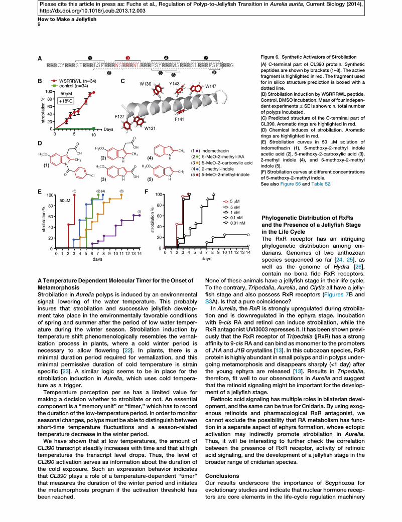
CL390 encodes a secreted protein with an unusual C-terminalpart where arginine repeats separate di- and tripeptides withthe sequences SF, LSF, WS, WL, FSY, WS, SL, and FSF (Fig-ure 6A). To directly test the function of CL390 in strobilation,we fed polyps with the full-length recombinant protein fusedto GFP (Figures S6A–S6D) and incubated the polyps in syn-thetic peptides (50 mM) representing subfragments of CL390(Figures 6A and S6E). Feeding causes formation of segment-like constriction below the head of Aurelia polyps (see Fig-ure S5D), but the segmentation does not progress further. Incontrast to that, synthetic peptide with the sequenceWSRRRWL induces strobilation within 72 hr of incubationat +18�C (Figure 6B). Other peptides tested do not induce stro-bilation (Figures 6A and S6E). These results indicate thatCL390 might be the precursor of the strobilation inducer,which becomes biologically active after being processed intosmaller fragments. As shown in Figure 6C, according toin silico structure prediction, the C-terminal part of CL390forms a helix where aromatic amino acids separated by threearginins are located in close proximity to each other (F127–W131 and Y143–W147). In case of the WSRRRWL peptide,indole rings of two tryptophans are also located in close prox-imity and inmutually perpendicular planes (Figure S6F). Such aclose positioning of the aromatic rings is similar to the corepart of the indole derivative indomethacin (Figure 6D), whichhas been recently shown to induce strobilation in the Japanesestrain of Aurelia aurita [14]. Indomethacin is an anti-inflamma-tory drug that functions via inhibition of cyclooxygenases(COX1/COX2). The transcriptome of Aurelia aurita and thegenomes ofNematostella,Hydra, andAcropora do not containhomologs of COX-1 and COX-2. Thus, in light of the observa-tions described above, the inductive activity of indomethacinmight be explained by its structural similarity to the naturallyoccurring strobilation hormone of Aurelia encoded by CL390.
In order to identify the minimal pharmacophore able to acti-vate strobilation, we screened indole and tryptophan deriva-tives and identified four substances with a strong capacity toinduce strobilation at 50 mM (see Figure S6G and Table S2
(B) Clusters with the expression profile predicted for the strobilation induce
Numbers, cluster identifiers; ES, early strobila; LS, late strobila; I, clusters wher
downregulation in ephyra. The heatmap represents EF1a normalized expressi
(C–G) CL390 is strongly upregulated during strobilation and is not expressed i
(H–L) CL112 is expressed in a subpopulation of endodermal cells during strob
(M–Q) CL631 is exclusively expressed in the segments of strobila and in ephy
(R) Protein domain organization of CL390, CL112, and CL631. Putative signal pe
EGF-like domain (see also Figures S4C and S4D).
(S) CL390 protein. Arginine repeats are highlighted in gray. Cystein residues a
Polyp stage (C, H, M); early strobila (D, I, N); late strobila (E, J, O), late strobila u
See also Figure S4.
for the complete list of the compounds tested). As shown onFigure 6D, all active compounds share a core structure madeof an indole ring and methyl or carboxylic groups at positiontwo. Despite the structural similarity, compounds (1)–(5) havedrastically different inductive capacities in the Roscoff strainof Aurelia (Figure 6E). Indomethacin (1) induces strobilationonly after 6 days of incubation, while 5-methoxy-2-methylin-dole acetic acid (2) and 5-methoxyindole-2-carboxylic acid(3) do so after 4–5 days, 2-methylindole (4) after 3 days, and5-methoxy-2-methylindole (5) within 48 hr. Interestingly, at aconcentration of 50 mM, 5-methoxy-2-methylindole inducesstrobilationwith the same kinetics as feedingwith strobila seg-ments. The inductive capacity of 5-methoxy-2-methylindoleremains identical within a concentration range between50 mM and 5 nM (Figure 6F). Even at a concentration of 1 nM,strobilation is induced after 48 hr and all of the incubatedpolyps start tometamorphose within 8 days. Only at a concen-tration of 0.1 nM is the inductive effect considerably reduced,and it is completely absent at a concentration of 0.01 nM.Thus, the CL390 gene appears to encode the pre-
cursor of the strobilation hormone in Aurelia aurita and 5-methoxy-2-methylindole appears to represents its minimalpharmacophore.
Discussion
Molecular Toolkit of Strobilation in Aurelia
Alternation between larval and adult stages with drasticmorphological, physiological, and behavioral differences is afundamental metazoan feature [3]. Life cycles with variouslevels of complexity are present in all animal phyla, but themo-lecular machinery driving the transitions between life stageshas been deciphered in just a few model organisms [1–3, 11].In amphibians and arthropods, where the molecular basis of
metamorphosis is characterized in detail, the activation ofmetamorphosis has always two successive steps—a neuronalone and an endocrine one. In both animal groups, the coremodule of life-cycle regulation consists of two nuclear hor-mone receptors that cooperatively control the activation ofthe downstream metamorphosis genes (Figure 7A).In amphibians, thyroid-stimulating hormone (TSH) from the
pituitary gland regulates the production of thyroid hormone(TH), which induces tadpole transformation into juvenile frog[18, 19]. Thyroid hormone is a condensation product of twoiodinated tyrosins derived from a high-molecular-weight pro-tein precursor. The cascade of downstream genes leading tometamorphosis is activated by a heterodimer of thyroid hor-mone receptor (TR) and RxR. TR homologs are present inChordata, Urochordata, Cephalochordata, and Hemichordata[3]. In the protostomian lineage, TRs have been identified inthe plathelmynths Schistosoma mansoni, S. japonium, andSchmidtea mediterranea, as well as in the mollusk Lottiagigantea and the arthropod Daphnia pulex [20]. TR genes
r. Clusters representing the splice variants of the same gene are boxed.
e expression is strongly downregulated in ephyra; II, clusters without strong
on values after log2 transformation.
n ephyra.
ilation and in the manubrium of ephyra.
ra. Black arrowhead, segment border.
ptide sequence is shown in red; gray boxes, internal repeats; light-blue box,
re shown in red. Putative signal peptide sequence is underlined.
ndergoing ephyra morphogenesis (F, K, P), and ephyra (G, L, Q) are shown.
A
B
C D
E F G
H
I
J
Figure 5. Functional Analysis of the CL390 Gene
(A) Induction of strobilation at +10�C. 1d, noninduced polyp; 6d–19d, expression in polyps incubated for 6–19 days; VES, very early strobila with one
constriction; ES, early strobila; LS, late strobila; E, ephyra; EF1, elongation factor 1-alpha; RDH1/2, retinol dehydrogenese 1/2; RxR, retinoic X receptor.
Lanes were cropped from the original image shown in Figure S5A.
(B) Minimal duration required for the temperature induction. Strobilation curves and CL390 expression levels after 3, 6, and 9 days (+3d, +6d, and +9d in the
insert) are shown. Control, polyps continuously incubated at +10�C.(C) CL390 expression is exponentially increased at +10�C. Horizontal red line, threshold level for strobilation.
(D) CL390 expression in polyps treated with 9-cis RA (1 mM) at +18�C. Expression in nontreated animals (0) and after 3, 6, and 9 days of incubation.
(E–G) Expression of CL390 before the onset of segmentation (E). A close-up view of the area boxed in (E) is shown in (F). CL390 expression several hours
before the appearance of the first constriction is presented in (G). Black arrowheads, groups of CL390-positive cells.
(H) Local upregulated of CL390 in transplantation experiment. Dotted line, area of the tissue contact between the recipient polyp and the transplant;
arrowhead, position of the transplant.
(I) Systemic upregulation of CL390 after feeding with strobila segments.
(J) Influence ofCL390,CL112, andCL631 knockdown on the temperature-induced strobilation. dsRNAofCL1008 (ephyra-specific gene) was electroporated
as a control. Expression ofCL390 is downregulated for about 60% (quantitative RT-PCR data6 SD in the insert). N, total number of electroporated polyps in
three experiments. Strobilation curves compared by log-rank (Mantel-Cox) test (p = 0.0014).
Expression levels in (C) and (D) were determined by quantitative RT-PCR.Mean expression6 SD (n = 3). EF1awas used for equilibration. See also Figure S5.
Current Biology Vol 24 No 38
Please cite this article in press as: Fuchs et al., Regulation of Polyp-to-Jellyfish Transition in Aurelia aurita, Current Biology (2014),http://dx.doi.org/10.1016/j.cub.2013.12.003
have been lost in insects and nematodes [3] and have not beenidentified in cnidarians.
In insects, the major inducers of metamorphosis are pro-thoracicotropic hormone (PTTH) andecdysone (seeFigure 7A).PTTH is a neuropeptide that activates ecdysone production inthe prothoracic gland. The sequence of PTTH considerablydiffers in Manduca, Drosophila, and Bombyx mori [1], and ithas not been identified outside insects. In contrast to PTTH,ecdysone has a broader phylogenetic distribution, and itsderivatives serve as hormones throughout the arthropods[21]. In insects, similar to that in amphibians, the target genesresponsible for the metamorphosis are also activated by aheterodimer that in this case consists of Ultraspiracles (homol-ogous to RxR) and the ecdysone nuclear receptor (EcR).
In Aurelia, we show that RxR transcription factor is involvedin the polyp-to-jellyfish transition and a that secreted Aurelia-
specific protein (CL390) is the best candidate for the precursorof the strobilation hormone (Figure 7A). In the polyps incu-bated in 9-cis RA, the RxR and CL390 genes are upregulated,indicating an interaction between RxR and CL390. Thus, it isreasonable to propose that in Aurelia, similar to that in insectsand amphibians, the RxR receptor belongs to the core molec-ular module that regulates the polyp-to-jellyfish transition.There is also an additional striking analogy. Thyroid hormonein vertebrates is a condensation product of an aromatic aminoacid tyrosine. In Aurelia, the minimal pharmacophore of thestrobilation hormone is based on an indole ring structurethat is nothing else but the core element of another aromaticamino acid, tryptophan. Our data therefore suggest that thepolyp-to-jellyfish transition in Aurelia follows at least some ofthe general principles of metamorphosis regulation knownfrom more advanced animals.
A
B C
D
E F
Figure 6. Synthetic Activators of Strobilation
(A) C-terminal part of CL390 protein. Synthetic
peptides are shown by brackets (1–8). The active
fragment is highlighted in red. The fragment used
for in silico structure prediction is boxed with a
dotted line.
(B) Strobilation induction by WSRRRWL peptide.
Control, DMSO incubation.Mean of four indepen-
dent experiments6 SE is shown; n, total number
of polyps incubated.
(C) Predicted structure of the C-terminal part of
CL390. Aromatic rings are highlighted in red.
(D) Chemical induces of strobilation. Aromatic
rings are highlighted in red.
(E) Strobilation curves in 50 mM solution of
indomethacin (1), 5-methoxy-2-methyl indole
acetic acid (2), 5-methoxy-2-carboxylic acid (3),
2-methyl indole (4), and 5-methoxy-2-methyl
indole (5).
(F) Strobilation curves at different concentrations
of 5-methoxy-2-methyl indole.
See also Figure S6 and Table S2.
How to Make a Jellyfish9
Please cite this article in press as: Fuchs et al., Regulation of Polyp-to-Jellyfish Transition in Aurelia aurita, Current Biology (2014),http://dx.doi.org/10.1016/j.cub.2013.12.003
ATemperature Dependent Molecular Timer for theOnset of
MetamorphosisStrobilation in Aurelia polyps is induced by an environmentalsignal: lowering of the water temperature. This probablyinsures that strobilation and successive jellyfish develop-ment take place in the environmentally favorable conditionsof spring and summer after the period of low water temper-ature during the winter season. Strobilation induction bytemperature shift phenomenologically resembles the vernal-ization process in plants, where a cold winter period isnecessary to allow flowering [22]. In plants, there is aminimal duration period required for vernalization, and thisminimal permissive duration of cold temperature is strainspecific [23]. A similar logic seems to be in place for thestrobilation induction in Aurelia, which uses cold tempera-ture as a trigger.
Temperature perception per se has a limited value formaking a decision whether to strobilate or not. An essentialcomponent is a ‘‘memory unit’’ or ‘‘timer,’’ which has to recordthe duration of the low-temperature period. In order to monitorseasonal changes, polypsmust be able to distinguish betweenshort-time temperature fluctuations and a season-relatedtemperature decrease in the winter period.
We have shown that at low temperatures, the amount ofCL390 transcript steadily increases with time and that at hightemperatures the transcript level drops. Thus, the level ofCL390 activation serves as information about the duration ofthe cold exposure. Such an expression behavior indicatesthat CL390 plays a role of a temperature-dependent ‘‘timer’’that measures the duration of the winter period and initiatesthe metamorphosis program if the activation threshold hasbeen reached.
Phylogenetic Distribution of RxRs
and the Presence of a Jellyfish Stagein the Life Cycle
The RxR receptor has an intriguingphylogenetic distribution among cni-darians. Genomes of two anthozoanspecies sequenced so far [24, 25], aswell as the genome of Hydra [26],contain no bona fide RxR receptors.
None of these animals have a jellyfish stage in their life cycle.To the contrary, Tripedalia, Aurelia, and Clytia all have a jelly-fish stage and also possess RxR receptors (Figures 7B andS3A). Is that a pure coincidence?In Aurelia, the RxR is strongly upregulated during strobila-
tion and is downregulated in the ephyra stage. Incubationwith 9-cis RA and retinol can induce strobilation, while theRxR antagonist UVI3003 represses it. It has been shown previ-ously that the RxR receptor of Tripedalia (jRxR) has a strongaffinity to 9-cis RA and can bind as monomer to the promotersof J1A and J1B crystallins [13]. In this cubozoan species, RxRprotein is highly abundant in small polyps and in polyps under-going metamorphosis and disappears sharply (<1 day) afterthe young ephyra are released [13]. Results in Tripedalia,therefore, fit well to our observations in Aurelia and suggestthat the retinoid signaling might be important for the develop-ment of a jellyfish stage.Retinoic acid signaling has multiple roles in bilaterian devel-
opment, and the same can be true for Cnidaria. By using exog-enous retinoids and pharmacological RxR antagonist, wecannot exclude the possibility that RA metabolism has func-tion in a separate aspect of ephyra formation, whose ectopicactivation may indirectly promote strobilation in Aurelia.Thus, it will be interesting to further check the correlationbetween the presence of RxR receptor, activity of retinoicacid signaling, and the development of a jellyfish stage in thebroader range of cnidarian species.
ConclusionsOur results underscore the importance of Scyphozoa forevolutionary studies and indicate that nuclear hormone recep-tors are core elements in the life-cycle regulation machinery
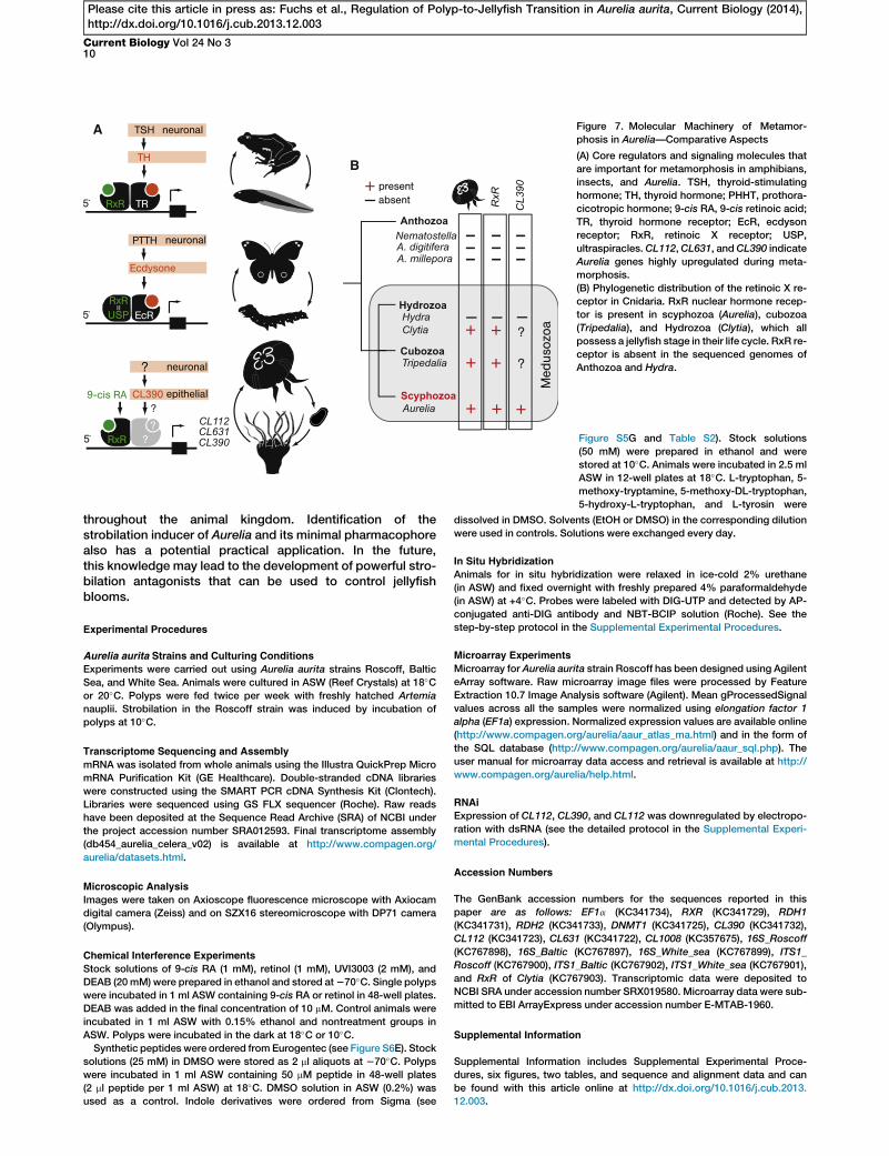
A
B
Figure 7. Molecular Machinery of Metamor-
phosis in Aurelia—Comparative Aspects
(A) Core regulators and signaling molecules that
are important for metamorphosis in amphibians,
insects, and Aurelia. TSH, thyroid-stimulating
hormone; TH, thyroid hormone; PHHT, prothora-
cicotropic hormone; 9-cis RA, 9-cis retinoic acid;
TR, thyroid hormone receptor; EcR, ecdyson
receptor; RxR, retinoic X receptor; USP,
ultraspiracles.CL112,CL631, andCL390 indicate
Aurelia genes highly upregulated during meta-
morphosis.
(B) Phylogenetic distribution of the retinoic X re-
ceptor in Cnidaria. RxR nuclear hormone recep-
tor is present in scyphozoa (Aurelia), cubozoa
(Tripedalia), and Hydrozoa (Clytia), which all
possess a jellyfish stage in their life cycle. RxR re-
ceptor is absent in the sequenced genomes of
Anthozoa and Hydra.
Current Biology Vol 24 No 310
Please cite this article in press as: Fuchs et al., Regulation of Polyp-to-Jellyfish Transition in Aurelia aurita, Current Biology (2014),http://dx.doi.org/10.1016/j.cub.2013.12.003
throughout the animal kingdom. Identification of thestrobilation inducer of Aurelia and its minimal pharmacophorealso has a potential practical application. In the future,this knowledge may lead to the development of powerful stro-bilation antagonists that can be used to control jellyfishblooms.
Experimental Procedures
Aurelia aurita Strains and Culturing Conditions
Experiments were carried out using Aurelia aurita strains Roscoff, Baltic
Sea, and White Sea. Animals were cultured in ASW (Reef Crystals) at 18�Cor 20�C. Polyps were fed twice per week with freshly hatched Artemia
nauplii. Strobilation in the Roscoff strain was induced by incubation of
polyps at 10�C.
Transcriptome Sequencing and Assembly
mRNA was isolated from whole animals using the Illustra QuickPrep Micro
mRNA Purification Kit (GE Healthcare). Double-stranded cDNA libraries
were constructed using the SMART PCR cDNA Synthesis Kit (Clontech).
Libraries were sequenced using GS FLX sequencer (Roche). Raw reads
have been deposited at the Sequence Read Archive (SRA) of NCBI under
the project accession number SRA012593. Final transcriptome assembly
(db454_aurelia_celera_v02) is available at http://www.compagen.org/
aurelia/datasets.html.
Microscopic Analysis
Images were taken on Axioscope fluorescence microscope with Axiocam
digital camera (Zeiss) and on SZX16 stereomicroscope with DP71 camera
(Olympus).
Chemical Interference Experiments
Stock solutions of 9-cis RA (1 mM), retinol (1 mM), UVI3003 (2 mM), and
DEAB (20 mM) were prepared in ethanol and stored at270�C. Single polyps
were incubated in 1 ml ASW containing 9-cis RA or retinol in 48-well plates.
DEAB was added in the final concentration of 10 mM. Control animals were
incubated in 1 ml ASW with 0.15% ethanol and nontreatment groups in
ASW. Polyps were incubated in the dark at 18�C or 10�C.Synthetic peptides were ordered from Eurogentec (see Figure S6E). Stock
solutions (25 mM) in DMSO were stored as 2 ml aliquots at 270�C. Polypswere incubated in 1 ml ASW containing 50 mM peptide in 48-well plates
(2 ml peptide per 1 ml ASW) at 18�C. DMSO solution in ASW (0.2%) was
used as a control. Indole derivatives were ordered from Sigma (see
Figure S5G and Table S2). Stock solutions
(50 mM) were prepared in ethanol and were
stored at 10�C. Animals were incubated in 2.5 ml
ASW in 12-well plates at 18�C. L-tryptophan, 5-methoxy-tryptamine, 5-methoxy-DL-tryptophan,
5-hydroxy-L-tryptophan, and L-tyrosin were
dissolved in DMSO. Solvents (EtOH or DMSO) in the corresponding dilution
were used in controls. Solutions were exchanged every day.
In Situ Hybridization
Animals for in situ hybridization were relaxed in ice-cold 2% urethane
(in ASW) and fixed overnight with freshly prepared 4% paraformaldehyde
(in ASW) at +4�C. Probes were labeled with DIG-UTP and detected by AP-
conjugated anti-DIG antibody and NBT-BCIP solution (Roche). See the
step-by-step protocol in the Supplemental Experimental Procedures.
Microarray Experiments
Microarray for Aurelia aurita strain Roscoff has been designed using Agilent
eArray software. Raw microarray image files were processed by Feature
Extraction 10.7 Image Analysis software (Agilent). Mean gProcessedSignal
values across all the samples were normalized using elongation factor 1
alpha (EF1a) expression. Normalized expression values are available online
(http://www.compagen.org/aurelia/aaur_atlas_ma.html) and in the form of
the SQL database (http://www.compagen.org/aurelia/aaur_sql.php). The
user manual for microarray data access and retrieval is available at http://
www.compagen.org/aurelia/help.html.
RNAi
Expression of CL112, CL390, and CL112 was downregulated by electropo-
ration with dsRNA (see the detailed protocol in the Supplemental Experi-
mental Procedures).
Accession Numbers
The GenBank accession numbers for the sequences reported in this
paper are as follows: EF1a (KC341734), RXR (KC341729), RDH1
(KC341731), RDH2 (KC341733), DNMT1 (KC341725), CL390 (KC341732),
CL112 (KC341723), CL631 (KC341722), CL1008 (KC357675), 16S_Roscoff
(KC767898), 16S_Baltic (KC767897), 16S_White_sea (KC767899), ITS1_
Roscoff (KC767900), ITS1_Baltic (KC767902), ITS1_White_sea (KC767901),
and RxR of Clytia (KC767903). Transcriptomic data were deposited to
NCBI SRA under accession number SRX019580. Microarray data were sub-
mitted to EBI ArrayExpress under accession number E-MTAB-1960.
Supplemental Information
Supplemental Information includes Supplemental Experimental Proce-
dures, six figures, two tables, and sequence and alignment data and can
be found with this article online at http://dx.doi.org/10.1016/j.cub.2013.
12.003.

How to Make a Jellyfish11
Please cite this article in press as: Fuchs et al., Regulation of Polyp-to-Jellyfish Transition in Aurelia aurita, Current Biology (2014),http://dx.doi.org/10.1016/j.cub.2013.12.003
Acknowledgments
We thank Antje Thomas for the help with microscopy, Thomas Walter for
constructing jellyfish facility, and Elke Blohm-Sievers for the help with
microarrays. Tatiana Shaposhnikova and Gerhard Jarms provided the ani-
mal lines. Ivan Gluzdikov, Ludmila Kuznetzova, and Rainer Herges gave
important advice in organic chemistry. Ulrich Technau, Johanna Kraus,
and David Fredman shared with us highly valuable nonpublished Clytia
and Aurelia data. We are grateful to Diethard Tautz for the opportunity to
use research facilities at MPI for Evolutionary Biology in Plon. K.K. thanks
Oleg Chaga, Ivan Tikhomirov, and Ivan Rudsky for inspiring discussions.
Our work was financially supported by DFG Clusters ‘‘Future Ocean’’ and
‘‘Inflammation at Interfaces.’’ K.K. is supported by RFBR (RVVJ) grant
13-04-01795. W.W. was supported by China Scholarship Council.
Received: May 2, 2013
Revised: August 19, 2013
Accepted: December 3, 2013
Published: January 16, 2014
References
1. McBrayer, Z., Ono, H., Shimell, M., Parvy, J.P., Beckstead, R.B.,Warren,
J.T., Thummel, C.S., Dauphin-Villemant, C., Gilbert, L.I., and O’Connor,
M.B. (2007). Prothoracicotropic hormone regulates developmental
timing and body size in Drosophila. Dev. Cell 13, 857–871.
2. Brown, D.D., and Cai, L. (2007). Amphibian metamorphosis. Dev. Biol.
306, 20–33.
3. Laudet, V. (2011). The origins and evolution of vertebrate metamor-
phosis. Curr. Biol. 21, R726–R737.
4. Hofmann, D.K., Neumann, R., and Henne, K. (1978). Strobilation,
budding and initiation of scyphistomamorphogenesis in the rhizostome
Cassiopea andromeda (Cnidaria, Scyphozoa). Mar. Biol. 47, 161–176.
5. Leitz, T., and Wagner, T. (1993). The marine bacterium Alteromonas es-
pejiana induces metamorphosis of the hydroid Hydractinia echinata.
Mar. Biol. 115, 173–178.
6. Kroiher, M., Siefker, B., and Berking, S. (2000). Induction of segmenta-
tion in polyps of Aurelia aurita (Scyphozoa, Cnidaria) into medusae
and formation of mirror-image medusa anlagen. Int. J. Dev. Biol. 44,
485–490.
7. Miyake, H., Terazaki, M., and Kakinuma, Y. (2002). On the polyps of
the common jellyfish Aurelia aurita in Kagoshima Bay. J. Oceanogr.
58, 451–459.
8. Collins, A.G. (2002). Phylogeny of Medusozoa and the evolution of
cnidarian life cycles. J. Evol. Biol. 15, 418–432.
9. Collins, A.G., Schuchert, P., Marques, A.C., Jankowski, T., Medina, M.,
and Schierwater, B. (2006). Medusozoan phylogeny and character
evolution clarified by new large and small subunit rDNA data and an
assessment of the utility of phylogenetic mixture models. Syst. Biol.
55, 97–115.
10. Seipel, K., and Schmid, V. (2006). Mesodermal anatomies in cnidarian
polyps and medusae. Int. J. Dev. Biol. 50, 589–599.
11. Schmich, J., Trepel, S., and Leitz, T. (1998). The role of GLWamides in
metamorphosis ofHydractinia echinata. Dev. Genes Evol. 208, 267–273.
12. Muller, W.A. (1984). Retinoids and pattern formation in a hydroid.
J. Embryol. Exp. Morphol. 81, 253–271.
13. Kostrouch, Z., Kostrouchova, M., Love, W., Jannini, E., Piatigorsky, J.,
and Rall, J.E. (1998). Retinoic acid X receptor in the diploblast,
Tripedalia cystophora. Proc. Natl. Acad. Sci. USA 95, 13442–13447.
14. Kuniyoshi, H., Okumura, I., Kuroda, R., Tsujita, N., Arakawa, K., Shoji, J.,
Saito, T., and Osada, H. (2012). Indomethacin induction of metamor-
phosis from the asexual stage to sexual stage in the moon jellyfish,
Aurelia aurita. Biosci. Biotechnol. Biochem. 76, 1397–1400.
15. Moller, H. (1980). Population dynamics of Aurelia auritamedusae in Kiel
Bight, Germany (FRG). Mar. Biol. 60, 123–128.
16. Berking, S., Czech, N., Gerharz, M., Herrmann, K., Hoffmann, U., Raifer,
H., Sekul, G., Siefker, B., Sommerei, A., and Vedder, F. (2005). A newly
discovered oxidant defence system and its involvement in the develop-
ment of Aurelia aurita (Scyphozoa, Cnidaria): reactive oxygen species
and elemental iodine control medusa formation. Int. J. Dev. Biol. 49,
969–976.
17. Schroth, W., Jarms, G., Streit, B., and Schierwater, B. (2002). Speciation
and phylogeography in the cosmopolitan marine moon jelly, Aurelia sp.
BMC Evol. Biol. 2, 1.
18. Gudernatsch, J.F. (1912). Feeding experiments on tadpoles. I. The influ-
ence of specific organs given as food on growth and differentiation, a
contribution to the knowledge of organs with internal section. Arch.
Entwicklungsmech. Org. 35, 457–483.
19. Furlow, J.D., and Neff, E.S. (2006). A developmental switch induced by
thyroid hormone: Xenopus laevis metamorphosis. Trends Endocrinol.
Metab. 17, 40–47.
20. Wu,W., Niles, E.G., and LoVerde, P.T. (2007). Thyroid hormone receptor
orthologues from invertebrate species with emphasis on Schistosoma
mansoni. BMC Evol. Biol. 7, 150.
21. Nakagawa, Y., and Henrich, V.C. (2009). Arthropod nuclear receptors
and their role in molting. FEBS J. 276, 6128–6157.
22. Song, J., Angel, A., Howard, M., and Dean, C. (2012). Vernalization - a
cold-induced epigenetic switch. J. Cell Sci. 125, 3723–3731.
23. Heggie, L., and Halliday, K.J. (2005). The highs and lows of plant life:
temperature and light interactions in development. Int. J. Dev. Biol.
49, 675–687.
24. Putnam, N.H., Srivastava, M., Hellsten, U., Dirks, B., Chapman, J.,
Salamov, A., Terry, A., Shapiro, H., Lindquist, E., Kapitonov, V.V., et al.
(2007). Sea anemone genome reveals ancestral eumetazoan gene
repertoire and genomic organization. Science 317, 86–94.
25. Shinzato, C., Shoguchi, E., Kawashima, T., Hamada, M., Hisata, K.,
Tanaka, M., Fujie, M., Fujiwara, M., Koyanagi, R., Ikuta, T., et al.
(2011). Using the Acropora digitifera genome to understand coral
responses to environmental change. Nature 476, 320–323.
26. Chapman, J.A., Kirkness, E.F., Simakov, O., Hampson, S.E., Mitros, T.,
Weinmaier, T., Rattei, T., Balasubramanian, P.G., Borman, J., Busam,
D., et al. (2010). The dynamic genome of Hydra. Nature 464, 592–596.