reflex increase in blood pressure during the intracoronary...
TRANSCRIPT

Reflex Increase in Blood Pressure during the IntracoronaryAdministration of Adenosine in ManDavid A. Cox, Joseph A. Vita, Charles B. Treasure, R. David Fish, Andrew P. Selwyn, and Peter GanzDepartment of Medicine, Cardiovascular Division, Brigham and Women's Hospital,Harvard Medical School, Boston, Massachusetts 02115
Abstract Methods
Infusion of adenosine (0.022-2.2 mg/min) into the left anteriordescending (LAD) coronary artery of 26 patients produced adose-dependent increase in blood pressure without a change inheart rate. At adenosine 2.2 mg/min, systolic pressure rose by21.0±2.2 mmHgfrom 134±43 mmHg(P < 0.001) and dia-stolic pressure increased by 10.4±1.1 mmHgfrom 76±1.9mmHg(P < 0.001). The rise in arterial pressure was asso-ciated with a 22±3.4% increase in systemic vascular resistance(P < 0.01) and no change in cardiac output (-2.8±43%, P= NS). Plasma norepinephrine levels rose by 40±14% from105±9 pg/ml (P < 0.05) and epinephrine levels by 119±31%from 37±9 pg/ml (P < 0.01). Right atrial infusion of adenosineproduced insignificant hemodynamic effects, suggesting thatsystemic spillover of adenosine was not responsible for theobserved effects. In 20 cardiac transplant patients with dener-vated hearts, LAD infusion of adenosine (2.2 mg/min) pro-duced no change in systolic pressure (-0.1±1.6 mmHgfrom139±3.4 mmHg,P = NS) and a decrement in diastolic pres-sure (-4.7±1.2 mmHgfrom 98±2.5 mmHg,P < 0.01). Thus,infusion of adenosine into the LAD coronary artery causes areflex increase in arterial pressure due to a rise in systemicvascular resistance, probably as a result of increased sympa-thetic discharge. This reflex pathway may be of importance indisease states such as myocardial ischemia, in which myocar-dial adenosine levels are elevated.
Introduction
Adenosine is an agent whose direct effect is potent relaxationof vascular smooth muscle in vitro and in vivo (1 -3). Systemicadministration produces hypotension in conscious animals (4,5) and in anesthetized humans (6). However, in initiating astudy of coronary artery dilation in conscious patients we oWserved a hypertensive response elicited by the intracoronaryadministration of adenosine. This hypertensive response wasof particular interest as increases in blood pressure are knownto accompany myocardial ischemia (7), a condition in whichmyocardial adenosine accumulates (8). The purpose of thisstudy was to describe the blood pressure rise elicited by theintracoronary administration of adenosine and to determineunderlying mechanisms.
Address correspondence to Dr. Peter Ganz, Cardiovascular Division,Brigham and Women's Hospital, 75 Francis Street, Boston, MA02115.
Received for publication 18 October 1988 and in revised form 1February 1989.
Patients studiedInnervated hearts. 26 patients undergoing diagnostic cardiac catheter-ization for evaluation of chest pain syndromes were studied. 13 of thesepatients have been included in a preliminary report that describes theeffects of adenosine on coronary blood flow (9). Patients with unstableangina, recent myocardial infarction, valvular heart disease, elevatedleft ventricular filling pressures, or impaired left ventricular systolicfunction were excluded. Patients with a focal left anterior descending(LAD)' coronary artery stenosis of 50% or greater diameter narrowingwere not studied. Five patients had an angiographically documentedstenosis of > 50% in the right or left circumflex coronary artery, butnone received angiographically evident collateral blood flow from theLAD. There were 20 menand 6 womenranging in age from 17 to 72 yr(mean 48 yr).
Denervated hearts. 20 cardiac transplant recipients undergoing aroutine annual cardiac catheterization were studied between 1 and 3 yrposttransplantation. Patients with uncontrolled hypertension were ex-cluded. There were 15 males and 5 females ranging in age from 19 to62 yr (mean 44 yr). Written informed consent was obtained from allpatients before the diagnostic catheterization, in accordance withguidelines established by the Committee for the Protection of HumanSubjects at Brigham and Women's Hospital.
Study designAll vasoactive medications were discontinued 18-24 h before cardiaccatheterization, except for unrestricted use of sublingual nitroglycerin,which was withheld 1 h before catheterization. None of these patientswere taking dipyridamole or methylxanthine medications before thestudy. No effort was made to control caffeine ingestion.
Diagnostic right and left heart catheterization and coronary angiog-raphy were performed by a standard percutaneous femoral approach.After completion of the diagnostic catheterization, an additional 5,000U of heparin was given intravenously and an 8F guiding catheter waspositioned at the ostium of the left coronary artery. A 20-mHz pulsedDoppler crystal mounted on the tip of a 2.5F catheter (Millar Instru-ments, Inc., Houston, TX) was advanced through the guiding catheterinto the proximal or middle segment of the LAD. The use of this deviceto assess intracoronary blood flow velocity has been described in detail(10). The Doppler catheter was connected to a photographic multi-channel oscillographic recorder (model VR16; Electronics for Medi-cine, Pleasantville, NY) to display phasic and mean velocity waveforms. Before beginning the experimental protocol, the position of theDoppler flow velocity catheter and the range gate control were adjustedto optimize the audio flow velocity signal and the phasic flow velocitywaveform. The Doppler catheter position and the range gate controlwere not changed thereafter.
Serial 2-min intracoronary infusions were administered via thecentral lumen of the Doppler catheter in the following sequence: con-trol (0.9% sodium chloride); three concentrations of adenosine (0.022,0.22, and 2.2 mg/min); and repeat control (0.9% sodium chloride for 5min). Assuming a baseline blood flow in the LADof 80 ml/min (1 1),these doses of adenosine would give final blood concentrations of 10-6,10-5, and I0-O M, but with the increase in blood flow at higher doses
1. Abbreviations used in this paper: ANOVA, analysis of variance;LAD, left anterior descending coronary artery.
592 D. A. Cox, J. A. Vita, C. B. Treasure, R. D. Fish, A. P. Selwyn, and P. Ganz
J. Clin. Invest.© The American Society for Clinical Investigation, Inc.0021-9738/89/08/0592/05 $2.00Volume 84, August 1989, 592-596
the actual concentrations would be proportionately lower. At the endof each infusion, coronary arteriography was performed in biplaneorthogonal views with the use of a power injection of nonionic contrastmedium, iohexol (Omnipaque; Winthrop-Breon, New York, NY).Throughout each infusion the heart rate, arterial pressure, coronaryblood flow velocity, and electrocardiogram (lead I) were monitoredcontinuously. Adenosine (Sigma Chemical Co., St. Louis, MO) wasdissolved in 0.9% sodium chloride and cold-filtered under sterile con-ditions. The purity was confirmed by HPLC.
In six patients with innervated hearts, arterial blood samples forblood gas measurements were drawn under control conditions and justbefore the end of the infusion of the peak dose (2.2 mg/min) of intra-coronary adenosine.
In five patients with innervated hearts, a model 7F Swan-Ganzcatheter was advanced to the pulmonary artery to allow collection ofarterial and mixed venous blood samples for determination of cardiacoutput by the Fick technique. Samples for cardiac output determina-tions were obtained under control conditions and just before the end ofthe peak dose (2.2 mg/min) of intracoronary adenosine.
Paired LADand right atrial infusions of adenosine. In five patientswith innervated hearts, a Swan-Ganz catheter was positioned in theright atrium for a 2-min infusion of adenosine at 2.2 mg/min withcontinuous recording of heart rate, arterial pressure, and electrocar-diogram (lead I). After a 5-min recovery period each of these patientsreceived infusions into the LAD of three doses of adenosine (0.022,0.22, and 2.2 mg/min) as described above, to allow paired comparisonsof the responses to right atrial and intracoronary infusions.
Intracoronary acetylcholine. In five patients acetylcholine was in-fused for 2 min at 15 mcg/min into the LAD via a Doppler catheter toincrease coronary blood flow. The coronary blood flow and arterialpressure responses of five patients who exhibited epicardial coronaryartery dilation or minimal constriction without a flow-limiting nar-rowing were analyzed.
Plasma catecholamines. In nine patients with innervated heartsand seven patients with denervated transplanted hearts, samples ofblood for plasma catecholamine determinations were obtained from afemoral artery under control conditions and just before the end of thepeak dose (2.2 mg/min) of intracoronary adenosine. Blood for cate-cholamines was collected into iced tubes containing EGTAand re-duced glutathione and assayed radioenzymatically (12).
Quantitative coronary angiography. Analysis of LAD dimensionsat the Doppler catheter tip was performed by quantitative coronaryangiography in all patients with suitable measurements of coronaryflow velocity. The technique has been described previously in detail(13). In six patients with innervated hearts, the dimensions of a leftcircumflex epicardial arterial segment were analyzed during the LADinfusion of adenosine at 2.2 mg/min.
Estimates of coronary blood flow changes. Estimates of relativechanges in coronary blood flow were made by correcting relativechanges in mean coronary blood flow velocity, as measured directly bythe Doppler catheter for changes from control in estimated vesselcross-sectional area at the catheter tip, as determined from the changein diameter measured by quantitative angiography in the optimal sin-gle plane view. This is a modification of the technique previouslydescribed ( 13).
Statistical analysisDose response data were analyzed by analysis of variance (ANOVA),and Bonferroni t tests were applied to determine which values weredifferent from baseline. Statistical significance of linear regressionanalyses was assessed by ANOVA. Parameters that were measured atonly one dose of adenosine (cardiac output, systemic vascular resis-tance, plasma catecholamines levels, and arterial blood gas measure-ments) were analyzed by paired t tests. Data that were not normallydistributed were analyzed by the Wilcoxon signed rank test. Groupdifferences were analyzed by unpaired t tests. All null hypotheses weretwo tailed and the criterion of significance was P < 0.05. The data arepresented as mean±SEM.
Results
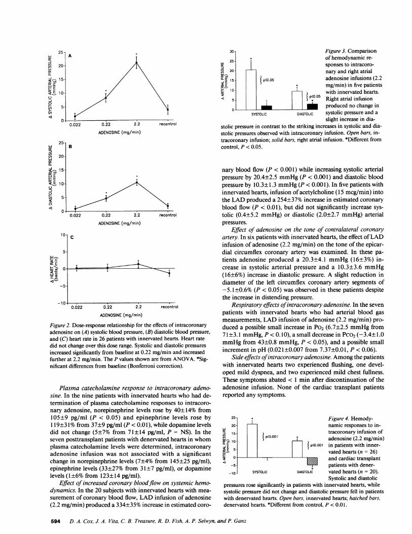
Hemodynamic effects of intracoronary adenosine in patientswith innervated hearts. Infusion of adenosine over the range0.022 to 2.2 mg/min into the LADof patients with innervatedhearts produced a dose-dependent increase in systolic and dia-stolic arterial pressure without a significant change in heartrate (Figs. 1 and 2). At the highest dose of adenosine (2.2mg/min) systolic arterial pressure rose by 21.0±2.2 mmHgfrom 134±4.3 mmHg(P < 0.001) and diastolic pressure in-creased by 10.4±1.1 mmHgfrom 76±1.9 mmHg(P< 0.001),while heart rate did not change (1.3±1.1 from 67±1.7 beats/min, P = NS). Each of these parameters returned to baseline (P= NS) during the recontrol measurement 5 min after discon-tinuing the last adenosine infusion. In the five patients withmeasurements of cardiac output, adenosine (2.2 mg/min) pro-duced a 22±3.4% increase (P < 0.01) in systemic vascularresistance from 1,045±131 dyn s cm-5 without a change incardiac output (-2.8±4.3% from 7.4±0.8 liters/min, P = NS).
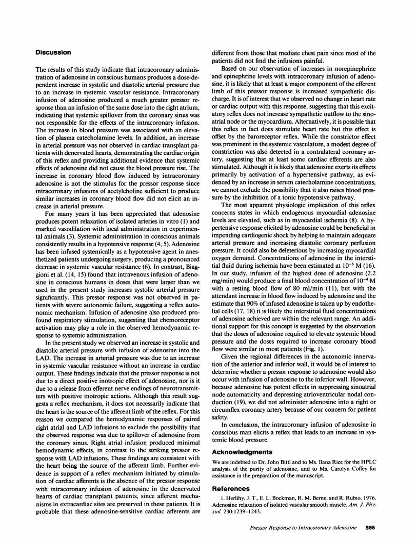
Paired LAD and right atrial infusions of adenosine. Fivepatients with innervated hearts received infusions of adenosineinto the LAD (2.2 mg/min) and into the right atrium (2.2mg/min) (Fig. 3). Systolic arterial pressure rose to a greaterextent (P < 0.05) with LAD infusion (20.4±4.8 mmHg)thanwith right atrial infusion (2.0±2.8 mmHg). There was also agreater increase (P < 0.05) in diastolic pressure with LADinfusion (9.4±2.0 mmHg) than with right atrial infusion(2.8±0.8 mmHg).
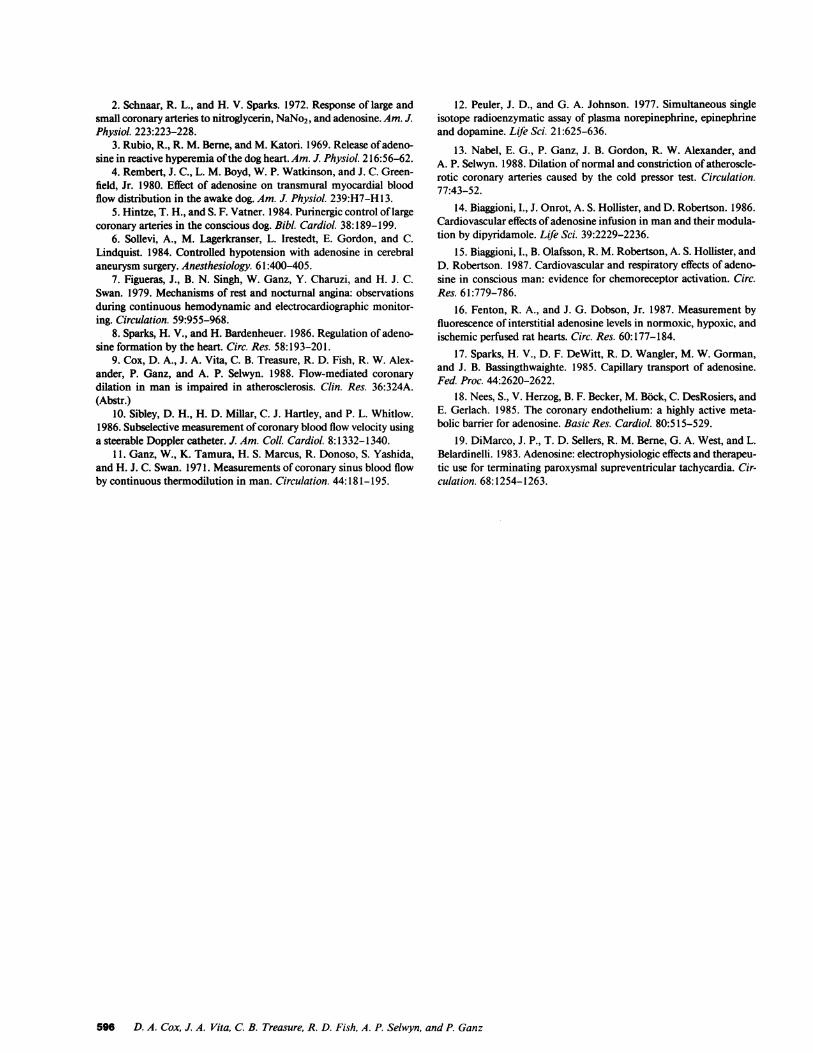
Hemodynamic effects of intracoronary adenosine in pa-tients with denervated hearts. In marked contrast to patientswith innervated hearts was the response observed in cardiactransplant patients with denervated hearts (Fig. 4), in whomadenosine (2.2 mg/min) produced no change in systolic arte-rial pressure (-0.1 ± 1.6 mmHgfrom 139±3.4 mmHg,P = NS)and a decrement in diastolic pressure (-4.7±1.2 mmHgfrom98±2.5 mmHg, P < 0.01) with no change in heart rate(-0.2±0.8 from 86±2.5 beats/min, P = NS).
ADENOSINEDOSERESPONSE
Coronary F/ow VelocDoppler Shift (Ak
200 .1.zt
A h~~ I
J,iootRv~'~
aty IA
Control 0.022 0.22 2.2
/ntrfcoronA A ?e(mg/nVln)
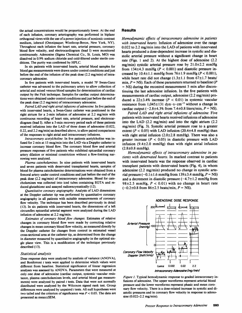
Figure 1. Typical hemodynamic response to graded intracoronary in-fusions of adenosine. The upper waveforms represent arterial bloodpressure and the lower waveforms represent phasic and mean coro-nary flow velocity. There is a dose-related increase in systolic and di-astolic pressures and in coronary flow velocity in response to adeno-sine (0.022-2.2 mg/min).
Pressor Response to Intracoronary Adenosine 593
251 -tr
cn 20-
< 15-
WEI E 10-0
0(; 5.u/
25-w
cn 20-w0.Q-< . 15-
HE< E 10-0
0C/) 50Q
c
tz Ecr-U)
I a
<_
A
/ Y~~~~~~~~~~~~00.022 0.22 2.2 recontrol
ADENOSINE(mg/min)
0.022 0.22 2.2 recontrol
ADENOSINE(mg/min)
10- c
5-
0- I I
-5-
-10-0.022 0.22 2.2 recontrol
ADENOSINE(mg/min)
Figure 2. Dose-response relationship for the effects of intracoronaryadenosine on (A) systolic blood pressure, (B) diastolic blood pressure,and (C) heart rate in 26 patients with innervated hearts. Heart ratedid not change over this dose range. Systolic and diastolic pressuresincreased significantly from baseline at 0.22 mg/min and increasedfurther at 2.2 mg/min. The P values shown are from ANOVA. *Sig-nificant differences from baseline (Bonferroni correction).
Plasma catecholamine response to intracoronary adeno-sine. In the nine patients with innervated hearts who had de-termination of plasma catecholamine responses to intracoro-nary adenosine, norepinephrine levels rose by 40±14% from105±9 pg/ml (P < 0.05) and epinephrine levels rose by1 19±31% from 37±9 pg/ml (P < 0.01), while dopamine levelsdid not change (5±7% from 71±14 pg/ml, P = NS). In theseven posttransplant patients with denervated hearts in whomplasma catecholamine levels were determined, intracoronaryadenosine infusion was not associated with a significantchange in norepinephrine levels (7±4% from 145±25 pg/ml),epinephrine levels (33±27% from 31±7 pg/ml), or dopaminelevels (1±6% from 123± 14 pg/ml).
Effect of increased coronary bloodflow on systemic hemo-dynamics. In the 20 subjects with innervated hearts with mea-surement of coronary blood flow, LAD infusion of adenosine(2.2 mg/min) produced a 334±35% increase in estimated coro-
30- Figure 3. Comparison
25- of hemodynamic re-sponses to intracoro-
20- nary and right atrial15- p(o.o5 adenosine infusions (2.2
a:E T mg/min) in five patients=10- with innervated hearts.<5- *P(005 Right atrial infusion
o- ___ produced no change inSYSTOLIC DIASTOLIC systolic pressure and a
slight increase in dia-stolic pressure in contrast to the striking increases in systolic and dia-stolic pressures observed with intracoronary infusion. Open bars, in-tracoronary infusion; solid bars, right atrial infusion. *Different fromcontrol, P < 0.05.
nary blood flow (P < 0.001) while increasing systolic arterialpressure by 20.4±2.5 mmHg(P < 0.001) and diastolic bloodpressure by 10.3±1.3 mmHg(P < 0.001). In five patients withinnervated hearts, infusion of acetylcholine (15 mcg/min) intothe LADproduced a 254±37% increase in estimated coronaryblood flow (P < 0.01), but did not significantly increase sys-tolic (0.4±5.2 mmHg) or diastolic (2.0±2.7 mmHg) arterialpressures.
Effect of adenosine on the tone of contralateral coronaryartery. In six patients with innervated hearts, the effect of LADinfusion of adenosine (2.2 mg/min) on the tone of the epicar-dial circumflex coronary artery was examined. In these pa-tients adenosine produced a 20.3±4.1 mmHg(16±3%) in-crease in systolic arterial pressure and a 10.3±3.6 mmHg(16±6%) increase in diastolic pressure. A slight reduction indiameter of the left circumflex coronary artery segments of-5.1±0.6% (P < 0.05) was observed in these patients despitethe increase in distending pressure.
Respiratory effects of intracoronary adenosine. In the sevenpatients with innervated hearts who had arterial blood gasmeasurements, LAD infusion of adenosine (2.2 mg/min) pro-duced a possible small increase in P02 (6.7±2.5 mmHgfrom71±3.1 mmHg,P < 0. 10), a small decrease in PCO2(-3.4± 1.0mmHgfrom 43±0.8 mmHg,P < 0.05), and a possible smallincrement in pH (0.021±0.007 from 7.37±0.01, P < 0.06).
Side effects of intracoronary adenosine. Amongthe patientswith innervated hearts two experienced flushing, one devel-oped mild dyspnea, and two experienced mild chest fullness.These symptoms abated < 1 min after discontinuation of theadenosine infusion. None of the cardiac transplant patientsreported any symptoms.
25 * Figure 4. Hemody-20 namic responses to in-
D 15 tracoronary infusion of.I10 Si<0° adenosine (2.2 mg/min)i~ E 5 T {p<O.001 in patients with inner-
vated hearts (n = 26)a _ and cardiac transplant
-5 patients with dener--10 SYSTOUC DIASTOLIC vated hearts (n = 20).
Systolic and diastolicpressures rose significantly in patients with innervated hearts, whilesystolic pressure did not change and diastolic pressure fell in patientswith denervated hearts. Open bars, innervated hearts; hatched bars,denervated hearts. *Different from control, P < 0.01.
594 D. A. Cox, J. A. Vita, C. B. Treasure, R. D. Fish, A. P. Selwyn, and P. Ganz
IB
Discussion
The results of this study indicate that intracoronary adminis-tration of adenosine in conscious humans produces a dose-de-pendent increase in systolic and diastolic arterial pressure dueto an increase in systemic vascular resistance. Intracoronaryinfusion of adenosine produced a much greater pressor re-sponse than an infusion of the same dose into the right atrium,indicating that systemic spillover from the coronary sinus wasnot responsible for the effects of the intracoronary infusion.The increase in blood pressure was associated with an eleva-tion of plasma catecholamine levels. In addition, an increasein arterial pressure was not observed in cardiac transplant pa-tients with denervated hearts, demonstrating the cardiac originof this reflex and providing additional evidence that systemiceffects of adenosine did not cause the blood pressure rise. Theincrease in coronary blood flow induced by intracoronaryadenosine is not the stimulus for the pressor response sinceintracoronary infusions of acetylcholine sufficient to producesimilar increases in coronary blood flow did not elicit an in-crease in arterial pressure.
For many years it has been appreciated that adenosineproduces potent relaxation of isolated arteries in vitro (1) andmarked vasodilation with local administration in experimen-tal animals (3). Systemic administration in conscious animalsconsistently results in a hypotensive response (4, 5). Adenosinehas been infused systemically as a hypotensive agent in anes-thetized patients undergoing surgery, producing a pronounceddecrease in systemic vascular resistance (6). In contrast, Biag-gioni et al. (14, 15) found that intravenous infusion of adeno-sine in conscious humans in doses that were larger than weused in the present study increases systolic arterial pressuresignificantly. This pressor response was not observed in pa-tients with severe autonomic failure, suggesting a reflex auto-nomic mechanism. Infusion of adenosine also produced pro-found respiratory stimulation, suggesting that chemoreceptoractivation may play a role in the observed hemodynamic re-sponse to systemic administration.
In the present study we observed an increase in systolic anddiastolic arterial pressure with infusion of adenosine into theLAD. The increase in arterial pressure was due to an increasein systemic vascular resistance without an increase in cardiacoutput. These findings indicate that the pressor response is notdue to a direct positive inotropic effect of adenosine, nor is itdue to a release from efferent nerve endings of neurotransmit-ters with positive inotropic actions. Although this result sug-gests a reflex mechanism, it does not necessarily indicate thatthe heart is the source of the afferent limb of the reflex. For thisreason we compared the hemodynamic responses of pairedright atrial and LAD infusions to exclude the possibility thatthe observed response was due to spillover of adenosine fromthe coronary sinus. Right atrial infusion produced minimalhemodynamic effects, in contrast to the striking pressor re-sponse with LAD infusions. These findings are consistent withthe heart being the source of the afferent limb. Further evi-dence in support of a reflex mechanism initiated by stimula-tion of cardiac afferents is the absence of the pressor responsewith intracoronary infusion of adenosine in the denervatedhearts of cardiac transplant patients, since afferent mecha-nisms in extracardiac sites are preserved in these patients. It isprobable that these adenosine-sensitive cardiac afferents are
different from those that mediate chest pain since most of thepatients did not find the infusions painful.
Based on our observation of increases in norepinephrineand epinephrine levels with intracoronary infusion of adeno-sine, it is likely that at least a major component of the efferentlimb of this pressor response is increased sympathetic dis-charge. It is of interest that we observed no change in heart rateor cardiac output with this response, suggesting that this excit-atory reflex does not increase sympathetic outflow to the sino-atrial node or the myocardium. Alternatively, it is possible thatthis reflex in fact does stimulate heart rate but this effect isoffset by the baroreceptor reflex. While the constrictor effectwas prominent in the systemic vasculature, a modest degree ofconstriction was also detected in a contralateral coronary ar-tery, suggesting that at least some cardiac efferents are alsostimulated. Although it is likely that adenosine exerts its effectsprimarily by activation of a hypertensive pathway, as evi-denced by an increase in serum catecholamine concentrations,we cannot exclude the possibility that it also raises blood pres-sure by the inhibition of a tonic hypotensive pathway.
The most apparent physiologic implication of this reflexconcerns states in which endogenous myocardial adenosinelevels are elevated, such as in myocardial ischemia (8). A hy-pertensive response elicited by adenosine could be beneficial inimpending cardiogenic shock by helping to maintain adequatearterial pressure and increasing diastolic coronary perfusionpressure. It could also be deleterious by increasing myocardialoxygen demand. Concentrations of adenosine in the intersti-tial fluid during ischemia have been estimated at 10-6 M(16).In our study, infusion of the highest dose of adenosine (2.2mg/min) would produce a final blood concentration of l0-' Mwith a resting blood flow of 80 ml/min (11), but with theattendant increase in blood flow induced by adenosine and theestimate that 90%of infused adenosine is taken up by endothe-lial cells (17, 18) it is likely the interstitial fluid concentrationsof adenosine achieved are within the relevant range. An addi-tional support for this concept is suggested by the observationthat the doses of adenosine required to elevate systemic bloodpressure and the doses required to increase coronary bloodflow were similar in most patients (Fig. 1).
Given the regional differences in the autonomic innerva-tion of the anterior and inferior wall, it would be of interest todetermine whether a pressor response to adenosine would alsooccur with infusion of adenosine to the inferior wall. However,because adenosine has potent effects in suppressing sinoatrialnode automaticity and depressing atrioventricular nodal con-duction (19), we did not administer adenosine into a right orcircumflex coronary artery because of our concern for patientsafety.
In conclusion, the intracoronary infusion of adenosine inconscious man elicits a reflex that leads to an increase in sys-temic blood pressure.
AcknowledgmentsWeare indebted to Dr. John Bittl and to Ms. Ilana Rice for the HPLCanalysis of the purity of adenosine, and to Ms. Carolyn Coffey forassistance in the preparation of the manuscript.
References1. Herlihy, J. T., E. L. Bockman, R. M. Berne, and R. Rubio. 1976.
Adenosine relaxation of isolated vascular smooth muscle. Am. J. Phy-
siol. 230:1239-1243.
Pressor Response to Intracoronary Adenosine 595
2. Schnaar, R. L., and H. V. Sparks. 1972. Response of large andsmall coronary arteries to nitroglycerin, NaNo2, and adenosine. Am. J.Physiol. 223:223-228.
3. Rubio, R., R. M. Berne, and M. Katori. 1969. Release of adeno-sine in reactive hyperemia of the dog heart. Am. J. Physiol. 216:56-62.
4. Rembert, J. C., L. M. Boyd, W. P. Watkinson, and J. C. Green-field, Jr. 1980. Effect of adenosine on transmural myocardial bloodflow distribution in the awake dog. Am. J. Physiol. 239:H7-H1 3.
5. Hintze, T. H., and S. F. Vatner. 1984. Purinergic control of largecoronary arteries in the conscious dog. Bibl. Cardiol. 38:189-199.
6. Sollevi, A., M. Lagerkranser, L. Irestedt, E. Gordon, and C.Lindquist. 1984. Controlled hypotension with adenosine in cerebralaneurysm surgery. Anesthesiology. 61:400-405.
7. Figueras, J., B. N. Singh, W. Ganz, Y. Charuzi, and H. J. C.Swan. 1979. Mechanisms of rest and nocturnal angina: observationsduring continuous hemodynamic and electrocardiographic monitor-ing. Circulation. 59:955-968.
8. Sparks, H. V., and H. Bardenheuer. 1986. Regulation of adeno-sine formation by the heart. Circ. Res. 58:193-201.
9. Cox, D. A., J. A. Vita, C. B. Treasure, R. D. Fish, R. W. Alex-ander, P. Ganz, and A. P. Selwyn. 1988. Flow-mediated coronarydilation in man is impaired in atherosclerosis. Clin. Res. 36:324A.(Abstr.)
10. Sibley, D. H., H. D. Millar, C. J. Hartley, and P. L. Whitlow.1986. Subselective measurement of coronary blood flow velocity usinga steerable Doppler catheter. J. Am. Coll. Cardiol. 8:1332-1340.
11. Ganz, W., K. Tamura, H. S. Marcus, R. Donoso, S. Yashida,and H. J. C. Swan. 1971. Measurements of coronary sinus blood flowby continuous thermodilution in man. Circulation. 44:181-195.
12. Peuler, J. D., and G. A. Johnson. 1977. Simultaneous singleisotope radioenzymatic assay of plasma norepinephrine, epinephrineand dopamine. Life Sci. 21:625-636.
13. Nabel, E. G., P. Ganz, J. B. Gordon, R. W. Alexander, andA. P. Selwyn. 1988. Dilation of normal and constriction of atheroscle-rotic coronary arteries caused by the cold pressor test. Circulation.77:43-52.
14. Biaggioni, I., J. Onrot, A. S. Hollister, and D. Robertson. 1986.Cardiovascular effects of adenosine infusion in manand their modula-tion by dipyridamole. Life Sci. 39:2229-2236.
15. Biaggioni, I., B. Olafsson, R. M. Robertson, A. S. Hollister, andD. Robertson. 1987. Cardiovascular and respiratory effects of adeno-sine in conscious man: evidence for chemoreceptor activation. Circ.Res. 61:779-786.
16. Fenton, R. A., and J. G. Dobson, Jr. 1987. Measurement byfluorescence of interstitial adenosine levels in normoxic, hypoxic, andischemic perfused rat hearts. Circ. Res. 60:177-184.
17. Sparks, H. V., D. F. DeWitt, R. D. Wangler, M. W. Gorman,and J. B. Bassingthwaighte. 1985. Capillary transport of adenosine.Fed. Proc. 44:2620-2622.
18. Nees, S., V. Herzog, B. F. Becker, M. Bbck, C. DesRosiers, andE. Gerlach. 1985. The coronary endothelium: a highly active meta-bolic barrier for adenosine. Basic Res. Cardiol. 80:515-529.
19. DiMarco, J. P., T. D. Sellers, R. M. Berne, G. A. West, and L.Belardinelli. 1983. Adenosine: electrophysiologic effects and therapeu-tic use for terminating paroxysmal supreventricular tachycardia. Cir-culation. 68:1254-1263.
596 D. A. Cox, J. A. Vita, C. B. Treasure, R. D. Fish, A. P. Selwyn, and P. Ganz