redalyc.sturge-weber syndrome: a case report
TRANSCRIPT

RSBO Revista Sul-Brasileira de Odontologia
ISSN: 1806-7727
Universidade da Região de Joinville
Brasil
da Conceição, Joanna G.; dos Santos, Luiz Felipe G.; Pimentel de Sá Bahia, Thaís; Silva, Vanessa de
A. S.; Barbosa Ramos, Maria Eliza; Israel, Mônica
Sturge-Weber syndrome: a case report
RSBO Revista Sul-Brasileira de Odontologia, vol. 8, núm. 4, octubre-diciembre, 2011, pp. 469-472
Universidade da Região de Joinville
Joinville, Brasil
Available in: http://www.redalyc.org/articulo.oa?id=153022060017
How to cite
Complete issue
More information about this article
Journal's homepage in redalyc.org
Scientific Information System
Network of Scientific Journals from Latin America, the Caribbean, Spain and Portugal
Non-profit academic project, developed under the open access initiative
Case Report Article
Sturge-Weber syndrome: a case report
Joanna G. da Conceição1
Luiz Felipe G. dos Santos1
Thaís Pimentel de Sá Bahia1
Vanessa de A. S. Silva1
Maria Eliza Barbosa Ramos1
Mônica Israel2
Corresponding author:Thaís Pimentel de Sá BahiaAvenida Lucio Costa, n.º 4.420, bloco 9, apto. 302 – Barra da TijucaCEP 22630-011 – Rio de Janeiro – RJ – BrasilE-mail: [email protected]
1 Department of Dentistry, University of Rio de Janeiro State – Rio de Janeiro – RJ – Brazil.2 Department of Dentistry, Federal University of Rio de Janeiro State – Rio de Janeiro – RJ – Brazil.
Received for publication: August 17, 2010. Accepted for publication: January 27, 2011.
Abstract
Introduction: The Sturge-Weber Syndrome, also known as encephalotrigeminal angiomatosis, is a rare vascular neurocutaneous alteration. The main clinical features of this syndrome are facial vascular cutaneous naevus, usually unilateral, which often follows the outline distribution of trigeminal nerve. Objective: To report a clinical case of Sturge-Weber Syndrome in a 29-yeral-old male patient who presented oral manifestations related to the syndrome. Case report and conclusion: The patient reported that he had presented a cutaneous vascular nevus on the face during childhood as well as epileptic crisis episodes. However, he had no ophthalmic alterations. Sturge-Weber syndrome is a systemic condition commonly affecting oral cavity through vascular lesions, therefore, dentists’ knowledge is extremely important to provide an adequate dental treatment without complications.
Keywords: Sturge-Weber syndrome; angiomatosis; hemangioma.
ISSN: Printed version: 1806-7727Electronic version: 1984-5685RSBO. 2011 Oct-Dec;8(4):469-72
Introduction
S t u r g e - W e b e r s y n d r o m e i s a r a r e developmental nonhereditary congenital condition [2, 9], characterized by vascular hamartoma
involv ing brain and face t issues [10]. A lso known as encephalotrigeminal angiomatosis [1], Sturge-Weber syndrome is a rare vascular neurocutaneous alteration presenting oral and systemic signals.
Conceição et al.Sturge-Weber syndrome: a case report470 –
The main clinical features of this syndrome are: port-wine stain in the face (naevus flammeus), usually unilateral, which often follows the outline distribution of trigeminal nerve [10, 14]; and venous angiomas in leptomeninges over the cerebral-cortex and ipsilateral gyriform calcification, leading to epilepsy and contralateral hemiparesthesia. Other manifestations may be related to this syndrome such as: glaucoma and mental retardation [12].
Neurological involvement is related to the presence of leptomeningeal angiomas involving the pia mater. It is believed that this syndrome’s possible cause be a vascular plexus around the cephalic portion of the neural tube [13].
The disease’s most frequent feature is the presence of epileptic crisis ranging from 74% to 90% of the cases [3]. The later the epilepsy onset, the lesser is the patient’s probability of developing neuropsychomotor retardation [4].
Among the disease’s oral manifestations, it can be emphasized the morphological and histological alterations in the periodontal tissues, and eventually, pulp involvement has been reported [8, 11]. However the feature most commonly found in oral cavity is gingival hemangioma, which frequently affects the ipsilateral area of maxilla, mandible, mouth’s floor, lips, palate, tongue and jugal mucosa [6].
It is important that the clinician know this disease’s clinical features and complications that may occur. Surgical procedures in patients presenting this syndrome should be carefully conducted due to increased risk of hemorrhage, even if they are considered as simple [1].
The aim of this study is to report a clinical case of Weber-Sturge syndrome in a male, 29-year-old patient presenting oral manifestations related to the syndrome.
Case report
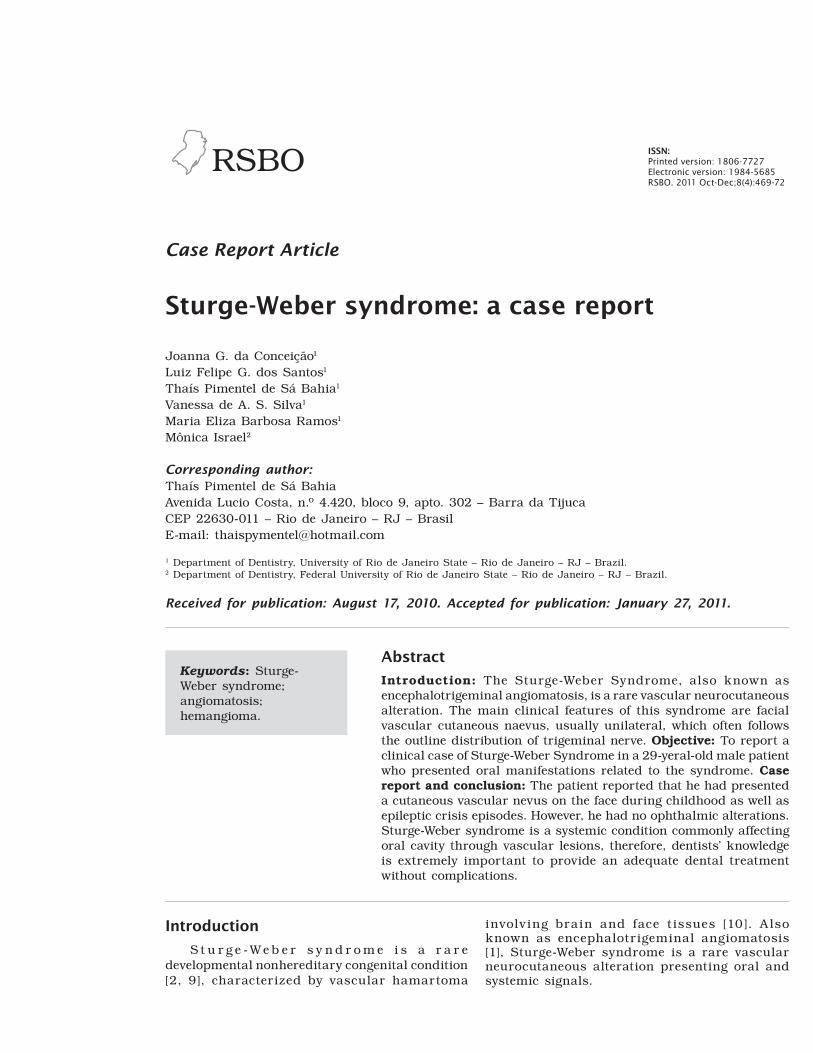
A male, leucoderm, 29-year-old pat ient was referred to the Stomatology Clinics of the School of Dentistry (University of Rio de Janeiro State) for routine dental treatment. At extra-oral physical examination, a reddish lesion similar to hemangiona was observed at the vermillion border of the lower lip causing a volume increase at this anatomical area (figure 1). Diagnosis hypothesis was confirmed after vitropression execution.
Figure 1 – Lesion in vermillion border of lower lip
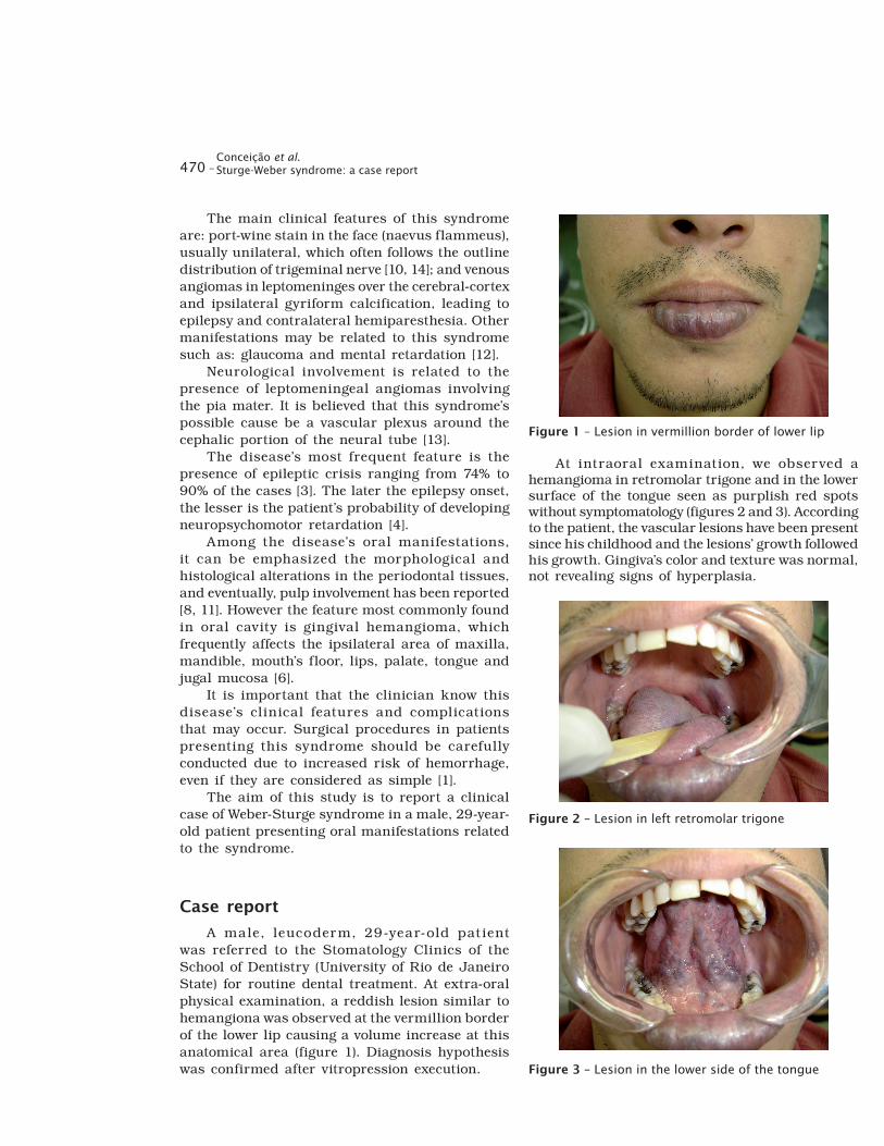
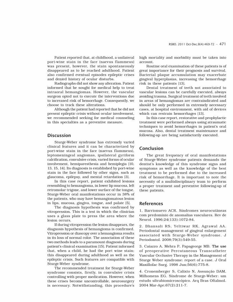
At intraoral examination, we observed a hemangioma in retromolar trigone and in the lower surface of the tongue seen as purplish red spots without symptomatology (figures 2 and 3). According to the patient, the vascular lesions have been present since his childhood and the lesions’ growth followed his growth. Gingiva’s color and texture was normal, not revealing signs of hyperplasia.
Figure 2 – Lesion in left retromolar trigone
Figure 3 – Lesion in the lower side of the tongue
RSBO. 2011 Oct-Dec;8(4):469-72 – 471
Patient reported that, at childhood, a unilateral port-wine stain in the face (naevus f lammeus) was present, however, the stain spontaneously disappeared as to he reached adulthood. Patient also confirmed eventual episodes epileptic crises and denied history of ocular disturbs.
Radiographs did not show any alteration. Patient informed that he sought for medical help to treat intraoral hemangiomas. However, the vascular surgeon opted not to execute the interventions due to increased risk of hemorrhage. Consequently, we choose to track these alterations.
Although the patient had reported that he did not present epileptic crisis without ocular involvement, we recommended seeking for medical counseling in this specialties as a preventive measure.
Discussion
Sturge-Weber syndrome has extremely varied clinical features and it can be characterized by port-wine stain in the face (naevus f lammeus), leptomeningeal angiomas, ipsilateral gyriform calcification, convulsive crisis, varied forms of ocular involvement, hemiparesthesia and hemiplegia [10, 13, 15, 14]. Its diagnosis is established by port-wine stain in the face followed by other signs, such as glaucoma, epilepsy, and mental retardation [3].
In this case report, patient exhibited lesions resembling to hemangioma, in lower lip mucosa, left retromolar trigone, and lower surface of the tongue. Sturge-Weber oral manifestations occur in 38% of the patients, who may have hemangiomatous lesion in lips, mucosa, gingiva, tongue, and palate [5].
The diagnosis hypothesis was confirmed by vitropression. This is a test in which the clinician uses a glass plate to press the area where the lesion occurs.
If during vitropression the lesion disappears, the diagnosis hypothesis of hemangioma is confirmed. Vitropression or diascopy over a hemangioma results in its loss of normal color. The association of these two methods leads to a paramount diagnosis during patient’s clinical examination [15]. Patient informed that, when a child, he had the port wine stain; this disappeared during adulthood as well as the epileptic crisis. Such features are compatible with Sturge-Weber syndrome.
The recommended treatment for Sturge-Weber syndrome consists, firstly, in convulsive crisis controlling with proper medication. However, when these crises become uncontrollable, neurosurgery is necessary. Notwithstanding, this procedure’s
high mortality and morbidity must be taken into account.
Routine oral examination of these patients is of great importance for their prognosis and survival. Bacterial plaque accumulation may exacerbate gingival hyperplasias, increasing the hemorrhage risk in these patients [13].
Dental treatment of teeth not associated to vascular lesions can be carefully executed, always avoiding trauma. Surgical treatment of teeth involved in areas of hemangiomas are contraindicated and should be only performed in extremely necessary cases, at hospital environment, with aid of devices which can restrain hemorrhages [13].
In this case report, restorative and prophylactic treatment were performed always using atraumatic techniques to avoid hemorrhages in patient’s oral mucosa. Also, dental treatment maintenance and following-up are being satisfactorily executed.
Conclusion
The great frequency of oral manifestations of Sturge-Weber syndrome patients demands the dentist’s knowledge of this syndrome signs and symptoms as well as the knowledge of the best treatment to be performed due to the increased risk of hemorrhage. It is important to note the necessity of a multidisciplinary team to perform a proper treatment and preventive following-up of these patients.
References
1. Barrionuevo ACR. Síndromes neurocutâneas com predomínio de anomalias vasculares. Rev de Neurol. 1996;24(133):1072-84.
2. Bhansali RS, Yeltiwar RK, Agrawal AA. Periodontal management of gingival enlargement associated with Sturge-Weber syndrome. J Periodontol. 2008;79(3):549-55.
3. Caiazzo A, Mehra P, Papageorge MB. The useThe use of preoperative Percutaneous Transcatheter Vascular Occlusive Therapy in the Management of Sturge Weber syndrome: report of a case. J Oral Maxillofac Surg. 1998 Jun;56(6):775-8.
4. Cronemberger S, Calixto N, Assunção DAM, Milhomens EG. Síndrome de Sturge-Weber: um estudo ultrabiomicroscópico. Arq Bras Oftalmol. 2004 Mar-Apr;67(2):211-7.

Conceição et al.Sturge-Weber syndrome: a case report472 –
5. Dualibi SE, Vieira MBEIP, Riviriego RA. Proposta de tratamento odontológico em paciente portador de Síndrome de Sturge-Weber (relato de caso clínico). Rev Paul Odontol. 1989 Nov-Dec;11(6):2, 4, 6.
6. Gasparini G, Perugini M, Vetrano S, Cassoni A, Fini G. Angiodysplasia with osteohypertrophy affecting the oromaxillofacial area: clinical f indings. The J of Craniofac Surg. 2001 Sep;12(5):485-9.
7. Güngör HC, Altay N, Kaymaz FF. Pulpal tissuePulpal tissue in bilateral talon cusps of primary central incisors: report of a case. Oral Surg Oral Med Oral Pathol Oral Radiol Endod. 2000 Feb;89(2):231-5.
8. Jacobs J, Levan P, Olivier A, Andermann F, Dubeau F. Late-onset epilepsy in a surgically-treated Sturge-Weber patient. Epileptic Disord. 2008;10(4):312-8.
9. Neville B, Damm D, Allen C, Bouquot JE. Patologia oral e maxilofacial. 2. ed. Rio de Janeiro:Rio de Janeiro: Guanabara-Koogan; 2002.
10. Palheta Neto FX, Vieira Junior MA, Ximenes LS, Jacob CCS, Rodrigues Junior AGR, Palheta ACP et al. Aspectos clínicos da Síndrome de Sturge-Weber. Arq Int Otorrinolaringol. 2008;12(4):565-70.
11. Pozzati E, Padovani R, Frank F, Gaist G. Leptomeningeal angiomatosis and aplasia congenita of the scalp. J Neurosurg. 1983 Jun;58(6):937-40.J Neurosurg. 1983 Jun;58(6):937-40.1983 Jun;58(6):937-40.
12. Santos S, Cavalheiro L. Síndrome de Sturge-Weber: relato de caso dos achados da avaliação fonoaudiológica. Rev Cefac. 2010 Jan-Rev Cefac. 2010 Jan-Feb;12(1):161-70.
13. Sen Y, Dilber E, Odemis E, Ahmetoglu A, Aynaci FM. Sturge Weber syndrome in a 14-year-Sturge Weber syndrome in a 14-year-old girl without facial naevus. Eur J Pediatr. 2002 Sep;161(9):505-6.
14. Shafer WG, Hine MK, Levy BM. A textbook of oral pathology. 4. ed. Harcourt Asia Pub; 2002. p. 157-8.
15. Silva FM, Portolan FBAM, Figueiredo PJ. Hemangioma. Rev Fac Odontol Lins. 2000;12Rev Fac Odontol Lins. 2000;122000;12 (1, 2):57-9.