red and white lesions of the oral mucosa. hereditary white lesions reactive/inflammatory white...
TRANSCRIPT
RED AND WHITE LESIONS OFTHE ORAL MUCOSA
HEREDITARY WHITE LESIONS REACTIVE/INFLAMMATORY WHITE LESIONS INFECTIOUS WHITE LESIONS AND WHITE AND RED LESIONS IDIOPATHIC “TRUE” LEUKOPLAKIA BOWEN’S DISEASE ERYTHROPLAKIA ORAL LICHEN PLANUS LICHENOID REACTIONS LUPUS ERYTHEMATOSUS (SYSTEMIC AND DISCOID) DEVELOPMENTAL WHITE LESIONS: ECTOPIC LYMPHOID
TISSUE FORDYCE’S GRANULES GINGIVAL AND PALATAL CYSTS OF
THE NEWBORN AND ADULT MISCELLANEOUS LESIONS
LEUKOEDEMA
Etiology Unknown Benign; common in general
population, with racial clustering in Blacks
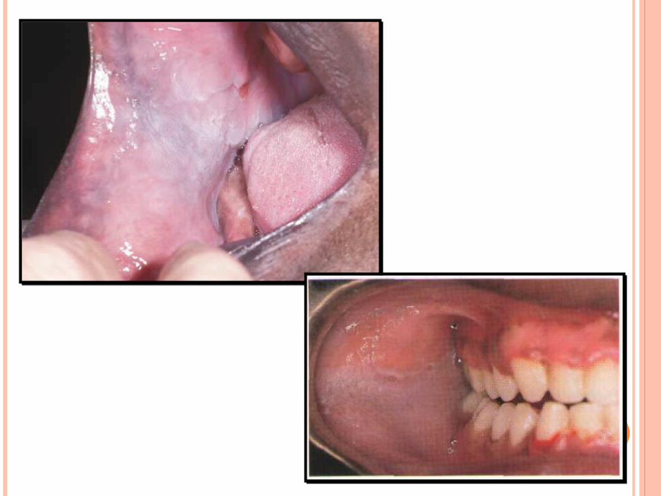
Clinical Presentation Symmetric, asymptomatic. Buccal mucosa involved by gray-
white, diffuse, milky surface with an opalescent quality.
Wrinkled surface features at rest. Dissipation of changes with
stretching of mucosa.
Diagnosis Clinical recognition is sufficient. Biopsy findings will show marked
intracellular edema of spinous layer. Individual cells with clear cytoplasm and
compact nuclei. Normal basal cell layer.
Differential Diagnosis Cheek chewing Hereditary benign intraepithelial
dyskeratosis White sponge nevus Lichen planus Candidiasis
Treatment None necessary; no relation to
dysplasia /carcinoma Reassurance
Prognosis Excellent
WHITE SPONGE NEVUS
Etiology Hereditary (autosomal-dominant)
disorder of keratinization affecting nonkeratinizing oral, esophageal, and anogenital mucosal epithelium.
Point mutations in the keratin 4 and/or 13 genes
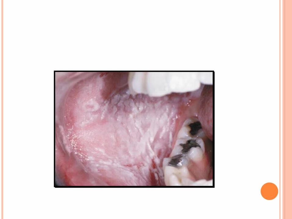
Clinical Presentation Asymptomatic Deeply folded, thickened, white mucosa Buccal mucosa chiefly affected No functional impairment Increased prominence during second
decade
Microscopic Findings Parakeratosis, acanthosis, intracellular
edema Perinuclear condensation of keratin
Diagnosis Clinical appearance Family history Microscopic findings
Differential Diagnosis Idiopathic leukoplakia Chemical/thermal burn Chronic low-grade trauma (morsicatio)
Treatment None required No malignant potential
Prognosis Excellent
HEREDITARY BENIGN INTRAEPITHELIAL DYSKERATOSIS
Etiology It is a rare, autosomal dominant
hereditary condition. Also known as Witkop’s disease.
Clinical Presentation Early onset of bulbar conjunctivitis and oral white
lesions. Preceding the bulbar conjunctivitis are foamy
gelatinous plaques that represent the ocular counterpart of the oral mucosal lesions.
Oral lesions consist of soft, asymptomatic, white folds and plaques of spongy mucosa.
Areas characteristically involved include the buccal and labial mucosa and labial commissures, as well as the floor of the mouth and lateral surfaces of the tongue, gingiva, and palate. The dorsum of the tongue is usually spared.
Patients may complain of photophobia, especially in early life. Blindness, secondary to corneal vascularization, has been reported.
Diagnosis Clinical appearance Family history Microscopic findingsDifferential Diagnosis Idiopathic leukoplakia Chemical/thermal burn Chronic low-grade trauma (morsicatio) Lichen planus Lubus erythematosus
Treatment None required For evaluation and treatment of
the ocular lesions, the patient should be referred to an ophthalmologist.
Prognosis Excellent