scoring and grading b-mode synovitis and doppler findings ... · scoring and grading b-mode...
TRANSCRIPT

ScoringandGradingB-ModeSynovitisandDopplerfindingsinpediatricMSKUS
JohannesRothMDPhDFRCPCRhMSUS

Pathology- DefinitionSynovitis
•SynovitisonultrasonographyinchildrenB-modeandDoppler
•Dependingonthejoint,synovitiscanbediagnosedonthebasisofB-modeabnormalitiesalone
•AbnormalB-modeincludessynovialeffusionorsynovialhypertrophy

Pathology- DefinitionSynovitis ctd.
• Synovialeffusionisdefinedasabnormal,intraarticular,an-orhypoechoic fluidthatisdisplaceable
• SynovialHypertrophyisdefinedasabnormal,intraarticular,hypoechoic materialthatisnon-displaceable
• AbnormalDopplersignalswithinanareaofsynovialhypertrophy
• PhysiologicDopplersignalscanbepresentinanyareaofthejoint
hypo- andanechoic (non-)displaceable Doppler

Scores
B-ModeandDoppler
Rationale:
1) Serialclinicalassessmentandcomparability
2) Research

Howtoreport?
1)Descriptive– “Effusioninthetibiotalar jointwithincreasedDoppler”,measure?
2)Qualitative– Mild,moderate,severe
3)Semiquantiative Scoring
4)Pixelcountorother(RI)?

Challenges
• Variabilityinequipment
• Solution:Ensureoptimizationofsettings,especiallyfrequencyandPRF
• Donotusestandardizedsettings!
• But:paygreatattentiontolowflowsettingsandfrequency

UltrasoundScoringSystems

Semi-quantitative US-Score
4 step semi quantitative grading system (Grad 0 – 3) separately for:
•Joint effusion•Synovial hypertrophy•Bone erosions•Power Doppler activity
of 5 selected joints unilateral: MCP II, III, PIP II and MTP I and II from dorsal
Interobserver Kappa agreement 0.48 – 0.68
Szkudlarek M et al: Arthritis Rheum. 2001;44:2018-23

Semi-quantitative Synovitis – Score
Grade 0: no elevation of joint capsule, no synovitisGrade 1: sligth elevation of joint capsule, mild synovitis, parallel to bone lineGrade 2: paralel elevation of joint capsule to skin, moderate synovitisGrade 3: convex elevation of joint capsule severe synovitis
Sum scores of synovitis of MCP II –V and PIP II – VAssess dorsal and volar
Scheel AK, Backhaus M et al., Arthritis Rheum 2005: 52; 733-43

Pathology– ScoresAdultsB-Mode
ScheelAK,BackhausMetal.,ArthritisRheum2005:52;733-43

Pathology– ScoresAdultsDoppler
Scheel AK, Backhaus M et al., Arthritis Rheum 2005: 52; 733-43

Grade 0 or normal: no synovial hypertrophy, no Doppler signal
Grade 1 or minimal: minimal synovial hypertrophy, with (or without) no more than grade 1 Doppler signal
Grade 2 or moderate: moderate synovial hypertrophy with no more than grade 2 Doppler signalminimal synovial hypertrophy and grade 2 Doppler signal
Grade 3 or severe: severe synovial hypertrophy with or without Doppler signalminimal/moderate synovial hypertrophy and grade 3 Doppler signal
D´Agostino MA, et al. J Rheumatol 2009 Naredo E, et al. J Rheumatol 2011
Composite score

US Scores 7 – 12 – 28 – 44 – 78 joints
High correlation coefficients between all these scores for grey scale and Power Doppler
Hammer-H et al ACR 2011
Howmanyjointstoassess?
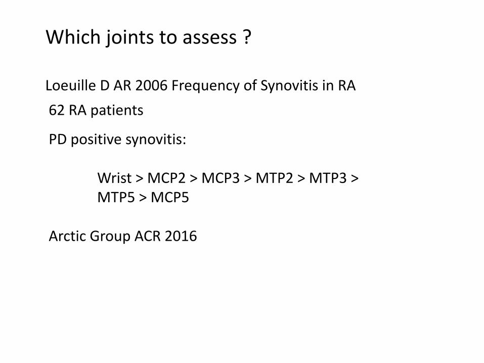
Whichjointstoassess?
Loeuille DAR2006FrequencyofSynovitisinRA62RApatients
PDpositivesynovitis:
Wrist>MCP2>MCP3>MTP2>MTP3>MTP5>MCP5
ArcticGroupACR2016

based on suggested frequency of involvement25, feasibility8, orrepresentative value of target joints9, or were developed in alogistic regression model26. Two reduced joint counts seemedto present good validity issues: the 12-joint count proposed byNaredo, et al26 and the 7-joint count by Backhaus, et al25.Joints selected in the proposed 12-joint count are wrist,MCP-2, MCP-3, knee, ankle, and elbow evaluated bilateral-ly26. Examining the other proposed joint counts, we found thatsome of them included a minimal number of 7 joints in theirscoring systems and, in particular, joints featured in the 7-jointcount25 combination [i.e., wrist, MCP-2, MCP-3, PIP-2, PIP-3,metatarsophalangeal (MTP)-2, and MTP-5] were included inthe global synovitis scores of 50% (7 of 14) of the articles. Inorder to evaluate the applicability of the 7-joint count in anoth-er dataset, we analyzed data from Naredo and colleagues byusing the proposed selection of 7 joints from Backhaus25.Comparative results on responsiveness by using the 2 jointcounts are presented in Table 4. The use of the 7-joint count inthe new dataset also showed good responsiveness; however,the application of this joint count bilaterally (14 instead of 7
joints) was characterized by higher sensitivity to change,which was closer to that observed with the evaluation of all ini-tially evaluated joints (i.e., 44 joints).
DISCUSSION
The prospect of developing a global ultrasound joint score isattractive, in that it might potentially be able to more objec-tively reflect the “real” level of synovitis, and hence diseaseactivity of patients with RA, compared with conventionalclinical measures, i.e., disease activity indices. In order to beaccepted as an objective tool, ultrasound must demonstratereliability and sensitivity to change, and the evaluation of several joints must also appear feasible. This review hasdemonstrated that ultrasound is a worthwhile tool for assess-ing global joint inflammation in RA.
This review has highlighted that discrepancies were pres-ent among studies, relating to the definition and detection ofsynovitis, and in the composition of joints included in theglobal evaluation of disease activity. The variability of defini-tion and detection of synovitis at joint level within the studies
2059Mandl, et al: US scoring systems of RA synovitis
Personal non-commercial use only. The Journal of Rheumatology Copyright © 2011. All rights reserved.
Table 3. Metric properties of the studies.
Year Study Validity Responsiveness Reliability FeasibilityFace Content Construct Criterion
2003 Szudlarek9 Yes Yes Yes NA NA Yes Yes2004 Taylor13 Yes Yes Yes NA Yes NA Yes2005 Naredo27 Yes Yes Yes NA NA Yes Bo2005 Scheel8 Yes Yes Yes NA NA NA Yes2005 Naredo28 Yes Yes Yes NA NA NA Yes2005 Naredo29 Yes Yes Yes NA NA Unclear Yes2008 Hameed30 Yes Yes Yes NA NA NA Yes2008 Ozgocmen31 Yes Yes Yes NA NA No Yes2008 Naredo26 Yes Yes Yes NA Yes Yes Yes/No*2008 Naredo32 Yes Yes Yes NA Yes Yes Yes2009 Scire33 Yes Yes Yes NA Yes Yes Yes2009 Backhaus25 Yes Yes Yes NA Yes Unclear Yes2009 Dougados34 Yes Yes Yes NA Yes Yes Yes/No*2010 Balsa35 Yes Yes Yes NA Unclear Yes No
* Several scoring systems were evaluated in the study, feasibility varied with number of joints assessed. NA: not assessed in study.
Table 4. Evaluation of responsiveness of the 7-joint score developed by Backhaus, et al25 in an additional dataset obtained from Naredo, et al26. Data for thisanalysis is courtesy of Marina Backhaus and Esperanza Naredo.
Joint Count Gray-scale Synovitis Power Doppler ActivityMean decrease‡ SRM Mean decrease‡ SRM
(95% CI) (95% CI)
Wrist, MCP2, MCP3, knee ankle, elbow (bilateral)* 2.5 (2.0–2.9) 0.925 2.0 (1.6–2.4) 0.797Simplified 12-joint PDUS model (12 joints 24 recesses)# 2.3 (1.9–2.8) 0.881 1.8 (1.3–2.2) 0.717Wrist, MCP2, MCP3, PIP2, PIP3, MTP2, MTP5 (unilateral right)** 1.5 (1.2–1.7) 0.842 1.0 (0.8–1.2) 0.678Wrist, MCP2, MCP3, PIP2, PIP3, MTP2, MTP5 (unilateral left)** 1.3 (1.0–1.6) 0.732 0.7 (0.5–1.0) 0.581Wrist, MCP2, MCP3, PIP2, PIP3, MTP2, MTP5 (bilateral)† 2.8 (2.3–3.4) 0.890 1.8 (1.3–2.2) 0.72144 joints†† 7.4 (6.1–8.8) 0.934 4.6 (3.5–5.6) 0.732
* Joints selected in the 12-count from Naredo, et al26; # for detailed list of recesses see Naredo, et al 26, ** 7-joint count 25, see Backhaus, et al for details onrecesses and scoring modalities; † 7-joint count evaluated bilaterally; †† 44 joints include bilateral shoulder, elbow, wrist, MCP1-5, PIP-5, hip, knee, ankle,tarsal, MTP1-5; ‡ sensitivity to change; SRM: standardized response mean; MCP: metacarpophalangeal joint; PDUS: power Doppler ultrasound.
RheumatologyThe Journal of on March 5, 2016 - Published by www.jrheum.orgDownloaded from
Mandl PetalJRheum2013
Conclusion:Smallandatleastonelargejointbilateral

Challenges
• Scoresdifferintheviewstheyinclude,MCPdorsalvsvolar
• Doesthescoringsystemworkforallviews?
• Doesthescoringsystemworkforlargejoints?
• 4viewsaresummarizedfortheMCPresultinginamaxscoreof12comparedtooneviewforthekneeresultinginamaxscoreof3
• Noimagesarekept,Kaeley etalAR2016significantreaderdrift

Backhaus Large Joint Score BMC Musculoskeletal Disorders 2013
GSUSSynovitis by GSUS analyzed semiquantitatively from 0 to 3 (0 = absence, 1 = mild, 2 = moderate, 3 = severe) Grade 1 small abnormal hypoechoic/anechoic line beneath capsule.Grade 2 joint capsule elevated parallel to the joint area. Grade 3 strong convex distension of the joint.
PDUSGrade 0 = no intraarticular color signal, Grade 1 = up to 3 color signals representing only low flowGrade 2 = greater than grade 1 to < 50% of the intraarticular areaGrade 3 = > 50% of the intraarticular area filled with color signals.


WhereDoWeLookforSynovitis?
Recessesarelaxcapsularareaswherefluidandsynovialhypertrophyaccumulate.FavoritejointsandPDsensitivity:
MCP:Dorsal>Volar
PIP:Volar>Dorsal
Wrist:Dorsal
Elbow:LateralandAnnularRecess>Posterior>Anterior
Knee:Parapatellar >Parameniscal >>Suprapatellar
MTP– Dorsalrecessescontainsmallamountsoffluidnormally

Synovitis MCP
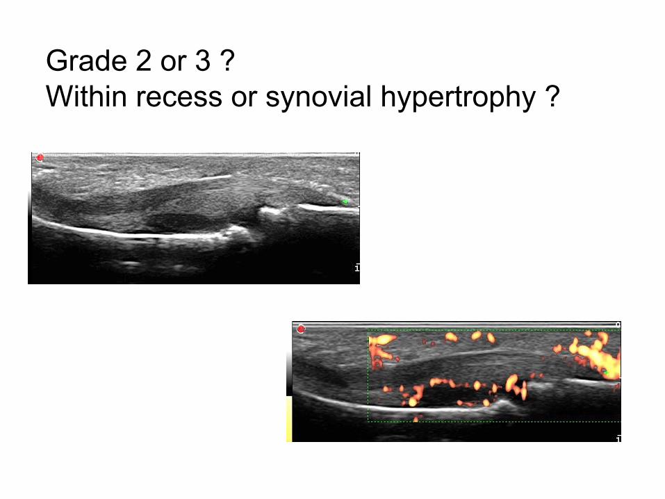
Grade 2 or 3 ?Within recess or synovial hypertrophy ?

WhichcolorshouldthePixelsbein?

Kaeley etal– Tocilizumabstudy
BluecolorforPowerDoppler
DetailedscoringsystemB-modeandDoppler

Collado-PetalRheumatology2013
ScoresPediatric
reducedjointpowerDopplerUS(PDUS)score
validity,feasibility,reliabilityandsensitivitytochangecomparedwitha44jointsPDUSassessmentinJIA
42childrenwithactiveJIArequiringmodifiedtherapy
greyscale(GS)synovitisandpowerDopplersignal4-pointsemiquantitativescale
calculationofUScompositeindicesandUScompositejointcounts

PedSynS 4-pointsemiquantitative scaleofGSsynovitis
0 absence,normaljointrecess
1 mild,synovitis fillingthejointrecessbetweenperiarticular epiphyseschangefromangle-shapedtoaplateau-shapedrecess
2 moderate,convexshapeofthejointrecesswithoutextensionoverthebonediaphysis
3 marked,convexshapeofthejointrecesswithextensiontoatleastoneofthebonediaphyses
Collado-PetalRheumatology2013

Collado P, et al. Rheumatology 2013;52:1477–84.
Grade 0
Grade 1
Grade 2
Grade 3
0 absence, normal joint recess
1 mild, synovitis filling the joint recess between periarticular epiphyses, change from angle-shapedto a plateau-shaped recess
2 moderate, convex shape of the joint recess without extension over the bone diaphysis
3 marked, convex shape of the joint recess withextension to at least one of the bone diaphyses
Theproblem:

• Grade0=normal
• Grade1=mild,synovitisfillingthejointrecessbetweenperiarticularepiphysesthatleadstoachangefromtheangle-shapedrecesstoaplateau-shapedrecess
• Grade2=convexshapeoftherecesswithoutextensiontothebonediaphysisnortothewholelengthofashortbone
• Grade3=convexshapeoftherecesswithextensiontothebonediaphysisorthewholelengthofashortbone
Scoresuggestion

PowerDopplerScore
0absence,nosynovialflow
1mild,single-vesselsignal
2moderate,confluentvessels
3marked,vesselsignalsinmorethanhalfoftheIntraarticulararea
Tenosynovitisandbursitiswerescoredusingadichotomousassessment(0,absence;1,presence)inbothGSandPDscoringsystems
Collado-PetalRheumatology2013

CurrentPediatricProjects
1) Omeractongoing– globalscoringsystem
2) PRES
3) PANLAR– enthesis
4) CARRA– jointbyjoint
5) Collaborativeprojectsusing12jointscore

Reader&Manual&Version&1&–&February&6th&2016&&
2&&
1"#"Overview"of"Joints"and"Images"Joint Imaging specification Total Number of Images
Elbow • A sagittal scan over the humeroulnar, humeroradial and dorsal elbow joint will be done.
• 3 images in B-Mode and 3 images in Doppler-Mode, total 6 images.
Wrist • A sagittal scan over the dorsal aspect of the medial and ulnar part of the wrist will be done and the radiocarpal, midcarpal as well as the carpometacarpal joints will be evaluated.
• The radioulnar joint will be evaluated as well in an additional scan.
• A dorsal transverse scan of the wrist will be done to evaluate the extensor tendon sheaths
• 4 images in B-Mode and 4 images Doppler-Mode, total 8 images.
Fingers • The subject will be in a sitting position with the hands palm-side down or dorsum-side down (depending on whether dorsal or volar assessment is done) in a neutral position on an examination table.
• A sagittal scan over the dorsal and volar aspect of the MCP 2 and 3 joint will be done. In addition a transverse scan on the dorsal and volar aspect will be done for MCP 2 and 3.
• 8 images in B-Mode and 8 images Doppler-Mode, total 16 images
Knee • The subject will be in a supine position with the lower extremities parallel to each other. A measurement will be taken in 30 degrees flexed position.
• The measurement will be done sagittally over the suprapatellar recess, with the probe positioned cranial to the superior edge of the patella.
• Subsequently a medial and lateral parapatellar scan will be done.
• 3 images in B-Mode and 3 images Doppler-Mode, total 6 images
Ankle • The subject will be in supine position with the knee in 90 degrees flexion.
• A sagittal scan over the medial aspect of the tibiotalar joint will be done. In addition in 3 transverse scans the tendon sheath and cross sectional area of the Tibialis anterior, Extensor Hallucis Longus, Extensor Digitorum at the level of the talar cartilage, the Tibialis Posterior, Flexor Digitorum, Flexor Hallucis Longus at the level of the sustentaculum and the Peroneus Tendons at the level of the supramalleolar and trochlea (where they separate) will be assessed.
• In a separate medial and lateral scan the subtalar joint will be assessed.
• Finally the talonavicular joint will be assessed in a sagittal scan.
• 7 images in B-Mode and 7 images Doppler-Mode, total 14 images

Reader&Manual&Version&1&–&February&6th&2016&&
13&&
Grade"1"–"mild"
Distension& of& synovial& recess& originating& from& the& radiocarpal& or& midcarpal& joint& or& both& with& the&superior&border&of&the&recess&remaining¶llel&to&the&respective&bone&line&and&filling&less&than&50&%&of&the&potential&space&below&the&beta&line.&
&
Grade"2"–"moderate"
Distension& of& synovial& recess& originating& from& the& radiocarpal& or& midcarpal& joint& or& both& with& the&superior&border&of&the&recess&convex&and&filling&50&%&or&more&of&the&potential&space&below&the&beta&line&and& remaining&deep& to& the&beta& line.&Distension&of& the" recess&may&be&present& in& the& radiocarpal& joint&only,&in&the&intercarpal&joint&only,&or&in&both&joints.&
&
Grade"3"–"severe&
Distension& of& synovial& recess& originating& from& the& radiocarpal& or& midcarpal& joint& or& both& with& the&superior&border&of&the&recess&convex&and&extending&above&the&beta&line.&&
&
&& &
& &
&
Radius''''''''''''''Lunate'''''''''& !!!!!!!!!!!!!!!!!!!!!!!!!!!!!!Capitate& !!!!!!!!!!!!!!!!!!!!!!!!!!&!!!!!!!!Metc
& &
&& &
& &
&
Radius''''''''''''''Lunate#########& !!!!!!!!!!!!!!!!!!!!!!!!!!!!!!Capitate& !!!!!!!!!!!!!!!!!!!!!!!!!!&!!!!!!!!Metc
& &
&& &
& &
&
Radius''''''''''''''Lunate'''''''''& !!!!!!!!!!!!!!!!!!!!!!!!!!!!!!Capitate& !!!!!!!!!!!!!!!!!!!!!!!!!!&!!!!!!!!Metc
& &

ScoringDoppler
Grade0NoDopplersignalsexceptforfeedingvesselscrossingsynovialrecesses.Afeedingvesselisdefinedasasinglevesselshowingcontinuityinenteringthebone/cartilage
Grade1SingleSignals(uptothree)
Grade2Confluentsignalsinlessthan50%ofsynovialhypertrophy/hyperplasia
Grade3Confluentsignalsinmorethan50%ofsynovialhypertrophy/hyperplasia
Reader&Manual&Version&1&–&February&6th&2016&&
32&&
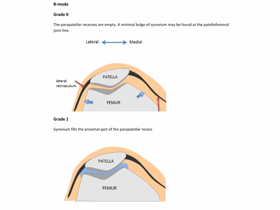
4.4.2."Knee"–"parapatellar""
B6mode"
Grade"0"
The¶patellar&recesses&are&empty.&A&minimal&bulge&of&synovium&may&be&found&at&the&patellofemoral&joint&line.&&
"
Grade"1"
Synovium&fills&the&proximal&part&of&the¶patellar&recess&
"
"""""""""" " "

Reader&Manual&Version&1&–&February&6th&2016&&
33&&
Grade"2"
Synovium&fills&part&of&the¶patellar&recess&or&extends&throughout&the&recess&filling&<50%&of&its&expected&full&volume.&
""""""""""""""""""" "
"
Grade"3""
Synovium&occupies&>50%&of&the&expected&full&volume&of&the&recess&
&
"
"
"

Enthesis ScoresAdults
Hugevariabilityinscores
B-modevsDoppler,DopplerinareaofabnormalB-mode?
Tendon,Enthesis,Bursae,otherstructures
Inclusionofparametersofdamage?
Varioussumscoresweighingparametersdifferently
RESEARCH ARTICLE Open Access
Ultrasound in the evaluation of enthesitis: statusand perspectivesFrédérique Gandjbakhch1, Lene Terslev2, Fredrick Joshua3, Richard J Wakefield4, Esperanza Naredo5 andMaria Antonietta D’Agostino6*, for OMERACT Ultrasound Task Force
Abstract
Introduction: An increasing number of studies have applied ultrasound to the evaluation of entheses inspondyloarthritis patients. However, no clear agreement exists on the definition of enthesitis, on the number andchoice of entheses to examine and on ultrasound technique, which may all affect the results of the examination.The objectives of this study were to first determine the level of homogeneity in the ultrasound definitions for theprincipal lesions of enthesitis in the published literature and second, to evaluate the metric properties ofultrasound for detecting enthesitis according to the OMERACT filter.
Methods: Search was performed in PUBMED and EMBASE. Both grey-scale and Doppler definitions of enthesitis,including describing features of enthesitis, were collected and metrological qualities of studies were assessed.
Results: After selection, 48 articles were analyzed. The definition of ultrasound enthesitis and elementary featuresvaried among authors. Grey-scale enthesitis was characterized by increasing thickness (94% of studies),hypoechogenicity (83%), enthesophytes (69%), erosions (67%), calcifications (52%), associated bursitis (46%) andcortical irregularities (29%). Only 46% of studies reported the use of Doppler. High discrepancies were observed onfrequency, type of probe and Doppler mode used. Face and content validity were the most frequently evaluatedcriteria (43%) followed by reliability (29%) and responsiveness (19%).
Conclusions: Ultrasound has evidence to support face, content validity and reliability for the evaluation ofenthesitis, though there is a lack of well-reported methodology in most of the studies. Consensus on elementarylesions and standardization of exam is needed to determine the ultrasound definition of enthesitis in grey-scaleand in Doppler for future applications.
Keywords: Systematic literature review, scoring system, ultrasound, power Doppler, enthesitis, enthesopathy, spon-dyloarthritis, ankylosing spondylitis, OMERACT filter
IntroductionEnthesitis, that is, the inflammation of insertions of ten-dons, ligaments and capsules into the bone, is the char-acteristic sign of ankylosing spondylitis and relatedpathologies, which are commonly regrouped as spondy-loarthritis (SPA). The functioning enthesis dissipatesstress over a wide area, including the insertion, immedi-ately adjacent tendon and adjacent bone. The soft tissuecomponents of an enthesis have traditionally been
evaluated by clinical examination based on the presenceof tenderness and/or swelling while X-rays have beenused to assess associated bony changes. The accuracy ofthese methods, however, is uncertain, which is why newimaging techniques such as ultrasound and magneticresonance imaging (MRI) have been sought. The role ofMRI for assessing the spectrum of pathology in SPA hasrecently been reported [1,2]. This technique has beenmost commonly used to assess axial disease. The MRIpattern of SPA enthesitis has been described as a diffusebone edema adjacent to enthesis, associated with sur-rounding soft tissue edema [3]. However, MRI lacks sen-sitivity and specificity for peripheral enthesitis [4]. Thiscan be explained because changes in the fibrous part of
* Correspondence: [email protected] Department, Université Paris Ouest-Versailles-Saint Quentinen Yvelines, Hôpital Ambroise Paré, APHP, UPRES EA 2506, 9 avenue CharlesDe Gaulle 92100 Boulogne-Billancourt, FranceFull list of author information is available at the end of the article
Gandjbakhch et al. Arthritis Research & Therapy 2011, 13:R188http://arthritis-research.com/content/13/6/R188
© 2011 Gandjbakhch et al.; licensee BioMed Central Ltd. This is an open access article distributed under the terms of the CreativeCommons Attribution License (http://creativecommons.org/licenses/by/2.0), which permits unrestricted use, distribution, andreproduction in any medium, provided the original work is properly cited.

Scoring Enthesis Adults - OptionThickness
<1 mm of increase exceeding the threshold of normal values grade 1
1 mm or greater but less than 2 mm of increase grade 2
2 mm or greater grade 3
Erosions
max diameter (grade 1, >0 mm but <2 mm; grade 2, ≥2 mm and <3 mm; grade 3, ≥3 mm)
Hypoechogenicity, enthesophytes, calcifications and bursa semiquantitative scores (mild
changes: grade 1, moderate changes: grade 2 and severe changes: grade 3).
Intraentheseal Doppler signals
0: no Doppler signal, 1: mild (≤ 2 punctiform Doppler signals with no confluent Doppler
signal), 2: moderate (2–4 punctiform Doppler signal or 1 confluent Doppler signal), 3:
marked (> 4 punctiform Doppler signals or > 1 confluent Doppler signal).
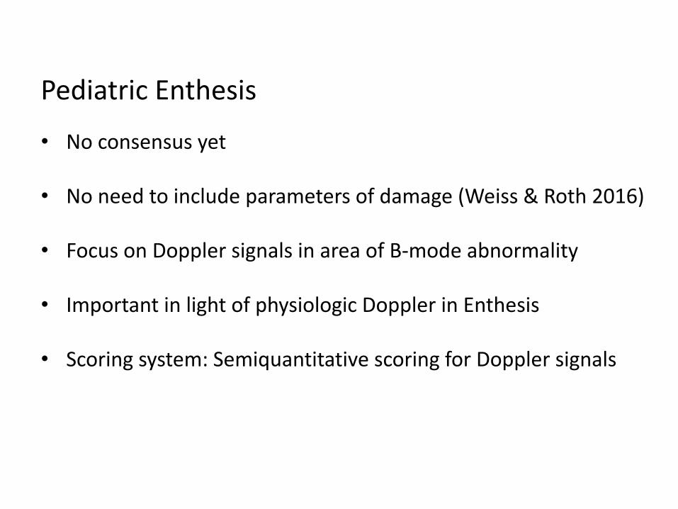
PediatricEnthesis
• Noconsensusyet
• Noneedtoincludeparametersofdamage(Weiss&Roth2016)
• FocusonDopplersignalsinareaofB-modeabnormality
• ImportantinlightofphysiologicDopplerinEnthesis
• Scoringsystem:Semiquantitative scoringforDopplersignals
