inflammation and adaptive immunity in parkinson’s...
TRANSCRIPT
Inflammation and Adaptive Immunityin Parkinson’s Disease
R. Lee Mosley, Jessica A. Hutter-Saunders, David K. Stone, and Howard E. Gendelman
Movement Disorders Program, Department of Pharmacology and Experimental Neuroscience, Center forNeurodegenerative Disorders, University of Nebraska Medical Center, Omaha, Nebraska 68198
Correspondence: [email protected]
The immune system is designed to protect the host from infection and injury. However, whenan adaptive immune response continues unchecked in the brain, the proinflammatory innatemicroglial response leads to the accumulation of neurotoxins and eventual neurodegenera-tion. What drives such responses are misfolded and nitrated proteins. Indeed, the antigen inParkinson’s disease (PD) is an aberrant self-protein, although the adaptive immune responsesare remarkably similar in a range of diseases. Ingress of lymphocytes and chronic activationof glial cells directlyaffect neurodegeneration. With this understanding, new therapies aimedat modulating the immune system’s response during PD could lead to decreased neuronalloss and improved clinical outcomes for disease.
Parkinson’s disease (PD) is the second mostcommon neurodegenerative disorder affect-
ing the elderly. Pathologically, the disease ischaracterized by the cytoplasmic accumulationof proteinaceous aggregates called Lewy bodies(LBs), which are mainly comprised of a-syn-uclein (a-syn) and ubiquitin (Spillantini et al.1997, 1998). Progressive degeneration of dopa-minergic neurons in the substantia nigra (SN)pars compacta and their projections into thecaudate nucleus leads to substantial decreasesin dopamine levels, which manifest as restingtremor, bradykinesia, rigidity, and gait dysfunc-tion (Dauer and Przedborski 2003). Currently,no curative treatments or treatments that in-terdict disease progression exist. Although theetiology of PD remains unknown, abundantevidence implicates immune system abnormal-ities and central nervous system (CNS) inflam-
mation in disease pathobiology (McGeer et al.1988a; Stone et al. 2009; Kosloski et al. 2010).Harnessing inflammatory responses throughtargeted modulation of innate and adaptive im-mune responses has gained increasing interestin recent years as a potential therapeutic strat-egy. The interplay between innate and adaptiveimmunity in the pathobiology of PD, the evolu-tion and change in such immune responses, andthe means to alter it to the benefit of the dis-eased, is the focus of this article.
ADAPTIVE IMMUNITY AND THE CNS
William Hickey wrote, “vertebrates possess twobodily systems capable of learning and remem-bering: the nervous system and the immunesystem” (Hickey 2001; Weiner 2008). The CNSwas once thought to be an “immune privileged”
Editor: Serge Przedborski
Additional Perspectives on Parkinson’s Disease available at www.perspectivesinmedicine.org
Copyright # 2012 Cold Spring Harbor Laboratory Press; all rights reserved; doi: 10.1101/cshperspect.a009381
Cite this article as Cold Spring Harb Perspect Med 2012;2:a009381
1
ww
w.p
ersp
ecti
vesi
nm
edic
ine.
org
on July 15, 2018 - Published by Cold Spring Harbor Laboratory Press http://perspectivesinmedicine.cshlp.org/Downloaded from
site, in which immune cells of the peripherycould not enter or rarely entered, and thus thetwo systems had little to no interaction. This hy-pothesis was supported by the early observationthat tissue grafts in the eye or brain survivedlonger than grafts in other areas of the body(Medawar 1948). However, today, evidence ofan interactive adaptive immune system andthe CNS abounds. Indeed, communication be-tween the CNS and peripheral immune systemis much more fluid than previously consideredand, as such, may substantially affect diseaseprogression in neurological disorders (Ferrariand Tarelli 2011). Peripheral immune responsescan trigger inflammation and exacerbation ofCNS degeneration in several neurodegenerativediseases such as Alzheimer’s disease (AD), mul-tiple sclerosis (MS), amyotrophic lateral sclero-sis (ALS), stroke, and prion-mediated diseases(Cunningham et al. 2005a,b; Kamer et al.2008; Fiala and Veerhuis 2009; Holmes et al.2009; Lee et al. 2009a; McColl et al. 2009; Realeet al. 2009; Stoll and Bendszus 2009; Teeling andPerry 2009; Heesen et al. 2010; Perry 2010), andparticularly PD (Hasegawa et al. 2000; Arai et al.2006). In those disorders, increasing inflam-mation and breakdown of the blood–brain bar-rier (BBB) forces increased communication be-tween the CNS and peripheral immune systemsas evidenced in several neurodegenerative dis-eases with increased leukocyte migration withinthe brain parenchyma (Stolp and Dziegielewska2009). Under infectious or inflammatory con-ditions, peripheral immune cells have relativelyunfettered access to the CNS. These immunecells influence neuroinflammation and neuro-degeneration not only in a paracrine fashion,but also in an endocrine fashion. In turn,the CNS is capable of influencing the immuneresponse to pathogens in the periphery throughthe neuroendocrine system. Thus, the immunesystem is not only charged with protectingthe CNS from pathogens and injury, but isalso capable of affecting the functions and ho-meostasis of resident CNS cells, for better orworse. Furthermore, researchers are beginningto harness the neurotrophic effects of the im-mune system to aid in repair and regenerationin the CNS.
Even under normal conditions, activated Tand B lymphocytes patrol the CNS in low num-bers, whereas naıve lymphocytes are excluded(Hickey 1999; Togo et al. 2002; Engelhardt andRansohoff 2005). Although fewer activated Tcells infiltrate the normal CNS than other tis-sues (Yeager et al. 2000), this may be owing tothe low level of adhesion molecules expressedon endothelial cells under normal conditions(Hickey 2001), whereas increased expressionof adhesion molecules leads to increased lym-phocyte infiltration. When cytokines such asinterleukin (IL)-1 and tumor necrosis factor(TNF)-a are secreted by activated glia in thebrain, or are present in circulating blood, per-meability of the BBB is increased and the ex-pression of cellular adhesion molecules (suchas selectins) on microvascular endothelial cellsare up-regulated (Wong et al. 1999). ActivatedT cells and B cells are then able to extravasateand migrate to the site of neuronal injury in in-creased numbers (Aloisi et al. 1999; McGeer andMcGeer 2003; Olson and Miller 2004).
Indeed, abnormalities in the BBB have beenshown where T-cell infiltration occurs in neuro-AIDS (Petito and Cash 1992; Petito et al. 2003),AD (Rogers et al. 1988; Togo et al. 2002; Desaiet al. 2007), and PD (Farkas et al. 2000). Fur-thermore, whereas the CNS lacks a definedlymphatic system, antigens do exit the CNSvia arachnoid villi, cranial nerves, and spinalnerve root ganglia to lymph (Cserr and Knopf1992). Once in the lymph, these antigens maybe taken up by dendritic cells, processed, andpresented to T and B cells to mobilize an adap-tive immune response to the CNS. Whereasacute neuroinflammation is beneficial to re-gaining homeostasis and normal function ofthe CNS after injury or infection, chronic neu-roinflammation is damaging to the CNS andmay initiate or amplify neurodegeneration as-sociated with HIV-1 encephalitis, AD, or PD.
Cross-Regulation of Adaptive and InnateImmunity in the CNS
Innate immunity consists of the immune mech-anisms that are encoded in the germline and arepossessed at birth, and work in a “nonspecific”
R.L. Mosley et al.
2 Cite this article as Cold Spring Harb Perspect Med 2012;2:a009381
ww
w.p
ersp
ecti
vesi
nm
edic
ine.
org
on July 15, 2018 - Published by Cold Spring Harbor Laboratory Press http://perspectivesinmedicine.cshlp.org/Downloaded from
manner, for immediate defense against micro-bial infection, notably sepsis (Perry 2011; Ste-arns-Kurosawa et al. 2011). A host’s first lineof defense consists of physical barriers such asskin- and cell-regulated enzymes used to clearpathogens and debris, and serves to remove for-eign substances by phagocytosis, to recruit im-mune cells to sites of infection, to activate thecomplement cascade, but most importantly, toprocess and present antigens for activation ofand recognition by the adaptive immune re-sponse (Kim 2005; Filias et al. 2011; Sakaguchi2011; Sly and Holt 2011; Veerhuis et al. 2011).Its conservation is matched only by its simplic-ity, except for a broad range of self–nonselfpattern-recognition receptors. Such immuneactivation functions are through nonspecific,generic recognition of common cell signalingpathways shared through a host of endogenousand exogenous factors. These pathways aregaining considerable interest in therapeutic de-velopment (Goldman 2007; Basith et al. 2011).Cell debris and foreign matter within the CNSengage toll-like receptors (TLRs), which are ex-pressed by microglia, astrocytes, oligodendro-cytes, as well as by neurons (Lv et al. 2011; Zur-olo et al. 2011). Engagement of TLRs activatessignaling cascades that result in proinflamma-tory cytokine and chemokine production andin effects on the brain directly or indirectlythrough glial or BBB function (Franklin et al.2011; Greenwood et al. 2011; Holman et al.2011; Kacimi et al. 2011).
The innate immune system also is linked toits adaptive arm through the abilities to providerequired “signals” for antigen presentation andto act as final effectors by T-cell-mediated re-sponses in the CNS. The interrelationships be-tween innate and adaptive immunity permitthe host to recognize environmental and exoge-nous cues and work in concert to protect andsustain the host. Central to the innate immunenetwork are microglia (Perry 2011). They se-crete both anti- and proinflammatory cytokinesand chemokines together with other factors thatregulate not only adaptive immunity, but alsoneural function and neural homeostasis. Thosemicroglial factors found in the brain, cerebro-spinal fluid (CSF), and peripheral blood in-
clude transforming growth factor beta (TGF-b), IL-1 alpha/beta (IL-1a/b), IL-6, IL-10,IL-12, IL-23, and TNF-a, many chemokines(RANTES/CCL5, MCP-1/CCL2, and IP-10/CXCL10), proteolytic enzymes, matrix metallo-proteinases, complement, growth factors, andglutamate (Griffin et al. 1989; Dickson et al.1993; Moore and Thanos 1996; Qiu et al.1997). Moreover, COX-2 is present as increasedlevels of TRAF family member-associated NFkBactivator (TANK) and NFKB1 in the SN, andIL-15, RANTES, and IL-10 levels are signifi-cantly elevated in brains and peripheral circula-tion in PD patients (Blum-Degen et al. 1995;Teismann et al. 2003; Rentzos et al. 2007,2009; Reynolds et al. 2008a). Furthermore, in-nate immunity regulates lymphocyte infiltra-tion into the CNS. Cytokines, such as IL-1band TNF-a, secreted by activated glia or endo-thelial cells increase BBB permeability (Desaiet al. 2007), and the expression of cellularadhesion molecules (such as E-selectin) on mi-crovascular endothelial cells are up-regulated(Wong et al. 1999), together increase perme-ability of the BBB and increase homing, extra-vasation, and activation of lymphocytes.
Of the antiinflammatory cytokines pro-duced by T cells, macrophages, and microglia,TGF-b modulates injurious responses to thebrain (Finch et al. 1993) and suppresses proin-flammatory microglia and T-cell responses(Sakaguchi 2004), and as such may represent aneuroprotective host response (Chao et al.1994). However, TGF-b released from thedamaged brain microvasculature contributesto inflammation by increasing expression ofendothelial IL-1b and TNF-a (Grammas andOvase 2001). The engagement of TLRs leadsto translocation of nuclear factor-k light-chainenhancer of activated B cells (NF-kB) and AP-1 to the nucleus where they induce transcriptionof a broad range of innate immune proteins.Once activated, microglia secrete both neuro-trophic and neurotoxic factors (Zhang andFedoroff 1996; Glezer et al. 2007); proinflam-matory cytokines including IL-1a, IL-1b, andTNF-a (Giulian et al. 1986; Sawada et al. 1989);and neurotrophins including nerve growth fac-tor (NGF) and neurotrophin 3 (NT-3) (Elkabes
Inflammation and Immunity in Parkinson’s Disease
Cite this article as Cold Spring Harb Perspect Med 2012;2:a009381 3
ww
w.p
ersp
ecti
vesi
nm
edic
ine.
org
on July 15, 2018 - Published by Cold Spring Harbor Laboratory Press http://perspectivesinmedicine.cshlp.org/Downloaded from
et al. 1996; Heese et al. 1998). However, duringchronic inflammation, the neurotoxic effectsof microglia are proposed to eventually out-compete the neurotrophic effects, thus increas-ing neurodegeneration (Rock et al. 2004). Inturn, increased inflammation and BBB per-meability allow naıve T cells greater entry andaccessibility to activated microglia that in theacute phase induce proinflammatory T-cell re-sponses, and can, under chronic conditions,perpetuate the inflammatory state by engag-ing and activating polarized proinflammatoryeffector T cells that have expanded in the per-iphery and ingressed to sites of neurodegen-eration.
Induction of Innate and Adaptive ImmuneActivation by Misfolded Proteins
Evidence abounds for the involvement of mis-folded proteins in the pathology of neurode-generative diseases, as well as in the activation ofmicroglia and antigen-presenting cells (APCs)that function to induce the adaptive immunearm. In PD, LBs are associated with activatedmicroglia and dopaminergic neuronal death.LBs are comprised mostly of a-syn, ubiquitin,and neurofilament (Goldman et al. 1983; Spill-antini et al. 1997, 1998; Jellinger 2007) and post-translationally modified forms of a-syn havean increased propensity to aggregate (Uverskyet al. 2005; Cavallarin et al. 2010). These a-synspecies are created by ubiquitination (Shimuraet al. 2001), phosphorylation (Fujiwara et al.2002), or oxidation and nitration (Giassonet al. 2000), and are found in LB inclusions, ex-traneuronally in PD brains (Lee 2008), and inthe periphery of PD patients (Beach et al.2010). Abnormal species of a-syn found inPD patients are also present in LBs of othersynucleinopathy-affected brains, including ADand multiple-system atrophy (MSA) (Dudaet al. 2000; Giasson et al. 2000; Cavallarinet al. 2010). These observations are supportedby in vitro data and animal models of PDin which overexpression of native or mutatedforms of a-syn show that aberrant specieshave an increased propensity to aggregate(Parihar et al. 2009; Koprich et al. 2010). Aggre-
gation of a-syn is also caused by genetic muta-tions. Although most cases of PD have no familyhistory of disease, point mutations in the geneencoding a-syn (SNCA; OMIM 163890) arelinked to autosomal-dominant parkinsonism(PARK1, OMIM 168601) (Polymeropoulos etal. 1996, 1997; Kruger et al. 1998; Zarranz etal. 2004), as are duplications and triplicationsof the SNCA gene (PARK4, OMIM 605543)(Singleton et al. 2003; Chartier-Harlin et al.2004), all of which present increased aggrega-tion of a-syn (Narhi et al. 1999; Li et al. 2001;Uversky 2007). Taken together, these observa-tions led to the a-syn burden hypothesis, whichposits that sporadic PD results from the inabil-ity to clear a-syn, whereas familial PD resultsfrom overproduction of normal a-syn, muta-tions in a-syn that prevent or slow clearance,or mutations in other proteins that normally as-sist in a-syn clearance (McGeer and McGeer2008). McGeer further hypothesized that dis-ease can be eliminated with the reduction ofa-syn production or prevention of a-syn aggre-gation. Thus, vaccines currently being devel-oped for PD, target a-syn in order to increaseclearance of aggregated and aberrant forms ofthe protein. However, because evidence sup-ports a nonneuronal cell autonomous theoryfor PD progression whereby neuroinflammationis strongly implicated (Dawson 2008), immuno-therapeutic strategies also incorporate means bywhich to attenuate the neuroinflammatorycomponent.
Furthermore, reactive oxygen species pro-duced by activated microglia increases nitrationof a-syn and neuronal cell death (Shavali et al.2006); and in turn, immune T cells that rec-ognize nitrated a-syn (N-a-syn) enhance theneurotoxic activities of microglia in the acute1-methyl-4-phenyl-1,2,3,6-tetrahydropyridine(MPTP) mouse model of nigrostriatal degener-ation (Reynolds et al. 2008b). Activated T cellsand B cells are then able to enter the CNSmore readily and migrate to the site of neuronalinjury (Aloisi et al. 1999; McGeer and McGeer2003; Olson and Miller 2004). Indeed, increasedBBB permeability is found in both AD andPD, allowing for increased lymphocytic ingress(Rogers et al. 1988; Farkas et al. 2000; Togo et al.
R.L. Mosley et al.
4 Cite this article as Cold Spring Harb Perspect Med 2012;2:a009381
ww
w.p
ersp
ecti
vesi
nm
edic
ine.
org
on July 15, 2018 - Published by Cold Spring Harbor Laboratory Press http://perspectivesinmedicine.cshlp.org/Downloaded from
2002; Desai et al. 2007). In this way, activatedinnate immune cells of the CNS can affect theadaptive immune system in the periphery andrecruit cells to the CNS. However, another wayin which the adaptive immune system may beactivated during neurodegeneration is throughthe escape of CNS proteins into the periphery.Indeed, aberrant species of disease-specificproteins, including phosphorylated a-syn, arepresent in tissues outside the CNS in PD pa-tients (Beach et al. 2010). The occurrence ofthese aberrant forms of a-syn in the periphery,such as the gastrointestinal tract and drainingcervical lymph nodes, presents a possible meansfor exposure to the protein and subsequentactivation of the adaptive immune system.
ADAPTIVE IMMUNITY IN PD
From an earlier Perspectives in Biology and Med-icine (Johns Hopkins University Press), Abram-sky and colleagues broached the possibilitythat PD may arise from autoimmune blockadeof striatal dopamine receptor function (Abram-sky and Litvin 1978). Although clinical evi-dence has not sufficiently supported thispossibility, many studies have implicated theadaptive immune system in PD progression.With high glia/neuron ratios of 3:1 in the brain(Lawson et al. 1990) and the density of micro-glia contained within the SN, the highest ofany region in the brain (Kim et al. 2000), any in-crease in the inflammatory status of the patientmay additively, if not synergistically amplifyneuroinflammation. Since 1988, when McGeerand colleagues found HLA-DR-positive (acti-vated) microglia phagocytosing-free neurome-lanin in post mortem PD SN (McGeer et al.1988a), activated microglial consistently havebeen observed in PD patients, whereas othershave shown higher expression levels of poly-morphic major histocompatibility complex(MHC) class II (MHC II) molecules, HLA-DRand HLA-DQ, expressed by monocytes in theCSF and peripheral blood of PD patients com-pared with controls (McGeer et al. 1988b; Fiszeret al. 1994a; Lampe et al. 2003). More recently,genome-wide association studies (GWASs) ofPD patients (Hamza et al. 2010; Saiki et al.
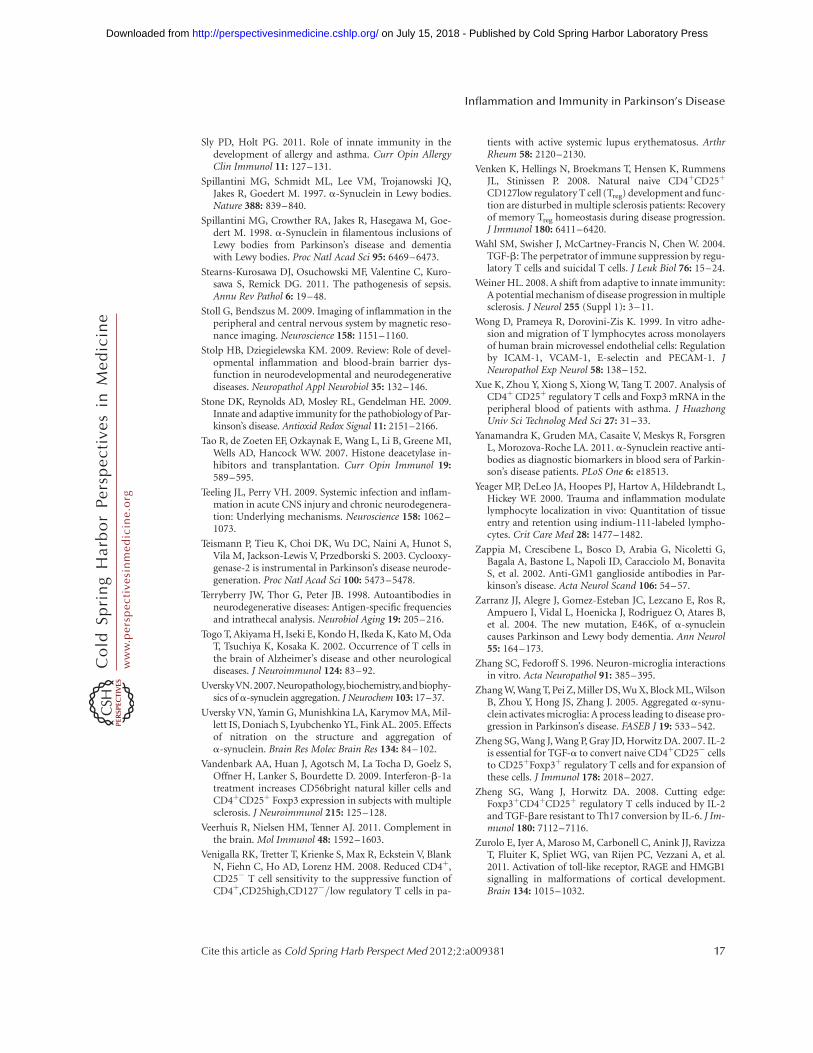
2010; Nalls et al. 2011; Puschmann et al. 2011;Simon-Sanchez et al. 2011), including a meta-analysis of the GWASs (Nalls et al. 2011), veri-fied an increased relative risk for PD and expres-sion of HLA-DR or HLA-DQ MHC II mole-cules, leading to the designation of HLA-DRAas PARK18 (Hamza et al. 2010). Increased sus-ceptibility to PD owing to MHC II moleculescould reflect increased neuroinflammation as-sociated with up-regulation of those moleculesor alternatively, could represent immune re-sponses to self- or weak nonself-antigens. Acti-vated microglia and monocytes in PD brainsand CFS secrete proinflammatory and neuro-toxic cytokines and chemokines that disruptthe BBB and attract lymphocytes to the site ofneuronal injury. Indeed, levels of IL-1b, IL-6,and TNF-a are elevated in the CFS of PD pa-tients (Blum-Degen et al. 1995; Gonzalez-Scarano and Baltuch 1999), and intercellularadhesion molecule-1 (ICAM-1)-positive gliaare also increased in the SN of PD brains (Mi-klossy et al. 2006). Together, these data supportthe hypothesis that activation of cells of the in-nate immune system, such as microglia andmonocytes, directly contribute to the pathobi-ology of PD. Furthermore, it has been shownthat these cells are activated by overexpressionof a-syn or aberrant forms of a-syn. Aberrantposttranslational modifications of a-syn, suchas nitration (N-a-syn), can be found in LB in-clusions of PD brains (Giasson et al. 2000)and cause the protein to aggregate more readily(Uversky et al. 2005). Aggregated a-syn acti-vates microglia (Zhang et al. 2005), whichhave been shown to produce nitric oxide andsuperoxide in mice and inducible nitric oxidesynthase (iNOS) in humans, which increasesnitration of a-syn and perpetuates the proin-flammatory innate immune response in PD(Fig. 1) (Hunot et al. 1996; Gao et al. 2008).Nitrated a-syn in turn amplifies activation ofmicroglia and antigen-presenting cells thatcorrespondingly up-regulate both humoraland cell-mediated responses to nitrated a-synin the MPTP model (Benner et al. 2008).
Although autoantibodies against dopamineneuron antigens are present in sera and CSFof PD patients (McRae-Degueurce et al. 1988;
Inflammation and Immunity in Parkinson’s Disease
Cite this article as Cold Spring Harb Perspect Med 2012;2:a009381 5
ww
w.p
ersp
ecti
vesi
nm
edic
ine.
org
on July 15, 2018 - Published by Cold Spring Harbor Laboratory Press http://perspectivesinmedicine.cshlp.org/Downloaded from
Dahlstrom et al. 1990; Kunas et al. 1995), therole of the humoral adaptive immune systemhas only recently begun to be investigated indepth. In addition to a variety of antibodiesdirected against globally expressed tissue anti-gens such as heat shock protein (HSP)-65 andHSP-70 (Fiszer et al. 1996), PD patients also
show brain-associated autoantibodies includ-ing those directed against, GM1, S100B, glialfibrillar acidic protein (GFAP), NGF, neurofila-ment, myelin basic protein, tau, Ab, and neu-ronal calcium channels, as well as a-syn andits modified and fibriliary forms (Elizan et al.1983; Karcher et al. 1986; Appel et al. 1994;
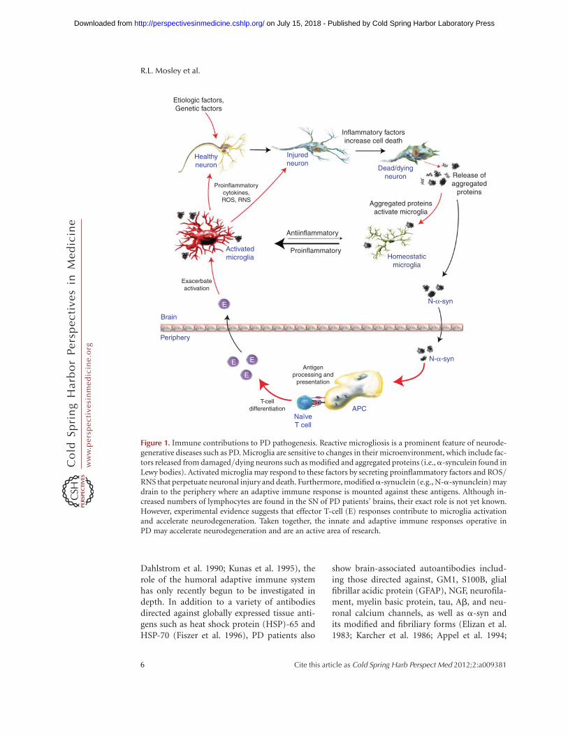
Etiologic factors,Genetic factors
Release ofaggregated
proteins
Aggregated proteinsactivate microglia
Inflammatory factorsincrease cell death
Proinflammatorycytokines,ROS, RNS
E
E E
EAntigen
processing andpresentation
Exacerbateactivation
T-celldifferentiation
Antiinflammatory
ProinflammatoryActivatedmicroglia
N-α-syn
N-α-syn
APCNaïveT cell
Brain
Periphery
Healthyneuron
Injuredneuron
Dead/dyingneuron
Homeostaticmicroglia
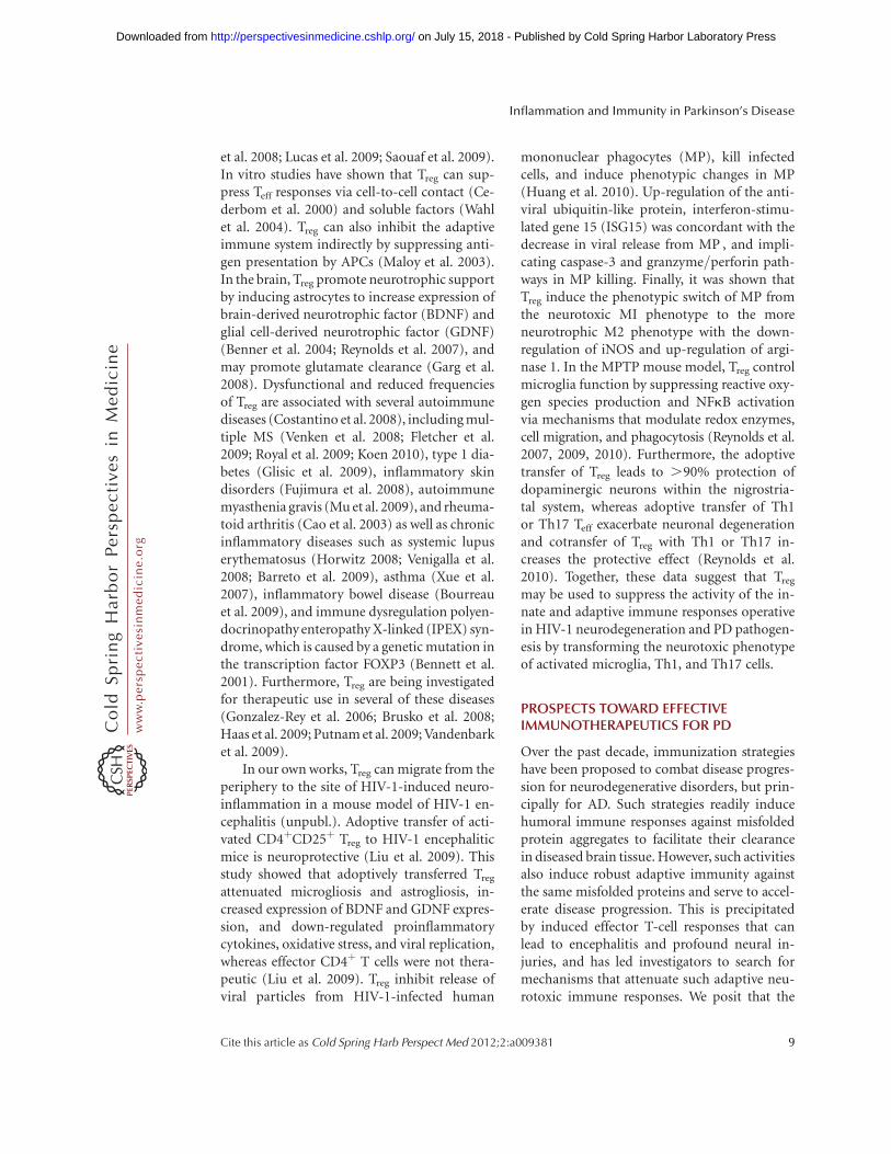
Figure 1. Immune contributions to PD pathogenesis. Reactive microgliosis is a prominent feature of neurode-generative diseases such as PD. Microglia are sensitive to changes in their microenvironment, which include fac-tors released from damaged/dying neurons such as modified and aggregated proteins (i.e.,a-synculein found inLewy bodies). Activated microglia may respond to these factors by secreting proinflammatory factors and ROS/RNS that perpetuate neuronal injury and death. Furthermore, modifieda-synuclein (e.g., N-a-synunclein) maydrain to the periphery where an adaptive immune response is mounted against these antigens. Although in-creased numbers of lymphocytes are found in the SN of PD patients’ brains, their exact role is not yet known.However, experimental evidence suggests that effector T-cell (E) responses contribute to microglia activationand accelerate neurodegeneration. Taken together, the innate and adaptive immune responses operative inPD may accelerate neurodegeneration and are an active area of research.
R.L. Mosley et al.
6 Cite this article as Cold Spring Harb Perspect Med 2012;2:a009381
ww
w.p
ersp
ecti
vesi
nm
edic
ine.
org
on July 15, 2018 - Published by Cold Spring Harbor Laboratory Press http://perspectivesinmedicine.cshlp.org/Downloaded from
Terryberry et al. 1998; Poletaev et al. 2000;Zappia et al. 2002; Papachroni et al. 2007; Gru-den et al. 2011; Yanamandra et al. 2011). Im-munohistochemical staining of tissues fromidiopathic and familial PD patients show dopa-minergic neurons within the SN bind IgG, butnot IgM, whereas tissues from age-matchedcontrols and nonnigral control tissues showno detectable bound immunoglobulins (Orret al. 2005). In one study, IgG reacted with30% of the dopaminergic neurons within thenigra, and yielded positive correlations withnumbers of MHC IIþ and CD64þ (FcgRI) reac-tive microglia, yet yielded a negative correlationwith disease duration. In PD patients, approxi-mately 4% of the pigmented neurons containedLewy bodies and all pigmented, Lewy body-containing neurons showed detectable IgGand a-syn within inclusions. This pattern ofantibody reactivity was consistent with acti-vated microglia and destruction of dopaminer-gic neurons in PD. Together these data suggestthat endogenous antibodies of unknown specif-icity have the capacity to cross the BBB and bindcognate antigens expressed by dopaminergicneurons. Of interest, deletion of the FcgR bygenetic ablation inhibits microglial activationand dopaminergic cell death in animal modelsof PD (He et al. 2002). Moreover, levels of anti-bodies to a-syn and catecholamine-derivedmelanin (neuromelanin) are increased in PDpatients with antineuromelanin immunoglob-ulin binding shown to be more active in earlydisease (Double et al. 2009). Opsonization ofcells or damaged neurons with antibody, tar-gets the cells for phagocytosis and degradationby phagocytic macrophages, and also can ac-tivate the complement system, a major media-tor of immune/inflammatory reactions. Inter-estingly, activation of the complement systemmay also be involved in neuronal death. Micro-glia are the only cells within the SN that expressthe initial recognition component of comple-ment, C1q (Depboylu et al. 2011). Moreover,compared with controls, PD patients show in-creased areas of C1q-opsonized extracellulardepositions of neuromelanin within the paren-chyma, and C1q-expressing phagocytic micro-glia surround those areas, as well as cells around
the luminal surfaces of the vasculature that ex-press neuromelanin and C1q, suggesting a rolefor antiself antibodies and C1q-mediated clear-ing pathways in PD.
Along with activated microglia and astro-cytes, T cells may also comprise components ofPD pathobiology, although the mechanism(s)by which they effect disease remains enigmatic.Early autopsy evidence within the SN of PD pa-tients showed increased numbers of CD8þ Tcells in close proximity to activated microgliaand degenerating neurons (McGeer et al.1988a). More recently, both CD4þ and CD8þ
T cells have been discovered within the SN ofPD patients (Brochard et al. 2009). Although adysfunctional BBB in PD patients may showsome leakiness (Kortekaas et al. 2005), thismay not be sufficient to allow unrestrictedlymphocyte infiltration because CD4/CD8 ra-tios were 1:4.8 (Brochard et al. 2009) comparedwith the typical 2:1 ratio expected for peripheralT cells performing surveillance functions. Thus,the mechanism by which these T cells gain ac-cess to the SN, their activation state, and theirfunction are questions that remain to be an-swered.
Peripheral immune aberrations, particu-larly in lymphocyte subsets, are abundant inPD patients. Total numbers of lymphocyteshave been shown to be diminished by 17%,whereas CD19þ B cells are diminished as muchas 35% and CD3þ T cells are diminished by22% (Bas et al. 2001). Among CD3þ T cells,numbers of CD4þ T cells have been shown tobe diminished by 31%, whereas numbers ofCD8þ T cells are not significantly changed. Agreater loss of naıve helper CD4þ T cells(CD45RAþ) and either unchanged or increasedlevels of effector/memory helper T-cell subset(CD29þ or CD45R0þ) have also been observed.Selective loss of CD4þCD45RAþ cells are alsodetected in other neuoropathological-associ-ated disorders such as MS and Down’s syn-drome, suggesting a common immunologicalabnormality in neurological disorders (Fiszeret al. 1994a; Crucian et al. 1995). Increased fre-quencies of activated CD4þ T cells expressingFas (Hisanaga et al. 2001) and increased IFN-g-producing Th1 cells, decreased IL-4-producing
Inflammation and Immunity in Parkinson’s Disease
Cite this article as Cold Spring Harb Perspect Med 2012;2:a009381 7
ww
w.p
ersp
ecti
vesi
nm
edic
ine.
org
on July 15, 2018 - Published by Cold Spring Harbor Laboratory Press http://perspectivesinmedicine.cshlp.org/Downloaded from
Th2 cells, and a decrease in CD4þCD25þ T cellshave been found in the peripheral blood of PDpatients (Baba et al. 2005), whereas circulatingIL-15, RANTES, and IL-10 are significantly ele-vated in PD patients compared with controls(Rentzos et al. 2007, 2009). Evidence of in-creased mutual coexpression of CD4 and CD8by CD45R0þ T cells, increased expression ofCD25 (a chain of the high-affinity IL-2 recep-tor) and TNF-a receptors, and diminished ex-pression of IFN-g receptors suggest that theseT-cell subsets from PD patients are indeed acti-vated. In addition to T cells that express a andb chains of the T-cell receptor (TCRabþT cells),elevated frequencies of T-cell populations ex-pressing g and d chains of the T-cell receptor(TCRgdþT cells) also have been found in theCSF of PD patients (Fiszer et al. 1994b) and arethought to play a regulatory role in CNS inflam-mation (Ponomarev and Dittel 2005; Bennettand Stuve 2009; Blink and Miller 2009). More-over, a large proportion of the TCRgdþ T cellsalso express CD25, suggesting these CSF-ob-tained T cells are preferentially activated in PDpatients (Fiszer et al. 1994b). One way in whichthe adaptive immune system could be mobilizedto infiltrate the CNS during PD is through thedrainage of aberrant forms of a-syn into thelymphatic system where the protein could acti-vate lymphocytes. Indeed, in MPTP-intoxicatedmice, a-syn drains to cervical lymph nodeswhere it activates antigen-presenting cells andT cells (Benner et al. 2008). An influx of a-syn-specific Th1 or Th17 effector T cells into thebrain during PD could increase the inflamma-tory phenotype and neurotoxic response of mi-croglia near dopaminergic neurons by increas-ing the concentration of proinflammatorymolecules in the SN (Reynolds et al. 2010). Takentogether, increased frequencies of memory andactivated peripheral T-cell subsets, as well asthose cells within the nigra of PD patients, sug-gest putative roles of T cells in disease progres-sion, if not PD etiology. Although those roleshave yet to be delineated, activated effector Tcells (Teff ) or regulatory T cells (Treg), the latteralso showing an effector/memory T-cell pheno-type, may migrate to the foci of inflammation inPD patients and either exacerbate or attenuate
PD-associated neuroinflammatory responsesand neurodegeneration. Thus, Treg have the ca-pacity to keep the disorder in check during theasymptomatic phase, whereas Teff can acceleratedisease progression (Fig. 1). Whether T-cell ab-errations in PD patients reflect specifically ac-tivated effector or regulatory T-cell subsets andto what antigen(s) those T cells are induced, re-quire answers to develop more precise immune-based therapeutic strategies.
ADAPTIVE IMMUNITY FOR THERAPEUTICGAIN IN PD
It is likely that the adaptive immune system’sresponse to disease in the CNS is similar in arange of neurodegenerative diseases. Thus, ther-apeutic strategies aimed at modulating the im-mune response during disease may be applica-ble to several neurodegenerative diseases. Herewe will discuss recent approaches taken to mod-ulate the adaptive immune system for diseasetherapy.
Treg are an important subset of CD4þ T cellsthat are known to maintain self-tolerance,prevent autoimmunity, and regulate immunehomeostasis by attenuating excessive inflamma-tion caused by pathogens or injury (Sakaguchiet al. 1995; Cederbom et al. 2000; Kipniset al. 2002; Hori et al. 2003; Sakaguchi 2004;Coombes et al. 2005; Kim et al. 2007; Bourreauet al. 2009). They are identified by the expres-sion of CD4 and CD25 cell-surface markersand by the transcription factor forkhead boxP3 (FoxP3) in mice (Hall et al. 1990; Fontenotet al. 2003; Hori et al. 2003), and the expressionof FOXP3, CD4, CD25, CD39, CD49d, and alack of CD127 in humans (Fletcher et al. 2009;Kleinewietfeld et al. 2009). Although naturallyoccurring Treg mature in the thymus, naıveCD4þ-stimulated T cells in the periphery canbe polarized into an inducible Treg (iTreg) phe-notype under certain conditions. For example,TGF-b, IL-2, IL-10, and all-trans retinoic acidare known to polarize T cells to iTreg (Zhenget al. 2007, 2008; Khattar et al. 2009; Lee et al.2009b), whereas histone deacetylase inhibitorsare known to increase proliferation and sup-pressor activity of Treg (Tao et al. 2007; Johnson
R.L. Mosley et al.
8 Cite this article as Cold Spring Harb Perspect Med 2012;2:a009381
ww
w.p
ersp
ecti
vesi
nm
edic
ine.
org
on July 15, 2018 - Published by Cold Spring Harbor Laboratory Press http://perspectivesinmedicine.cshlp.org/Downloaded from
et al. 2008; Lucas et al. 2009; Saouaf et al. 2009).In vitro studies have shown that Treg can sup-press Teff responses via cell-to-cell contact (Ce-derbom et al. 2000) and soluble factors (Wahlet al. 2004). Treg can also inhibit the adaptiveimmune system indirectly by suppressing anti-gen presentation by APCs (Maloy et al. 2003).In the brain, Treg promote neurotrophic supportby inducing astrocytes to increase expression ofbrain-derived neurotrophic factor (BDNF) andglial cell-derived neurotrophic factor (GDNF)(Benner et al. 2004; Reynolds et al. 2007), andmay promote glutamate clearance (Garg et al.2008). Dysfunctional and reduced frequenciesof Treg are associated with several autoimmunediseases (Costantino et al. 2008), including mul-tiple MS (Venken et al. 2008; Fletcher et al.2009; Royal et al. 2009; Koen 2010), type 1 dia-betes (Glisic et al. 2009), inflammatory skindisorders (Fujimura et al. 2008), autoimmunemyasthenia gravis (Mu et al. 2009), and rheuma-toid arthritis (Cao et al. 2003) as well as chronicinflammatory diseases such as systemic lupuserythematosus (Horwitz 2008; Venigalla et al.2008; Barreto et al. 2009), asthma (Xue et al.2007), inflammatory bowel disease (Bourreauet al. 2009), and immune dysregulation polyen-docrinopathy enteropathy X-linked (IPEX) syn-drome, which is caused by a genetic mutation inthe transcription factor FOXP3 (Bennett et al.2001). Furthermore, Treg are being investigatedfor therapeutic use in several of these diseases(Gonzalez-Rey et al. 2006; Brusko et al. 2008;Haas et al. 2009; Putnam et al. 2009; Vandenbarket al. 2009).
In our own works, Treg can migrate from theperiphery to the site of HIV-1-induced neuro-inflammation in a mouse model of HIV-1 en-cephalitis (unpubl.). Adoptive transfer of acti-vated CD4þCD25þ Treg to HIV-1 encephaliticmice is neuroprotective (Liu et al. 2009). Thisstudy showed that adoptively transferred Treg
attenuated microgliosis and astrogliosis, in-creased expression of BDNF and GDNF expres-sion, and down-regulated proinflammatorycytokines, oxidative stress, and viral replication,whereas effector CD4þ T cells were not thera-peutic (Liu et al. 2009). Treg inhibit release ofviral particles from HIV-1-infected human
mononuclear phagocytes (MP), kill infectedcells, and induce phenotypic changes in MP(Huang et al. 2010). Up-regulation of the anti-viral ubiquitin-like protein, interferon-stimu-lated gene 15 (ISG15) was concordant with thedecrease in viral release from MP , and impli-cating caspase-3 and granzyme/perforin path-ways in MP killing. Finally, it was shown thatTreg induce the phenotypic switch of MP fromthe neurotoxic MI phenotype to the moreneurotrophic M2 phenotype with the down-regulation of iNOS and up-regulation of argi-nase 1. In the MPTP mouse model, Treg controlmicroglia function by suppressing reactive oxy-gen species production and NFkB activationvia mechanisms that modulate redox enzymes,cell migration, and phagocytosis (Reynolds et al.2007, 2009, 2010). Furthermore, the adoptivetransfer of Treg leads to .90% protection ofdopaminergic neurons within the nigrostria-tal system, whereas adoptive transfer of Th1or Th17 Teff exacerbate neuronal degenerationand cotransfer of Treg with Th1 or Th17 in-creases the protective effect (Reynolds et al.2010). Together, these data suggest that Treg
may be used to suppress the activity of the in-nate and adaptive immune responses operativein HIV-1 neurodegeneration and PD pathogen-esis by transforming the neurotoxic phenotypeof activated microglia, Th1, and Th17 cells.
PROSPECTS TOWARD EFFECTIVEIMMUNOTHERAPEUTICS FOR PD
Over the past decade, immunization strategieshave been proposed to combat disease progres-sion for neurodegenerative disorders, but prin-cipally for AD. Such strategies readily inducehumoral immune responses against misfoldedprotein aggregates to facilitate their clearancein diseased brain tissue. However, such activitiesalso induce robust adaptive immunity againstthe same misfolded proteins and serve to accel-erate disease progression. This is precipitatedby induced effector T-cell responses that canlead to encephalitis and profound neural in-juries, and has led investigators to search formechanisms that attenuate such adaptive neu-rotoxic immune responses. We posit that the
Inflammation and Immunity in Parkinson’s Disease
Cite this article as Cold Spring Harb Perspect Med 2012;2:a009381 9
ww
w.p
ersp
ecti
vesi
nm
edic
ine.
org
on July 15, 2018 - Published by Cold Spring Harbor Laboratory Press http://perspectivesinmedicine.cshlp.org/Downloaded from
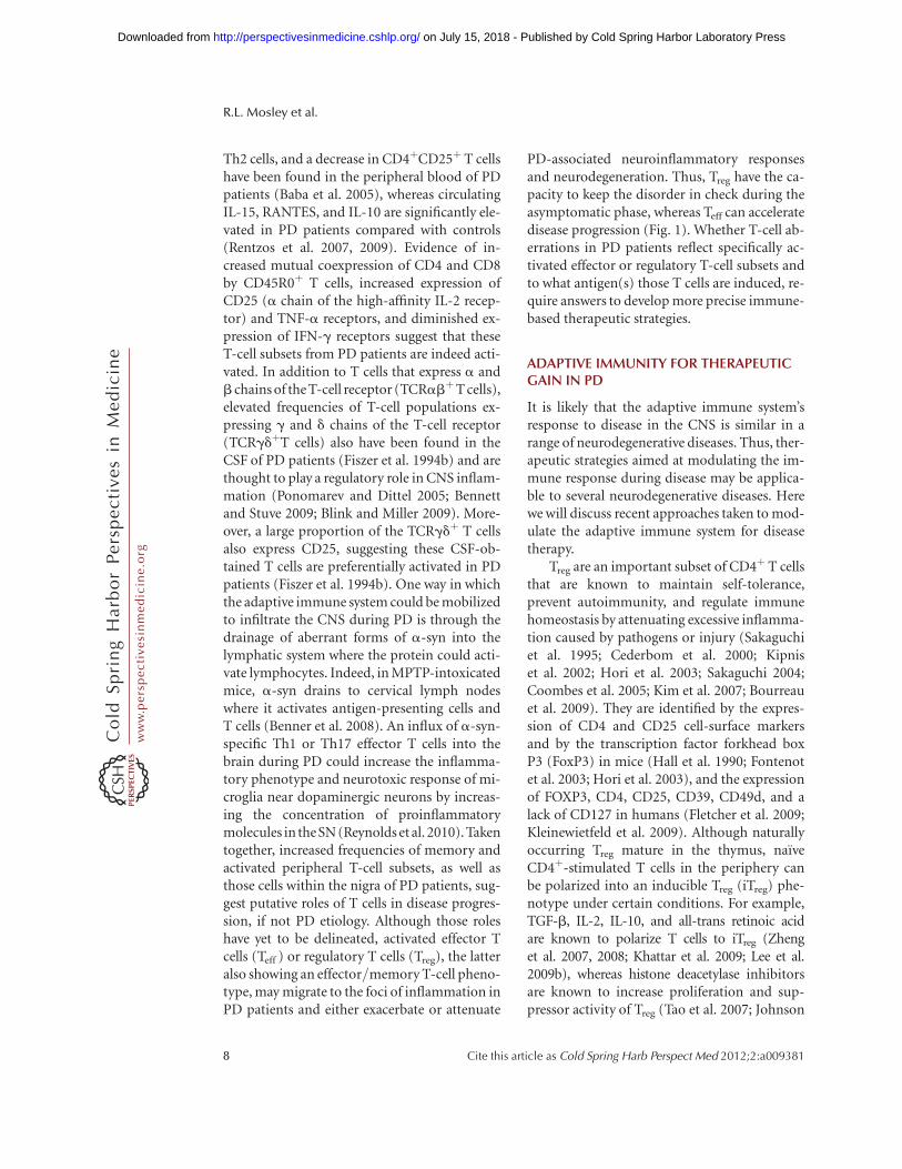
Etiologic factors,Genetic factors
ROS/RNSproduction
Antigenclearance
Phenotypemodulation
R
RR
RR
R
E
E
Activationsuppression/
killing ofactivated cells
Functionaltransformation
Reprogrammingsignals
Inhibitionof maturation
T-celldifferentiation
Inducersof Treg
Antigenpresentation
Antiinflammatory
Proinflammatory
Repair/recovery
Neurotrophins
Healthyneuron
Activatedmicroglia
Brain
Periphery
Homeostaticmicroglia
Healthyneuron
α-syn
N-α-syn
N-α-syn
N-α-syn
APC
NaïveT cell
Injuredneuron
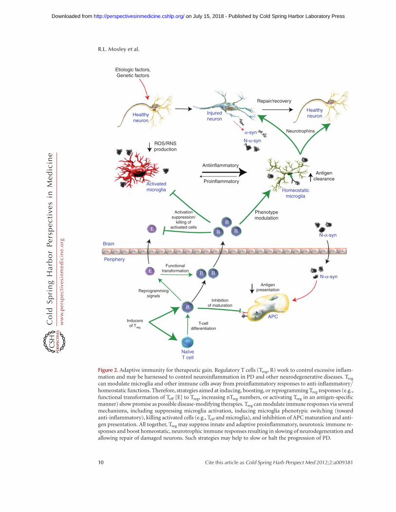
Figure 2. Adaptive immunity for therapeutic gain. Regulatory T cells (Treg, R) work to control excessive inflam-mation and may be harnessed to control neuroinflammation in PD and other neurodegenerative diseases. Treg
can modulate microglia and other immune cells away from proinflammatory responses to anti-inflammatory/homeostatic functions. Therefore, strategies aimed at inducing, boosting, or reprogramming Treg responses (e.g.,functional transformation of Teff [E] to Treg, increasing nTreg numbers, or activating Treg in an antigen-specificmanner) show promise as possible disease-modifying therapies. Treg can modulate immune responses via severalmechanisms, including suppressing microglia activation, inducing microglia phenotypic switching (towardanti-inflammatory), killing activated cells (e.g., Teff and microglia), and inhibition of APC maturation and anti-gen presentation. All together, Treg may suppress innate and adaptive proinflammatory, neurotoxic immune re-sponses and boost homeostatic, neurotrophic immune responses resulting in slowing of neurodegeneration andallowing repair of damaged neurons. Such strategies may help to slow or halt the progression of PD.
R.L. Mosley et al.
10 Cite this article as Cold Spring Harb Perspect Med 2012;2:a009381
ww
w.p
ersp
ecti
vesi
nm
edic
ine.
org
on July 15, 2018 - Published by Cold Spring Harbor Laboratory Press http://perspectivesinmedicine.cshlp.org/Downloaded from
Treg responses in PD are dysfunctional duringadvanced disease states. As such, this serves toshift the balance from regulatory to effectorT-cell activities, and yields an inability to at-tenuate ongoing neurotoxic inflammatory events(Fig. 2). If clearance of misfolded proteins canbe achieved with subsequent immune mod-ulation and restoration of Treg responses, im-proved therapeutic outcomes for neuronal pro-tection would be realized. This may be achievedthrough advances in immune regulation used toachieve a homeostatic glial response for thera-peutic gain.
SUMMARY AND CONCLUSIONS
No doubt, the role of inflammation in PD isnow well appreciated. This is supported bydata seen in a growing number of laboratoryand animal investigations as well as in humanstudies. Inflammation is linked to the extracel-lular appearance of brain protein modificationsand aberrant protein misfolding, which remainprominent hallmarks of PD-associated dopa-minergic neurodegeneration. During disease,dopaminergic neurons accumulate a-synucleintogether with other misfolded proteins seen asintracellular Lewy body inclusions. On injuryor death, neurons release these proteins to thesurrounding neuroenvironment and the modi-fied proteins find their way to the peripherallymphatic system. In an attempt to clear and di-gest cellular debris, microglia and blood-bornemacrophages infiltrate sites of neuronal injuryand death and convert to a proinflammatoryactivated state. Misfolded and modified a-synuclein species draining from these sites alsohave the capability to activate antigen-present-ing cells in peripheral lymphoid tissues induc-ing effector neurotoxic T-cell responses. Thisnormally serves to facilitate debris clearanceand repair functions. Indeed, under steady-stateconditions and early in disease, activated mi-croglia and adaptive immune responses are lim-ited. Nonetheless, as disease becomes more ro-bust, activated microglia are seen in abundantnumbers removed from proximate areas ofneuronal death. This may be attributable tothe failure of activated microglia to return to a
homeostatic state. Thus, disease-affected micro-glia are in a constant activation flux, oscillatingbetween homeostatic and activation states. In-terestingly, whereas this cell maintains an acti-vated amoeboid phenotype, microglial func-tional profiles can encompass a broad rangeof responses that range from an M1 (proinflam-matory neurotoxic) to M2 (anti-inflammatoryneuroprotective) activities. Such evolution incell function may be driven by the depositionand release of nondegradable modified proteinsor by infiltrating T lymphocytes. Once modifiedprotein species enters the peripheral lymphoidtissues, activate antigen-presenting cells, andare presented as neoepitopes, adaptive immuneresponses are induced. Subsequent inductionof effector T-cell-mediated responses may pro-foundly affect microglial states or directly killneurons. As disease evolves, these responsesemerge from a well-regulated neuroprotectivestate to loss of regulatory function and disease.In this scenario, the engagement of therapeuticstrategies that dampen inflammatory and neuro-toxic profiles by induction or repair of aberrantregulatory T-cell responses could provide ameans to reverse the neurodegenerative process.Thus, modulation of inflammatory responsesfor therapeutic gain remains an ever-pressing di-rective to improve disease outcomes.
REFERENCES�Reference is also in this collection.
Abramsky O, Litvin Y. 1978. Autoimmune response todopamine-receptor as a possible mechanism in thepathogenesis of Parkinson’s disease and schizophrenia.Perspect Biol Med 22: 104–114.
Aloisi F, Ria F, Columba-Cabezas S, Hess H, Penna G, Ado-rini L. 1999. Relative efficiency of microglia, astrocytes,dendritic cells and B cells in naive CD4þ T cell primingand Th1/Th2 cell restimulation. Eur J Immunol 29:2705–2714.
Appel SH, Smith RG, Alexianu M, Engelhardt J, Mosier D,Colom L, Stefani E. 1994. Neurodegenerative disease:Autoimmunity involving calcium channels. Ann NYAcad Sci 747: 183–194.
Arai H, Furuya T, Mizuno Y, Mochizuki H. 2006. Inflamma-tion and infection in Parkinson’s disease. Histol Histopa-thol 21: 673–678.
Baba Y, Kuroiwa A, Uitti RJ, Wszolek ZK, Yamada T. 2005.Alterations of T-lymphocyte populations in Parkinsondisease. Parkinsonism Relat Disord 11: 493–498.
Inflammation and Immunity in Parkinson’s Disease
Cite this article as Cold Spring Harb Perspect Med 2012;2:a009381 11
ww
w.p
ersp
ecti
vesi
nm
edic
ine.
org
on July 15, 2018 - Published by Cold Spring Harbor Laboratory Press http://perspectivesinmedicine.cshlp.org/Downloaded from
Barreto M, Ferreira RC, Lourenco L, Moraes-Fontes MF,Santos E, Alves M, Carvalho C, Martins B, Andreia R, Vi-ana JF, et al. 2009. Low frequency of CD4þCD25þ Treg inSLE patients: A heritable trait associated with CTLA4 andTGFb gene variants. BMC Immunol 10: 5.
Bas J, Calopa M, Mestre M, Mollevi DG, Cutillas B, Ambro-sio S, Buendia E. 2001. Lymphocyte populations in Par-kinson’s disease and in rat models of parkinsonism.J Neuroimmunol 113: 146–152.
Basith S, Manavalan B, Lee G, Kim SG, Choi S. 2011. Toll-like receptor modulators: A patent review (2006–2010).Expert Opin Ther Pat 21: 927–944.
Beach TG, Adler CH, Sue LI, Vedders L, Lue L, White CL III,Akiyama H, Caviness JN, Shill HA, Sabbagh MN, et al.2010. Multi-organ distribution of phosphorylateda-syn-uclein histopathology in subjects with Lewy body disor-ders. Acta Neuropathol 119: 689–702.
Benner EJ, Mosley RL, Destache CJ, Lewis TB, Jackson-Lewis V, Gorantla S, Nemachek C, Green SR, PrzedborskiS, Gendelman HE. 2004. Therapeutic immunization pro-tects dopaminergic neurons in a mouse model of Parkin-son’s disease. Proc Natl Acad Sci 101: 9435–9440.
Benner EJ, Banerjee R, Reynolds AD, Sherman S, PisarevVM, Tsiperson V, Nemachek C, Ciborowski P, Przedbor-ski S, Mosley RL, et al. 2008. Nitrated a-synuclein im-munity accelerates degeneration of nigral dopaminergicneurons. PLoS One 3: e1376.
Bennett JL, Stuve O. 2009. Update on inflammation, neuro-degeneration, and immunoregulation in multiple sclero-sis: Therapeutic implications. Clin Neuropharmacol 32:121–132.
Bennett CL, Christie J, Ramsdell F, Brunkow ME, FergusonPJ, Whitesell L, Kelly TE, Saulsbury FT, Chance PF, OchsHD. 2001. The immune dysregulation, polyendocrinop-athy, enteropathy, X-linked syndrome (IPEX) is causedby mutations of FOXP3. Nat Gen 27: 20–21.
Blink SE, Miller SD. 2009. The contribution of gd T cellsto the pathogenesis of EAE and MS. Curr Mol Med 9:15–22.
Blum-Degen D, Muller T, Kuhn W, Gerlach M, Przuntek H,Riederer P. 1995. Interleukin-1 band interleukin-6 areelevated in the cerebrospinal fluid of Alzheimer’s andde novo Parkinson’s disease patients. Neurosci Lett 202:17–20.
Bourreau E, Ronet C, Darcissac E, Lise MC, Sainte Marie D,Clity E, Tacchini-Cottier F, Couppie P, Launois P. 2009.Intralesional regulatory T-cell suppressive function dur-ing human acute and chronic cutaneous leishmaniasisdue to Leishmania guyanensis. Infect Immun 77: 1465–1474.
Brochard V, Combadiere B, Prigent A, Laouar Y, Perrin A,Beray-Berthat V, Bonduelle O, Alvarez-Fischer D, Calle-bert J, Launay JM, et al. 2009. Infiltration of CD4þ lym-phocytes into the brain contributes to neurodegenerationin a mouse model of Parkinson disease. J Clin Invest 119:182–192.
Brusko TM, Putnam AL, Bluestone JA. 2008. Human regu-latory T cells: Role in autoimmune disease and therapeu-tic opportunities. Immunol Rev 223: 371–390.
Cao D, Malmstrom V, Baecher-Allan C, Hafler D, KlareskogL, Trollmo C. 2003. Isolation and functional character-ization of regulatory CD25brightCD4þ T cells from the
target organ of patients with rheumatoid arthritis. Eur JImmunol 33: 215–223.
Cavallarin N, Vicario M, Negro A. 2010. The role of phos-phorylation in synucleinopathies: Focus on Parkinson’sdisease. CNS Neurol Disord Drug Targets 9: 471–481.
Cederbom L, Hall H, Ivars F. 2000. CD4þCD25þ regulatoryT cells down-regulate co-stimulatory molecules onantigen-presenting cells. Eur J Immunol 30: 1538–1543.
Chao CC, Hu S, Kravitz FH, Tsang M, Anderson WR, Peter-son PK. 1994. Transforming growth factor-b protects hu-man neurons against b-amyloid-induced injury. MolecChem Neuropathol 23: 159–178.
Chartier-Harlin MC, Kachergus J, Roumier C, Mouroux V,Douay X, Lincoln S, Levecque C, Larvor L, Andrieux J,Hulihan M, et al. 2004. a-Synuclein locus duplicationas a cause of familial Parkinson’s disease. Lancet 364:1167–1169.
Coombes JL, Robinson NJ, Maloy KJ, Uhlig HH, Powrie F.2005. Regulatory T cells and intestinal homeostasis. Im-munol Rev 204: 184–194.
Costantino CM, Baecher-Allan CM, Hafler DA. 2008. Hu-man regulatory T cells and autoimmunity. Eur J Immunol38: 921–924.
Crucian B, Dunne P, Friedman H, Ragsdale R, Pross S, Wi-den R. 1995. Alterations in levels of CD282/CD8þ sup-pressor cell precursor and CD45ROþ/CD4þ memory Tlymphocytes in the peripheral blood of multiple sclerosispatients. Clin Diagn Lab Immunol 2: 249–252.
Cserr HF, Knopf PM. 1992. Cervical lymphatics, the blood-brain barrier and the immunoreactivity of the brain: Anew view. Immunol Today 13: 507–512.
Cunningham C, Wilcockson DC, Boche D, Perry VH.2005a. Comparison of inflammatory and acute-phase re-sponses in the brain and peripheral organs of the ME7model of prion disease. J Virol 79: 5174–5184.
Cunningham C, Wilcockson DC, Campion S, Lunnon K,Perry VH. 2005b. Central and systemic endotoxin chal-lenges exacerbate the local inflammatory response andincrease neuronal death during chronic neurodegenera-tion. J Neurosci 25: 9275–9284.
Dahlstrom A, Wigander A, Lundmark K, Gottfries CG,Carvey PM, McRae A. 1990. Investigations on auto-anti-bodies in Alzheimer’s and Parkinson’s diseases, usingdefined neuronal cultures. J Neural Transm Suppl 29:195–206.
Dauer W, Przedborski S. 2003. Parkinson’s disease: Mecha-nisms and models. Neuron 39: 889–909.
Dawson TM. 2008. Non-autonomous cell death in Parkin-son’s disease. Lancet Neurol 7: 474–475.
Depboylu C, Schafer MK, Arias-Carrion O, Oertel WH,Weihe E, Hoglinger GU. 2011. Possible involvement ofcomplement factor C1q in the clearance of extracellularneuromelanin from the substantia nigra in Parkinsondisease. J Neuropathol Exp Neurol 70: 125–132.
Desai BS, Monahan AJ, Carvey PM, Hendey B. 2007. Blood-brain barrier pathology in Alzheimer’s and Parkinson’sdisease: Implications for drug therapy. Cell Transplant16: 285–299.
Dickson D, Lee S, Mattiace L, Yen S, Brosnan C. 1993. Mi-croglia and cytokines in neurological disease, with specialreference to AIDS and Alzheimer’s disease. Glia 7: 75–83.
R.L. Mosley et al.
12 Cite this article as Cold Spring Harb Perspect Med 2012;2:a009381
ww
w.p
ersp
ecti
vesi
nm
edic
ine.
org
on July 15, 2018 - Published by Cold Spring Harbor Laboratory Press http://perspectivesinmedicine.cshlp.org/Downloaded from
Double KL, Rowe DB, Carew-Jones FM, Hayes M, Chan DK,Blackie J, Corbett A, Joffe R, Fung VS, Morris J, et al.2009. Anti-melanin antibodies are increased in sera inParkinson’s disease. Exp Neurol 217: 297–301.
Duda JE, Giasson BI, Chen Q, Gur TL, Hurtig HI, Stern MB,Gollomp SM, Ischiropoulos H, Lee VM, Trojanowski JQ.2000. Widespread nitration of pathological inclusions inneurodegenerative synucleinopathies. Am J Pathol 157:1439–1445.
Elizan TS, Casals J, Yahr MD. 1983. Antineurofilament anti-bodies in postencephalitic and idiopathic Parkinson’sdisease. J Neurol Sci 59: 341–347.
Elkabes S, DiCicco-Bloom EM, Black IB. 1996. Brain micro-glia/macrophages express neurotrophins that selectivelyregulate microglial proliferation and function. J Neurosci16: 2508–2521.
Engelhardt B, Ransohoff RM. 2005. The ins and outs ofT-lymphocyte trafficking to the CNS: Anatomical sitesand molecular mechanisms. Trends Immunol 26: 485–495.
Farkas E, De Jong GI, Apro E, De Vos RA, Steur EN, LuitenPG. 2000. Similar ultrastructural breakdown of cerebro-cortical capillaries in Alzheimer’s disease, Parkinson’sdisease, and experimental hypertension. What is thefunctional link? Ann NY Acad Sci 903: 72–82.
Ferrari CC, Tarelli R. 2011. Parkinson’s disease and systemicinflammation. Parkinsons Dis 2011: 436813.
Fiala M, Veerhuis R. 2009. Biomarkers of inflammation andamyloid-bphagocytosis in patients at risk of Alzheimerdisease. Exp Gerontol 45: 57–63.
Filias A, Theodorou GL, Mouzopoulou S, Varvarigou AA,Mantagos S, Karakantza M. 2011. Phagocytic ability ofneutrophils and monocytes in neonates. BMC Pediatr11: 29.
Finch CE, Laping NJ, Morgan TE, Nichols NR, PasinettiGM. 1993. TGF-b1 is an organizer of responses to neuro-degeneration. J Cell Biochem 53: 314–322.
Fiszer U, Mix E, Fredrikson S, Kostulas V, Link H. 1994a.Parkinson’s disease and immunological abnormalities:Increase of HLA-DR expression on monocytes in cere-brospinal fluid and of CD45ROþ T cells in peripheralblood. Acta Neurol Scand 90: 160–166.
Fiszer U, Mix E, Fredrikson S, Kostulas V, Olsson T, Link H.1994b. gdþ T cells are increased in patients with Parkin-son’s disease. J Neurol Sci 121: 39–45.
Fiszer U, Fredrikson S, Czlonkowska A. 1996. Humoral re-sponse to hsp 65 and hsp 70 in cerebrospinal fluid in Par-kinson’s disease. J Neurol Sci 139: 66–70.
Fletcher JM, Lonergan R, Costelloe L, Kinsella K, Moran B,O’Farrelly C, Tubridy N, Mills KHG. 2009. CD39þ
Foxp3þ regulatory T cells suppress pathogenic Th17 cellsand are impaired in multiple sclerosis. J Immunol 183:7602–7610.
Fontenot JD, Gavin MA, Rudensky AY. 2003. Foxp3 pro-grams the development and function of CD4þCD25þ
regulatory T cells. Nat Immunol 4: 330–336.
Franklin BS, Ishizaka ST, Lamphier M, Gusovsky F, HansenH, Rose J, Zheng W, Ataide MA, de Oliveira RB, Golen-bock DT, et al. 2011. Therapeutical targeting of nucleicacid-sensing Toll-like receptors prevents experimentalcerebral malaria. Proc Natl Acad Sci 108: 3689–3694.
Fujimura T, Okuyama R, Ito Y, Aiba S. 2008. Profiles ofFoxp3þ regulatory T cells in eczematous dermatitis, psor-iasis vulgaris and mycosis fungoides. Br J Dermatol 158:1256–1263.
Fujiwara H, Hasegawa M, Dohmae N, Kawashima A, Mas-liah E, Goldberg MS, Shen J, Takio K, Iwatsubo T. 2002.a-Synuclein is phosphorylated in synucleinopathy le-sions. Nat Cell Biol 4: 160–164.
Gao HM, Kotzbauer PT, Uryu K, Leight S, Trojanowski JQ,Lee VM. 2008. Neuroinflammation and oxidation/nitra-tion of a-synuclein linked to dopaminergic neurodegen-eration. J Neurosci 28: 7687–7698.
Garg SK, Banerjee R, Kipnis J. 2008. Neuroprotective im-munity: T cell-derived glutamate endows astrocytes witha neuroprotective phenotype. J Immunol 180: 3866–3873.
Giasson BI, Duda JE, Murray IV, Chen Q, Souza JM, HurtigHI, Ischiropoulos H, Trojanowski JQ, Lee VM. 2000.Oxidative damage linked to neurodegeneration by selec-tivea-synuclein nitration in synucleinopathy lesions. Sci-ence 290: 985–989.
Giulian D, Baker TJ, Shih LC, Lachman LB. 1986. Interleu-kin 1 of the central nervous system is produced by ame-boid microglia. J Exp Med 164: 594–604.
Glezer I, Simard AR, Rivest S. 2007. Neuroprotective role ofthe innate immune system by microglia. Neuroscience147: 867–883.
Glisic S, Klinker M, Waukau J, Jailwala P, Jana S, Basken J,Wang T, Alemzadeh R, Hagopian W, Ghosh S. 2009. Ge-netic association of HLA DQB1 with CD4þCD25þ(high)T-cell apoptosis in type 1 diabetes. Genes Immun 10:334–340.
Goldman M. 2007. Translational mini-review series on Toll-like receptors: Toll-like receptor ligands as novel pharma-ceuticals for allergic disorders. Clin Exp Immunol 147:208–216.
Goldman JE, Yen SH, Chiu FC, Peress NS. 1983. Lewy bodiesof Parkinson’s disease contain neurofilament antigens.Science 221: 1082–1084.
Gonzalez-Rey E, Fernandez-Martin A, Chorny A, DelgadoM. 2006. Vasoactive intestinal peptide induces CD4þ,CD25þ T regulatory cells with therapeutic effect incollagen-induced arthritis. Arthritis Rheum 54: 864–876.
Gonzalez-Scarano F, Baltuch G. 1999. Microglia as media-tors of inflammatory and degenerative diseases. AnnuRev Neurosci 22: 219–240.
Grammas P, Ovase R. 2001. Inflammatory factors are ele-vated in brain microvessels in Alzheimer’s disease. Neuro-biol Aging 22: 837–842.
Greenwood J, Heasman SJ, Alvarez JI, Prat A, Lyck R,Engelhardt B. 2011. Review: Leucocyte-endothelial cellcrosstalk at the blood-brain barrier: A prerequisite forsuccessful immune cell entry to the brain. NeuropatholAppl Neurobiol 37: 24–39.
Griffin WS, Stanley LC, Ling C, White L, MacLeod V, PerrotLJ, White CLIII, Araoz C. 1989. Brain interleukin 1 andS-100 immunoreactivity are elevated in Down syndromeand Alzheimer disease. Proc Natl Acad Sci 86: 7611–7615.
Gruden MA, Sewell RD, Yanamandra K, Davidova TV,Kucheryanu VG, Bocharov EV, Bocharova OR, PolyschukVV, Sherstnev VV, Morozova-Roche LA. 2011. Immuno-protection against toxic biomarkers is retained during
Inflammation and Immunity in Parkinson’s Disease
Cite this article as Cold Spring Harb Perspect Med 2012;2:a009381 13
ww
w.p
ersp
ecti
vesi
nm
edic
ine.
org
on July 15, 2018 - Published by Cold Spring Harbor Laboratory Press http://perspectivesinmedicine.cshlp.org/Downloaded from
Parkinson’s disease progression. J Neuroimmunol 233:221–227.
Haas J, Korporal M, Balint B, Fritzsching B, Schwarz A,Wildemann B. 2009. Glatiramer acetate improves regula-tory T-cell function by expansion of naive CD4þCD25þ
FOXP3þCD31þ T-cells in patients with multiple sclero-sis. J Neuroimmunol 216: 113–117.
Hall BM, Pearce NW, Gurley KE, Dorsch SE. 1990. Specificunresponsiveness in rats with prolonged cardiac allograftsurvival after treatment with cyclosporine. III. Furthercharacterization of the CD4þ suppressor cell and itsmechanisms of action. J Exp Med 171: 141–157.
Hamza TH, Zabetian CP, Tenesa A, Laederach A, Monti-murro J, Yearout D, Kay DM, Doheny KF, Paschall J,Pugh E, et al. 2010. Common genetic variation in theHLA region is associated with late-onset sporadic Parkin-son’s disease. Nat Genet 42: 781–785.
Hasegawa Y, Inagaki T, Sawada M, Suzumura A. 2000. Im-paired cytokine production by peripheral blood mono-nuclear cells and monocytes/macrophages in Parkinson’sdisease. Acta Neurol Scand 101: 159–164.
He Y, Le WD, Appel SH. 2002. Role of Fcga receptors in ni-gral cell injury induced by Parkinson disease immuno-globulin injection into mouse substantia nigra. Exp Neu-rol 176: 322–327.
Heese K, Hock C, Otten U. 1998. Inflammatory signals in-duce neurotrophin expression in human microglial cells.J Neurochem 70: 699–707.
Heesen C, Schulz KH, Fiehler J, Von der Mark U, Otte C,Jung R, Poettgen J, Krieger T, Gold SM. 2010. Correlatesof cognitive dysfunction in multiple sclerosis. Brain Be-hav Immun 24: 1148–1155.
Hickey WF. 1999. Leukocyte traffic in the central nervoussystem: The participants and their roles. Semin Immunol11: 125–137.
Hickey WF. 2001. Basic principles of immunological surveil-lance of the normal central nervous system. Glia 36:118–124.
Hisanaga K, Asagi M, Itoyama Y, Iwasaki Y. 2001. Increase inperipheral CD4 brightþ CD8 dullþ T cells in Parkinsondisease. Arch Neurol 58: 1580–1583.
Holman DW, Klein RS, Ransohoff RM. 2011. The blood-brain barrier, chemokines and multiple sclerosis. BiochimBiophys Acta 1812: 220–230.
Holmes C, Cunningham C, Zotova E, Woolford J, Dean C,Kerr S, Culliford D, Perry VH. 2009. Systemic inflamma-tion and disease progression in Alzheimer disease. Neu-rology 73: 768–774.
Hori S, Nomura T, Sakaguchi S. 2003. Control of regulatoryT cell development by the transcription factor Foxp3. Sci-ence 299: 1057–1061.
Horwitz DA. 2008. Regulatory T cells in systemic lupuserythematosus: Past, present and future. Arthritis ResTher 10: 227.
Huang X, Stone DK, Yu F, Zeng Y, Gendelman HE. 2010.Functional proteomic analysis for regulatory T cell sur-veillance of the HIV-1 infected macrophage. J ProteomeRes 9: 6759–6773.
Hunot S, Boissiere F, Faucheux B, Brugg B, Mouatt-PrigentA, Agid Y, Hirsch EC. 1996. Nitric oxide synthase and
neuronal vulnerability in Parkinson’s disease. Neuro-science 72: 355–363.
Jellinger KA. 2007. More frequent Lewy bodies but less fre-quent Alzheimer-type lesions in multiple system atrophyas compared to age-matched control brains. Acta Neuro-pathol 114: 299–303.
Johnson J, Pahuja A, Graham M, Hering B, Hancock WW,Bansal-Pakala P. 2008. Effects of histone deacetylase in-hibitor SAHA on effector and FOXP3þregulatory T cellsin rhesus macaques. Transpl Proc 40: 459–461.
Kacimi R, Giffard RG, Yenari MA. 2011. Endotoxin-acti-vated microglia injure brain derived endothelial cellsvia NF-kB, JAK-STAT and JNK stress kinase pathways. JInflamm (Lond) 8: 7.
Kamer AR, Dasanayake AP, Craig RG, Glodzik-Sobanska L,Bry M, de Leon MJ. 2008. Alzheimer’s disease and pe-ripheral infections: The possible contribution from pe-riodontal infections, model and hypothesis. J AlzheimersDis 13: 437–449.
Karcher D, Federsppiel BS, Lowenthal FD, Frank F, Lowen-thal A. 1986. Anti-neurofilament antibodies in blood ofpatients with neurological diseases. Acta Neuropathol72: 82–85.
Khattar M, Chen W, Stepkowski SM. 2009. Expanding andconverting regulatory T cells: A horizon for immuno-therapy. Arch Immunol Ther Exp 57: 199–204.
Kim J. 2005. Review of the innate immune response in acnevulgaris: Activation of Toll-like receptor 2 in acne triggersinflammatory cytokine responses. Dermatology 211:193–198.
Kim WG, Mohney RP, Wilson B, Jeohn GH, Liu B, Hong JS.2000. Regional difference in susceptibility to lipopolysac-charide-induced neurotoxicity in the rat brain: Role ofmicroglia. J Neurosci 20: 6309–6316.
Kim JM, Rasmussen JP, Rudensky AY. 2007. RegulatoryT cells prevent catastrophic autoimmunity throughoutthe lifespan of mice. Nat Immunol 8: 191–197.
Kipnis J, Mizrahi T, Hauben E, Shaked I, Shevach E,Schwartz M. 2002. Neuroprotective autoimmunity: Nat-urally occurring CD4þCD25þ regulatory T cells suppressthe ability to withstand injury to the central nervous sys-tem. Proc Natl Acad Sci 99: 15620–15625.
Kleinewietfeld M, Starke M, Di Mitri D, Borsellino G, Bat-tistini L, Rotzschke O, Falk K. 2009. CD49d provides ac-cess to “untouched” human Foxp3þ Treg free of contam-inating effector cells. Blood 113: 827–836.
Koen V. 2010. Disturbed regulatory T cell homeostasis inmultiple sclerosis. Trends Mol Med 16: 58.
Koprich JB, Johnston TH, Reyes MG, Sun X, Brotchie JM.2010. Expression of human A53T a-synuclein in the ratsubstantia nigra using a novel AAV1/2 vector producesa rapidly evolving pathology with protein aggregation,dystrophic neurite architecture and nigrostriatal degener-ation with potential to model the pathology of Parkin-son’s disease. Mol Neurodegener 5: 43.
Kortekaas R, Leenders KL, van Oostrom JC, Vaalburg W,Bart J, Willemsen AT, Hendrikse NH. 2005. Blood-brainbarrier dysfunction in parkinsonian midbrain in vivo.Ann Neurol 57: 176–179.
Kosloski LM, Ha DM, Hutter JA, Stone DK, PichlerMR, Reynolds AD, Gendelman HE, Mosley RL. 2010.
R.L. Mosley et al.
14 Cite this article as Cold Spring Harb Perspect Med 2012;2:a009381
ww
w.p
ersp
ecti
vesi
nm
edic
ine.
org
on July 15, 2018 - Published by Cold Spring Harbor Laboratory Press http://perspectivesinmedicine.cshlp.org/Downloaded from
Adaptive immune regulation of glial homeostasis as animmunization strategy for neurodegenerative diseases. JNeurochem 114: 1261–1276.
Kruger R, Kuhn W, Muller T, Woitalla D, Graeber M, Kosel S,Przuntek H, Epplen JT, Schols L, Riess O. 1998. Ala30Promutation in the gene encoding a-synuclein in Parkin-son’s disease. Nat Genet 18: 106–108.
Kunas RC, McRae A, Kesselring J, Villiger PM. 1995. Anti-dopaminergic antibodies in a patient with a complexautoimmune disorder and rapidly progressing Parkin-son’s disease. J Allergy Clin Immunol 96: 688–690.
Lampe JB, Gossrau G, Herting B, Kempe A, Sommer U, Fus-sel M, Weber M, Koch R, Reichmann H. 2003. HLA typ-ing and Parkinson’s disease. Eur Neurol 50: 64–68.
Lawson LJ, Perry VH, Dri P, Gordon S. 1990. Heterogeneityin the distribution and morphology of microglia in thenormal adult mouse brain. Neuroscience 39: 151–170.
Lee SJ. 2008. Origins and effects of extracellulara-synuclein:Implications in Parkinson’s disease. J Molec Neurosci 34:17–22.
Lee JK, Tran T, Tansey MG. 2009a. Neuroinflammation inParkinson’s disease. J Neuroimmune Pharmacol 4: 419–429.
Lee YK, Mukasa R, Hatton RD, Weaver CT. 2009b. Develop-mental plasticity of Th17 and Treg cells. Curr Opin Immu-nol 21: 274–280.
Li J, Uversky VN, Fink AL. 2001. Effect of familial Parkin-son’s disease point mutations A30P and A53T on thestructural properties, aggregation, and fibrillation of hu-man a-synuclein. Biochemistry 40: 11604–11613.
Liu J, Gong N, Huang X, Reynolds AD, Mosley RL, Gendel-man HE. 2009. Neuromodulatory activities of CD4þ
CD25þ regulatory T cells in a murine model of HIV-1-as-sociated neurodegeneration. J Immunol 182: 3855–3865.
Lucas JL, Mirshahpanah P, Haas-Stapleton E, Asadullah K,Zollner TM, Numerof RP. 2009. Induction of Foxp3þ
regulatory T cells with histone deacetylase inhibitors.Cell Immunol 257: 97–104.
Lv M, Liu Y, Zhang J, Sun L, Liu Z, Zhang S, Wang B, Su D,Su Z. 2011. Roles of inflammation response in microgliacell through Toll-like receptors 2/interleukin-23/inter-leukin-17 pathway in cerebral ischemia/reperfusioninjury. Neuroscience 176: 162–172.
Maloy KJ, Salaun L, Cahill R, Dougan G, Saunders NJ,Powrie F. 2003. CD4þCD25þ T(R) cells suppress innateimmune pathology through cytokine-dependent mecha-nisms. J Exp Med 197: 111–119.
McColl BW, Allan SM, Rothwell NJ. 2009. Systemic infec-tion, inflammation and acute ischemic stroke. Neuro-science 158: 1049–1061.
McGeer EG, McGeer PL. 2003. Inflammatory processes inAlzheimer’s disease. Prog Neuropsychopharmacol BiolPsychiatry 27: 741–749.
McGeer PL, McGeer EG. 2008. Glial reactions in Parkinson’sdisease. Mov Disord 23: 474–483.
McGeer EG, Singh EA, McGeer PL. 1988a. Peripheral-typebenzodiazepine binding in Alzheimer disease. AlzheimerDis Assoc Disord 2: 331–336.
McGeer PL, Itagaki S, Boyes BE, McGeer EG. 1988b. Reac-tive microglia are positive for HLA-DR in the substantia
nigra of Parkinson’s and Alzheimer’s disease brains. Neu-rology 38: 1285–1291.
McRae-Degueurce A, Rosengren L, Haglid K, Booj S, Gott-fries CG, Granerus AC, Dahlstrom A. 1988. Immunocy-tochemical investigations on the presence of neuron-specific antibodies in the CSF of Parkinson’s diseasecases. Neurochem Res 13: 679–684.
Medawar PB. 1948. Immunity to homologous grafted skin;the fate of skin homografts transplanted to the brain, tosubcutaneous tissue, and to the anterior chamber of theeye. Br J Exp Pathol 29: 58–69.
Miklossy J, Doudet DD, Schwab C, Yu S, McGeer EG,McGeer PL. 2006. Role of ICAM-1 in persisting inflam-mation in Parkinson disease and MPTP monkeys. ExpNeurol 197: 275–283.
Moore S, Thanos S. 1996. The concept of microglia in rela-tion to central nervous system disease and regeneration.Progr Neurobiol 48: 441–460.
Mu L, Sun B, Kong Q, Wang J, Wang G, Zhang S, Wang D,Liu Y, Liu Y, An H, et al. 2009. Disequilibrium of T helpertype 1, 2 and 17 cells and regulatory T cells during the de-velopment of experimental autoimmune myastheniagravis. Immunology 128: e826–e836.
Nalls MA, Plagnol V, Hernandez DG, Sharma M, SheerinUM, Saad M, Simon-Sanchez J, Schulte C, Lesage S,Sveinbjornsdottir S, et al. 2011. Imputation of sequencevariants for identification of genetic risks for Parkinson’sdisease: A meta-analysis of genome-wide associationstudies. Lancet 377: 641–649.
Narhi L, Wood SJ, Steavenson S, Jiang Y, Wu GM, Anafi D,Kaufman SA, Martin F, Sitney K, Denis P, et al. 1999.Both familial Parkinson’s disease mutations acceleratea-synuclein aggregation. J Biol Chem 274: 9843–9846.
Olson JK, Miller SD. 2004. Microglia initiate central nervoussystem innate and adaptive immune responses throughmultiple TLRs. J Immunol 173: 3916–3924.
Orr CF, Rowe DB, Mizuno Y, Mori H, Halliday GM. 2005. Apossible role for humoral immunity in the pathogenesisof Parkinson’s disease. Brain 128: 2665–2674.
Papachroni KK, Ninkina N, Papapanagiotou A, Hadjigeor-giou GM, Xiromerisiou G, Papadimitriou A, KalofoutisA, Buchman VL. 2007. Autoantibodies to a-synucleinin inherited Parkinson’s disease. J Neurochem 101: 749–756.
Parihar MS, Parihar A, Fujita M, Hashimoto M, GhafourifarP. 2009. a-Synuclein overexpression and aggregation ex-acerbates impairment of mitochondrial functions byaugmenting oxidative stress in human neuroblastomacells. Int J Biochem Cell Biol 41: 2015–2024.
Perry VH. 2010. Contribution of systemic inflammation tochronic neurodegeneration. Acta Neuropathol 120: 277–286.
� Perry VH. 2011. Innate inflammation in Parkinson’s disease.Cold Spring Harb Perspect Med doi: 10.1101/cshper-spect.a009373.
Petito CK, Cash KS. 1992. Blood-brain barrier abnormal-ities in the acquired immunodeficiency syndrome: Im-munohistochemical localization of serum proteins inpostmortem brain. Ann Neurol 32: 658–666.
Petito CK, Adkins B, McCarthy M, Roberts B, Khamis I.2003. CD4þ and CD8þ cells accumulate in the brains of
Inflammation and Immunity in Parkinson’s Disease
Cite this article as Cold Spring Harb Perspect Med 2012;2:a009381 15
ww
w.p
ersp
ecti
vesi
nm
edic
ine.
org
on July 15, 2018 - Published by Cold Spring Harbor Laboratory Press http://perspectivesinmedicine.cshlp.org/Downloaded from
acquired immunodeficiency syndrome patients with hu-man immunodeficiency virus encephalitis. J Neurovirol 9:36–44.
Poletaev AB, Morozov SG, Gnedenko BB, Zlunikin VM,Korzhenevskey DA. 2000. Serum anti-S100b, anti-GFAPand anti-NGF autoantibodies of IgG class in healthy per-sons and patients with mental and neurological disor-ders. Autoimmunity 32: 33–38.
Polymeropoulos MH, Higgins JJ, Golbe LI, Johnson WG,Ide SE, Di Iorio G, Sanges G, Stenroos ES, Pho LT, SchafferAA, et al. 1996. Mapping of a gene for Parkinson’s diseaseto chromosome 4q21–q23. Science 274: 1197–1199.
Polymeropoulos MH, Lavedan C, Leroy E, Ide SE, Dehejia A,Dutra A, Pike B, Root H, Rubenstein J, Boyer R, et al.1997. Mutation in thea-synuclein gene identified in fam-ilies with Parkinson’s disease. Science 276: 2045–2047.
Ponomarev ED, Dittel BN. 2005. gdT cells regulate the ex-tent and duration of inflammation in the central nervoussystem by a Fas ligand-dependent mechanism. J Immunol174: 4678–4687.
Puschmann A, Verbeeck C, Heckman MG, Soto-OrtolazaAI, Lynch T, Jasinska-Myga B, Opala G, Krygowska-WajsA, Barcikowska M, Uitti RJ, et al. 2011. Human leukocyteantigen variation and Parkinson’s disease. ParkinsonismRelat Disord 17: 376–378.
Putnam AL, Brusko TM, Lee MR, Liu W, Szot GL, Ghosh T,Atkinson MA, Bluestone JA. 2009. Expansion of humanregulatory T-cells from patients with type 1 diabetes. Dia-betes 58: 652–662.
Qiu WQ, Ye Z, Kholodenko D, Seubert P, Selkoe DJ. 1997.Degradation of amyloid b-protein by a metalloproteasesecreted by microglia and other neural and non-neuralcells. J Biol Chem 272: 6641–6646.
Reale M, Iarlori C, Thomas A, Gambi D, Perfetti B, Di Ni-cola M, Onofrj M. 2009. Peripheral cytokines profile inParkinson’s disease. Brain Behav Immun 23: 55–63.
Rentzos M, Nikolaou C, Andreadou E, Paraskevas GP,Rombos A, Zoga M, Tsoutsou A, Boufidou F, Kapaki E,Vassilopoulos D. 2007. Circulating interleukin-15 andRANTES chemokine in Parkinson’s disease. Acta NeurolScand 116: 374–379.
Rentzos M, Nikolaou C, Andreadou E, Paraskevas GP, Rom-bos A, Zoga M, Tsoutsou A, Boufidou F, Kapaki E, Vassi-lopoulos D. 2009. Circulating interleukin-10 and inter-leukin-12 in Parkinson’s disease. Acta Neurol Scand119: 332–337.
Reynolds AD, Banerjee R, Liu J, Gendelman HE, Mosley RL.2007. Neuroprotective activities of CD4þCD25þ regula-tory T cells in an animal model of Parkinson’s disease.J Leuk Biol 82: 1083–1094.
Reynolds AD, Glanzer JG, Kadiu I, Ricardo-Dukelow M,Chaudhuri A, Ciborowski P, Cerny R, Gelman B, ThomasMP, Mosley RL, et al. 2008a. Nitrated a-synuclein-acti-vated microglial profiling for Parkinson’s disease. J Neu-rochem 104: 1504–1525.
Reynolds AD, Kadiu I, Garg SK, Glanzer JG, Nordgren T, Ci-borowski P, Banerjee R, Gendelman HE. 2008b. Nitrateda-synuclein and microglial neuroregulatory activities.J Neuroimmune Pharmacol 3: 59–74.
Reynolds AD, Stone DK, Mosley RL, Gendelman HE. 2009.Proteomic studies of nitrated a-synuclein microglia
regulation by CD4þCD25þ T Cells. J Proteome Res 8:3497–3511.
Reynolds AD, Stone DK, Hutter JA, Benner EJ, Mosley RL,Gendelman HE. 2010. Regulatory T cells attenuate th17cell-mediated nigrostriatal dopaminergic neurodegener-ation in a model of Parkinson’s disease. J Immunol 184:2261–2271.
Rock RB, Gekker G, Hu S, Sheng WS, Cheeran M, Lokens-gard JR, Peterson PK. 2004. Role of microglia in centralnervous system infections. Clin Microbiol Rev 17: 942–964.
Rogers J, Luber-Narod J, Styren SD, Civin WH. 1988. Ex-pression of immune system-associated antigens by cellsof the human central nervous system: Relationship tothe pathology of Alzheimer’s disease. Neurobiol Aging 9:339–349.
Royal W III, Mia Y, Li H, Naunton K. 2009. Peripheral bloodregulatory T cell measurements correlate with serum vi-tamin D levels in patients with multiple sclerosis. J Neuro-immunol 213: 135–141.
Saiki M, Baker A, Williams-Gray CH, Foltynie T, GoodmanRS, Taylor CJ, Compston DA, Barker RA, Sawcer SJ, GorisA. 2010. Association of the human leucocyte antigen re-gion with susceptibility to Parkinson’s disease. J NeurolNeurosurg Psychiatry 81: 890–891.
Sakaguchi S. 2004. Naturally arising CD4þ regulatory T cellsfor immunologic self-tolerance and negative control ofimmune responses. Ann Rev Immunol 22: 531–562.
Sakaguchi S. 2011. Regulatory T cells: History and perspec-tive. Methods Mol Biol 707: 3–17.
Sakaguchi S, Sakaguchi N, Asano M, Itoh M, Toda M. 1995.Immunologic self-tolerance maintained by activated Tcells expressing IL-2 receptor a-chains (CD25). Break-down of a single mechanism of self-tolerance causes var-ious autoimmune diseases. J Immunol 155: 1151–1164.
Saouaf SJ, Li B, Zhang G, Shen Y, Furuuchi N, Hancock WW,Greene MI. 2009. Deacetylase inhibition increases regula-tory T cell function and decreases incidence and severityof collagen-induced arthritis. Exp Mol Pathol 87: 99–104.
Sawada M, Kondo N, Suzumura A, Marunouchi T. 1989.Production of tumor necrosis factor-aby microglia andastrocytes in culture. Brain Res 491: 394–397.
Shavali S, Combs CK, Ebadi M. 2006. Reactive macrophagesincrease oxidative stress anda-synuclein nitration duringdeath of dopaminergic neuronal cells in co-culture: Rele-vance to Parkinson’s disease. Neurochem Res 31: 85–94.
Shimura H, Schlossmacher MG, Hattori N, Frosch MP,Trockenbacher A, Schneider R, Mizuno Y, Kosik KS,Selkoe DJ. 2001. Ubiquitination of a new form of a-syn-uclein by parkin from human brain: Implications for Par-kinson’s disease. Science 293: 263–269.
Simon-Sanchez J, van Hilten JJ, van de Warrenburg B, PostB, Berendse HW, Arepalli S, Hernandez DG, de Bie RM,Velseboer D, Scheffer H, et al. 2011. Genome-wide asso-ciation study confirms extant PD risk loci among theDutch. Eur J Hum Genet 19: 655–661.
Singleton AB, Farrer M, Johnson J, Singleton A, Hague S,Kachergus J, Hulihan M, Peuralinna T, Dutra A, Nuss-baum R, et al. 2003. a-Synuclein locus triplication causesParkinson’s disease. Science 302: 841.
R.L. Mosley et al.
16 Cite this article as Cold Spring Harb Perspect Med 2012;2:a009381
ww
w.p
ersp
ecti
vesi
nm
edic
ine.
org
on July 15, 2018 - Published by Cold Spring Harbor Laboratory Press http://perspectivesinmedicine.cshlp.org/Downloaded from
Sly PD, Holt PG. 2011. Role of innate immunity in thedevelopment of allergy and asthma. Curr Opin AllergyClin Immunol 11: 127–131.
Spillantini MG, Schmidt ML, Lee VM, Trojanowski JQ,Jakes R, Goedert M. 1997. a-Synuclein in Lewy bodies.Nature 388: 839–840.
Spillantini MG, Crowther RA, Jakes R, Hasegawa M, Goe-dert M. 1998. a-Synuclein in filamentous inclusions ofLewy bodies from Parkinson’s disease and dementiawith Lewy bodies. Proc Natl Acad Sci 95: 6469–6473.
Stearns-Kurosawa DJ, Osuchowski MF, Valentine C, Kuro-sawa S, Remick DG. 2011. The pathogenesis of sepsis.Annu Rev Pathol 6: 19–48.
Stoll G, Bendszus M. 2009. Imaging of inflammation in theperipheral and central nervous system by magnetic reso-nance imaging. Neuroscience 158: 1151–1160.
Stolp HB, Dziegielewska KM. 2009. Review: Role of devel-opmental inflammation and blood-brain barrier dys-function in neurodevelopmental and neurodegenerativediseases. Neuropathol Appl Neurobiol 35: 132–146.
Stone DK, Reynolds AD, Mosley RL, Gendelman HE. 2009.Innate and adaptive immunity for the pathobiology of Par-kinson’s disease. Antioxid Redox Signal 11: 2151–2166.
Tao R, de Zoeten EF, Ozkaynak E, Wang L, Li B, Greene MI,Wells AD, Hancock WW. 2007. Histone deacetylase in-hibitors and transplantation. Curr Opin Immunol 19:589–595.
Teeling JL, Perry VH. 2009. Systemic infection and inflam-mation in acute CNS injury and chronic neurodegenera-tion: Underlying mechanisms. Neuroscience 158: 1062–1073.
Teismann P, Tieu K, Choi DK, Wu DC, Naini A, Hunot S,Vila M, Jackson-Lewis V, Przedborski S. 2003. Cyclooxy-genase-2 is instrumental in Parkinson’s disease neurode-generation. Proc Natl Acad Sci 100: 5473–5478.
Terryberry JW, Thor G, Peter JB. 1998. Autoantibodies inneurodegenerative diseases: Antigen-specific frequenciesand intrathecal analysis. Neurobiol Aging 19: 205–216.
Togo T, Akiyama H, Iseki E, Kondo H, Ikeda K, Kato M, OdaT, Tsuchiya K, Kosaka K. 2002. Occurrence of T cells inthe brain of Alzheimer’s disease and other neurologicaldiseases. J Neuroimmunol 124: 83–92.
UverskyVN.2007.Neuropathology,biochemistry,andbiophy-sics of a-synuclein aggregation. J Neurochem 103: 17–37.
Uversky VN, Yamin G, Munishkina LA, Karymov MA, Mil-lett IS, Doniach S, Lyubchenko YL, Fink AL. 2005. Effectsof nitration on the structure and aggregation ofa-synuclein. Brain Res Molec Brain Res 134: 84–102.
Vandenbark AA, Huan J, Agotsch M, La Tocha D, Goelz S,Offner H, Lanker S, Bourdette D. 2009. Interferon-b-1atreatment increases CD56bright natural killer cells andCD4þCD25þ Foxp3 expression in subjects with multiplesclerosis. J Neuroimmunol 215: 125–128.
Veerhuis R, Nielsen HM, Tenner AJ. 2011. Complement inthe brain. Mol Immunol 48: 1592–1603.
Venigalla RK, Tretter T, Krienke S, Max R, Eckstein V, BlankN, Fiehn C, Ho AD, Lorenz HM. 2008. Reduced CD4þ,CD252 T cell sensitivity to the suppressive function ofCD4þ,CD25high,CD1272/low regulatory T cells in pa-
tients with active systemic lupus erythematosus. ArthrRheum 58: 2120–2130.
Venken K, Hellings N, Broekmans T, Hensen K, RummensJL, Stinissen P. 2008. Natural naive CD4þCD25þ
CD127low regulatory T cell (Treg) development and func-tion are disturbed in multiple sclerosis patients: Recoveryof memory Treg homeostasis during disease progression.J Immunol 180: 6411–6420.
Wahl SM, Swisher J, McCartney-Francis N, Chen W. 2004.TGF-b: The perpetrator of immune suppression by regu-latory T cells and suicidal T cells. J Leuk Biol 76: 15–24.
Weiner HL. 2008. A shift from adaptive to innate immunity:A potential mechanism of disease progression in multiplesclerosis. J Neurol 255 (Suppl 1): 3–11.
Wong D, Prameya R, Dorovini-Zis K. 1999. In vitro adhe-sion and migration of T lymphocytes across monolayersof human brain microvessel endothelial cells: Regulationby ICAM-1, VCAM-1, E-selectin and PECAM-1. JNeuropathol Exp Neurol 58: 138–152.
Xue K, Zhou Y, Xiong S, Xiong W, Tang T. 2007. Analysis ofCD4þ CD25þ regulatory T cells and Foxp3 mRNA in theperipheral blood of patients with asthma. J HuazhongUniv Sci Technolog Med Sci 27: 31–33.
Yanamandra K, Gruden MA, Casaite V, Meskys R, ForsgrenL, Morozova-Roche LA. 2011. a-Synuclein reactive anti-bodies as diagnostic biomarkers in blood sera of Parkin-son’s disease patients. PLoS One 6: e18513.
Yeager MP, DeLeo JA, Hoopes PJ, Hartov A, Hildebrandt L,Hickey WF. 2000. Trauma and inflammation modulatelymphocyte localization in vivo: Quantitation of tissueentry and retention using indium-111-labeled lympho-cytes. Crit Care Med 28: 1477–1482.
Zappia M, Crescibene L, Bosco D, Arabia G, Nicoletti G,Bagala A, Bastone L, Napoli ID, Caracciolo M, BonavitaS, et al. 2002. Anti-GM1 ganglioside antibodies in Par-kinson’s disease. Acta Neurol Scand 106: 54–57.
Zarranz JJ, Alegre J, Gomez-Esteban JC, Lezcano E, Ros R,Ampuero I, Vidal L, Hoenicka J, Rodriguez O, Atares B,et al. 2004. The new mutation, E46K, of a-synucleincauses Parkinson and Lewy body dementia. Ann Neurol55: 164–173.
Zhang SC, Fedoroff S. 1996. Neuron-microglia interactionsin vitro. Acta Neuropathol 91: 385–395.
Zhang W, Wang T, Pei Z, Miller DS, Wu X, Block ML, WilsonB, Zhou Y, Hong JS, Zhang J. 2005. Aggregated a-synu-clein activates microglia: A process leading to disease pro-gression in Parkinson’s disease. FASEB J 19: 533–542.
Zheng SG, Wang J, Wang P, Gray JD, Horwitz DA. 2007. IL-2is essential for TGF-a to convert naive CD4þCD252 cellsto CD25þFoxp3þ regulatory T cells and for expansion ofthese cells. J Immunol 178: 2018–2027.
Zheng SG, Wang J, Horwitz DA. 2008. Cutting edge:Foxp3þCD4þCD25þ regulatory T cells induced by IL-2and TGF-bare resistant to Th17 conversion by IL-6. J Im-munol 180: 7112–7116.
Zurolo E, Iyer A, Maroso M, Carbonell C, Anink JJ, RavizzaT, Fluiter K, Spliet WG, van Rijen PC, Vezzani A, et al.2011. Activation of toll-like receptor, RAGE and HMGB1signalling in malformations of cortical development.Brain 134: 1015–1032.
Inflammation and Immunity in Parkinson’s Disease
Cite this article as Cold Spring Harb Perspect Med 2012;2:a009381 17
ww
w.p
ersp
ecti
vesi
nm
edic
ine.
org
on July 15, 2018 - Published by Cold Spring Harbor Laboratory Press http://perspectivesinmedicine.cshlp.org/Downloaded from
November 15, 20112012; doi: 10.1101/cshperspect.a009381 originally published onlineCold Spring Harb Perspect Med
R. Lee Mosley, Jessica A. Hutter-Saunders, David K. Stone and Howard E. Gendelman Inflammation and Adaptive Immunity in Parkinson's Disease
Subject Collection Parkinson's Disease
Functional Neuroanatomy of the Basal Ganglia
ObesoJosé L. Lanciego, Natasha Luquin and José A. Dysfunction in Parkinson's Disease
as a Model to Study MitochondrialDrosophila
Ming Guo
GeneticsAnimal Models of Parkinson's Disease: Vertebrate
DawsonYunjong Lee, Valina L. Dawson and Ted M. Pathways
and DJ-1 and Oxidative Stress and Mitochondrial Parkinsonism Due to Mutations in PINK1, Parkin,
Mark R. CooksonInnate Inflammation in Parkinson's Disease
V. Hugh PerryProgrammed Cell Death in Parkinson's Disease
Katerina Venderova and David S. Park
NeuropathologyParkinson's Disease and Parkinsonism:
Dennis W. DicksonDiseaseGenomics and Bioinformatics of Parkinson's
al.Sonja W. Scholz, Tim Mhyre, Habtom Ressom, et
Parkinson's DiseasePhysiological Phenotype and Vulnerability in
Sanchez, et al.D. James Surmeier, Jaime N. Guzman, Javier
DiseaseMotor Control Abnormalities in Parkinson's
CortésPietro Mazzoni, Britne Shabbott and Juan Camilo
ManagementFeatures, Diagnosis, and Principles of Clinical Approach to Parkinson's Disease:
João Massano and Kailash P. Bhatia
Parkinson's Disease: Gene Therapies
Patrick AebischerPhilippe G. Coune, Bernard L. Schneider and
The Role of Autophagy in Parkinson's DiseaseMelinda A. Lynch-Day, Kai Mao, Ke Wang, et al.
Functional Neuroimaging in Parkinson's Disease
EidelbergMartin Niethammer, Andrew Feigin and David
Parkinson's DiseaseDisruption of Protein Quality Control in
PetrucelliCasey Cook, Caroline Stetler and Leonard
Key QuestionsLeucine-Rich Repeat Kinase 2 for Beginners: Six
Lauren R. Kett and William T. Dauer
http://perspectivesinmedicine.cshlp.org/cgi/collection/ For additional articles in this collection, see
Copyright © 2012 Cold Spring Harbor Laboratory Press; all rights reserved
on July 15, 2018 - Published by Cold Spring Harbor Laboratory Press http://perspectivesinmedicine.cshlp.org/Downloaded from