diffuse pigmented villonodular synovitis in knee joint ... · r ev bras ortop. 2017;52(4):450–457...
TRANSCRIPT
r e v b r a s o r t o p . 2 0 1 7;5 2(4):450–457
SOCIEDADE BRASILEIRA DEORTOPEDIA E TRAUMATOLOGIA
www.rbo.org .br
Update Article
Diffuse pigmented villonodular synovitis in kneejoint: diagnosis and treatment�
Eduardo Frois Temponia,∗, Antônio Augusto Guimarães Barrosa,Vinícius Oliveira Paganinia, Victor Atsushi Kasuya Barbosaa, Roger Badetb,Lúcio Honório de Carvalho Júniora,c
a Hospital Madre Teresa, Belo Horizonte, MG, Brazilb Pôle Ostéo Articulaire Santé et Sport, Bourgoin Jallieu, Francec Universidade Federal de Minas Gerais, Faculdade de Medicina, Departamento do Aparelho Locomotor, Belo Horizonte, MG, Brazil
a r t i c l e i n f o
Article history:
Received 29 May 2016
Accepted 25 July 2016
Available online 24 June 2017
Keywords:
Synovitis, pigmented villonodular
Knee
Radiotherapy
a b s t r a c t
Pigmented villonodular synovitis is a rare proliferative condition of the synovium. Although
the condition can present in any joint, the knee is the most commonly affected site. Despite
being a benign condition, pigmented villonodular synovitis is often aggressive, with marked
extra-articular extension in some cases. Monoarticular involvement occurs in two forms:
localized and diffuse. The latter is more common, with a high recurrence rate. There is no
standard method of management of this lesion. Open surgery is a classical and effective
method for treatment. Arthroscopic synovectomy, however, has gained popularity, and has
several advantages over the open technique particularly in exclusively articular cases. The
combined approach is suggested in cases with extra-articular involvement. Synovectomy
through any approach may prevent secondary osteoarthritis and subsequent joint arthro-
plasty. Internal irradiation or external beam radiation as an adjuvant treatment to surgical
synovectomy appears to decrease the rate of local recurrence in diffuse cases. The authors
observed a great heterogeneity in reporting of functional results, and specific conclusions
should not be drawn. Each patient should be managed in accordance with his/her particular
condition.
© 2016 Sociedade Brasileira de Ortopedia e Traumatologia. Published by Elsevier EditoraLtda. This is an open access article under the CC BY-NC-ND license (http://
creativecommons.org/licenses/by-nc-nd/4.0/).
� Study conducted at Hospital Madre Teresa, Belo Horizonte, MG, Brazil.∗ Corresponding author.
E-mails: [email protected], [email protected] (E.F. Temponi).http://dx.doi.org/10.1016/j.rboe.2017.06.0082255-4971/© 2016 Sociedade Brasileira de Ortopedia e Traumatologia. Published by Elsevier Editora Ltda. This is an open access articleunder the CC BY-NC-ND license (http://creativecommons.org/licenses/by-nc-nd/4.0/).
r e v b r a s o r t o p . 2 0 1 7;5 2(4):450–457 451
Sinovite vilonodular pigmentada difusa no joelho: diagnóstico etratamento
Palavras-chave:
Sinovite pigmentada vilonodular
Joelho
Radioterapia
r e s u m o
A sinovite vilonodular pigmentada é uma rara condicão proliferativa da membrana sinovial.
Apesar de a doenca poder estar presente em qualquer articulacão, o joelho é o local
mais frequentemente afetado. Ainda que doenca benigna, geralmente tem comportamento
agressivo, pode ter extensão extra-articular em alguns casos. O acometimento monoartic-
ular ocorre em duas formas: localizada ou difusa. A forma difusa é mais comum e tem alta
taxa de recorrência. Não há método padronizado para o manejo dessa lesão. O tratamento
cirúrgico aberto é o método clássico e efetivo. A sinovectomia artroscópica, entretanto, tem
ganhado popularidade e tem diversas vantagens sobre a técnica aberta, principalmente
em casos exclusivamente articulares. A abordagem combinada é sugerida em casos com
envolvimento extra-articular. A sinovectomia pode prevenir a osteoartrose secundária e o
subsequente tratamento reconstrutivo. A radioterapia usada como tratamento adjuvante
à sinovectomia parece diminuir a taxa de recorrência local na forma difusa da doenca.
Os autores encontraram grande heterogeneidade na forma como os resultados funcionais
foram reportados e não se deve chegar a conclusões específicas. Cada paciente deve ser
manejado de acordo com suas particularidades.
© 2016 Sociedade Brasileira de Ortopedia e Traumatologia. Publicado por Elsevier
Editora Ltda. Este e um artigo Open Access sob uma licenca CC BY-NC-ND (http://
I
PtaotIltfWt
mThehbMldLlie
rajmgl
ntroduction
igmented villonodular synovitis (PVNS) is a rare prolifera-ive process that affects the synovial joints, tendon sheaths,nd bursas. In 1852, Chassaignac1 reported the first casef a lesion in the flexor tendon sheath of the second andhird fingers; this was subsequently reported in other joints.n 1941, Jaffe et al.2 coined the term “pigmented villonodu-ar synovitis”; subsequently, Granowitz et al.3 expanded theerminology, distinguishing the localized (LPVNS) and dif-use (DPVNS) forms from other synovial lesions. Recently, the
orld Health Organization has defined PVNS and giant cellumor to be equivalent terms.4,5
The estimated incidence of PVNS ranges around 1.8 perillion.6,7 It is usually monoarticular, affecting large joints.
he knee is the most affected site (28%–70%), but cases in theip, ankle, shoulder, and elbow are often observed.5,6,8 The dis-ase presents in two forms, localized or diffuse, and both typesave similar appearance: a synovial membrane characterizedy inflammation and presence of hemosiderin deposits.3,9
icroscopically, it is characterized by the presence of lipid-aden macrophages, multinucleated giant cells, hemosiderineposits, and proliferation of fibroblasts and stromal cells.PVNS is characterized by discrete or pedunculated nodularesions. In turn, DPVNS is the most common presentation,nvolving intra-articular tissues; it may have extra-articularxtension, behaving as a chronic process.10–12
In the last 100 years, little progress has been madeegarding treatment. The goal of PVNS treatment is to removell synovial tissue in order to relieve pain, decrease the risk ofoint destruction, and prevent local recurrence. Several treat-
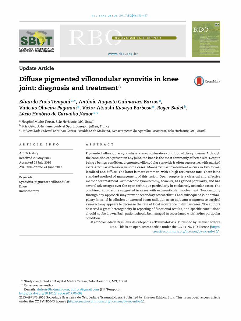
ent options have been proposed for this disease in cases ofenicular involvement, ranging from observation and radicalocal surgery to total knee arthroplasty (Fig. 1).4,5,8,13,14
creativecommons.org/licenses/by-nc-nd/4.0/).
Etiology and pathophysiology
The etiology of PVNS is still unknown. Some authors sug-gest that the disease occurs as a result of baseline traumaand subsequent bleeding in the affected joint.13–20 This the-ory is supported by the fact that patients with hemophiliapresent progressive destruction of the cartilage during thenatural course of the disease. However, studies that pro-duced PVNS-like histological findings by injecting iron orblood into the joint were unable to replicate the classic lipid-laden histiocytes and giant cells.15 However, most studiesreported a history of trauma in less than one-third of patients.Abnormal metabolic activity has also been indicated as anadjuvant event in the inflammation observed in PVNS, butthis is an inconsistent finding.16 There are also reports inthe literature that PVNS may be a neoplastic process. Severalauthors suggested the presence of chromosome 7 trisomy andclonal rearrangements as a cause.11,17 Some reports, albeitrare, indicate the occurrence of malignant transformationand metastases in patients initially diagnosed with PVNS.17,18
Despite the case reports of malignant PVNS and aneuploidy,there is evidence against the theory that PVNS is a neoplas-tic process. In their analysis, Oehler et al.9 observed a strongevidence of chronic inflammation. Their findings were basedon the presence of a cellular marker of inflammation witha heterogeneous population of mononuclear cells. They alsopostulated that the presence of large amounts of iron in thelesion would stimulate synoviocytes and fibroblasts to developmacrophage-like characteristics.
Histologically, LPVNS and DPVNS are similar. However,they differ in clinical presentation, prognosis, and response
to treatment. In the knee, LPVNS is more frequently observedin the anterior compartment.12 Flandry et al.19 reported thatmost lesions begin in the meniscocapsular junction. The most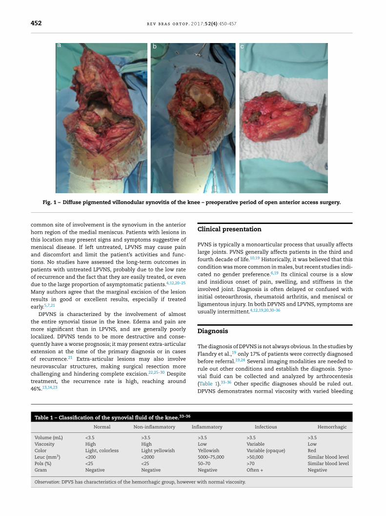
452 r e v b r a s o r t o p . 2 0 1 7;5 2(4):450–457
knee
Fig. 1 – Diffuse pigmented villonodular synovitis of thecommon site of involvement is the synovium in the anteriorhorn region of the medial meniscus. Patients with lesions inthis location may present signs and symptoms suggestive ofmeniscal disease. If left untreated, LPVNS may cause painand discomfort and limit the patient’s activities and func-tions. No studies have assessed the long-term outcomes inpatients with untreated LPVNS, probably due to the low rateof recurrence and the fact that they are easily treated, or evendue to the large proportion of asymptomatic patients.4,12,20–25
Many authors agree that the marginal excision of the lesionresults in good or excellent results, especially if treatedearly.5,7,21
DPVNS is characterized by the involvement of almostthe entire synovial tissue in the knee. Edema and pain aremore significant than in LPVNS, and are generally poorlylocalized. DPVNS tends to be more destructive and conse-quently have a worse prognosis; it may present extra-articularextension at the time of the primary diagnosis or in casesof recurrence.21 Extra-articular lesions may also involveneurovascular structures, making surgical resection more
challenging and hindering complete excision.22,25–30 Despitetreatment, the recurrence rate is high, reaching around46%.13,14,23Table 1 – Classification of the synovial fluid of the knee.33–36
Normal Non-inflammatory Infl
Volume (mL) <3.5 >3.5
Viscosity High High
Color Light, colorless Light yellowish
Leuc (mm3) <200 <2000
Pols (%) <25 <25
Gram Negative Negative
Observation: DPVS has characteristics of the hemorrhagic group, however w
– preoperative period of open anterior access surgery.
Clinical presentation
PVNS is typically a monoarticular process that usually affectslarge joints. PVNS generally affects patients in the third andfourth decade of life.10,19 Historically, it was believed that thiscondition was more common in males, but recent studies indi-cated no gender preference.6,19 Its clinical course is a slowand insidious onset of pain, swelling, and stiffness in theinvolved joint. Diagnosis is often delayed or confused withinitial osteoarthrosis, rheumatoid arthritis, and meniscal orligamentous injury. In both DPVNS and LPVNS, symptoms areusually intermittent.4,12,19,20,30–36
Diagnosis
The diagnosis of DPVNS is not always obvious. In the studies byFlandry et al.,19 only 17% of patients were correctly diagnosedbefore referral.19,24 Several imaging modalities are needed to
rule out other conditions and establish the diagnosis. Syno-vial fluid can be collected and analyzed by arthrocentesis(Table 1).33–36 Other specific diagnoses should be ruled out.DPVNS demonstrates normal viscosity with varied bleedingammatory Infectious Hemorrhagic
>3.5 >3.5 >3.5Low Variable LowYellowish Variable (opaque) Red5000–75,000 >50,000 Similar blood level50–70 >70 Similar blood levelNegative Often + Negative
ith normal viscosity.
r e v b r a s o r t o p . 2 0 1 7;5 2(4):450–457 453
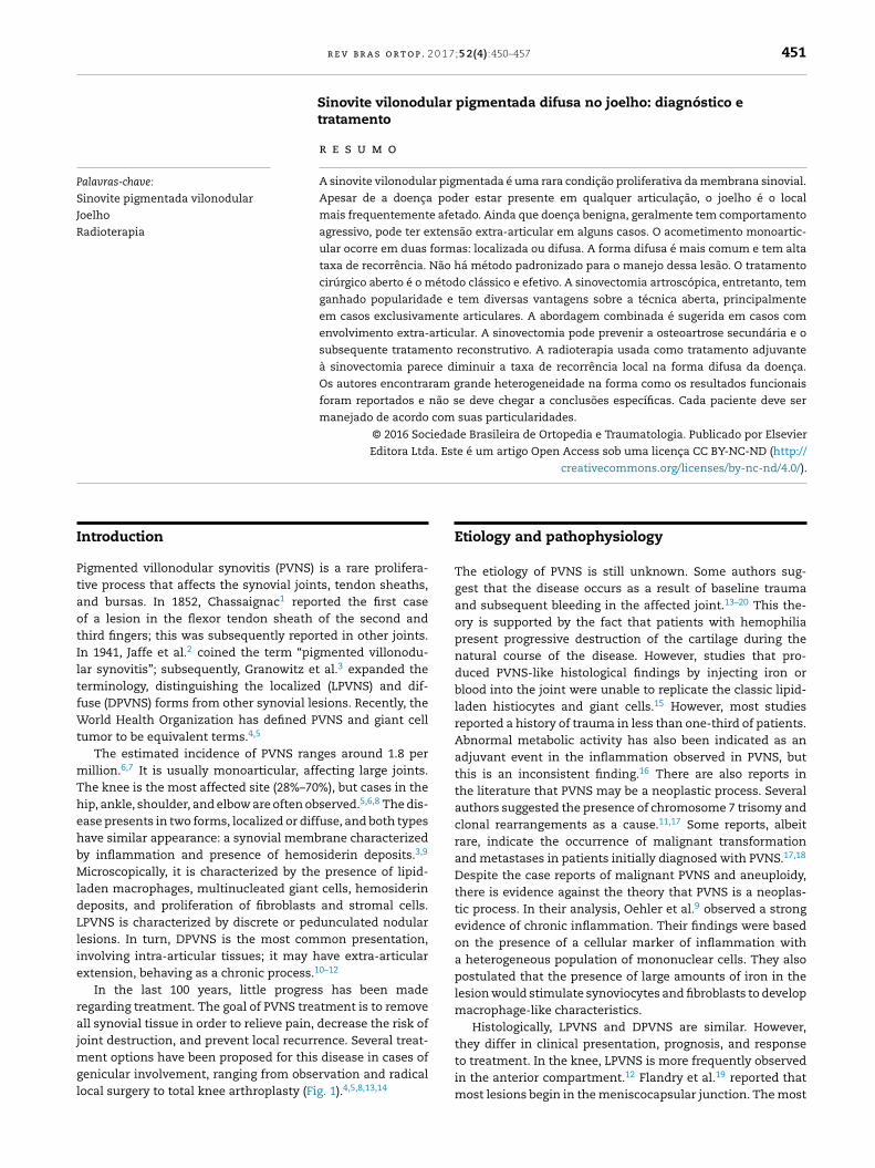
Fig. 2 – Knee radiograph of a diffuse pigmented villonodular synovitis of the knee in (A) antero-posterior and (B) profile,evidencing areas of bone destruction.
Fig. 3 – Magnetic resonance imaging of a knee with diffuse pigmented villonodular synovitis of the knee. A and B, coronalcut in T1; C, sagittal section in T1; D, T1 axial cut with areas of diffuse synovitis with tibial and femoral bone invasion,r
psTcdiri
esulting in significant joint destruction.
atterns, and radiographs can be useful (Fig. 2), as they mayhow periarticular erosions with a thin layer of reactive bone.he late radiographic finding of joint space narrowing indi-ates loss of articular cartilage, which may be difficult toifferentiate from primary osteoarthrosis. Radiographic find-
ngs can be observed in up to 30% of patients.4,19,20 Moreecently, magnetic resonance imaging (MRI) has become themaging modality of choice for diagnosing DPVNS.25 This is a
non-invasive examination with high accuracy; furthermore, itassesses the extent of the disease and distinguishes betweenthe diffuse and localized variants. The high hemosiderincontent causes the lesion to have an irregular or extensiveaspect, with low signal in the T1- and T2-weighted images.
Both LPVNS and DPVNS may present with joint effusion. InDPVNS, there is a poorly localized mass or synovial thicken-ing with varying degrees of periarticular erosion. Classically, it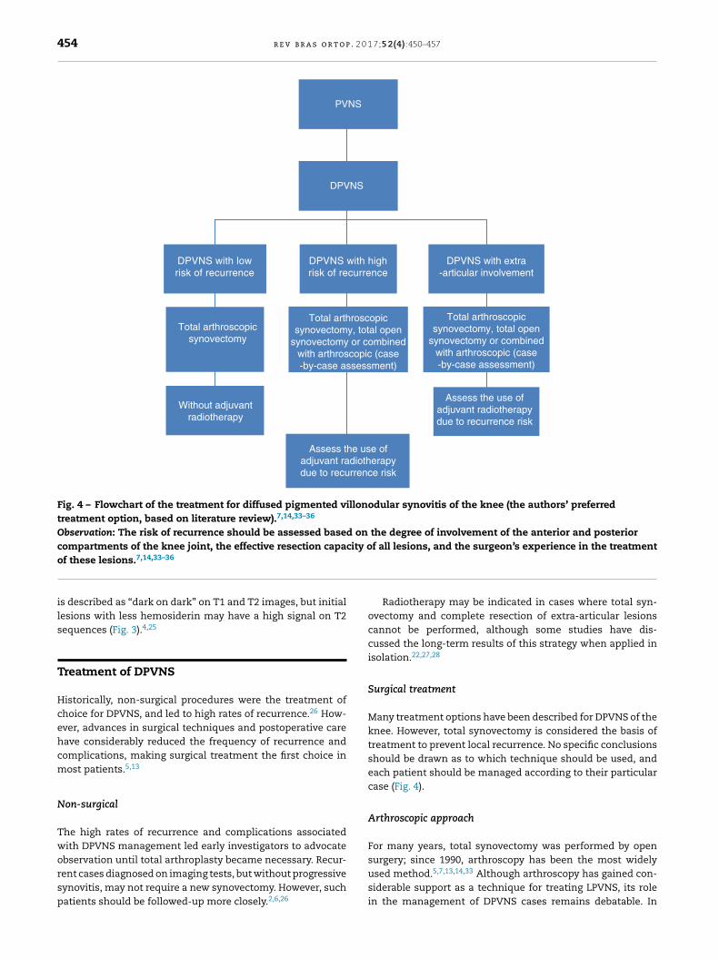
454 r e v b r a s o r t o p . 2 0 1 7;5 2(4):450–457
PVNS
DPVNS
DPVNS with lowrisk of recurrence
Total arthroscopicsynovectomy
Without adjuvantradiotherapy
Assess the use ofadjuvant radiotherapydue to recurrence risk
Assess the use ofadjuvant radiotherapydue to recurrence risk
Total arthroscopicsynovectomy, total open
synovectomy or combinedwith arthroscopic (case-by-case assessment)
Total arthroscopicsynovectomy, total open
synovectomy or combinedwith arthroscopic (case-by-case assessment)
DPVNS with highrisk of recurrence
DPVNS with extra-articular involvement
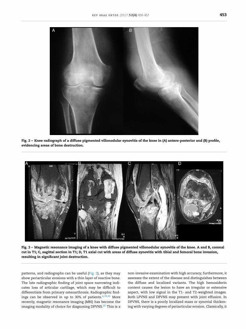
Fig. 4 – Flowchart of the treatment for diffused pigmented villonodular synovitis of the knee (the authors’ preferredtreatment option, based on literature review).7,14,33–36
Observation: The risk of recurrence should be assessed based on the degree of involvement of the anterior and posteriorcompartments of the knee joint, the effective resection capacity of all lesions, and the surgeon’s experience in the treatment
of these lesions.7,14,33–36is described as “dark on dark” on T1 and T2 images, but initiallesions with less hemosiderin may have a high signal on T2sequences (Fig. 3).4,25
Treatment of DPVNS
Historically, non-surgical procedures were the treatment ofchoice for DPVNS, and led to high rates of recurrence.26 How-ever, advances in surgical techniques and postoperative carehave considerably reduced the frequency of recurrence andcomplications, making surgical treatment the first choice inmost patients.5,13
Non-surgical
The high rates of recurrence and complications associatedwith DPVNS management led early investigators to advocate
observation until total arthroplasty became necessary. Recur-rent cases diagnosed on imaging tests, but without progressivesynovitis, may not require a new synovectomy. However, suchpatients should be followed-up more closely.2,6,26Radiotherapy may be indicated in cases where total syn-ovectomy and complete resection of extra-articular lesionscannot be performed, although some studies have dis-cussed the long-term results of this strategy when applied inisolation.22,27,28
Surgical treatment
Many treatment options have been described for DPVNS of theknee. However, total synovectomy is considered the basis oftreatment to prevent local recurrence. No specific conclusionsshould be drawn as to which technique should be used, andeach patient should be managed according to their particularcase (Fig. 4).
Arthroscopic approach
For many years, total synovectomy was performed by open
surgery; since 1990, arthroscopy has been the most widelyused method.5,7,13,14,33 Although arthroscopy has gained con-siderable support as a technique for treating LPVNS, its rolein the management of DPVNS cases remains debatable. In
0 1 7
Drcrlmapebaii
riisrgtpttictskpftpasosroa
nroaa
O
TswiabptOtS
r e v b r a s o r t o p . 2
PVNS, the anterior compartment is typically involved andequires greater technical care from the surgeon, makingomplementary portals to the traditional portals made ante-iorly, and using 30◦ and 70◦ arthroscopes.33–36 Patients witharge masses in the popliteal fossa or extra-articular involve-
ent generally are not candidates for exclusively arthroscopicpproach. Arthroscopic treatment should be reserved foratients with limited, purely intra-articular disease.34–36 Dis-ase assessment and choice of best treatment schedule shoulde made with MRI before the definitive resection. If therthroscopic approach is selected, a complete synovectomy,ncluding the posterior compartments, should be performedn order to minimize the risk of recurrence.20,23,34
Two similar-sized groups of patients were identified in theeview by Auregan et al.34: local recurrences were observedn 28 of 124 patients (23%) in the open surgery group andn twenty of 124 (16%) in the group that underwent arthro-copic surgery, in mean follow-up periods of six and five years,espectively. The lower recurrence rate in the arthroscopicroup can be explained by the shorter follow-up period inhis group. Nonetheless, a higher rate of postoperative com-lications was reported in the group that underwent openotal synovectomy when compared with the group submit-ed to arthroscopy. Functional scores appear to be bettern the arthroscopic group; however, as discussed earlier, noonvincing conclusion can be drawn. This study concludedhat posterior open treatment, together with anterior arthro-copic synovectomy, is a viable approach for DPVNS of thenee, with low recurrence rates and few postoperative com-lications. Based on these results, the arthroscopic approachor arthroscopic synovectomy is recommended wheneverechnically possible. Colman et al.33 reported 48 cases ofatients treated using the arthroscopic, posterior open withnterior arthroscopic, or anterior open and posterior openynovectomy techniques. Recurrence rates were lower in thepen/arthroscopic group when compared with the arthro-copic or with the open/open groups: 9% vs. 62% vs. 64%,espectively. Osteoarthrosis progression was observed in 17%f the total of patients, 8% of whom underwent total kneerthroplasty during the follow-up period.33
Although arthroscopy is a less invasive procedure, it isot free of potential complications. In addition to the risk ofecurrence, arthroscopic excision presents the theoretical riskf joint tumor dispersion and portal contamination. A failedrthroscopic approach may cause extensive joint involvementnd extra-articular dissemination.20,21,23,34–36
pen approach
he open approach followed by complete synovectomy is thetandard surgical treatment for DPVNS of the knee. Patientsith extensive extra-articular involvement and large masses
n the popliteal fossa clearly are not suitable candidates forrthroscopic synovectomy. Moreover, open procedures shoulde considered for patients with disease in hard-to-reachlaces, such as the popliteal tendon sheath, between the gas-
rocnemius heads, and within the semimembranous bursa.pen treatment begins with an anterior approach, arthro-omy, and aggressive anterior synovectomy, followed by an-shaped posterior approach, protecting the neurovascular
;5 2(4):450–457 455
structures. Subsequently, joint and extra-articular explorationshould be conducted in order to avoid any remaining tumortissue.9,21,23,24,34–36
Old studies assessing open treatment reported excessivelyhigh rates of recurrence. Nonetheless, in these studies recur-rence probably indicated incomplete excisions of the lesionsand likely referred to inadequate surgical exposure. Flandryet al.19 reported a series of 25 knees with biopsy and provenDPVNS that were treated by double open approach. Theauthors reported a recurrence rate of 8% in a follow-up of 58months.
Open treatment, however, is not free of risks and com-plications. Compared with the arthroscopic procedure, opensynovectomy is associated with longer hospital stay andlonger rehabilitation period. One of the main criticisms ofthe open procedure is postoperative stiffness, which oftenrequires manipulation under anesthesia. In the study byFlandry et al.,19 the rate of postoperative stiffness was 24%.Therefore, many experts advocate the use of less invasive andless aggressive procedures.19,34
Combined open and arthroscopic approach
The combination of open and arthroscopic approach has notbeen well described in the literature. Patients with posteriorinvolvement associated with minimal anterior involvementmay benefit from anterior arthroscopic synovectomy com-bined with posterior open synovectomy. Another suitablescenario for the combined approach is in cases in whichtotal synovectomy by arthroscopic approach alone is impossi-ble. In this scenario, several authors suggest a combinationof anterior arthroscopic synovectomy and open posteriorsynovectomy.5,7,14 Moreover, arthroscopy may play an impor-tant role in the pre- and postoperative diagnosis, as well as inthe treatment of residual disease after open surgery. De Car-valho et al.14 reported that patients diagnosed with DPVNStreated with partial arthroscopic synovectomy combined withanterior open synovectomy presented a recurrence rate of12.5% in a mean follow-up of 8.6 years; no major complicationswere observed during follow-up.14
Adjuvant radiotherapy
Radiotherapy has been used for many years as an alternativeto surgical synovectomy in patients with nonspecific syn-ovitis. Radiation-induced synovectomy in the treatment ofDPVNS has been increasingly discussed, but with conflictingresults.14,23,28,29 From 1950 onwards, good results have beenreported with use of adjuvant external radiation in themanagement of recurrent DPVNS.30 Potential complicationsare associated with external radiation, including skin reac-tions, poor wound healing, joint stiffness, and neoplastictransformation.31–36
De Carvalho et al.14 assessed patients diagnosed withDPVNS treated with partial arthroscopic synovectomy com-bined with anterior open synovectomy followed by adjuvant
radiotherapy (2000 cGy). No cases of major postoperative com-plications or radiotherapy-related side effects were observed.No progression to arthrosis was detected. Recurrence wasobserved in only one patient (12.5%), in a mean follow-up
p . 2 0
r
1
1
1
1
1
1
1
1
456 r e v b r a s o r t o
period of 8.6 years.14 Blanco et al.29 described 22 patients withknee DPVNS treated with arthroscopic synovectomy com-bined with postoperative external radiotherapy (total doseof 2600 cGy). The recurrence rate was 13.62%; these patientsunderwent a second surgical procedure. Ustinova et al.31
described their experience with 24 patients with DPVNS. Radi-ation (in two doses, one of 1.2–1.5 cGy in five fractions and afocal dose of 16–20 cGy) was applied, since the affected syno-vial tissue had not been completely removed during surgery.No recurrences were observed in the follow-up period, whichranged from six months to six years, and occupational reha-bilitation was achieved in 87.5% of the patients.31 O’Sullivanet al.22 also reported the cases of 14 patients with intra-and extra-articular lesions who received adjuvant radiation.Those authors concluded that after DPVNS removal, the useof moderate external radiation was very effective in preven-ting recurrence, avoiding amputation in advanced cases andpreserving limb function.
Local radiotherapy can also be applied with intra-articularradioisotope injection. Chin et al.21 assessed a large numberof patients (n = 40) with DPVNS, each subjected to either openor arthroscopic surgery, but who presented recurrence. Thenumber of residual lesions in the groups treated with radio-therapy was lower than in the untreated group. Shabat et al.,in a mean follow-up of six years, assessed ten patients withDPVNS who underwent one or more partial synovectomiesand received intra-articular injection of yttrium isotope (90Y;15–25 mCi) between six and eight weeks after the last surgery.In nine patients, no evidence of recurrence was observed dur-ing follow-up, whereas in one patient the disease stabilized.32
Final considerations
DPVNS of the knee is a rare condition in which treat-ment may be associated with a significant risk of localrecurrence, postoperative complications, and functional lim-itations. Regarding local recurrence, there was no differencein the literature between open or arthroscopic total synovec-tomy for the treatment of DPVNS. However, a lower rate ofcomplications was reported after arthroscopic synovectomy.Incomplete synovectomy for the treatment of DPVNS shouldnot be performed in isolation, due to the high risk of recur-rence. Internal or external radiation as an adjuvant treatmentmethod to surgical synovectomy appears to decrease therate of local recurrence in cases of DPVNS. Cases of DPVNSwith extra-articular involvement should be treated with totalsynovectomy, open excision of extra-articular lesions, andadjuvant radiotherapy treatment.
Conflicts of interest
The authors declare no conflicts of interest.
Acknowledgments
To all researchers and to Dr. Guilherme Reis for having autho-rized the use of the archive images in this article.
1 7;5 2(4):450–457
e f e r e n c e s
1. Chassaignac EP. Cancer de la gaine des tendons. Gaz Hop CivMilit. 1852;25:185–6.
2. Jaffe HL, Lichtenstein I, Sutro CJ. Pigmented villonodularsynovitis, bursitis and tenosynovitis: a discussion of thesynovial and bursal equivalents of the tenosynovial lesioncommonly denoted as xanthoma, xanthogranuloma, giantcell tumor or myeloplaxoma of tendon sheath with someconsideration of this tendon sheath lesion itself. Arch Pathol.1941;31:731–65.
3. Granowitz SP, Mankin HJ. Localized pigmented villonodularsynovitis of the knee. Report of five cases. J Bone Joint SurgAm. 1967;49(1):122–8.
4. Jendrissek KA, Hotfiel T, Swoboda B, Soder S, Janka R.Pigmented villonodular synovitis: a rare differential diagnosisof synovial joint swelling. Z Rheumatol. 2016;75(2):157–65.
5. Rodriguez-Merchan EC. Review article: open versusarthroscopic synovectomy for pigmented villonodularsynovitis of the knee. J Orthop Surg (Hong Kong).2014;22(3):406–8.
6. Myers BW, Masi AT. Pigmented villonodular synovitis andtenosynovitis: a clinical epidemiologic study of 166 cases andliterature review. Medicine (Baltimore). 1980;59(3):223–38.
7. Yang B, Liu D, Lin J, Jin J, Weng XS, Qian WW, et al. Surgicaltreatment of diffuse pigmented villonodular synovitis of theknee. Zhongguo Yi Xue Ke Xue Yuan Xue Bao.2015;37(2):234–9.
8. Sherry JB, Anderson W. The natural history of pigmentedvillonodular synovitis of tendon sheaths. J Bone Joint SurgAm. 1955;37-A(5):1005–11.
9. Oehler S, Fassbender HG, Neureiter D, Meyer-Scholten C,Kirchner T, Aigner T. Cell populations involved in pigmentedvillonodular synovitis of the knee. J Rheumatol.2000;27(2):463–70.
0. de Visser E, Veth RP, Pruszczynski M, Wobbes T, Van de PutteLB. Diffuse and localized pigmented villonodular synovitis:evaluation of treatment of 38 patients. Arch Orthop TraumaSurg. 1999;119(7–8):401–4.
1. Perka C, Labs K, Zippel H, Buttgereit F. Localized pigmentedvillonodular synovitis of the knee joint: neoplasm or reactivegranuloma? A review of 18 cases. Rheumatology (Oxford).2000;39(2):172–8.
2. Bouguennec N, Meyer A, Graveleau N. Localized form ofpigmented villonodular synovitis of the knee: the meniscalmime. Orthop Traumatol Surg Res. 2014;100(2):251–4.
3. Mollon B, Griffin AM, Ferguson PC, Wunder JS,Theodoropoulos J. Combined arthroscopic and opensynovectomy for diffuse pigmented villonodular synovitis ofthe knee. Knee Surg Sports Traumatol Arthrosc.2016;24(1):260–6.
4. de Carvalho LH Jr, Soares LF, Goncalves MB, Temponi EF, deMelo Silva O Jr. Long-term success in the treatment of diffusepigmented villonodular synovitis of the knee with subtotalsynovectomy and radiotherapy. Arthroscopy.2012;28(9):1271–4.
5. Singh R, Grewal DS, Chakravarti RN. Experimental productionof pigmented villonodular synovitis in the knee and anklejoints of rhesus monkeys. J Pathol. 1969;98(2):137–42.
6. Hirohata K. Light microscopic and electron microscopicstudies of individual cells in pigmented villonodular synovitisand bursitis (Jaffe). Kobe J Med Sci. 1968;14(4):251–79.
7. Abdul-Karim FW, el-Naggar AK, Joyce MJ, Makley JT, Carter JR.
Diffuse and localized tenosynovial giant cell tumor andpigmented villonodular synovitis: a clinicopathologic andflow cytometric DNA analysis. Hum Pathol. 1992;23(7):729–35.
0 1 7
1
1
2
2
2
2
2
2
2
2
2
2
3
3
3
3
3
3
3
r e v b r a s o r t o p . 2
8. Bertoni F, Unni KK, Beabout JW, Sim FH. Malignant giant celltumor of the tendon sheaths and joints (malignant pigmentedvillonodular synovitis). Am J Surg Pathol. 1997;21(2):153–63.
9. Flandry F, Hughston JC, McCann SB, Kurtz DM. Diagnosticfeatures of diffuse pigmented villonodular synovitis of theknee. Clin Orthop Relat Res. 1994;(298):212–20.
0. Akgun I, Ogut T, Kesmezacar H, Dervisoglu S. Localizedpigmented villonodular synovitis of the knee. Orthopedics.2003;26(11):1131–5.
1. Chin KR, Barr SJ, Winalski C, Zurakowski D, Brick GW.Treatment of advanced primary and recurrent diffusepigmented villonodular synovitis of the knee. J Bone JointSurg Am. 2002;84(12):2192–202.
2. O’Sullivan B, Cummings B, Catton C, Bell R, Davis A, FornasierV, et al. Outcome following radiation treatment for high-riskpigmented villonodular synovitis. Int J Radiat Oncol Biol Phys.1995;32(3):777–86.
3. Nassar WA, Bassiony AA, Elghazaly HA. Treatment of diffusepigmented villonodular synovitis of the knee with combinedsurgical and radiosynovectomy. HSS J. 2009;5(1):19–23.
4. Flandry FC, Hughston JC, Jacobson KE, Barrack RL, McCannSB, Kurtz DM. Surgical treatment of diffuse pigmentedvillonodular synovitis of the knee. Clin Orthop Relat Res.1994;(300):183–92.
5. Dale K, Smith HJ, Paus AC, Refsum SB. Dynamic MR-imagingin the diagnosis of pigmented villonodular synovitis of theknee. Scand J Rheumatol. 2000;29(5):336–9.
6. Byers PD, Cotton RE, Deacon OW, Lowy M, Newman PH,Sissons HA, et al. The diagnosis and treatment of pigmentedvillonodular synovitis. J Bone Joint Surg Br. 1968;50(2):290–305.
7. Park G, Kim YS, Kim JH, Lee SW, Song SY, Choi EK, et al.
Low-dose external beam radiotherapy as a postoperativetreatment for patients with diffuse pigmented villonodularsynovitis of the knee: 4 recurrences in 23 patients followedfor mean 9 years. Acta Orthop. 2012;83(3):256–60.;5 2(4):450–457 457
8. Heyd R, Seegenschmiedt MH, Micke O. The role of externalbeam radiation therapy in the adjuvant treatment ofpigmented villonodular synovitis. Z Orthop Unfall.2011;149(6):677–82.
9. Blanco CE, Leon HO, Guthrie TB. Combined partialarthroscopic synovectomy and radiation therapy for diffusepigmented villonodular synovitis of the knee. Arthroscopy.2001;17(5):527–31.
0. Friedman M, Schwartz EE. Irradiation therapy of pigmentedvillonodular synovitis. Bull Hosp Joint Dis. 1957;18(1):19–32.
1. Ustinova VF, Podliashuk EL, Rodionova SS. Combinedtreatment of the diffuse form of pigmented villonodularsynovitis. Med Radiol (Mosk). 1986;31(3):27–31.
2. Shabat S, Kollender Y, Merimsky O, Isakov J, Flusser G, NyskaM, et al. The use of surgery and yttrium 90 in themanagement of extensive and diffuse pigmentedvillonodular synovitis of large joints. Rheumatology (Oxford).2002;41(10):1113–8.
3. Colman MW, Ye J, Weiss KR, Goodman MA, McGough RL 3rd.Does combined open and arthroscopic synovectomy fordiffuse PVNS of the knee improve recurrence rates? ClinOrthop Relat Res. 2013;471(3):883–90.
4. Auregan JC, Klouche S, Bohu Y, Lefevre N, Herman S, Hardy P.Treatment of pigmented villonodular synovitis of the knee.Arthroscopy. 2014;30(10):1327–41.
5. Nakahara H, Matsuda S, Harimaya K, Sakamoto A,Matsumoto Y, Okazaki K, et al. Clinical results of opensynovectomy for treatment of diffuse pigmented villonodularsynovitis of the knee: case series and review of literature.Knee. 2012;19(5):684–7.
6. Mollon B, Lee A, Busse JW, Griffin AM, Ferguson PC, Wunder
JS, et al. The effect of surgical synovectomy and radiotherapyon the rate of recurrence of pigmented villonodular synovitisof the knee: an individual patient meta-analysis. Bone Joint J.2015;97-B(4):550–7.