development and validation of a biosensor-based immunoassay for progesterone in bovine milk
TRANSCRIPT
Development and validation of a biosensor-based immunoassay
for progesterone in bovine milk
Els H. Gillis a, James P. Gosling b, Joseph M. Sreenan c, Marian Kane a,*
aNational Diagnostic Centre, National University of Ireland, Galway, IrelandbDepartment of Biochemistry, National University of Ireland, Galway, IrelandcAnimal Reproduction Department, Teagasc, Athenry, Co. Galway, Ireland
Received 30 January 2002; received in revised form 22 April 2002; accepted 2 May 2002
Abstract
We have developed a rapid automated immunoassay, using the BIACOREk surface plasmon resonance (SPR) biosensor, to
measure progesterone in bovine milk. The assay was designed as an inhibition assay with progesterone covalently immobilised
to the carboxymethyl dextran matrix of a CM5 sensor chip. A fixed amount of monoclonal anti-progesterone antibody 39C5H7
was mixed 9:1 with the sample and the amount of free antibody was then determined using biomolecular interaction analysis
(BIA) by injection of the mixture over the immobilised progesterone sensor surface. The assay was designed to cover the
concentration range 0.5 to 50 ng/ml. The limit of detection (LOD) was 3.56 ng/ml. Reproducibility of the assay was very good
with both intra-assay and inter-assay coefficients of variation < 5%. As results become available within minutes of injection and
the procedure involves fully automated instrumentation, we believe that this BIA assay for progesterone in milk could be used
in-line in the milking parlour and, thus, provide an important tool for reproductive management of dairy cattle to detect heat and
predict pregnancy.
D 2002 Elsevier Science B.V. All rights reserved.
Keywords: SPR; Biosensor assays; Progesterone; Heat detection
1. Introduction
Reproductive performance is a major factor affect-
ing the production and economic efficiency of dairy and
beef cow herds. According to Sreenan and Diskin
(1992), a dairy herd is reproductively efficient when
the calving interval is 365 days, when 90% of the cows
calve in a 10-week period andwhen 5%or less cows are
culled for reproductive failure. For herds using artificial
insemination (AI), failure to detect heat is one of the
major causes of a prolonged calving interval. Visual
observation is currently the primary method for heat
0022-1759/02/$ - see front matter D 2002 Elsevier Science B.V. All rights reserved.
PII: S0022 -1759 (02 )00166 -7
Abbreviations: AI, Artificial insemination; CV, Coefficient of
variation; BIA, Biomolecular interaction analysis; CMO, Carbox-
ymethyloxime; EDC, N-ethyl-NV(3-ethylaminopropyl) carbodiimide;
ELISA, Enzyme-linked immunoassay; DMF, N,N-dimethylforma-
mide; HBS-EP, HEPES-buffered saline; HEPES, N-[2-hydroxye-
thyl]piperazine-NV-[2-ethanesulfonic acid]; LOD, Limit of detection;
NHS, N-hydroxysuccinimide; SD, Standard deviation; SPR, Surface
plasmon resonance.* Corresponding author. Tel.: +353-91-586559; fax: +353-91-
586570.
E-mail address: [email protected] (M. Kane).
www.elsevier.com/locate/jim
Journal of Immunological Methods 267 (2002) 131–138
detection, but this is time consuming and repetitive.
Farmers frequently achieve a heat detection rate of only
35–70% and up to 20% of cows presented for insemi-
nation are not in heat (Sreenan andDiskin, 1992; Diskin
and Sreenan, 2000).
According to Senger (1994), an ideal system for
detecting oestrous should have the following charac-
teristics: (i) continuous surveillance of the cow; (ii)
accurate and automatic identification of the cow in
oestrous; (iii) operation for the productive life-time
of the cow; (iv) minimal labour requirements; and
(v) high accuracy and efficiency (95%) for identify-
ing the appropriate physiological events that corre-
late with oestrous or ovulation or both. Elevated
milk progesterone concentrations indicate luteal
dominance, while low amounts of progesterone are
associated with oestrous. Progesterone measurement
tests based on enzyme-linked immunoassays
(ELISA) are available as kits for on-farm use (Nebel,
1988). Most of these tests are, however, manual and
designed as qualitative tests for the confirmation of
oestrous and determination of pregnancy or non-
pregnancy, rather than providing a precise concen-
tration. Attempts at automation and in-line applica-
tion of quantitative enzyme immunoassays for
progesterone have been described (Koelsch et al.,
1994; Claycomb et al., 1998; Pemberton et al.,
2001), but, to date, such methods either suffer from
poor sensitivity or are still too cumbersome for use
in the milking parlour.
Biomolecular interaction analysis (BIA) from Bia-
core Ab uses the optical phenomenon of surface
plasmon resonance (SPR) to monitor biomolecular
interactions in real time without labelling. SPR meas-
ures changes in refractive index of the solution close
to the sensor surface, resulting from changes in the
mass concentration of molecules in the solution
(reviewed by Hashimoto, 2000; Markey, 2000). BIA-
CORE instrumentation was developed primarily for
use as a research tool, with a variety of applications
such as screening biological samples for binding
partners, the determination of kinetic and equilibrium
constants for complex formation, epitope mapping
and ligand fishing for known receptors. Recently, this
principle has also gained popularity as an analytical
tool for the determination of concentrations of analy-
tes in biological fluids. In 1993, Minunni and Mascini
described a BIACORE assay for the determination of
the pesticide, atrazine, in drinking water, while Ster-
nesjo et al. (1995) used a BIACORE to develop a
biosensor-based immunoassay for the detection of
sulfamethazine residues in milk. Since then, BIA
assays have been described for other veterinary drug
residues (Elliott et al., 1999; Gaudin and Maris,
2001), toxins (Mullett et al., 1998; Daly et al.,
2000) and vitamins (Bostrom Caselunghe and Linde-
berg, 2000; Indyk et al., 2000).
When compared to traditional techniques, BIA-
CORE technology offers several significant advan-
tages, such as high precision, speed, simplicity, and
automation (Mellgren et al., 1996). This paper
describes the development and validation, according
to principles set out by Wong et al. (1997), of a BIA
assay for real-time measurement of progesterone in
bovine milk.
2. Materials and methods
2.1. Instrumentation
A BIACORE 2000k biosensor instrument and
CM5 sensor chips (research grade) were used (Biacore
Ab, Uppsala, Sweden). The BIACORE 2000k was
controlled by BIACORE Control Software version
3.1.1 running underWindows 95. The software also in-
cludes an interface to the separate BIAevalution soft-
ware. The instrument running temperature was 25 jC.
2.2. Reagents
HEPES-buffered saline (HBS-EP) buffer pH7.4
(10 mM N-[2-hydroxyethyl]piperazine-NV-[2-ethane-sulfonic acid] (HEPES), 0.15 M NaCl, 3.4 mM
EDTA, 0.005% Surfactant P-20), and amine coupling
kit (containing N-hydroxysuccinimide (NHS), N-
ethyl-NV(3-ethylaminopropyl) carbodiimide (EDC),
and ethanolamine hydrochloride) were obtained from
Biacore Ab (Uppsala, Sweden). The Ridgeway milk
progesterone ELISA was purchased from Ridgeway
Science (Alvington, Gloucestershire, UK). Progester-
one-3-carboxymethyloxime (CMO) was purchased
from Steraloids (Wilton, NH, USA). Progesterone
(MW 314.5) and N,N-dimethylformamide (DMF)
were purchased from Sigma. All other reagents were
of analytical grade.
E.H. Gillis et al. / Journal of Immunological Methods 267 (2002) 131–138132

2.3. Monoclonal antibody
The monoclonal antibody used was that described
by O’Rorke et al. (1994). The antibody was raised
against 11a-hydroxyprogesterone hemisuccinate–
bovine serum albumin conjugate, which was prepared
by modification of the mixed anhydride method
(Erlanger et al., 1957). Ascites fluid from mice
infected with hybridoma cells secreting antibody
39C5H7 was diluted 1/5000 in HBS-EP before use.
2.4. Immobilisation of progesterone-3-CMO deriva-
tive
Simultaneous immobilisation of progesterone onto
all four flowcells of a CM5 sensor chip was performed
outside the biosensor system using conventional amine
coupling (Sternesjo et al., 1995). For covalent immo-
bilisation, carboxyl groups of the sensor chip surface
are activated by derivatisation with N-hydroxysuccini-
mide (NHS) mediated by N-ethyl-NV(3-ethylamino-
propyl) carbodiimide (EDC). The resulting active
ester groups will react spontaneously with amino
groups (Johnsson et al., 1991). EDC and NHS were
mixed (1:1) and 40 Al of the mixture were deposited on
the gold film for 10 min activation. An amine surface
was then prepared by placing 40 Al of 1M ethylene
diamine pH8.5 in contact with the activated surface for
20 min. Capping any remaining active carboxyl groups
was achieved by exposing the surface to 40 Al 1Methanolamine for 10 min. Progesterone-3-CMO (2 mg)
was dissolved in 900 Al DMF and mixed with 450 Al of10 mM sodium acetate solution pH4.6 containing 5 mg
EDC and 2 mg NHS. Progesterone was then immobi-
lised on the surface by placing 40 Al of this solution in
contact with the amine surface for approximately 15
min. Prior to use, the immobilised surface was condi-
tioned with repeated injections of 25 Al regenerationbuffer (10% acetonitrile in 50 mM NaOH) to remove
any non-covalently bound progesterone.
2.5. Milk samples
Milk samples were collected from dairy cows
throughout the oestrous cycle. Samples were collected
into 10 ml plastic containers, with one Lactab mark III
tablet (Thompson and Capper, Cheshire, UK) added as
a preservative or alternatively 15 Al of 0.068 M sodium
dichromate solution. Samples were then stored at 4 jC.Before analysis, samples were heated for 40 min in a
water bath at 21 jC and dispersed by gentle mixing on a
rock and roller for 10 min. No difference was noted
between the results obtained in the BIACORE assay
with preserved or unpreserved milk samples.
2.6. Assay
Progesterone standards were prepared by dilution in
preserved whole milk from an oestrous animal. The
concentrations used to construct standard curves were
0.5, 1, 2, 5, 10, 20, and 50 ng/ml (i.e. 3.18, 6.36, 15.9,
31.8, 63.6 and 159 nmol/l). The curves were fitted
using a four-parameter equation with BIAevaluation
software.
The assay was designed as an inhibition assay. A
fixed amount of the monoclonal anti-progesterone
antibody was mixed 9:1 with the standard/sample.
The amount of free antibodies remaining was then
determined by injection of 50 Al of the mixture over
the progesterone surface at a rate of 25 Al/min. After
each measurement the surface was regenerated by
injection of 25 Al of 10% acetonitrile in 50 mM
NaOH at the same flow rate. The analysis cycle time,
including regeneration was 5 min 8 s.
3. Results
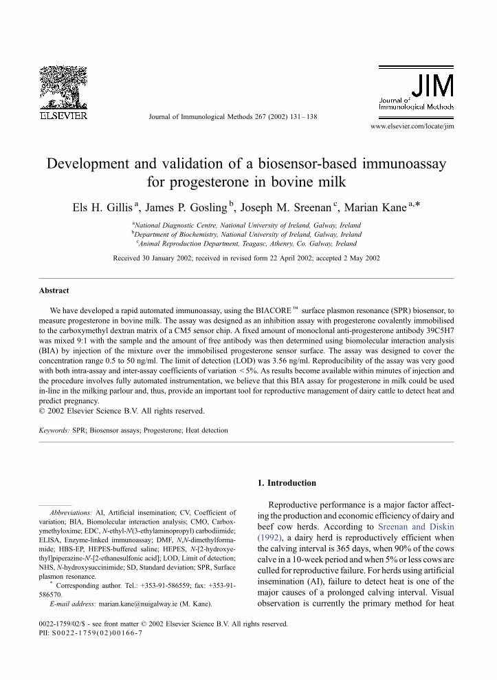
3.1. Stability of sensor chip during regeneration
Regeneration performance was evaluated by re-
peated analysis of a negative whole milk sample using
the standard assay procedure. Baseline readings and
antibody binding responses are shown in Fig. 1. The
baseline response showed less than 1% change over
129 cycles and the binding capacity was also unaf-
fected, with less than 5% change being observed from
the beginning to the end of the analysis. The progester-
one sensor surface went through 3600 regeneration
cycles over a 9-month period before any loss in binding
capacity was observed.
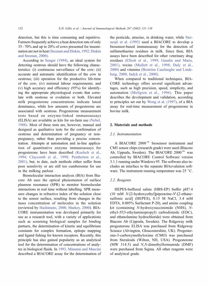
3.2. Limit of detection
The assay was designed to cover the concentration
range 0.5 to 50 ng/ml. The standard curve in Fig. 2
E.H. Gillis et al. / Journal of Immunological Methods 267 (2002) 131–138 133
represents the mean of 10 curves, obtained separately,
eachwith triplicate concentrations of the standards. The
precision of the response for each standard was < 7%
coefficient of variation (CV). The limit of detection
(LOD), determined as the concentration corresponding
to the mean response for negative whole milk minus
three times the standard deviation (SD),was 3.56 ng/ml.
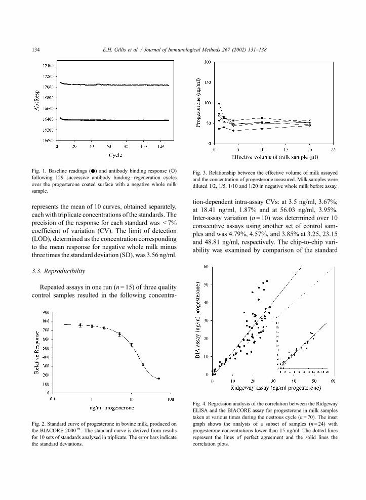
3.3. Reproducibility
Repeated assays in one run (n= 15) of three quality
control samples resulted in the following concentra-
tion-dependent intra-assay CVs: at 3.5 ng/ml, 3.67%;
at 18.41 ng/ml, 1.87% and at 56.03 ng/ml, 3.95%.
Inter-assay variation (n = 10) was determined over 10
consecutive assays using another set of control sam-
ples and was 4.79%, 4.57%, and 3.85% at 3.25, 23.15
and 48.81 ng/ml, respectively. The chip-to-chip vari-
ability was examined by comparison of the standard
Fig. 2. Standard curve of progesterone in bovine milk, produced on
the BIACORE 2000k. The standard curve is derived from results
for 10 sets of standards analysed in triplicate. The error bars indicate
the standard deviations.
Fig. 3. Relationship between the effective volume of milk assayed
and the concentration of progesterone measured. Milk samples were
diluted 1/2, 1/5, 1/10 and 1/20 in negative whole milk before assay.
Fig. 1. Baseline readings (.) and antibody binding response (o)
following 129 successive antibody binding– regeneration cycles
over the progesterone coated surface with a negative whole milk
sample.
Fig. 4. Regression analysis of the correlation between the Ridgeway
ELISA and the BIACORE assay for progesterone in milk samples
taken at various times during the oestrous cycle (n= 70). The inset
graph shows the analysis of a subset of samples (n= 24) with
progesterone concentrations lower than 15 ng/ml. The dotted lines
represent the lines of perfect agreement and the solid lines the
correlation plots.
E.H. Gillis et al. / Journal of Immunological Methods 267 (2002) 131–138134

curves obtained with three sensor chips coated at
different times. The standard curves obtained were
identical when overlaid (data not shown).
3.4. Linearity/parallelism
Milk samples from five animals were diluted 1/2,
1/5, 1/10 and 1/20 in negative whole milk before
assay. The amount of progesterone measured was
directly related to the effective milk volume assayed
over the range examined (Fig. 3), indicating minimal
interference of the milk matrix. The lowest effective
volume resulted in an over-estimation of progesterone
for some samples but these were below the LOD of
the assay. Regression analysis of the correlation
between actual and measured values of diluted sam-
ples confirmed linearity of the assay with R2 values
> 0.98.
3.5. Recovery
Samples containing various levels of endogenous
progesterone were spiked with 2, 5, 10 and 20 ng/ml
progesterone. The total concentration of progesterone
present in each sample was then determined to calcu-
late the recovery in each case. The percentage recov-
ery ranged from 88.9% to 112.4%, the mean was
101.7% (F 7.8 SD, n = 12).
3.6. Correlation with Ridgeway ELISA
Seventy samples collected at various times through-
out the oestrous cycle were analysed by both the
Fig. 5. Comparison of standard curves obtained by the BIA assay
(.) and the Ridgeway ELISA (o) for progesterone in bovine milk.
Duplicate readings for each assay are indicated on the graph.
Fig. 6. Progesterone levels estimated by the BIA (.) and the Ridgeway assay (o) in daily milk samples. These milk samples were collected for
26 days after artificial insemination. Heat was detected by visual observation.
E.H. Gillis et al. / Journal of Immunological Methods 267 (2002) 131–138 135
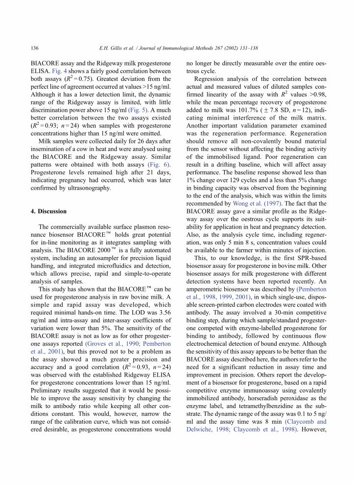
BIACORE assay and the Ridgeway milk progesterone
ELISA. Fig. 4 shows a fairly good correlation between
both assays (R2 = 0.75). Greatest deviation from the
perfect line of agreement occurred at values >15 ng/ml.
Although it has a lower detection limit, the dynamic
range of the Ridgeway assay is limited, with little
discrimination power above 15 ng/ml (Fig. 5). A much
better correlation between the two assays existed
(R2 = 0.93; n = 24) when samples with progesterone
concentrations higher than 15 ng/ml were omitted.
Milk samples were collected daily for 26 days after
insemination of a cow in heat and were analysed using
the BIACORE and the Ridgeway assay. Similar
patterns were obtained with both assays (Fig. 6).
Progesterone levels remained high after 21 days,
indicating pregnancy had occurred, which was later
confirmed by ultrasonography.
4. Discussion
The commercially available surface plasmon reso-
nance biosensor BIACOREk holds great potential
for in-line monitoring as it integrates sampling with
analysis. The BIACORE 2000k is a fully automated
system, including an autosampler for precision liquid
handling, and integrated microfluidics and detection,
which allows precise, rapid and simple-to-operate
analysis of samples.
This study has shown that the BIACOREk can be
used for progesterone analysis in raw bovine milk. A
simple and rapid assay was developed, which
required minimal hands-on time. The LOD was 3.56
ng/ml and intra-assay and inter-assay coefficients of
variation were lower than 5%. The sensitivity of the
BIACORE assay is not as low as for other progester-
one assays reported (Groves et al., 1990; Pemberton
et al., 2001), but this proved not to be a problem as
the assay showed a much greater precision and
accuracy and a good correlation (R2 = 0.93, n = 24)
was observed with the established Ridgeway ELISA
for progesterone concentrations lower than 15 ng/ml.
Preliminary results suggested that it would be possi-
ble to improve the assay sensitivity by changing the
milk to antibody ratio while keeping all other con-
ditions constant. This would, however, narrow the
range of the calibration curve, which was not consid-
ered desirable, as progesterone concentrations would
no longer be directly measurable over the entire oes-
trous cycle.
Regression analysis of the correlation between
actual and measured values of diluted samples con-
firmed linearity of the assay with R2 values >0.98,
while the mean percentage recovery of progesterone
added to milk was 101.7% (F 7.8 SD, n = 12), indi-
cating minimal interference of the milk matrix.
Another important validation parameter examined
was the regeneration performance. Regeneration
should remove all non-covalently bound material
from the sensor without affecting the binding activity
of the immobilised ligand. Poor regeneration can
result in a drifting baseline, which will affect assay
performance. The baseline response showed less than
1% change over 129 cycles and a less than 5% change
in binding capacity was observed from the beginning
to the end of the analysis, which was within the limits
recommended by Wong et al. (1997). The fact that the
BIACORE assay gave a similar profile as the Ridge-
way assay over the oestrous cycle supports its suit-
ability for application in heat and pregnancy detection.
Also, as the analysis cycle time, including regener-
ation, was only 5 min 8 s, concentration values could
be available to the farmer within minutes of injection.
This, to our knowledge, is the first SPR-based
biosensor assay for progesterone in bovine milk. Other
biosensor assays for milk progesterone with different
detection systems have been reported recently. An
amperometric biosensor was described by (Pemberton
et al., 1998, 1999, 2001), in which single-use, dispos-
able screen-printed carbon electrodes were coated with
antibody. The assay involved a 30-min competitive
binding step, during which sample/standard progester-
one competed with enzyme-labelled progesterone for
binding to antibody, followed by continuous flow
electrochemical detection of bound enzyme. Although
the sensitivity of this assay appears to be better than the
BIACORE assay described here, the authors refer to the
need for a significant reduction in assay time and
improvement in precision. Others report the develop-
ment of a biosensor for progesterone, based on a rapid
competitive enzyme immunoassay using covalently
immobilized antibody, horseradish peroxidase as the
enzyme label, and tetramethylbenzidine as the sub-
strate. The dynamic range of the assay was 0.1 to 5 ng/
ml and the assay time was 8 min (Claycomb and
Delwiche, 1998; Claycomb et al., 1998). However,
E.H. Gillis et al. / Journal of Immunological Methods 267 (2002) 131–138136

the re-usability of the test well was very poor (approx-
imately 15–20 cycles) due to noise from residual en-
zyme activity.
The BIACORE assay developed in this study is
based on covalently immobilised progesterone rather
than immobilised antibody, which accounts for the
much greater stability of the sensor surface compared
to the above methods. During this study, the sensor
withstood 3600 regeneration cycles before any
decrease in binding was observed, which would
enable twice daily measurements to be made on a
herd of 50 cows for a period of well over 1 month.
Since there are four flowcells, these results suggest
that the progesterone sensor surface has the potential
for at least 14000 regeneration cycles, which would,
however, have to be verified. The fact that the SPR
technology is incorporated into fully automated
instrumentation, which is commercially available, also
means that the assay described here could be much
more easily set up in the milking parlour for in-line
testing. Although the instrument model (BIACORE
2000k) used in this study is very expensive, it is
intended as a research tool. However, a less costly
BIACORE Qk system is also available. This is an
instrument dedicated for routine concentration analy-
sis, with greater automation, higher throughput rates
and immediate read-out of results and would be ideal
for on-farm applications.
The BIACORE assay has the potential to con-
tribute significantly to improved reproductive man-
agement of dairy herds by daily monitoring of milk
progesterone levels and, hence, accurate identifica-
tion of animals in heat, early pregnancy diagnosis
and detection of ovarian failure. Also, the introduc-
tion of BIACORE technology into the milking
parlour would facilitate continuous monitoring of
other analytes in milk, of relevance to both produc-
tive and reproductive performance and to milk
quality.
Acknowledgements
EHG is a recipient of a Walsh fellowship from
Teagasc. The BIACORE 2000k was purchased by
a research grant from the Irish Higher Education
Authority through its PRTLI-cycle 1 programme.
The authors would like to thank Dr. Chris Elliot
and Imelda Traynor, Chemical Surveillance Depart-
ment, Veterinary Sciences Division, Belfast, North-
ern Ireland for assistance with coating of the sensor
chip and for useful discussions.
References
Bostrom Caselunghe, M., Lindeberg, J., 2000. Biosensor-based de-
termination of folic acid in fortified food. Food Chem. 70, 523–
532.
Claycomb, R.W., Delwiche, M.J., 1998. Biosensor for on-line meas-
urement of bovine progesterone during milking. Biosens. Bio-
electron. 13, 1173–1180.
Claycomb, R.W., Delwiche, M.J., Munro, C.J., BonDurant, R.H.,
1998. Rapid enzyme immunoassay for measurement of bovine
progesterone. Biosens. Bioelectron. 13, 1165–1171.
Daly, S.J., Keating, G.J., Dillon, P.P., Manning, B.M., O’Kennedy,
R., Lee, H.A., Morgan, M.R., 2000. Development of surface
plasmon resonance-based immunoassay for aflatoxin B(1). J.
Agric. Food Chem. 48, 5097–5104.
Diskin, M.G., Sreenan, J.M., 2000. Expression and detection of
oestrus in cattle. Reprod. Nutr. Dev. 40, 481–491.
Elliott, C., Baxter, G., Crooks, S., McCaughey, W., 1999. The de-
velopment of a rapid immunobiosensor screening method for
the detection of residues of sulphadiazine. Food Agric. Immu-
nol. 11, 19–28.
Erlanger, B.F., Borek, F., Beiser, S.M., Lieberman, S., 1957. Ste-
roid–protein conjugates: I. Preparation and characterization of
conjugates of bovine serum albumin with testosterone and with
cortisone. J. Biol. Chem. 228, 127–713.
Gaudin, V., Maris, P., 2001. Development of a biosensor-based
immunoassay for screening of chloramphenicol residues in
milk. Food Agric. Immunol. 13, 77–86.
Groves, D.J., Sauer, M.J., Rayment, P., Foulkes, J.A., Morris, B.A.,
1990. The preparation of an ovine monoclonal antibody to pro-
gesterone. J. Endocrinol. 1990, 217–222.
Hashimoto, S., 2000. Principles of BIACORE. In: Nagata, K., Han-
da, H. (Eds.), Real-Time Analysis of Biomolecular Interac-
tions—Applications of BIACORE. Springer-Verlag, Tokyo, p.
22.
Indyk, H.E., Evans, E.A., Bostrom Caselunghe, M.C., Persson, B.S.,
Finglas, P.M.,Woollard, D.C., Filonzi, E.L., 2000. Determination
of biotin and folate in infant formula and milk by optical biosen-
sor-based immunoassay. J. AOAC 83, 1141–1148.
Johnsson, B., Lofas, S., Lindquist, G., 1991. Immobilisation of
proteins to carboxy methyl dextran-modified gold surface for
biospecific analysis in surface plasmon resonance sensors. Anal.
Biochem. 198, 268–277.
Koelsch, R.K., Aneshansley, D.J., Butler, W.R., 1994. Milk proges-
terone sensor for application with dairy cattle. J. Agric. Eng.
Res. 58, 115–120.
Markey, F., 2000. Principles of surface plasmon resonance. In: Na-
gata, K., Handa, H. (Eds.), Real-Time Analysis of Biomolecular
Interactions—Applications of BIACORE. Springer-Verlag,
Tokyo, p. 14.
E.H. Gillis et al. / Journal of Immunological Methods 267 (2002) 131–138 137

Mellgren, C., Sternesjo, A., Hammer, P., Bjork, L., Heeschen, W.,
1996. Comparison of biosensor, microbiological immunochem-
ical and physical methods for detection of sulfamethazine resi-
dues in raw milk. J. Food Prot. 58, 1223–1226.
Minunni, M., Mascini, M., 1993. Detection of pesticide in drinking
water using real-time biospecific interaction analysis (BIA).
Anal. Lett. 26, 1441–1460.
Mullett, W., Lai, E.P., Yeung, J.M., 1998. Immunoassay of fumo-
nisins by a surface plasmon resonance biosensor. Anal. Bio-
chem. 258, 161–167.
Nebel, R.L., 1988. On-farm milk progesterone tests. J. Dairy Sci.
71, 1682–1690.
O’Rorke, A., Kane, M.M., Gosling, J.P., Tallon, D.F., Fottrell, P.F.,
1994. Development and validation of a monoclonal antibody
enzyme immunoassay for measuring progesterone in saliva.
Clin. Chem. 40, 454–458.
Pemberton, R.M., Hart, J.P., Foulkes, J.A., 1998. Development of a
sensitive, selective electrochemical immunoassay for progester-
one in cow’s milk based on a disposable screen-printed ampero-
metric biosensor. Electrochim. Acta 43, 3567–3574.
Pemberton, R.M., Hart, J.P., Stoddard, P., Foulkes, J.A., 1999. A
comparison of 1-naphthyl phosphate and 4-aminophenyl phos-
phate as enzyme substrates for use with a screen-printed am-
perometric immunosensor for progesterone in cow’s milk.
Biosens. Bioelectron. 14, 495–503.
Pemberton, R.M., Hart, J.P., Mottram, T.T., 2001. An electrochem-
ical immunosensor for milk progesterone using a continuous
flow system. Biosens. Bioelectron. 16, 715–723.
Senger, P.L., 1994. The estrus detection problem: new concepts,
technologies, and possibilities. J. Dairy Sci. 77, 2745–2753.
Sreenan, J., Diskin, M., 1992. Breeding the Dairy Herd Teagasc,
Dublin.
Sternesjo, A., Mellgren, C., Bjorck, L., 1995. Determination of
sulfamethazine residues in milk by a surface plasmon reso-
nance-based biosensor assay. Anal. Biochem. 226, 175–181.
Wong, R.L., Mytych, D., Jacobs, S., Bordens, R., 1997. Validation
parameters for a novel biosensor assay which simultaneously
measures serum concentrations of a humanized monoclonal
antibody and detects induced antibodies. J. Immunol. Methods
209, 1–15.
E.H. Gillis et al. / Journal of Immunological Methods 267 (2002) 131–138138