amersham ecl advance western blotting detection kitblossombio.com/pdf/products/ug_ecladvance.pdf ·...
TRANSCRIPT

GE Healthcare Life Sciences
Amersham ECL Advance Western Blotting Detection KitProduct BookletCode: RPN2135

2
Page finder1. Legal 3
2. Handling 4 2.1. Safety warnings and precautions 4 2.2. Storage 4 2.3. Expiry 4
3. Components 5 3.1. Other materials required 5
4. Description 7
5. Critical parameters 8
6. Protocol 10 6.1. Electrophoresis and blotting 10 6.2. Blocking the membrane 11 6.3. Primary antibody incubation 12 6.4. Secondary antibody incubation 13 6.5. Streptavidin bridge incubation 14 6.6. Detection 15
7. Additional information 17 7.1. Stripping and reprobing membranes 17 7.2. Determination of optimum antibody concentration 18
8. Troubleshooting guide 22
9. Quality control 25
10. Related products 26
11. References 27

1. LegalGE, imagination at work and GE monogram are trademarks of General Electric Company.
Amersham, ECL, ECL Plus, Hybond, Hypercassette, Hyperfilm, Hyperprocessor, Hypertorch, ImageMaster, Molecular Dynamics, Rainbow, Sensitize and Storm are trademarks of GE Healthcare companies.
TMA-6 substrate was developed by Lumigen, Inc. and is distributed by GE Healthcare for western blotting under license from Lumigen, Inc. This component is covered by US patent numbers 5922558, 6696569, 6858733 and 7560556 and equivalent patents and patent applications in other countries and is sold under license from Lumigen, Inc.
All third party trademarks are the property of their respective owners.
© 2006 -2011 General Electric Company – All rights reserved. Previously published 2006
All goods and services are sold subject to the tems and conditions of sale of the company within GE Healthcare which supplies them. A copy of these terms and conditions is available on request.Contact your local GE Healthcare representative for the most current information.
http://www.gelifesciences.com/illustra
GE Healthcare UK Limited. Amersham Place, Little Chalfont, Buckinghamshire, HP7 9NA UK
3

4
2. Handling
2.1. Safety warnings and precautionsWarning: For research use only. Not recommended or intended for diagnosis of disease in humans or animals. Do not use internally or externally in humans or animals.
All chemicals should be considered as potentially hazardous. We therefore recommend that this product is handled only by those persons who have been trained in laboratory techniques and that it is used in accordance with the principles of good laboratory practice. Wear suitable protective clothing such as laboratory overalls, safety glasses and gloves. Care should be taken to avoid contact with skin or eyes. In the case of contact with skin or eyes wash immediately with water. See material safety data sheet(s) and/or safety statement(s) for specific advice.
Note: The protocol requires the use of Hydrochloric acid.
Warning: Hydrochloric Acid causes burns and is an irritant. Please follow the manufacturer’s safety data sheet relating to the safe handling and use of this material.
2.2. Storage On receipt all components should be stored in a refrigerator at 2–8°C.
2.3. Expiry The components of these products are stable for at least 3 months when stored under the recommended conditions.
The ECL Advance reagents are sensitive to prolonged exposure to light. Long term storage of the individual reagents should be in the light tight containers in which they are provided.

5
3. ComponentsECL Advance Solution A Solution containing Tris buffer in 3.2% (v/v) Ethanol, 50 ml.
ECL Advance Solution B Proprietary substrate in Tris buffer, 50 ml.
Sufficient for 1000 cm2 membrane.
ECL Advance Blocking Agent 40 g reagent for use as block and antibody diluent.
To be used at 0.2 ml/cm2 (20 ml per 10 cm x 10 cm blot).
3.1. Other materials requiredSolutions required The chemical reagents required for these solutions are available from GE Healthcare and are detailed in the current catalogue.
Phosphate buffered saline (PBS) pH7.5* 11.5 g di-Sodium Hydrogen Orthophosphate anhydrous (80 mM) 2.96 g Sodium Dihydrogen
Orthophosphate (20 mM) 5.84 g Sodium Chloride (100 mM)
Dissolve in distilled water and make up total volume to 1000 ml. Check pH
Tris buffered saline (TBS) pH7.6* 8 g Sodium Chloride 20 ml 1 M Tris HCl, pH 7.6. Dissolve in distilled water and make up total volume to 1000 ml. Check pH
Wash Buffer PBS-Tween (PBS-T) or TBS-Tween (TBS-T) Dilute the required amount of Tween™ 20 in the corresponding buffer. A 0.1% (v/v) Tween 20 concentration is suitable for most blotting applications.
Blocking solution and antibody diluent ECL Advance Blocking Agent (supplied with kit) 1. Shake the powdered block to ensure even distribution of components
6
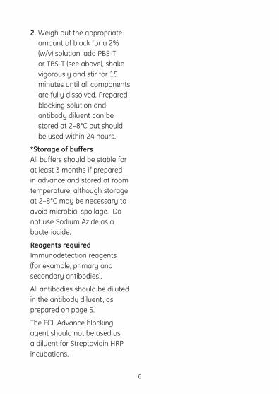
2. Weigh out the appropriate amount of block for a 2% (w/v) solution, add PBS-T or TBS-T (see above), shake vigorously and stir for 15 minutes until all components are fully dissolved. Prepared blocking solution and antibody diluent can be stored at 2–8°C but should be used within 24 hours.
*Storage of buffers All buffers should be stable for at least 3 months if prepared in advance and stored at room temperature, although storage at 2–8°C may be necessary to avoid microbial spoilage. Do not use Sodium Azide as a bacteriocide.
Reagents required Immunodetection reagents (for example, primary and secondary antibodies).
All antibodies should be diluted in the antibody diluent, as prepared on page 5.
The ECL Advance blocking agent should not be used as a diluent for Streptavidin HRP incubations.

4. DescriptionThe ECL Advance™ Western blotting detection kit from GE Healthcare provides an extremely sensitive non-radioactive method for the detection of immobilised antigens using Horseradish Peroxidase (HRP) labelled antibodies.
This system builds on the combined strengths of GE Healthcare and Lumigen Inc., in developing high performance detection products. Using Lumigen® TMA-6, a proprietary new substrate for horseradish peroxidase, ECL Advance provides a more intense light output at 440 nm which can be readily imaged using CCD imagers as well as with film.
The intense and sensitive signal produced with these substrates enable reduced antibody concentrations to be employed. This is a benefit where antibody supply is scarce. The new membrane block controls background noise whilst retaining a high level of signal intensity. The system is fully compatible with both Hybond™ ECL (nitrocellulose) and Hybond-P (PVDF) membranes.
Horseradish Peroxidase present on a Western blot is detected by its reaction with Peroxide, an enhancer molecule and the novel chemiluminescent substrate, all of which are present in the substrate reagents provided.
The sensitivity increase over ECL Plus Western blotting can be up to 10 fold, depending on the immunodetection system being used.
7

8
5. Critical parameters• Readtheentireprotocolthoroughlybeforeusingthekit.
• ECLAdvancewillprovideanincreaseinsensitivityoverECLPluson both nitrocellulose and PVDF membranes.
• ECLAdvanceisanextremelysensitivesystem.Forresultsshowing the best signal to noise ratio, it is essential to optimise the concentrations of both primary and secondary antibodies. Higher dilutions of antibodies are likely to be required when using ECL Advance in place of ECL Plus Western blotting reagents.
• Duringimmunodetection,asufficientvolumeofsolutionshouldbe used to adequately cover the membrane. Containers should be agitated gently on a mixer platform.
• TheuseofECLAdvanceBlockingAgentisstronglyrecommendedfor membrane blocking and all antibody diluents in order to reduce non-specific binding and background noise resulting from the high sensitivity of this reagent. Ensure the powdered blocking agent is shaken thoroughly before use.
NOTE: The ECL Advance blocking agent should not be used as a diluent for streptavidin HRP incubations.
• Whenwashing,thevolumeofwashbuffershouldbeaslargeaspossible; 4 ml of buffer per cm2 of membrane is suggested. Brief rinses of the membrane in wash buffer before incubating will improve washing efficiency.
• Ifexposuretimesoflessthan5secondsareroutinelyrequired,itis recommended that the antibodies used are further diluted as it is difficult to perform such short exposures.
• The‘workingmix’ofECLAdvancereagentsisstableforupto1month stored at 2–8°C. Therefore, the reagents can be mixed and stored before use or mixed immediately before use. If mixing

9
and storing the reagents, the container used should be similar to those provided. If the container is not light tight, a covering of foil should be used and the container should be stored at 2–8°C in the dark.
• Accidentalfreezingofthesubstrateswillnotcausedegradation.In the event that freezing occurs, thaw the solutions, mix well and store as recommended.

10
Protocol
1. Perform electrophoresis and blotting according to usual techniques. Proteins should be transferred to Hybond- P PVDF or Hybond ECL for optimum results. Blots may be used immediately or stored in a desiccator at 2–8°C for up to 3 months.
Notes
1. Hybond-P PVDF should be pre-wetted in 100% (v/v) Methanol, washed in distilled water for 5 minutes and equilibrated in transfer buffer for at least 10 minutes before blotting.
Hybond ECL should be pre- wetted in distilled water and equilibrated in transfer buffer for at least 10 minutes before blotting.
ECL Advance is also suitable for use with supported nitrocellulose such as Hybond C-Extra, this membrane should be prepared as for Hybond ECL.
6. Protocol
6.1. Electrophoresis and blotting

11
Protocol
1. Prepare sufficient blocking solution and antibody diluent (as described on page 5) for use as block and antibody diluent.
2. Block non-specific binding sites by immersing the membrane in blocking solution (see 6.2 step 1), for 1 hour at room temperature on an orbital shaker. Alternatively, membranes may be left in the blocking solution overnight in a refrigerator at 2–8°C, if more convenient.
3. Briefly rinse the membrane with two changes of wash buffer. (see page 5).
Notes
1. The combination of ECL Advance Blocking Agent and Tween 20 should be sufficient for most applications.
Optimum Tween 20 concentrations will vary to suit specific experiments, but 0.1% (v/v) Tween 20 is suitable for most blotting applications.
6.2. Blocking the membrane
12
Protocol
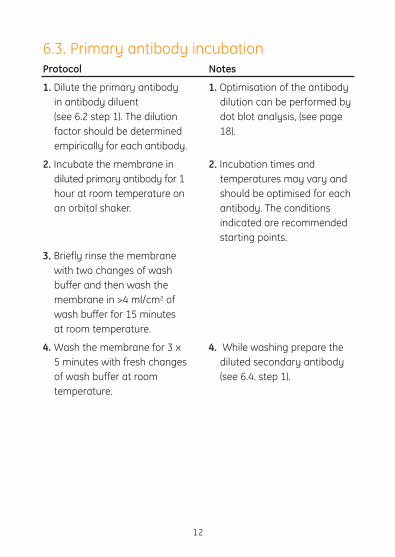
1. Dilute the primary antibody in antibody diluent (see 6.2 step 1). The dilution factor should be determined empirically for each antibody.
2. Incubate the membrane in diluted primary antibody for 1 hour at room temperature on an orbital shaker.
3. Briefly rinse the membrane with two changes of wash buffer and then wash the membrane in >4 ml/cm2 of wash buffer for 15 minutes at room temperature.
4. Wash the membrane for 3 x 5 minutes with fresh changes of wash buffer at room temperature.
Notes
1. Optimisation of the antibody dilution can be performed by dot blot analysis, (see page 18).
2. Incubation times and temperatures may vary and should be optimised for each antibody. The conditions indicated are recommended starting points.
4. While washing prepare the diluted secondary antibody (see 6.4. step 1).
6.3. Primary antibody incubation

13
Protocol
1. Dilute the HRP labelled secondary antibody or biotinylated antibody in antibody diluent (see 6.2.
step 1). If it is necessary to dilute the antibody in 2 lots of diluent, the first dilution can be made using PBS-T or TBS-T (up to 1:1000), but the final dilution should be made in antibody diluent. The dilution factor can be determined empirically for each antibody (see pages 18–20).
2. Incubate the membrane in the diluted secondary antibody for 1 hour at room temperature on an orbital shaker.
3. Briefly rinse the membrane with two changes of wash buffer and then wash the membrane in >4 ml/cm2 of wash buffer for 15 minutes at room temperature.
Notes
1. Use either an appropriate HRP labelled secondary antibody or a biotinylated secondary antibody.
2. Incubation times and temperatures may vary and should be optimised for each antibody. The conditions indicated are recommended starting points.
6.4. Secondary antibody incubation

14
Protocol
4. Wash the membrane for 3 x 5 minutes with fresh changes of wash buffer at room temperature.
Protocol
1. Dilute the streptavidin HRP conjugate or streptavidin- biotinylated HRP complex in PBS-T or TBS-T.
2. Incubate the membrane in the dilution for 15 minutes at room temperature on an orbital shaker.
3. Briefly rinse the membrane with two changes of wash buffer and then wash the membrane in >4 ml/cm2 of wash buffer for 15 minutes at room temperature.
4. Wash the membrane for 3 x 5 minutes with fresh changes of wash buffer at room temperature.
Notes
4. If using an HRP labelled secondary antibody proceeddirectly to 6.6. (detection) after this wash procedure. If using a biotinylated antibody, while washing, prepare the diluted Streptavidin HRP conjugate or complex (6.5. step 1)
Notes
1. The dilution factor should be determined empirically (see pages 18–20).
6.5. Streptavidin bridge incubation

15
Protocol
1. Remove the detection reagents from storage at 2–8°C and allow to equilibrate to room temperature before opening.
2. Mix detection solutions A and B in a ratio of 1:1 (for example, 2 ml solution A + 2 ml solution B) or use premixed solution. The final volume of detection reagent required is 0.1 ml/cm2.
3. Drain the excess wash buffer from the washed membranes and place protein side up on a sheet of SaranWrap™ or other suitable clean surface. Pipette the mixed detection reagent on to the membrane.
4. Incubate for 5 minutes at room temperature.
5. Drain off excess detection reagent by holding the membrane gently with forceps and touching the edge against a tissue. Place the blots protein side down on to a fresh piece of
Notes
2. If the mixed reagent is not to be used immediately protect it from exposure to the light either by wrapping in foil or storing in a dark place.
3. The reagents should cover the entire surface of the membrane, held by surface tension on to the surface of the membrane.
5. Close the SaranWrap around the membrane to form an envelope or use an alternative, suitable detection pocket. Avoid applying pressure on to the membrane.
6.6. Detection

16
Protocol 5. Continued. SaranWrap, wrap up the blots and gently smooth out any air bubbles.
6. Place the wrapped blots, protein side up, in an X-ray film cassette.
7. Place a sheet of autoradiography film (for example, Hyperfilm™ECL) on top of the membrane. Close the cassette and expose for 15 seconds.
8. Remove the film and replace with a second sheet of unexposed film. Develop the first piece of film immediately, and on the basis of its appearance estimate how long to continue the exposure of the second piece of film. Second exposures can vary from 1 minute to 1 hour.
Notes
6. Ensure there is no free detection reagent in the cassette, as the film must not get wet.
7. This stage should be carried out in a dark room using red safe lights. Do not move the film while it is being exposed.
8. The detected blots can also be exposed to Polaroid™ film using the ECL mini-camera (RPN2069), which is specifically designed for blots generated from mini-gel apparatus. The ECL mini-camera is suitable for blots up to 52 x 77 mm. Images can also be acquired using a CCD camera such as the Imagemaster™ VDS-CL (18-1130-55).

17
Protocol
1. Submerge the membrane in stripping buffer (100 mM 2-Mercaptoethanol, 2% (w/v) SDS, 62.5 mM Tris-HC1 pH 6.7) and incubate at 50°C for 30 minutes with occasional agitation.
2. Wash the membrane for 2 x 10 minutes in PBS-T or TBS-T at room temperature using large volumes of wash buffer.
3. Block the membrane in blocking solution (see page 5) for 1 hour at room temperature.
4. Repeat the immunodetection protocol, main protocol stages 3 to 6.
Notes
1. If more stringent conditions are required the incubation can be performed at 70°C or incubate for a longer time.
2. Membranes may be incubated with ECL Advance detection reagents and exposed to film to ensure removal of antibodies.
7. Additional information
7.1. Stripping and reprobing membranesThe complete removal of primary and secondary antibodies from the membrane is possible following the protocol outlined below. The membranes may be stripped of bound antibodies and reprobed several times. Membranes should be stored wet wrapped in SaranWrap in a refrigerator (2–8°C) after each immunodetection.

18
Protocol
1. Primary antibodies Dot blots are a quick and effective method of determining the optimum dilution of a primary antibody of unknown concentration. Alternatively, a Western blot can be prepared and then cut into several strips. It should be noted that some antibodies may require alternative blocking and washing steps to the following suggestions.
Notes
1. Spot a suitable amount of protein sample to a nitrocellulose or PVDF membrane and allow to air dry. Prepare one blot for each primary antibody dilution to be tested.
2. Incubate in blocking solution (see page 5) for 1 hour at room temperature with agitation.
3. Rinse the membranes briefly with two changes of wash buffer.4. Prepare several dilutions of primary antibody in antibody diluent: e.g. For both nitrocellulose and PVDF membranes: 1/10 000, 1/25 000, 1/50 000,
7.2. Determination of optimum antibody concentrationDue to the improved sensitivity of the ECL Advance detection reagents, optimisation of antibody concentrations is recommended to ensure the best results. In general, lower concentrations of both primary and secondary antibodies are required with ECL Advance compared to ECL Plus Western blotting reagents.
Outlined below are protocols for determining optimal antibody concentrations.

19
Protocol
2. Secondary antibodies
Notes
4. Continued. 1/75 000, 1/100 000. Incubate 1 blot in each dilution for 1 hour at room temperature with agitation.
5. Rinse blots in two changes of wash buffer, then wash for 1 x 15 minutes and 3 x 5 minutes in fresh changes of wash buffer.
6. Dilute the secondary antibody (using only one concentration) in antibody diluent and incubate the membranes for 1 hour at room temperature with agitation.
7. Wash as detailed in step 5.
8. Detect using ECL Advance detection reagents as detailed in section 6.6. of the main protocol. The antibody dilution which gives the best signal with the minimum background should be selected.
1. Prepare dot blots and block the membranes as detailed in steps 1 and 2.

20
Protocol
Notes
2. Dilute the primary antibody (using only one concentration) in antibody diluent and incubate the membranes for 1 hour at room temperature with agitation.
3. Wash as detailed in step 5 of protocol 1.
4. Prepare several dilutions of secondary antibody in antibody diluent: e.g. For both nitrocellulose and PVDF membranes: 1/50 000 1/100 000, 1/250 000, 1/500 000 Incubate 1 blot in each dilution for 1 hour at room temperature with agitation.
5. Wash as detailed in step 5 of protocol 1.
6. Detect using ECL Advance detection reagents as detailed in 6.6. of the main protocol. The antibody dilution which gives the best signal with minimum background should be selected.
21
Protocol
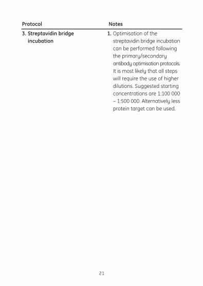
3. Streptavidin bridge incubation
Notes
1. Optimisation of the streptavidin bridge incubation can be performed following the primary/secondary antibody optimisation protocols. It is most likely that all steps will require the use of higher dilutions. Suggested starting concentrations are 1:100 000 – 1:500 000. Alternatively less protein target can be used.

22
Problems
1. No signal
2. Weak signal
3. Excessive diffuse signal
Possible causes and solutions
1. Check that transfer equipment is working properly and that the correct procedure has been followed.
2. Check protein transfer by staining the gel and/or membrane.
3. Some antigens may be affected by the treatments required for electrophoresis.
4. Target protein degradation may occur if the blots are stored incorrectly.
5. ECL Advance detection reagents may have become contaminated.
6. Incorrect storage of the ECL Advance detection reagents may cause a loss of signal.
1. Transfer efficiency may have been poor.
2. Insufficient protein was loaded on to the gel.
3. The concentration of primary and secondary antibodies could be too low; optimization is required.
4. Film exposure time may have been too short.
1. Too much protein was loaded onto the gel.
2. Electrophoresis and transfer protocols may need optimization.
8. Troubleshooting guide

23
Problems
3. Excessive diffuse signal continued.
4. White (negative) bands on the film
5. Uneven, spotted backgrounds
6. High backgrounds
Possible causes and solutions
3. The concentrations of primary and secondary antibodies could be too high; optimization is required.
1. Negative bands generally occur when protein target is in excess and antibody concentrations are too high. The effect is caused by substrate depletion. To rectify this either, reduce the amount of target loaded, use lower antibody concentrations or a combination of both.
1. Blotting technique requires optimization.
2. Areas of the blot may have dried during some of the incubations.
3. Incorrect handling can lead to contamination on the blots and/or membrane damage, which may cause non-specific signal.
1. The concentrations of primary and secondary antibodies could be too high; optimization is required.
2. Contamination can be transferred to the blots from electrophoresis and related equipment used in blot preparation.
3. Transfer and incubation buffers may have become contaminated and require replacing.
4. The blocking agent used was not freshly prepared, was too dilute or was incompatible with the application.

24
Possible causes and solutions
5. The level of Tween used in the blocking agent was not sufficient for the application performed.
6. The membranes were blocked for an insufficient time.
7. The type of membrane used was not compatible with non-radioactive systems.
8. The post antibody washes were not performed for a sufficient period of time or were not performed in a high enough volume.
9. There was insufficient Tween in the post antibody washes.
10. Insufficient changes of post antibody washes were used.
11. The film detection of the signal was allowed to over expose.
12. The level of signal is so high that the film has become completely overloaded.
13. The membrane was allowed to dry during one of the incubations.
Problems
6. High backgrounds continued
9. Quality controlEvery batch of ECL Advance detection reagents and ECL Advance blocking agent is functionally tested in a Western blotting application to ensure minimal batch to batch variability.
25

26
10. Related productsGE Healthcare offers a comprehensive range of Western blotting reagents and hardware all with proven compatibility to ensure reproducible high quality results. For a complete listing of products available see the current GE Healthcare catalogue or visit our web site at www.gehealthcare.com/lifesciences
ECL Western Blotting Detection Reagents For 4000 cm2 Membrane RPN2106
ECL Plus Western Blotting Detection Reagents For 1000 cm2 Membrane RPN2132Other pack sizes and detection reagents also available
ECL DualVue™ Western Blotting Markers RPN810Low-Range Rainbow™ MW Markers RPN755High-Range Rainbow MW Markers RPN756Full-Range Rainbow MW Markers (Recombinant) RPN800ECL Western Blotting MW Markers RPN2107
Hybond-P Membrane (PVDF, Pore Size 0.45 µm) RPN2020F
Hybond-ECL Membrane (Nitrocellulose, Pore Size 0.45 µm) RPN303D
Other membrane sizes also availableHyperfilm ECL 18 x 24, pack of 25 films Other film sizes are also available RPN2103K
Streptavidin-Biotinylated Horesradish Peroxidase Complex RPN1051Streptavidin Horseradish Peroxidase Conjugate RPN1231Mouse IgG, Horseradish Peroxidase-linked Whole Antibody (from Sheep), 1 ml and 100 µl NA931
Human IgG, Horseradish Peroxidase-linked Whole Antibody (from Sheep), 1 ml NA933
Rabbit IgG, Horseradish Peroxidase-linked Whole Antibody (from Donkey), 1 ml and 100 µl NA934

27
11. References1. ISACCSON, U. and WATERMARK, G., Anal. Chim. Acta. 68, 339–362
(1974).
2. WHITEHEAD, T. P. et al., Clin. Chem. 25, 1531–1546 (1979).
3. AKHAVAN-TAFTI, H. et al., Clin. Chem., 41, 1368–1369 (1995).
4. AKHAVAN-TAFTI, H. et al., Biolum. and Chemilum. Fundamentals and Applied Aspects, pp.199–202, Chichester, 1994.

imagination at work
RPN2135PL AF 02-2011
GE Healthcare offices:
GE Healthcare Bio-Sciences AB Björkgatan 30 751 84 Uppsala Sweden
GE Healthcare Europe GmbH Munzinger Strasse 5 D-79111 Freiburg Germany
GE Healthcare Bio-Sciences Corp 800 Centennial Avenue P.O. Box 1327 Piscataway NJ 08855-1327 USA
GE Healthcare Japan Corporation Sanken Bldg. 3-25-1 Hyakunincho Shinjuku-ku Tokyo 169-0073 Japan
For contact information for your local office, please visit: http://www.gelifesciences.com/contact
GE Healthcare UK Limited Amersham Place, Little Chalfont, Buckinghamshire, HP7 9NA UK http://www.gelifesciences.com/illustra
2899
2069