radiological signs (shënja radiologjike )-2
TRANSCRIPT
1
Sllavko K. Kallfa
Radiological Signs
(Shënja Radiologjike)
Përmbledhje Artikujsh nga
Radiopaedia.org
❷
2
inanimate object inspired X-Ray signs
champagne glass pelvis.......f. 3
coin lesion...............................................f.4
dinner fork deformity...........f.11
fishhook ureters............................f.13
goblet sign.............................................f.16
leather bottle stomach
Lincoln log vertebra...............f.17
napkin ring sign...........................f.26
rugger-jersey spine..................f.30
Tam O'Shanter sign...............f.38
water bottle sign..........................f.41
white pyramid sign..................f.45
3
Champagne glass pelvis
Dr Ayush Goel and Dr MT Niknejad et al.
The champagne glass pelvis is a helpful sign in achondroplasia which the iliac blades are
flattened, giving rise to a pelvic inlet and resembles a champagne glass. The acetabular angles
are increased, and the sacrosciatic notch is small.
References
1. Roche CJ, O'Keeffe DP, Lee WK et-al. Selections from the buffet of food signs in
radiology. Radiographics. 2002;22 (6): 1369-84. Radiographics (full text) -
doi:10.1148/rg.226025521 - Pubmed citation
2. Davies SG. Aids to Radiological Differential Diagnosis. Saunders Ltd. (2009)
ISBN:0702029793. Read it at Google Books - Find it at Amazon
Synonyms & Alternative Spellings
Synonyms or Alternative Spelling Include in Listings?
Champagne glass type pelvic inlet ✗
Champagne glass sign of pelvis ✗
Champagne glass appearance of pelvis
4
Coin lesion
Dr Henry Knipe et al.
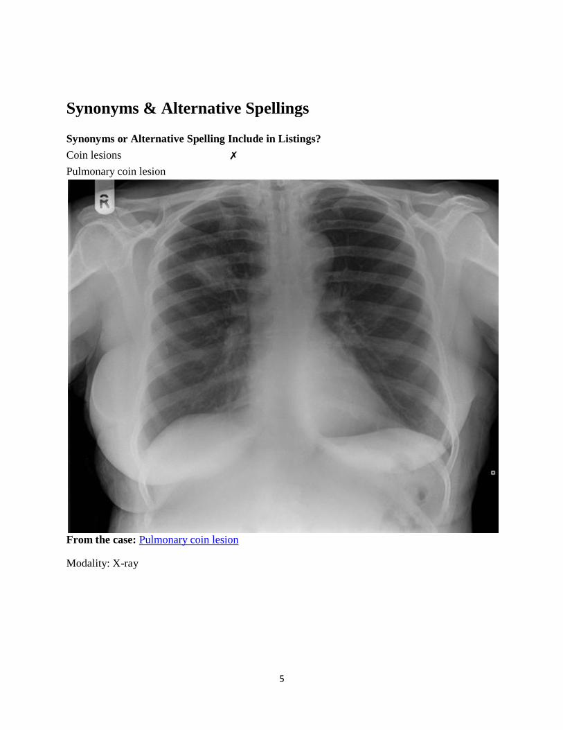
Coin lesion refers to a round or oval, well-circumscribed solitary pulmonary lesion. It is
typically 1-5 cm in diameter and calcification may or may not be present 1,3
. Typically but not
always the patient is asymptomatic 1.
Differential diagnosis
The differential diagnosis for such lesions is 1-3
:
primary lung malignancy - e.g. squamous cell carcinoma
metastases
infection
o pulmonary tuberculosis;
o Streptococcus sp., Staphlycoccus sp., or Klebisialla sp. infection
o hydatid cyst
o abscess
benign disease processes
o Wegener's granulomatosis
o pulmonary hamartoma
o pulmonary arteriovenous malformation
o rheumatoid nodule
o bronchogenic cyst
o bronchial adenoma
Take care not to call a "pseudo-coin lesion", which are caused by artifacts (e.g. button on
patient's clothing, unilateral nipple shadow, etc).
See also
solitary pulmonary nodule
References
1. The pulmonary "coin" lesion [editorial]. Radiology. 1950; 54(1):116-117.
2. Chowdhury R. Radiology at a glance. John Wiley (2013).
3. Parr LH. Coin lesions of the lung. J Natl Med Assoc. 1969;61 (2): 153-7. Free text at
pubmed - Pubmed citation
5
Synonyms & Alternative Spellings
Synonyms or Alternative Spelling Include in Listings?
Coin lesions ✗
Pulmonary coin lesion
From the case: Pulmonary coin lesion
Modality: X-ray
6
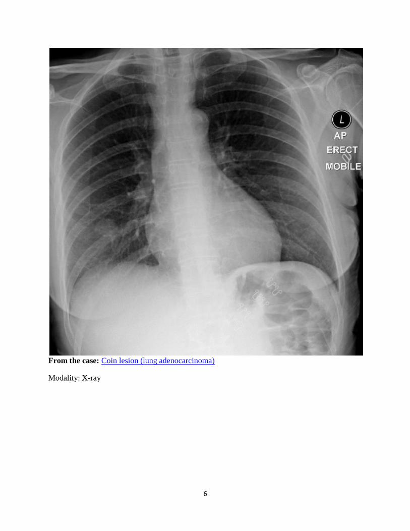
From the case: Coin lesion (lung adenocarcinoma)
Modality: X-ray
7
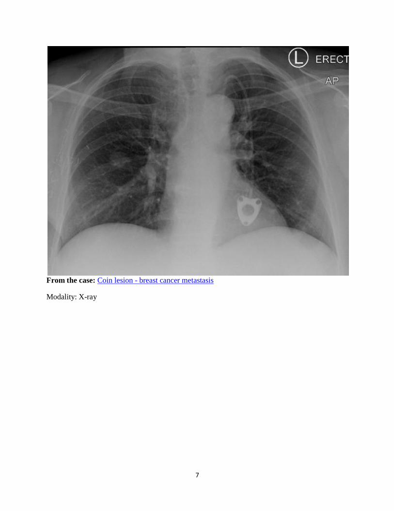
From the case: Coin lesion - breast cancer metastasis
Modality: X-ray
8
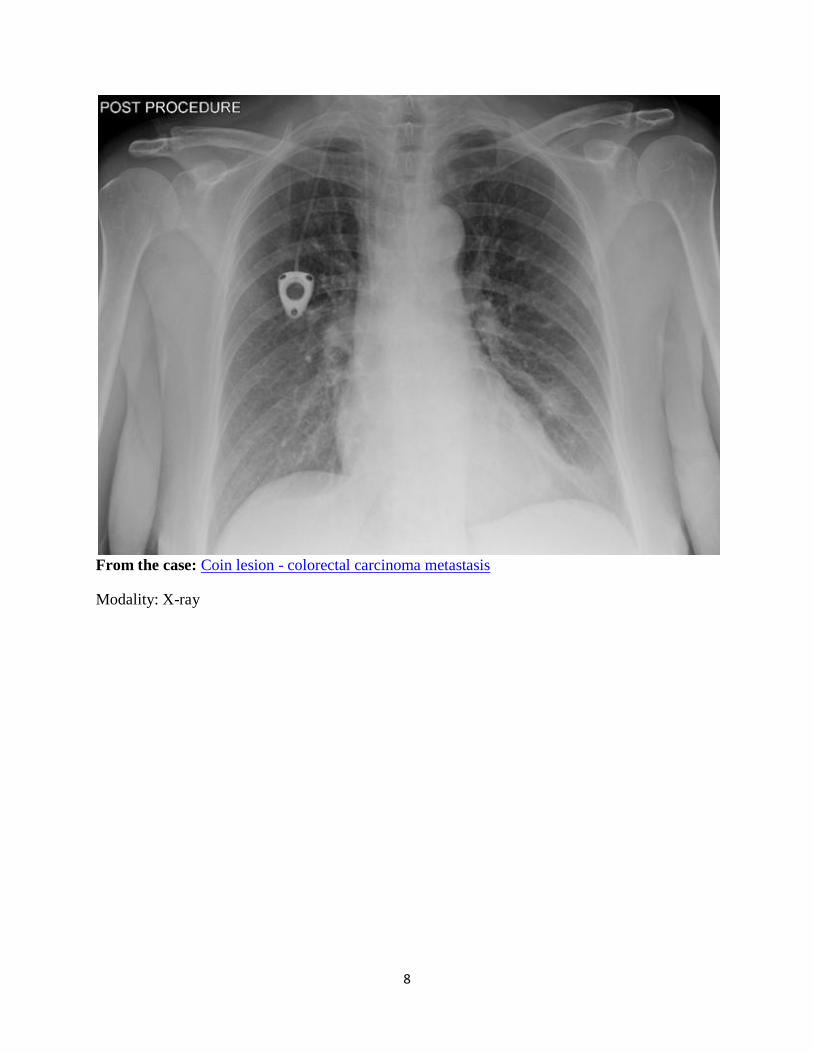
From the case: Coin lesion - colorectal carcinoma metastasis
Modality: X-ray
9
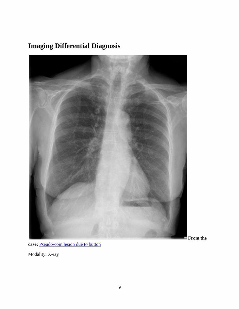
Imaging Differential Diagnosis
From the
case: Pseudo-coin lesion due to button
Modality: X-ray
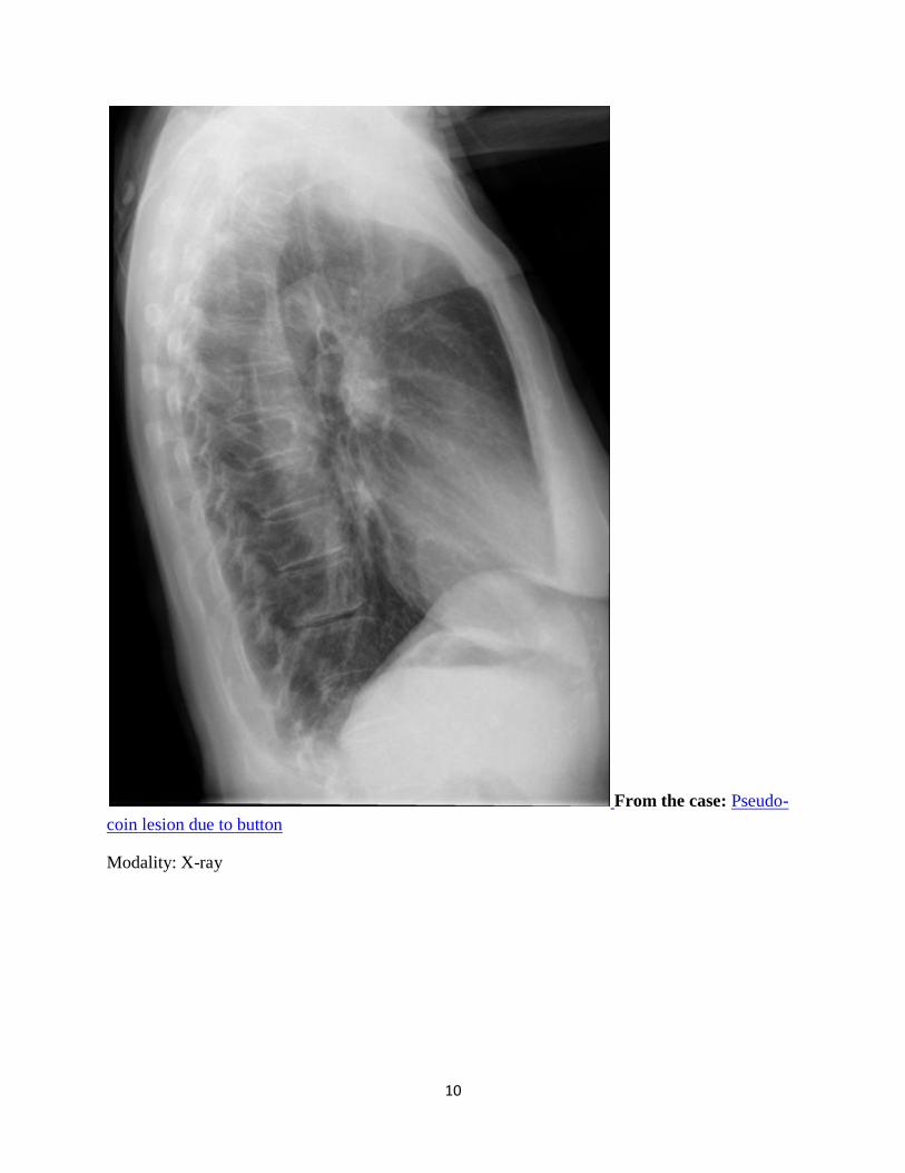
10
From the case: Pseudo-
coin lesion due to button
Modality: X-ray
11
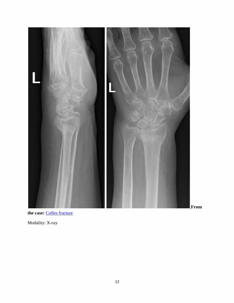
Dinner fork deformity
Dr Henry Knipe and Dr Frank Gaillard et al.
A dinner fork deformity (also known as a bayonet deformity) occurs as the result of a
malunited distal radial fracture, usually a Colles fracture. The distal fragment is dorsally
angulated, displaced and often also impacted. The term is descriptive, as the lateral view of the
wrist is similar to the shape of a fork, seen from the side, tines down.
See also
Colles fracture
upper extremity fractures
References
1. Hertling D, Kessler RM. Management of common musculoskeletal disorders, physical
therapy principles and methods. Philadelphia : Lippincott Williams & Wilkins, c2006.
(2006) ISBN:0781736269. Read it at Google Books - Find it at Amazon
2. Roche CJ, O'keeffe DP, Lee WK et-al. Selections from the buffet of food signs in
radiology. Radiographics. 22 (6): 1369-84. doi:10.1148/rg.226025521 - Pubmed citation
3.Maizlin ZV, Kuruvilla M, Clement JJ et-al. Radiologic signs of weapons and
munitions: How will noncombatants recognize them?. AJR Am J Roentgenol. 2010;195
(2): W96-104. doi:10.2214/AJR.09.4029 - Pubmed citation
Synonyms & Alternative Spellings
Synonyms or Alternative Spelling Include in Listings?
Bayonet deformity
13
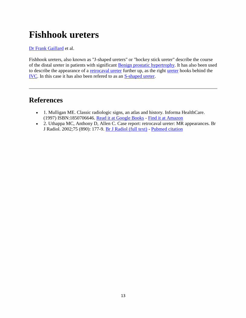
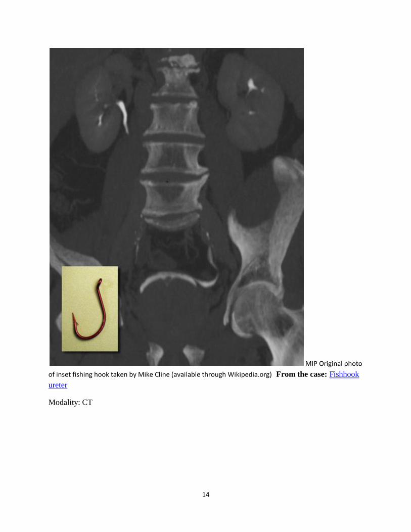
Fishhook ureters
Dr Frank Gaillard et al.
Fishhook ureters, also known as "J-shaped ureters" or "hockey stick ureter" describe the course
of the distal ureter in patients with significant Benign prostatic hypertrophy. It has also been used
to describe the appearance of a retrocaval ureter further up, as the right ureter hooks behind the
IVC. In this case it has also been refered to as an S-shaped ureter.
References
1. Mulligan ME. Classic radiologic signs, an atlas and history. Informa HealthCare.
(1997) ISBN:1850706646. Read it at Google Books - Find it at Amazon
2. Uthappa MC, Anthony D, Allen C. Case report: retrocaval ureter: MR appearances. Br
J Radiol. 2002;75 (890): 177-9. Br J Radiol (full text) - Pubmed citation
14
MIP Original photo
of inset fishing hook taken by Mike Cline (available through Wikipedia.org) From the case: Fishhook
ureter
Modality: CT
15
From the case:
Fishhook ureter
Modality: CT
16
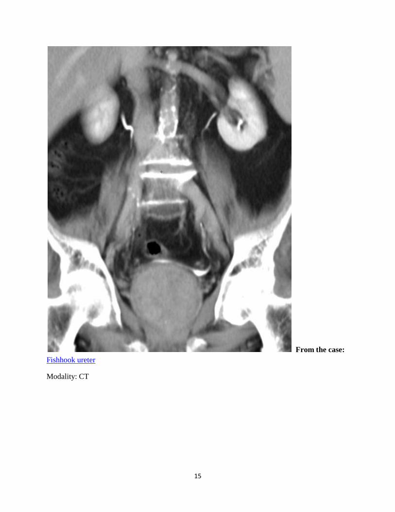
Goblet sign
Dr Yuranga Weerakkody and Dr Frank Gaillard et al.
The goblet sign (or champagne glass sign) refers to the appearance of the ureter when it is
focally dilated by an intraluminal mass. It is best seen when the ureter is opacified from below,
by a retrograde ureterogram. Presence of this sign indicates the pathology to be chronic,
permitting the lesion to be accommodated in the ureter.
Although most frequently caused by transitional cell carcinoma, a number of other entities can
lead to the same appearance 1:
transitional cell carcinoma of the ureter : most common
metastatic disease
endometriosis
References
1. Dyer RB, Chen MY, Zagoria RJ. Classic signs in uroradiology. Radiographics.
2004;24 Suppl 1 : S247-80. doi:10.1148/rg.24si045509 - Pubmed citation
Synonyms & Alternative Spellings
Synonyms or Alternative Spelling Include in Listings?
Champagne glass sign ✗
Edit Article Share
Photograph - glass goblet
17
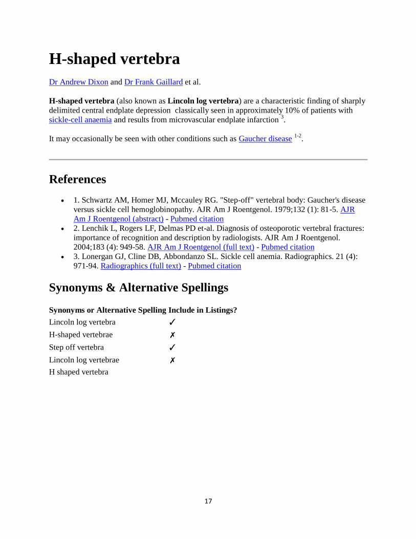
H-shaped vertebra
Dr Andrew Dixon and Dr Frank Gaillard et al.
H-shaped vertebra (also known as Lincoln log vertebra) are a characteristic finding of sharply
delimited central endplate depression classically seen in approximately 10% of patients with
sickle-cell anaemia and results from microvascular endplate infarction 3.
It may occasionally be seen with other conditions such as Gaucher disease 1-2
.
References
1. Schwartz AM, Homer MJ, Mccauley RG. "Step-off" vertebral body: Gaucher's disease
versus sickle cell hemoglobinopathy. AJR Am J Roentgenol. 1979;132 (1): 81-5. AJR
Am J Roentgenol (abstract) - Pubmed citation
2. Lenchik L, Rogers LF, Delmas PD et-al. Diagnosis of osteoporotic vertebral fractures:
importance of recognition and description by radiologists. AJR Am J Roentgenol.
2004;183 (4): 949-58. AJR Am J Roentgenol (full text) - Pubmed citation
3. Lonergan GJ, Cline DB, Abbondanzo SL. Sickle cell anemia. Radiographics. 21 (4):
971-94. Radiographics (full text) - Pubmed citation
Synonyms & Alternative Spellings
Synonyms or Alternative Spelling Include in Listings?
Lincoln log vertebra ✓
H-shaped vertebrae ✗
Step off vertebra ✓
Lincoln log vertebrae ✗
H shaped vertebra
18
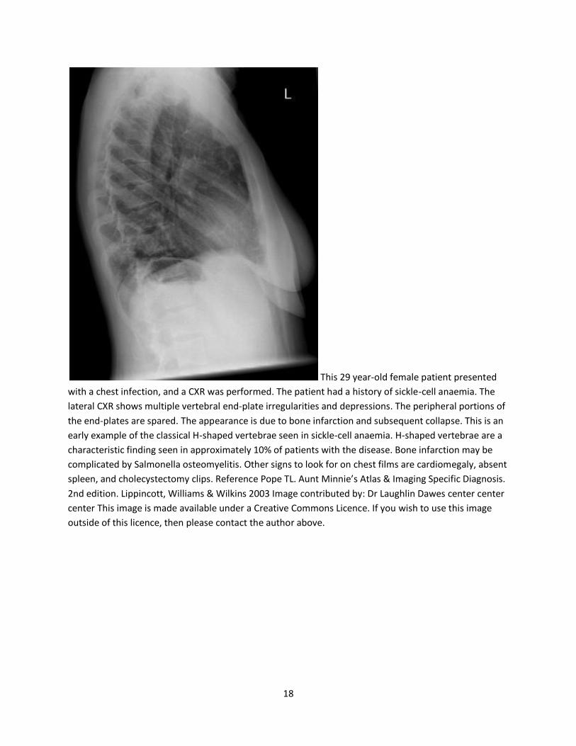
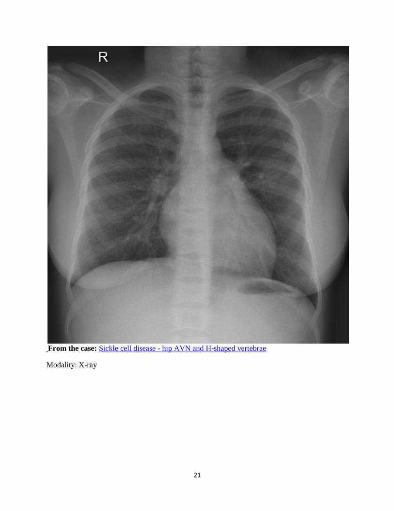
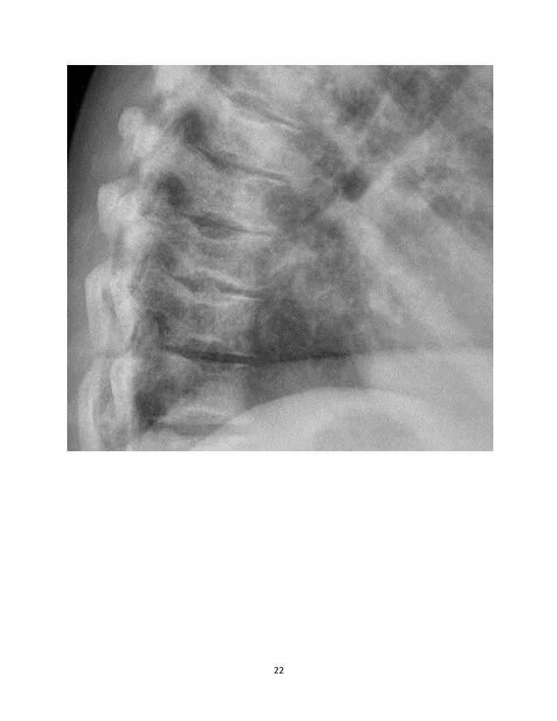
This 29 year-old female patient presented
with a chest infection, and a CXR was performed. The patient had a history of sickle-cell anaemia. The
lateral CXR shows multiple vertebral end-plate irregularities and depressions. The peripheral portions of
the end-plates are spared. The appearance is due to bone infarction and subsequent collapse. This is an
early example of the classical H-shaped vertebrae seen in sickle-cell anaemia. H-shaped vertebrae are a
characteristic finding seen in approximately 10% of patients with the disease. Bone infarction may be
complicated by Salmonella osteomyelitis. Other signs to look for on chest films are cardiomegaly, absent
spleen, and cholecystectomy clips. Reference Pope TL. Aunt Minnie’s Atlas & Imaging Specific Diagnosis.
2nd edition. Lippincott, Williams & Wilkins 2003 Image contributed by: Dr Laughlin Dawes center center
center This image is made available under a Creative Commons Licence. If you wish to use this image
outside of this licence, then please contact the author above.
21
From the case: Sickle cell disease - hip AVN and H-shaped vertebrae
Modality: X-ray
22
23
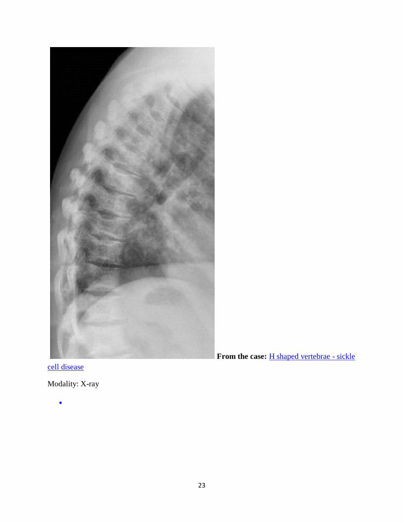
From the case: H shaped vertebrae - sickle
cell disease
Modality: X-ray
24
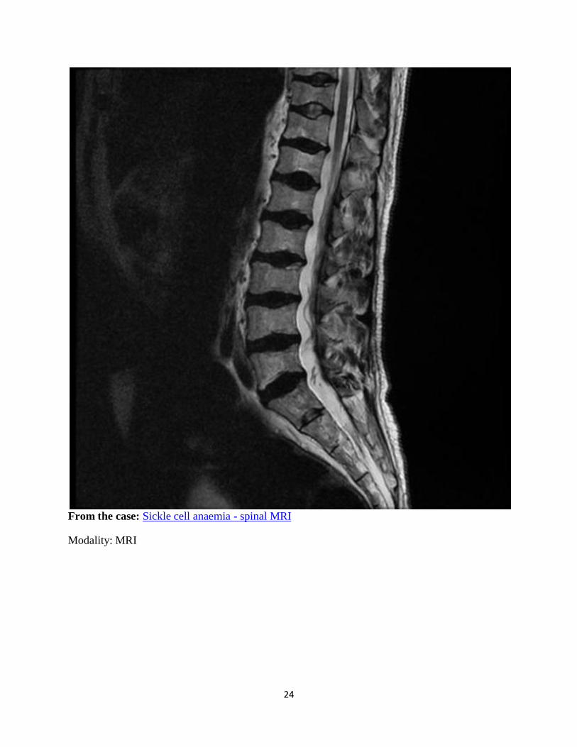
From the case: Sickle cell anaemia - spinal MRI
Modality: MRI
25
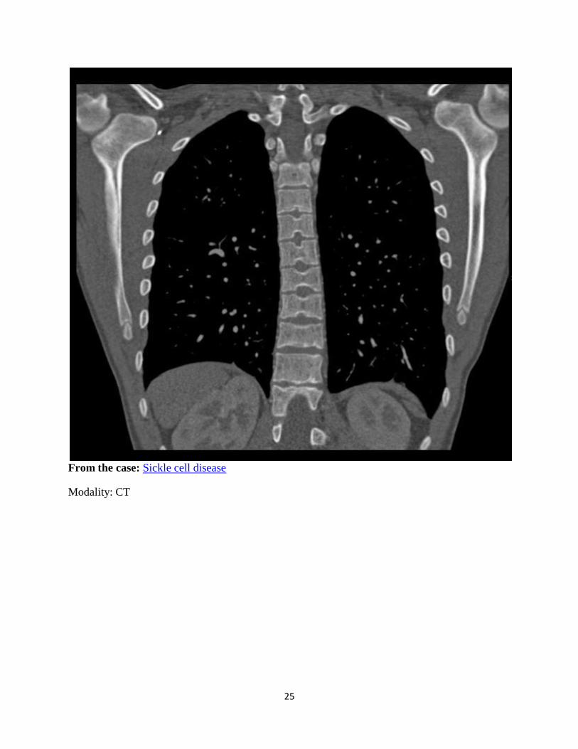
From the case: Sickle cell disease
Modality: CT
26
Apple core sign - colon
Dr Henry Knipe and Dr MT Niknejad et al.
The apple core sign, also known as a napkin ring sign (bowel), is most frequently associated
with constriction of the lumen of the colon by a stenosing annular colorectal carcinoma.
Differential diagnosis
The appearance of the apple-core lesion of the colon also can be caused by other diseases 3:
lymphoma with colonic involvement - appears more diffuse
Crohn’s disease
chronic ulcerative colitis
ischaemic colitis
Chlamydia infection
colonic tuberculosis
Helminthoma
colonic amoebiasis
colonic cytomegalovirus
villous adenoma
radiosurgery such as high doses of Cyberknife used for treating unresectable abdominal
malignancies
See also
napkin-ring sign (cardiac)
apple core sign (femur)
References
1. Mulligan ME. Classic radiologic signs, an atlas and history. Informa HealthCare.
(1997) ISBN:1850706646. Read it at Google Books - Find it at Amazon
2. Freyschmidt J. The apple core sign. Eur Radiol. 2002;12 (1): 245-7. Eur Radiol
(abstract) - doi:10.1007/s00330-001-1193-1 - Pubmed citation
3. Alzaraa A, Krzysztof K, Uwechue R et-al. Apple-core lesion of the colon: a case
report. Cases J. 2009;2 (1): 7275. doi:10.4076/1757-1626-2-7275 - Free text at pubmed -
Pubmed citation
27
Synonyms & Alternative Spellings
Synonyms or Alternative Spelling Include in Listings?
Napkin ring sign - colon ✓
Apple core sign of the colon ✗
Apple core appearance of the colon
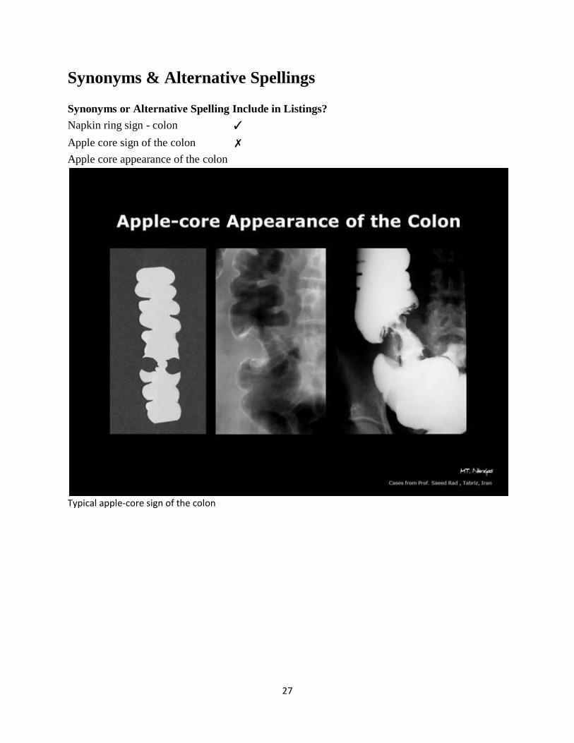
Typical apple-core sign of the colon
28
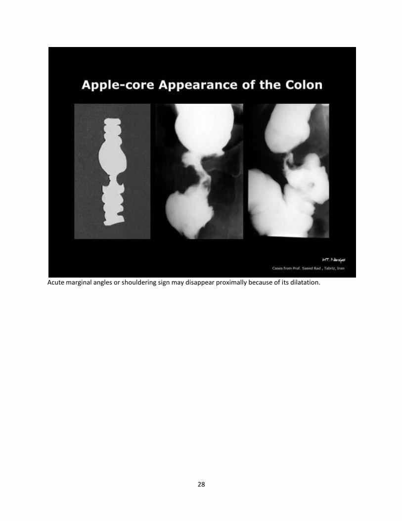
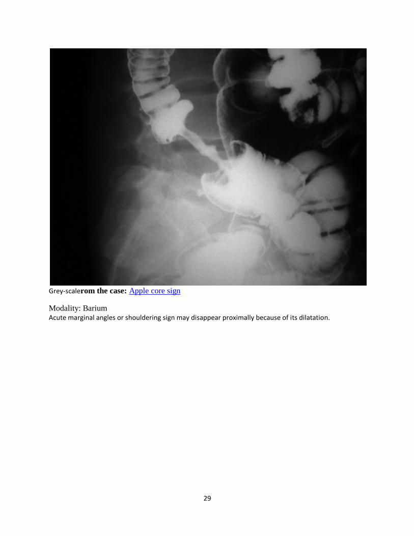
Acute marginal angles or shouldering sign may disappear proximally because of its dilatation.
29
Grey-scalerom the case: Apple core sign
Modality: Barium Acute marginal angles or shouldering sign may disappear proximally because of its dilatation.
30
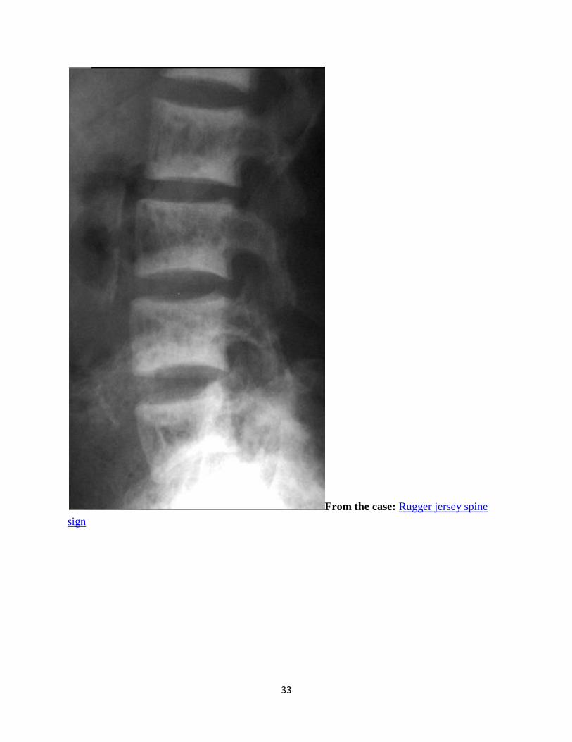
Rugger-jersey spine
Dr Frank Gaillard et al.
Rugger-jersey spine describes the prominent subendplate densities at multiple contiguous levels
to produce an alternating dense-lucent-dense appearance. This simulates the transverse bands of
a rugby jersey.
This term and pattern is distinctive for hyperparathyroidism.
Differential diagnosis
Paget disease - picture frame vertebral body
osteopetrosis - sandwich vertebral body
Note: some authors use the term rugger jersey for osteopetrosis, although most would assume the
term refers to hyperparathyroidism.
References
1. Terry R. Yochum, Lindsay J. Rowe. Yochum and Rowe's Essentials of skeletal
radiology. Philadelphia, Pa. : Lippincott Williams & Wilkins, c2005. ISBN:0781739462.
Read it at Google Books - Find it at Amazon
Synonyms & Alternative Spellings
Synonyms or Alternative Spelling Include in Listings?
Rugger jersey sign ✗
Rugby jersey sign
31
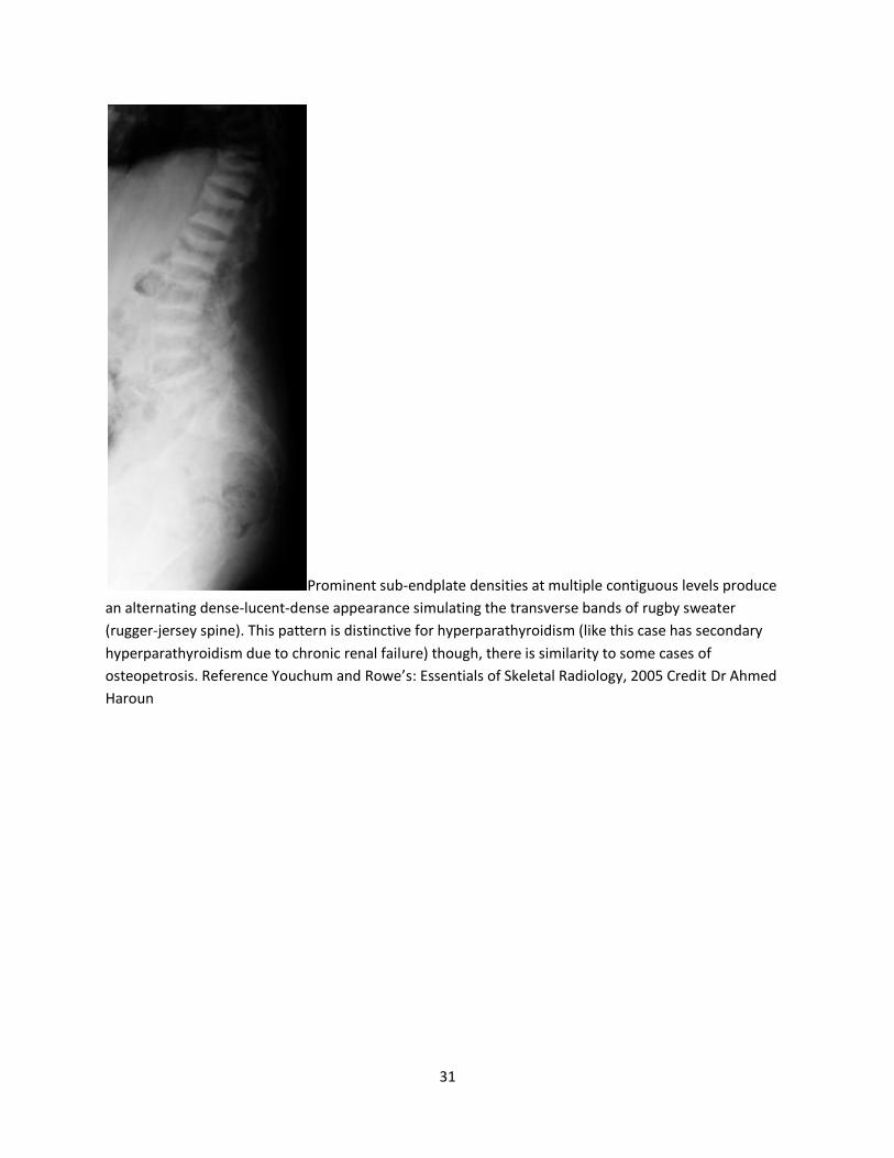
Prominent sub-endplate densities at multiple contiguous levels produce
an alternating dense-lucent-dense appearance simulating the transverse bands of rugby sweater
(rugger-jersey spine). This pattern is distinctive for hyperparathyroidism (like this case has secondary
hyperparathyroidism due to chronic renal failure) though, there is similarity to some cases of
osteopetrosis. Reference Youchum and Rowe’s: Essentials of Skeletal Radiology, 2005 Credit Dr Ahmed
Haroun
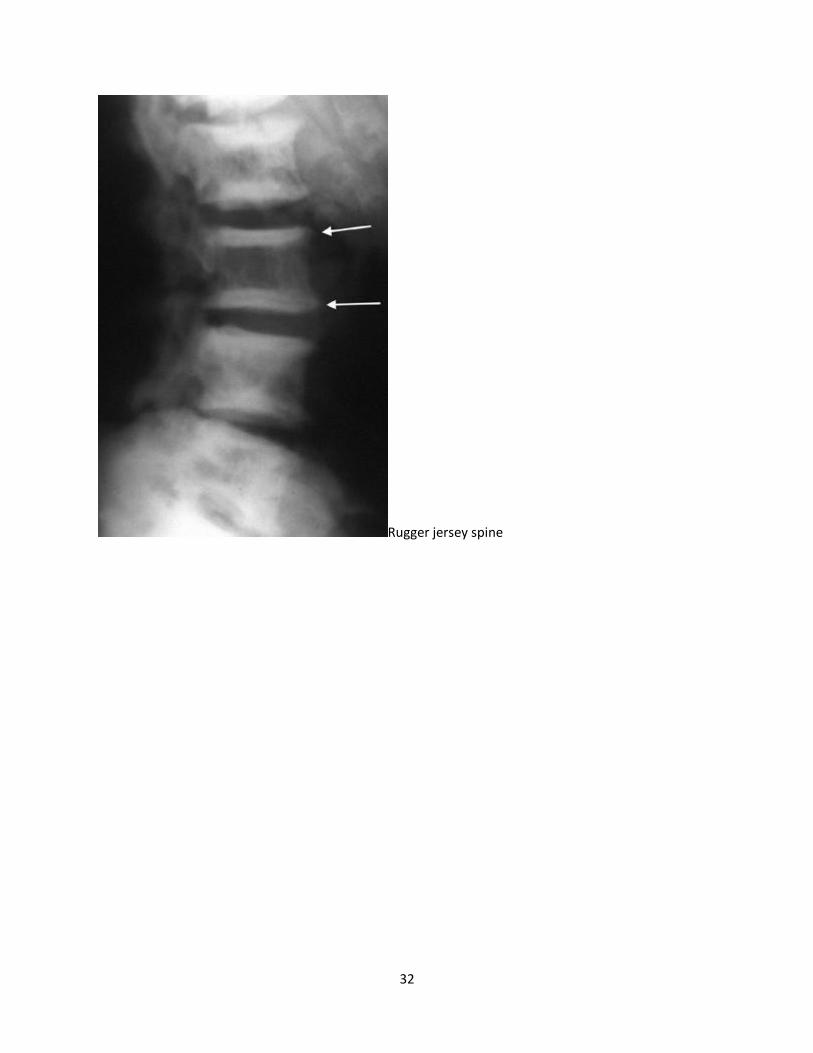
32
Rugger jersey spine
33
From the case: Rugger jersey spine
sign
34
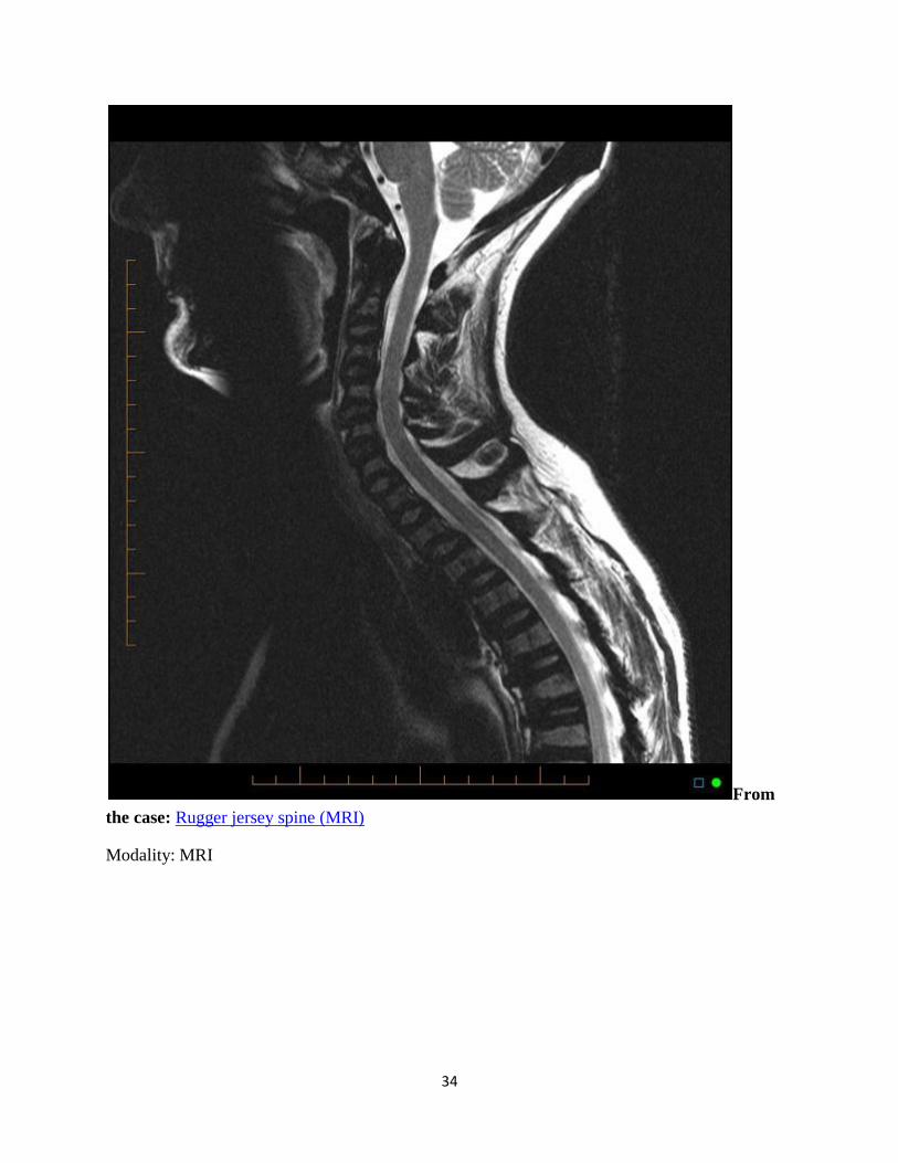
From
the case: Rugger jersey spine (MRI)
Modality: MRI
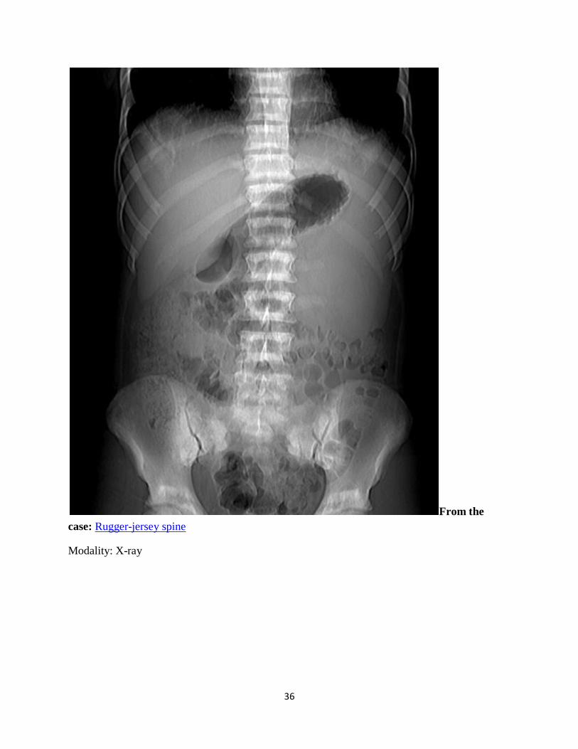
36
From the
case: Rugger-jersey spine
Modality: X-ray
37
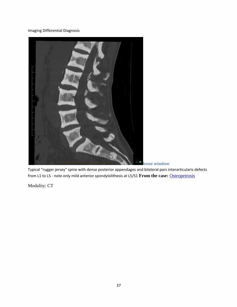
Imaging Differential Diagnosis
bone window
Typical "rugger jersey" spine with dense posterior appendages and bilateral pars interarticularis defects
from L1 to L5 - note only mild anterior spondylolithesis at L5/S1 From the case: Osteopetrosis
Modality: CT
38
Tam O'Shanter sign
Dr Frank Gaillard et al.
The "Tam O'Shanter" is a Scottish hat, named after the character in Robert Burn's 1 poem of
the same name. The appearances of advanced Paget's disease of the skull are similar in
appearance to the hat.
Paget's involvement of the skull, with widening of the diploic space and an overall enlargment of
the cranium, combinded with platybasia, leads to the appearnce of the skull falling over the facial
bones, such as a Tam O'Shanter hat would fall over the head.
Other Paget's disease related signs
blade of grass sign jigsaw pattern bone or mosaic pattern bone picture frame vertebra cotton wool appearance of bone banana fracture
Other inanimate object inspired signs.
References
1. Smith SE, Murphey MD, Motamedi K et-al. From the archives of the AFIP. Radiologic spectrum of Paget disease of bone and its complications with pathologic correlation. Radiographics. 2002;22 (5): 1191-216. Radiographics (full text) - Pubmed citation
2. Richards PS, Bargiota A, Corrall RJ. Paget's disease causing an Arnold-Chiari Type 1 malformation: radiographic findings. AJR Am J Roentgenol. 2001;176 (3): 816-7. doi:10.2214/ajr.176.3.1760816 - Pubmed citation
39
Synonyms & Alternative Spellings
Synonyms or Alternative Spelling Include in Listings?
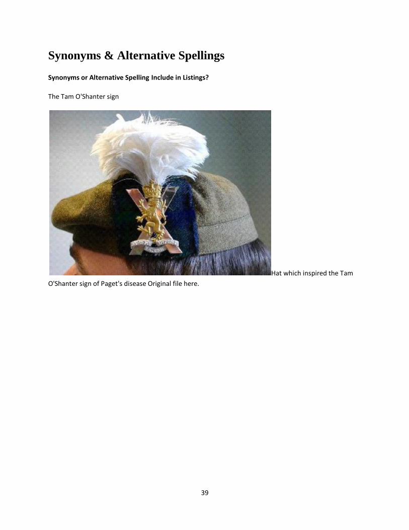
The Tam O'Shanter sign
Hat which inspired the Tam
O'Shanter sign of Paget's disease Original file here.
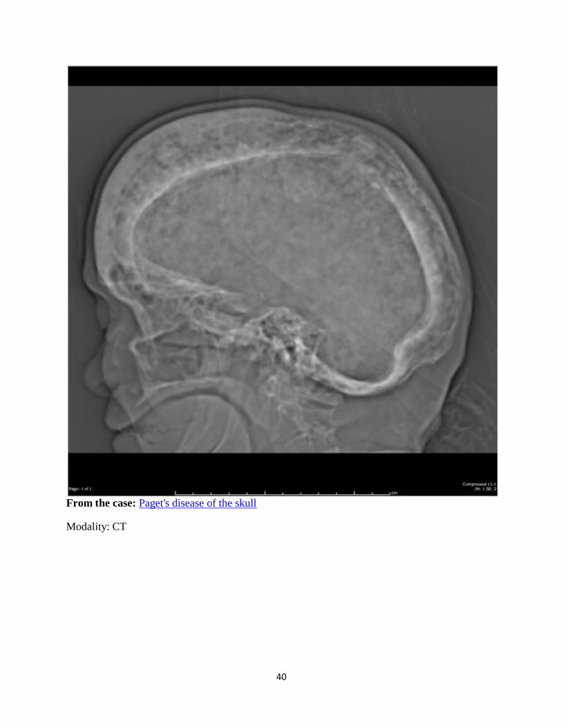
40
From the case: Paget's disease of the skull
Modality: CT
41
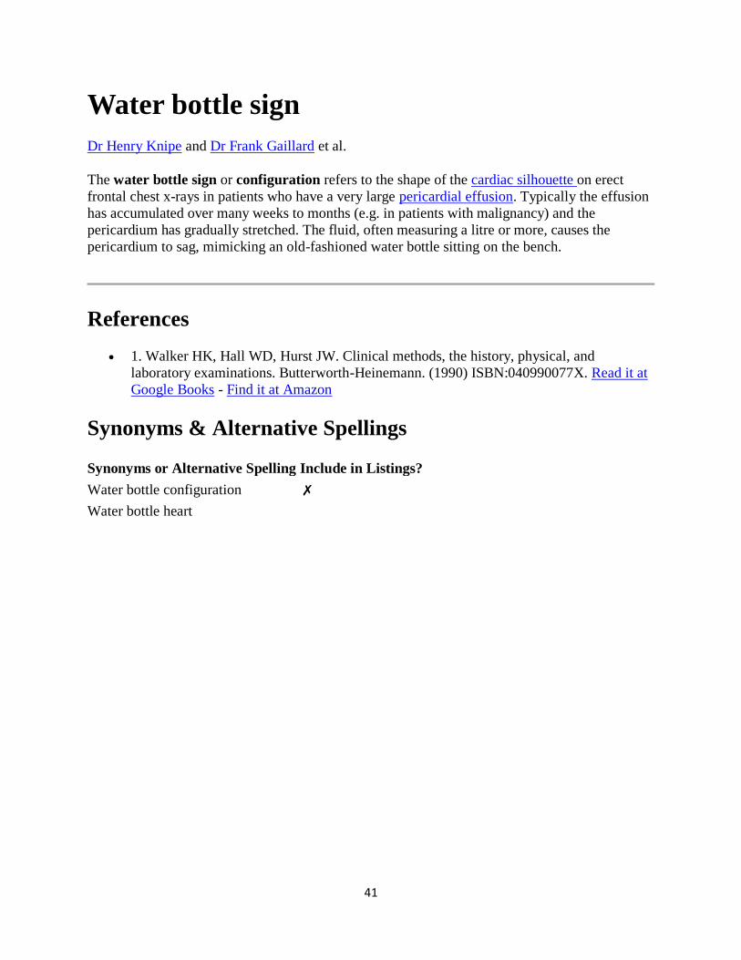
Water bottle sign
Dr Henry Knipe and Dr Frank Gaillard et al.
The water bottle sign or configuration refers to the shape of the cardiac silhouette on erect
frontal chest x-rays in patients who have a very large pericardial effusion. Typically the effusion
has accumulated over many weeks to months (e.g. in patients with malignancy) and the
pericardium has gradually stretched. The fluid, often measuring a litre or more, causes the
pericardium to sag, mimicking an old-fashioned water bottle sitting on the bench.
References
1. Walker HK, Hall WD, Hurst JW. Clinical methods, the history, physical, and
laboratory examinations. Butterworth-Heinemann. (1990) ISBN:040990077X. Read it at
Google Books - Find it at Amazon
Synonyms & Alternative Spellings
Synonyms or Alternative Spelling Include in Listings?
Water bottle configuration ✗
Water bottle heart
42
From the case: Pericardial effusion - water bottle sign
Modality: X-ray
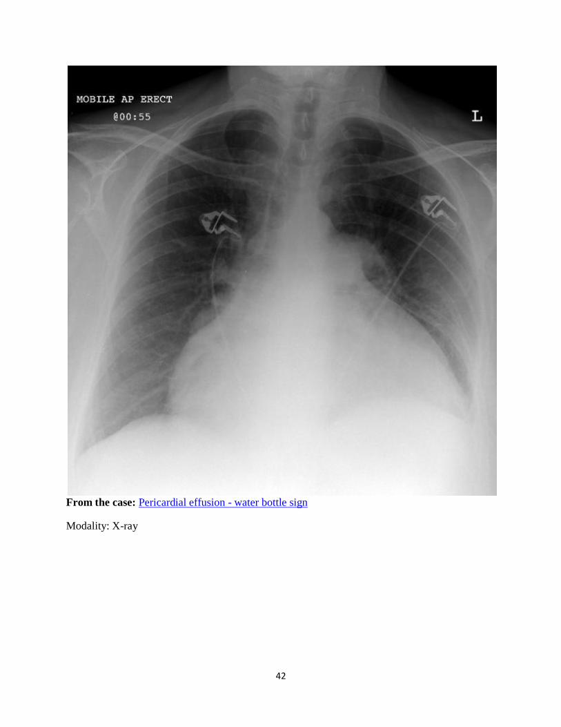
43
From the case: Pericardial effusion
Modality: X-ray
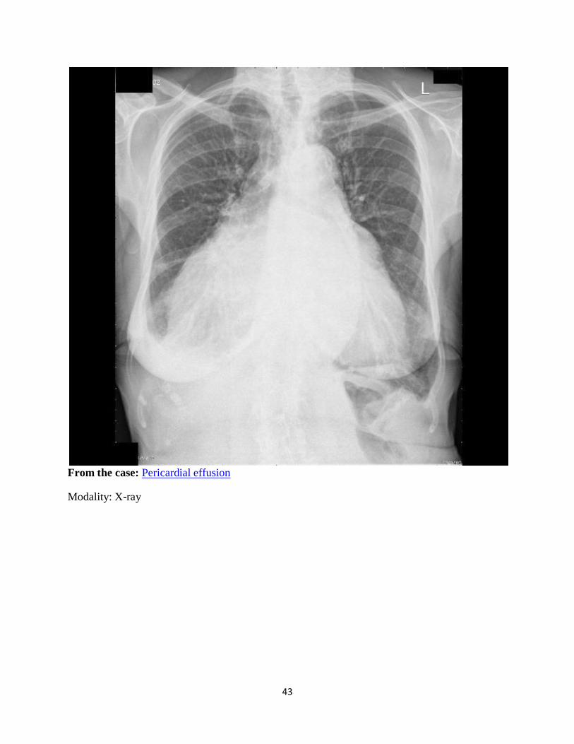
44
From the
case: Pericardial effusion
Modality: X-ray
45
White pyramid sign
Dr Henry Knipe and Dr Mohammed Al Khader.O.Thabet et al.
Medullary pyramids of kidney can be seen normally on unenhanced CT scans as high-
attenuation structures and this is known as the white pyramid sign:
bilateral high-attenuation renal pyramids are an occasional incidental normal finding
unilateral appearance of white pyramids is subtle secondary sign suggestive of
contralateral urinary tract obstruction
References
1. Webb WR, Brant W, Major N. Fundamentals of Body Ct. Saunders. (2006)
ISBN:1416000305. Read it at Google Books - Find it at Amazon
2. Dogra VS, MacLennan GT. Genitourinary Radiology: Kidney, Bladder and Urethra.
Springer. (2012) ISBN:1848002459. Read it at Google Books - Find it at Amazon
3. Step by Step in Emergency Radiology. Jaypee Brothers Medical Publishers.
ISBN:818061803X. Read it at Google Books - Find it at Amazon
