quorum sensing and quorum quenching in …studentsrepo.um.edu.my/4349/1/cxy_thesis.pdf · bacillus...
TRANSCRIPT

QUORUM SENSING AND QUORUM QUENCHING IN SELECTED
BACTERIA ISOLATED FROM DISEASED TILAPIA FISH
CHAN XIN YUE
THESIS SUBMITTED IN FULFILMENT OF THE
REQUIREMENTS FOR THE DEGREE OF MASTER OF SCIENCE
INSTITUTE OF BIOLOGICAL SCIENCES
UNIVERSITY OF MALAYA
KUALA LUMPUR
2013

iii
ABSTRACT
Living in a polymicrobial community, bacteria communicate within and among
species for interaction. This cell-cell communication in bacteria is termed quorum
sensing (QS). Among the QS signaling molecules, N˗acyl homoserine lactone (AHL) is
one of the well characterized QS signaling molecules produced and used by vast group
of Proteobacteria. This QS signaling molecule often associated with coordinating
certain group behavior of bacteria such as the bioluminescence, biofilm formation and
regulation of virulence factors expression. On the other hand, other than production,
some bacteria also degrade the QS signaling molecules and this is termed as quorum
quenching (QQ). It is believed that the bacteria possessed QQ as a strategy to survive in
the competitive environment against the QS bacteria.
In these studies, five bacteria are isolated from diseased tilapia fish namely
Bacillus sp. strain W2.2, Klebsiella oxytoca strain W4.2, Pseudomonas sp. strain W3,
Pseudomonas aeruginosa strain W3.1 and Serratia marcescens sp. strain W2.3. The QS
signaling molecules produced by these bacteria are characterized by thin layer
chromatography (TLC) and liquid chromatography mass spectrophotometry (LCMS).
Results showed that P. aeruginosa strain W3.1 produces a wide range of Pseudomonas
quinolone signaling (PQS), and AHLs including unusually long-chain N-(3-
oxohexadecanoyl)-L-homoserine lactone (OC16-HSL), but none of these compounds are
detected from Pseudomonas sp. strain W3. In contrast, Pseudomonas sp. strain W3
degraded N-butanoyl-L-homoserine lactone (C4-HSL) and N-hexanoyl-L-homoserine
lactone (C6-HSL). N-dodecanoyl-L-homoserine lactone (C12-HSL) was detected from
spent supernatant from S. marcescens strain W2.3 and this is the first report stating long
chain AHL produced by Serratia sp.. Bioassay using skimmed milk agar has confirmed

iv
that with the exception of Klebsiella sp. strain W4.2, all isolates showed proteolytic
activity, while assays using sheep blood agar showed that Bacillus sp. strain W2.2 and
Pseudomonas sp. strains W3 and P. aeruginosa strain W3.1 have hemolytic activity.

v
ABSTRAK
Bakteria berkomunikasi antara sama spesis dan juga kumpulan yang beza untuk
kehidupan. Komunikasi antara sel-sel bakteria ini dipanggil kuorum pengesanan (QS).
Antara molekul-molekul isyarat korum pengesanan, N˗acyl homoserine lactone (AHL)
merupakan isyarat korum pengesanan yang paling popular di mana ia dihasilkan dan
digunakan oleh sekumpulan besar Proteobacteria. Molekul isyarat ini sering dikaitkan
dengan menyelaraskan tingkah laku kumpulan bakteria tertentu seperti bioluminescence,
pembentukan biofilm dan memiliki faktor kebisaan. Selain daripada hasil, sesetengah
bakteria juga mengguna and memisahkan molekul isyarat kuorum pengesahan dan ini
dipanggil sebagai kuorum perencatan. Adalah dipercayai bahawa keupayaan kuorum
perencatan (QQ) bakteria adalah strategi untuk bertahan dan hidup di suasana yang
kompetitif.
Dalam kajian ini, 5 bakteria diasingkan daripada ikan tilapia yang sakit iaitu
Bacillus sp. W2.2, Klebsiella sp. W4.2, Pseudomonas sp. W3, Pseudomonas sp. W3.1
dan Serratia sp. W2.3. Isyarat molekul QS yang dihasilkan oleh bakteria ini dicirikan
oleh kromatografi lapisan nipis (TLC) dan kromatografi cecair mass spektrofotometri
(LCMS). Keputusan menunjukkan bahawa Pseudomonas sp. W3.1 menghasilkan
pelbagai isyarat quinolone Pseudomonas (PQS), dan pelbagai jenis AHLs termasuk
AHL dengan rantai panjang yang luar biasa, N-(3-oxohexadecanoyl)-L-homoserine
lactone (3-Oxo-C16-HSL). Walaubagaimanapun, isyarat luar biasa ini tidak dapat
dikesan daripada Pseudomonas sp. W3. Sebaliknya, Pseudomonas sp. W3 menguraikan
N-butanoylLhomoserine lactone (C4-HSL) dan N-hexanoyl-L-homoserine lactone (C6-
HSL). Selain daripada Pseudomonas sp. W3.1, N-dodecanoyl-L-homoserine lactone
(C12-HSL) dikesan dari kultur media yang dibelanjai oleh Serratia sp. W2.3 dan ini

vi
adalah laporan pertama menyatakan pengesanan AHL dengan rantai panjang yang luar
biasa dari Serratia sp.. Ujian bakteria dengan agar susu telah mengesahkan bahawa
kecuali Klebsiella sp. W4.2 semua bakteria menunjukkan aktiviti proteolitik, manakala
bagi agar darah biri-biri, Bacillus sp. W2.2 dan Pseudomonas sp. strain W3 dan W3.1
juga dipaparkan aktiviti hemolitik.

vii
ACKNOWLEDGEMENTS
I would like to send my gratitude to my supervisor, Dr. Chan Kok Gan, who
tirelessly provides new ideas, guidance and support for this project. He always spares
his time understanding the difficulties and the progress of the project despite of his tight
schedule. Again, I would like to thank him for the financial support that enabled my
project went without financial constrain.
Next, I would like to thank my lab manager, Ms. Yin Wai Fong for the well
management in the lab. She ensures all the constant supplies of necessity and the
reasonable working and storage area in the lab that provide us a comfortable working
environment.
Subsequently, I would like to express my gratitude to our collaborators, Dr. Davy
Chang. With the aid of Dr. Davy in analytical chemistry analysis and manuscript
preparation have successfully lead to the publication.
Deeply thanks University of Malaya for giving me a chance to further my study in
one of the world top University. In addition to that I would like to thank UM for the
financial support of my research.
Last but not least, I would like to thank all my lab members and my family
members who always lend me a hand when I am facing difficulties. They are always my
best companion and listener who provide the strongest support to me all the time.
viii
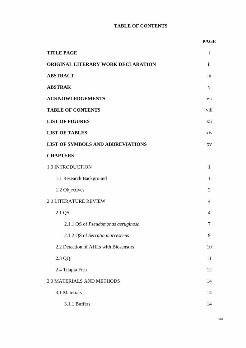
TABLE OF CONTENTS
PAGE
TITLE PAGE i
ORIGINAL LITERARY WORK DECLARATION ii
ABSTRACT iii
ABSTRAK v
ACKNOWLEDGEMENTS vii
TABLE OF CONTENTS viii
LIST OF FIGURES xii
LIST OF TABLES xiv
LIST OF SYMBOLS AND ABBREVIATIONS xv
CHAPTERS
1.0 INTRODUCTION 1
1.1 Research Background 1
1.2 Objectives 2
2.0 LITERATURE REVIEW 4
2.1 QS 4
2.1.1 QS of Pseudomonas aeruginosa 7
2.1.2 QS of Serratia marcescens 9
2.2 Detection of AHLs with Biosensors 10
2.3 QQ 11
2.4 Tilapia Fish 12
3.0 MATERIALS AND METHODS 14
3.1 Materials 14
3.1.1 Buffers 14

ix
3.1.1.1 Phosphate Buffer Saline (PBS) 14
3.1.1.2 TrisBase Acid Ethylenediaminetetracetic Acid
(TBE)
14
3.1.2 Culture Medium 15
3.1.2.1 LuriaBertani (LB) Medium 15
3.1.2.2 Agrobacterium (AB) Medium 15
3.1.2.3 Super Optimum Broth (SOC) 15
3.1.2.4 Skimmed Milk Agar 16
3.1.3 Reaction Mix 16
3.1.3.1 Polymerase Chain Reaction (PCR) Master Mix
for Gene Amplification
16
3.1.3.2 Ligation Mix 16
3.1.4 Biosensors and Strains 16
3.1.5 Primers 18
3.2 Methods 18
3.2.1 Isolation of Potential Fish Pathogen 18
3.2.2 Gram Stain 18
3.2.3 Genomic DNA Extraction 19
3.2.4 Agarose Gel Electrophoresis (AGE) 19
3.2.5 Bacteria Identification 19
3.2.6 PCR Product Purification 20
3.2.7 Competent Cell 20
3.2.8 Ligation and Transformation 21
3.2.9 Plasmid Extraction 22
3.2.10 Phylogenetic Analysis 22
x
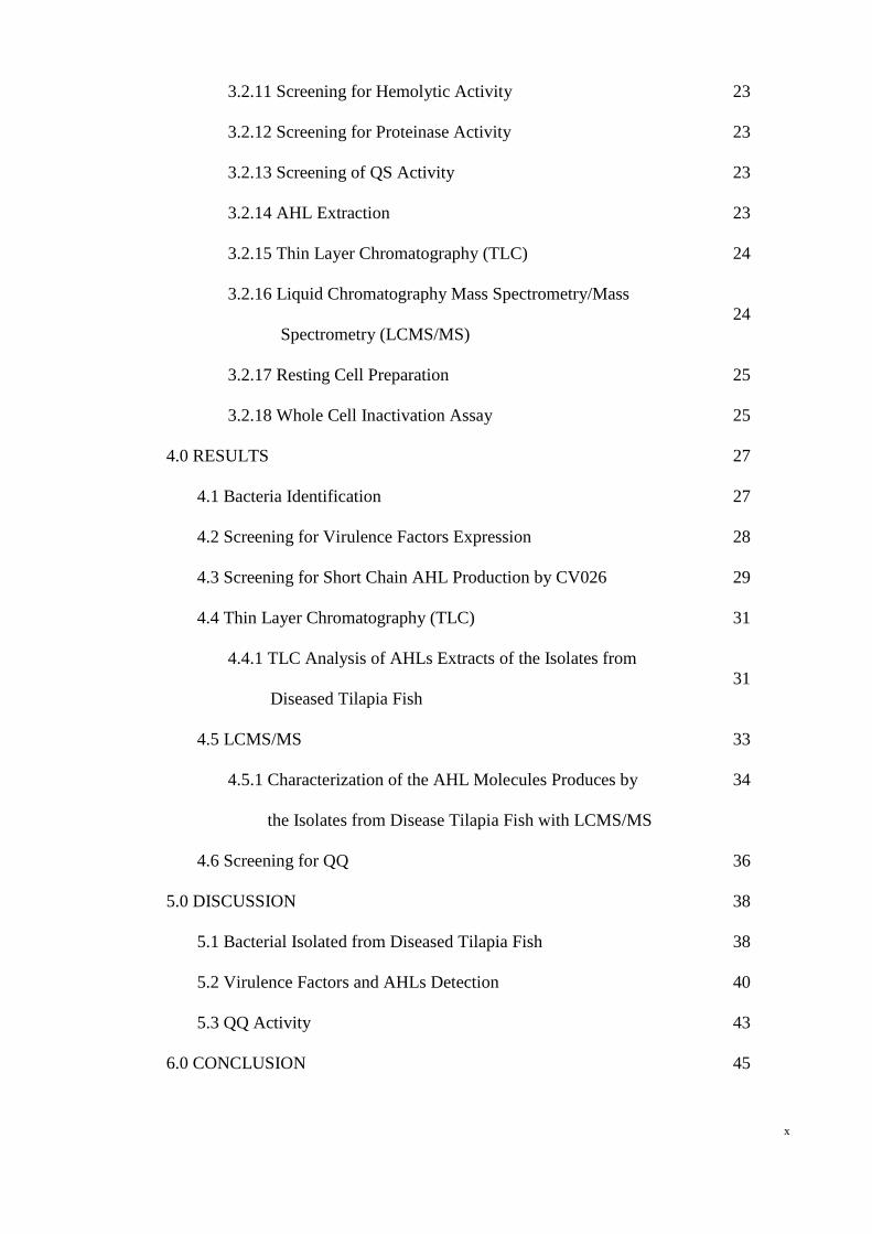
3.2.11 Screening for Hemolytic Activity 23
3.2.12 Screening for Proteinase Activity 23
3.2.13 Screening of QS Activity 23
3.2.14 AHL Extraction 23
3.2.15 Thin Layer Chromatography (TLC) 24
3.2.16 Liquid Chromatography Mass Spectrometry/Mass
Spectrometry (LCMS/MS)
24
3.2.17 Resting Cell Preparation 25
3.2.18 Whole Cell Inactivation Assay 25
4.0 RESULTS 27
4.1 Bacteria Identification 27
4.2 Screening for Virulence Factors Expression 28
4.3 Screening for Short Chain AHL Production by CV026 29
4.4 Thin Layer Chromatography (TLC) 31
4.4.1 TLC Analysis of AHLs Extracts of the Isolates from
Diseased Tilapia Fish
31
4.5 LCMS/MS 33
4.5.1 Characterization of the AHL Molecules Produces by
the Isolates from Disease Tilapia Fish with LCMS/MS
34
4.6 Screening for QQ 36
5.0 DISCUSSION 38
5.1 Bacterial Isolated from Diseased Tilapia Fish 38
5.2 Virulence Factors and AHLs Detection 40
5.3 QQ Activity 43
6.0 CONCLUSION 45

xi
REFERENCES 46

xii
LIST OF FIGURES
FIGURE PAGE
4.1 Phylogenetic analysis of 16s rDNA sequences of strain
W2.3: Serratia marcescens, W3.1: Pseudomonas
aeruginosa, W3: Pseudomonas sp., W2.2: Bacillus sp.
and W4.2: Klebsiella oxytoca. The evolutionary distance
is 0.05 changes per nucleotide position.
28
4.2 Screening of virulence factor, i.e. hemolysin, with (A)
5 % (v/v) sheep blood agar for hemolytic activity.
Bacillus sp. strain W2.2, Pseudomonas sp. strain W3 and
P. aeruginosa strain W3.1 produced hemolysin and
caused lysis of sheep blood. (B) 1.5 % (v/v) skimmed
milk agar for proteolytic activity. Bacillus sp. strain
W2.2, Pseudomonas sp. strain W3, P. aeruginosa strain
W3.1 and S. marcescens strain W2.3 produced protease
and degraded casein.
30
4.3 Screening of short chain AHL production with CV026.
The AHL produced by the bacteria will induced the
purple pigment formation of CV026. E. carotovora GS
101 is the positive control whereas E. carotovora PNP22.
Only P. aeruginosa strain W3.1 induced the purple
pigment formation.
31
4.4 Characterization of long chain AHL produced by P.
aeruginosa strain W3.1 and S. marcescens strain W2.3 by
Agrobacterium tumefaciens NTL4 pZLR4 with thin layer
33

xiii
chromatography. C12HSL is mark by as “**” while
OC16HSL is marked as “*”. P. aeruginos W3.1
produces of both C12HSL and OC16HSL which S.
marcescens W2.3 produces C12-HSL.
4.5 LCMS spectrum (A) P. aeruginosa strain W3.1 which
was detected to produce OC16HSL (m/z 354.26) and (B)
S. marcescens strain W2.3 which was detected to produce
C12HSL (m/z 284.2233)
35
4.6 Residual of (A) C4HSL and (B) C6HSL was incubated
with the bacteria for 0 and 24 hr. The AHL degradation
was determined by either the decrease of pigment size or
loss of pigment for the residual of 24 hr incubation.
37
xiv
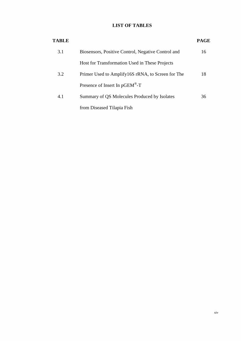
LIST OF TABLES
TABLE PAGE
3.1 Biosensors, Positive Control, Negative Control and
Host for Transformation Used in These Projects
16
3.2 Primer Used to Amplify16S rRNA, to Screen for The
Presence of Insert In pGEM®T
18
4.1 Summary of QS Molecules Produced by Isolates
from Diseased Tilapia Fish
36

xv
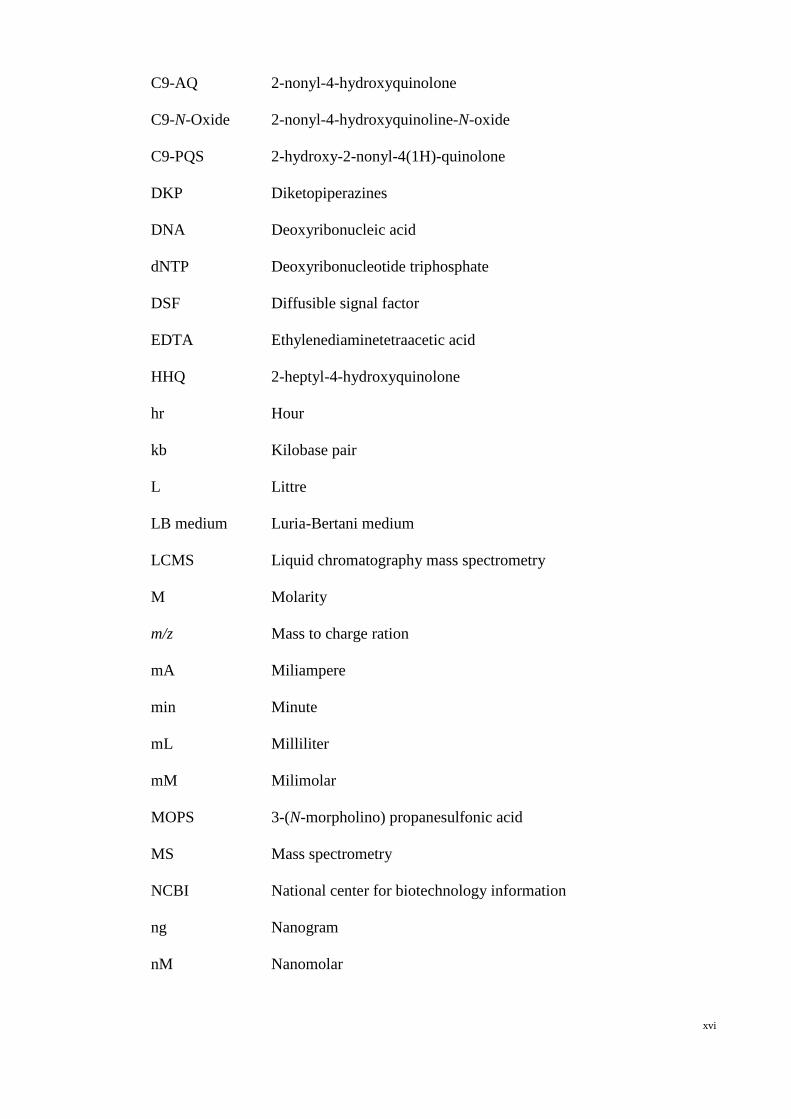
LIST OF SYMBOLS AND ABBREVIATIONS
% Percent
× Times
× g Gravity
°C Celsius
µg Microgram
µl Microlittre
µm Micron
µM Micromolar
AB medium Agrobacterium medium
ACN Acetonitrile
AGE Agarose gel electrophoresis
AHL Nacyl homoserine lactone
AHQ 2-alkyl-4(1H)-quinolones
AI2 Autoinducer 2
AIP Autoinducer peptide
C12HSL NdodecanoylLhomoserine lactone
C16HSL NhexadecanoylLhomoserine lactone
C4HSL NbutanoylLhomoserine lactone
C6HSL NhexanoylLhomoserine lactone
C7AQ 2-heptyl-4-hydroxyquinolone
C7HSL NheptanoylLhomoserine lactone
C7NOxide 2-heptyl-4-hydroxyquinolineN-oxide
C7PQS 2-heptyl-3-hydroxy-4(1H)-quinolone
C8HSL NoctanoylLhomoserine lactone
xvi
C9AQ 2-nonyl-4-hydroxyquinolone
C9NOxide 2-nonyl-4-hydroxyquinolineN-oxide
C9PQS 2-hydroxy-2-nonyl-4(1H)-quinolone
DKP Diketopiperazines
DNA Deoxyribonucleic acid
dNTP Deoxyribonucleotide triphosphate
DSF Diffusible signal factor
EDTA Ethylenediaminetetraacetic acid
HHQ 2-heptyl-4-hydroxyquinolone
hr Hour
kb Kilobase pair
L Littre
LB medium LuriaBertani medium
LCMS Liquid chromatography mass spectrometry
M Molarity
m/z Mass to charge ration
mA Miliampere
min Minute
mL Milliliter
mM Milimolar
MOPS 3-(N-morpholino) propanesulfonic acid
MS Mass spectrometry
NCBI National center for biotechnology information
ng Nanogram
nM Nanomolar
xvii
OC12HSL N(3oxododecanoyl)Lhomoserine lactone
OC14HSL N(3oxotetradecanoyl)Lhomoserine lactone
OC16HSL N(3oxohexadecanoyl)Lhomoserine lactone
OC6HSL N(3oxohexanoyl)Lhomoserine lactone
OC8HSL N(3oxooctanoyl)Lhomoserine lactone
OD Optical density
OHC16HSL 3hydroxyhexadecanoylLhomoserine lactone
PAME Palmitate methyl ester
PBS Phosphate Buffer Saline
PCR Polymerase chain reaction
PQS Pseudomonas quinolone signal
psi Pounds per square inch
QQ Quorum quenching
QS Quorum sensing
Rf Retardation factor
s Second
SAM S-adenosyl-L-methionine
SOC Super optimum broth
TBE Tris-Boric Acid Ethylenediaminetetraacetic acid
TLC Thin layer chromatography
TOF Time of flight
V Voltage
v/v Volume per volume
w/v Weight per volume
Xgal 5-bromo-4-chloro-indolyl-galactopyranoside

1
CHAPTER 1.0
INTRODUCTION
1.1 Research Background
Bacteria cell-cell communication is termed as quorum sensing (QS). It is a cell
density dependent system involving the signalling molecules production and exportation
to the extracellular environment (Joint, 2006). As the cell density increases, the QS
signalling molecules accumulate until a threshold the signalling molecule-receptor
protein complex will then coordinate certain group behavior of the bacteria (Dong &
Zhang, 2005; Waters & Bassler, 2005; Williams et al., 2007). For example, the
production of hemolysin and proteinases are the several phenotypes regulated by QS
(De Kievit & Iglewski, 2000; Swift et al., 1997).
QS is mediated by signalling molecules, such as N-acyl homoserine lactone
(AHL), autoinducer 2 (AI-2), and diffusible signal factor (DSF). Among various QS
signalling molecules, AHL which was produced by Proteobacteria is one of the most
commonly studied signalling molecules (Whitehead et al., 2006). The carbon-3 of the
acyl side chain can be an unsubstitute acyl side chain, 3-hydroxy or 3-oxo group (Hong
et al., 2012). Other than AHL, Pseudomonas harbors a second QS system which is
mediated by a different QS signalling molecule, i.e. Pseudomonas Quinolone Signal
(PQS) (Kim et al., 2010). The precursor of PQS, i.e. 2-heptyl-4-hydroxyquinolone
(HHQ) works hand in hand with PQS, intertwined with the AHL-mediated QS system,
forming a complex and versatile regulation mechanism, monitoring phenotypes such as
biofilm formation, exoproteases secretion, virulence factor production as well as

2
resistance against antibiotics (Déziel et al., 2004; Juhas et al., 2005; Kim et al., 2010;
McGrath et al., 2006; Wade et al., 2005).
In addition to QS, quorum quenching (QQ) activity which is the interference or
inactivation of QS system of the isolated bacteria was studied (Dong & Zhang, 2005).
According to Dong & Zhang, (2005), the degraded AHL was postulated to be utilized
by some bacteria as the source of carbon and nitrogen. Czajkowski & Jafra, (2009)
suggested interference of QS allows the QQ bacteria to gain competition advantage.
1.2 Objectives
In this study, bacteria were suspected to be the causative agent of an outbreak in
year 2009. It killed more than 50 % of the tilapia fish stock of Malaysia. The diseased
tilapia fish were delivered to our laboratory in 2009 from a local fish farm in
Terengganu, Malaysia. Five bacteria were isolated and identified from the diseased
tilapia fish. QS and QQ activities were performed on these potential fish pathogens.
3
OBJECTIVES
The four objectives in this study are as follow:
1. to identify the pathogens from the diseased tilapia fish,
2. to examine the production of virulence factors namely proteinase and
hemolysin from these isolates,
3. to characterize the AHL produced by isolates, and
4. to study QQ activity of from the isolates.

4
CHAPTER 2.0
LITERATURE REVIEW
2.1 QS
The discovery of bacteria communication signals has shift the perspective on
bacteria from an independent living unicellular organism to a complex community
where bacteria communication occurs among each other (Atkinson & Williams, 2009;
Waters & Bassler, 2005). These interactions happen both within the same species and
cross species (Waters & Bassler, 2005). This cell-cell communication was termed as
quorum sensing (QS) (Fuqua et al., 1994).
QS involve four steps where the bacteria synthesize the signalling molecules and
release them into the extracellular environment (Williams, 2007). As the population
density increases, the concentration of the signalling molecules in the extracellular
environment increases as well. When the concentration of the signalling molecules
reaches its threshold level, it will diffuse into the bacteria cells, binds with the receptor
protein. This signalling molecule- receptor protein will interact with the target promoter,
subsequently up-regulate or down-regulate certain phenotypes (Atkinson & Williams,
2009; Bassler, 2002; Dong & Zhang, 2005; Jayaraman & Wood, 2008; Williams, 2007).
The first reported QS-regulated bioactivity was the bioluminescence of Vibrio
fischeri which inhabited in the light organ of the Hawaiian bobtail squid (Eupryma
scolopes) (Callahan & Dunlap, 2000; Defoirdt et al., 2007; Fuqua & Greenberg, 2002).
A minute amount of V. fischeri is harbored in the light organ of the squid during day
5
time. The bacteria population gradually increases after hours of incubation as well as the
signalling molecules that trigger the luciferase expression when the signalling
molecules reach its threshold concentration. This bioluminescence was needed by the
squid to counterilluminate its shadow and avoid predation in the night (Callahan &
Dunlap, 2000; Defoirdt et al., 2007; Dunlap, 1999; Waters & Bassler, 2005). The
luminescence was “turned off” by the squid by pumping out large amount of bacteria
from its light organ, hence the bacteria has insufficient signalling molecules to trigger
the production of luciferase (Dunlap, 1999; Waters & Bassler, 2005).
Besides bioluminescence, QS regulates a vast array of phenotypes, such as the
production of chitinolytic enzymes, exoproteases as well as natural antibiotic violacein
by Chrombacterium violaceum (Chernin et al., 1998; McClean et al., 1997). QS too
regulates several phenotypes which are life-threatening such as biofilm formation,
production of virulence factors and swarming activity, in several notorious pathogens,
such as Pseudomonas aeruginosa (Cvitkovitch et al., 2003; De Kievit et al., 2001),
Serratia marcescens (Rice et al., 2005), Serratia liquefaciens (Labbate et al., 2004),
Burkholderia cepacia (Huber et al., 2001), Burkholderia cenocepacia (O'Grady et al.,
2012), Vibrio cholera (Hammer & Bassler, 2003), and Streptococcus mutans
(Cvitkovitch et al., 2003). More attention was given to the studies of bacteria QS when
it was learn that the expression of virulence factor for some of the bacteria are reported
to be QS regulated, for instance V. cholera (Hammer & Bassler, 2003), B. cenocepacia
(O'Grady et al., 2012), Pectobacter (Põllumaa et al., 2012) and P. aeruginosa.

6
There are various types of QS signalling molecules for example the AHL, 2-alkyl-
4(1H)-quinolones (AHQs), autoinducer peptide (AIP), DSF, palmitate methyl ester
(PAME), diketopiperazines (DKP) (Dong & Zhang, 2005; Kim et al., 2010; Lyon &
Novick, 2004; Uroz et al., 2008; Williams et al., 2007; Yin et al., 2012). Of all the
mentioned signalling molecules, AHL has been widely studied by most research
institutions. AHL is basically a group of signalling molecules which produced by most
Gram-negative bacteria, specifically Proteobacteria (Leadbetter & Greenberg, 2000;
Uroz et al., 2008). Several reports showed that single genus of bacteria may employ
more than one type of QS system for example, Pseudomonas, Burkholderia, and
Alteromonas have been reported to produce AHL as well as AHQ as QS signalling
molecules (Fletcher et al, 2007; McGrath et al., 2006).
In the AHL-mediated QS system, the AHL synthase encoded by luxI gene is
responsible for the production for AHL whereas the luxR gene which encodes for AHL
receptor play a crucial role in regulating QS-regulated phenotypes as well as facilitating
a positive autoinductive loop in driving the production of AHL (Atkinson & Williams,
2009). The AHL was made up of a lactone ring and acylated side chain (Williams et al.,
2007) where the lactone ring of AHL was synthesized by AHL synthase from S-
adenosyl-L-methionine (SAM) and its acyl chains was originated from lipid metabolism,
carried by various acyl-carrier proteins (Reading & Sperandio, 2005). This acyl side
chain normally range from 4 to 18 carbons where the longer acyl side chain stand a
chance to have unsaturated bond in the middle of the acyl chain (Fuqua & Greenberg,
2002). The third carbon (C3) of the acyl side chain could be fully reduced, fully
oxidized carbonyl or carry a hydroxyl group (Hong et al., 2012). Some other groups or
molecules such as cystein, biotin and fluorescence might be incorporated within the

7
AHL molecules as well and this has an effect on the binding affinity of modified-AHL
to its native AHL receptor (Fuqua & Greenberg, 2002).
2.1.1 QS of Pseudomonas aeruginosa
P. aeruginosa listed as one of the top three human opportunistic pathogen was
consistently found infecting immunocompromised host (Stover et al., 2000). It was
recorded as the most common Gramnegative bacteria responsible for nosocomial
infection (Van Delden & Iglewski, 1998). The resulting infection could cause morbidity
and mortality of the host yet the emergence of multidrug resistance strain makes
treatment more challenging (Erickson et al., 2002; Livermore, 2002). Besides human, P.
aeruginosa was recovered from sick tilapia fish in Eygpt and its close species
Pseudomonas angulliseptica was reported to cause more than 95 % mortality of the
infected fish (Eissa et al., 2010).
QS of P. aeruginosa has been studied extensively and was reported to be
regulating its virulence factor production, biofilm formation, swarming ability and
pyocyanin production (Dubern & Diggle, 2008; Köhler et al., 2000; Pesci et al., 1997).
Its genome carries at least two complete QS systems (Pesci et al., 1997). Las and Rhl
systems are responsible for AHL regulatory circuits (Zhu et al., 2004).
In brief, lasI gene is responsible for autoinducer
N(3oxododecanoyl)Lhomoserine actone (OC12HSL) synthesis in Las system (Smith
et al., 2002). The OC12HSL binds to its transcription factor LasRlike family protein

8
and the complex activates the expression of several virulence genes such as the lasB
gene that code for the production of elastase, toxA for exotoxin A, aprA for alkaline
protease and xcpP and xcpR for secretory pathway of P. aeruginosa (Passador et al.,
1993; Pesci et al., 1997; Storey et al., 1998; Van Delden & Iglewski, 1998). On top of
virulence gene, LasR/OC12HSL complex activates the expression of rhlR genes of
another QS system of P. aeruginosa, the Rhl system (Juhas et al., 2004; Van Delden &
Iglewski, 1998). rhlI synthesize NbutanoylLhomoserine lactone (C4HSL) which
binds with the rhlR cognate transcriptional factor activating the expression of genes that
encode for the synthesis of rhamnosyltransferase, elastase, alkaline protease, and
cyanide (Pearson et al., 1997; Smith et al., 2002; Van Delden & Iglewski, 1998). In
addition to C4HSL and OC12HSL, P. aeruginosa was reported to produce
N(3oxotetradecanoyl)Lhomoserine lactone (OC14HSL), however the mechanism
and function of this autoinducer was undetermined (Wong et al., 2012).
The third well known QS system of P. aeruginosa was termed as PQS system.
The active autoinducer involved in this system are 2-heptyl-3-hydroxy-4(1H)-quinolone,
commonly known as PQS and its immediate precursor, 2-heptyl-4-hydroxyquinolone
(HHQ) (Fletcher et al., 2007; Häussler & Becker, 2008). PQS belongs to the
4quinolone chemical family and its synthesis depends on pqsABCDE operon which is
also responsible for the synthesis of various type of hydroxyquinolone (Fletcher et al.,
2007; Gallagher et al., 2002; Wilder et al., 2011). PQS or HHQ binds to the receptor
protein, PqsR with high affinity, enhancing the binding of PqsR to pqsA promoter,
selfregulating the synthesis of PQS compound (Déziel et al., 2004; Fletcher et al., 2007;
Wade et al., 2005). This system works together with the AHL regulated QS system,
mainly the Rhl system, co-ordinating more than 100 genes expression including the

9
pyocynin production, secondary metabolites production and biofilm development
(Aendekerk et al., 2005; Déziel et al., 2004; Dubern & Diggle, 2008; McGrath et al.,
2006; Wade et al., 2005; Williams & Cámara, 2009).
2.1.2 QS of Serratia marcescens
S. marcescens belongs to Enterobacteriaceae family often associated with food
spoilage and causal agent of various diseases (Van Houdt et al., 2007; Wang et al.,
2012). It usually occurs as bloody red colonies due to the production of prodigiosin
(Thomson et al., 2002; Williams, 1973). The production of prodigiosin is regulated by
the QS system of S. marcescens (Thomson et al., 2002). Studies on S. marcescens strain
SS1, and strain MG1 (previously known as S. liquefaciens MG1) reviewed two AHL
based QS which are the Spn system and Swr system (Horng et al., 2002; Labbate et al.,
2004; Lindum et al., 1998; Van Houdt et al., 2007; Van Houdt et al., 2006; Wei & Lai,
2006; Williams, 1973).
In the study by Horng et al., (2002), spnR of S. marcescens SS1 acts contradict to
majority of the luxR in the QS system, as it negatively regulates the sliding activity,
pigment and nuclease production of S. marcescens SS1. This depressed nuclease gene
by spnR can be restore by N(3oxohexanoyl)Lhomoserine lactone (OC6HSL)
synthesize by spnI. Aside from OC6HSL, AHL synthase, spnI also synthesize
NhexanoylLhomoserine lactone (C6HSL), NheptanoylLhomoserine lactone
(C7HSL) and NoctanoylLhomoserine lactone (C8HSL) (Horng et al., 2002; Van
Houdt et al., 2007; Wei & Lai, 2006; Wei et al., 2006). Further studies on S.
marcescens strain SS1 shows that its spnR falls within the Tn3 family transposon

10
making it a mobile QS system that might transfer within different host (Wei et al.,
2006).
On the other hand, the main element involve in Swr system are the AHL synthase,
swrI, the translational regulators swrR and the signalling molecules, C4HSL and
C6HSL (Labbate et al., 2004; Van Houdt et al., 2007; Wei & Lai, 2006). The Swr
system positively regulates the production of biosurfactant with C4HSL which
facilitates the motility of Serratia (Labbate et al., 2004; Lindum et al., 1998). In
addition to the biosurfactant production, Swr too coordinates the biofilm maturation,
butanediol fermentation and extracellular lipase (Labbate et al., 2004; Lindum et al.,
1998; Rice et al., 2005; Van Houdt et al., 2006; Wei & Lai, 2006).
2.2 Detection of AHLs with Biosensors
Certain QS bacteria have been mutated and genetically modified to act as
biosensors for rapid screening for the presence of QS signalling molecules (Farrand et
al., 2002; Llamas et al., 2004; McClean et al., 1997). Most of the QS biosensors are
constructed by either modification of the bacterial QS gene or by the insertion of
plasmid reporter vector into the bacteria (Steindler & Venturi, 2007; Winson et al.,
2006). The presence and detection of QS signalling molecules was shown by possessing
certain characteristic such as pigment formation and bioluminescence (McClean et al.,
1997; Winson et al., 2006).

11
2.3 QQ
The interruption of QS activity was termed as QQ (Dong & Zhang, 2005). The
interruption can be enzymatically degradation of the QS signalling compound or by the
introduction of the antagonist that mimics the QS autoinducer which blocks the
autoinducer synthase or receptor protein (Adonizio et al., 2006; Czajkowski & Jafra,
2009; Dong & Zhang, 2005; Hong et al., 2012; Uroz et al., 2008). The QS antagonist
can be natural compound which are extracted and purified from plants (Chong et al.,
2011; Krishnan et al., 2012; Tan et al., 2012).
The enzymatic degradation of the AHL can be performed by 4 categories of
enzymes, namely the lactonase and decarboxylase that target and break the lactone ring,
while the acylase and deaminase target and cleave the acyl side chain (Dong & Zhang,
2005). To date only lactonase and acylase were found with an addition enzyme that
disturb but not disrupt the AHL based QS system (Chan et al., 2011; Dong & Zhang,
2005; Hong et al., 2012). These enzymes were frequently reported to be produced by
bacteria such as Bacillus cereus (lactonase), P. aeruginosa (acylase) and Burkholderia
(oxidoreductase) (Chan et al., 2010; Chan et al., 2011; Dong et al., 2002; Sio et al.,
2006). Studies suggest that bacteria degrades AHLs in the environment for several
reason including to compete for limited nutrient source, recycling the energy source
from AHL, and regulating its QS system (Czajkowski & Jafra, 2009; Haudecoeur et al.,
2009; Leadbetter & Greenberg, 2000; Park et al., 2008; Park et al., 2003).
12
2.4 Tilapia Fish
Tilapia Fish is the common name for approximately 70 species of fishes in
Cichlidae family (Gonzales & Brown, 2006). It is cultivated in both tropical and
subtropical countries and is supplied as food fish in countries around the world (Lee et
al., 2005). In year 2010, tilapia is reported as one of the major aquaculture production
that makes up to 7.6 % of the worldwide freshwater fish production (Bostock et al.,
2010). The major tilapia fish suppliers in the world include China, Eygpt, Philippines,
Indonesia, Thailand, Brazil, Bangladesh, Vietnam, Taiwan and Malaysia (Sayed, 2006;
Young & Muir, 2002).
The high growth rates, short generation time, ability of adapt to wide range of
environment, resistant to stress and diseases, feeding on low trophic levels and artificial
feeds has make it a popular aquaculture candidate in developing countries (Sayed, 2006).
According to several studies, tilapia fish able to tolerate a wide range of water salinity
where certain species can grow and reproduce normally in water with more than 50 %
of water salinity making it a good candidate for fish farmers, especially in countries
with limited freshwater source (El-Sayed, 2006). On the other hand, its lower protein
requirement with the credit of the high demand from the market resulting the extensive
cultivation of tilapia by fish farmers (El-Sayed, 2006; Fitzsimmons, 2000).
Malaysia being one of the major suppliers of farmed tilapia fish has increased the
cultivation volume of tilapia dramatically within 10 years. According to several reports,
the production of farmed tilapia fish in Malaysia increased by 15fold from 1345
million tonnes to 26872 tonnes from year 1992 to 2002 (Sayed, 2006). In year 2010,

13
Malaysia was estimated to produce 120000 million tonnes of tilapia fish (Josupeit,
2005). Most of this fish was sold to the domestic market of Malaysia, while a small
portion from the total harvested product was export as high value products, such as fish
fillets, to foreign market (Bostock et al., 2010; Fitzsimmons, 2000; Josupeit, 2005).
With the increasing demand for tilapia fish globally, tilapia fish has become an
important aquaculture product for Malaysia.

14
CHAPTER 3.0
MATERIALS AND METHODS
3.1 Materials
Chemicals used in this work were purchased from Sigma, USA; Merck, Germany;
Amresco, USA; BDH Ltd. UK; and BD DifcoTM Laboratories, USA. Solvents used in
this work were supplied by Fisher Scientific, UK. While AHL used were purchased
from Cayman, USA. Buffers and culture mediums were sterilized by autoclaving at
121 °C, 15 psi for 15 min before use, while the heat sensitive material including
inorganic supplement, antibiotics, glucose, skimmed milk and Xgal were filtered
sterilize using syringe filter (Sartorius, Germany) with pore size of 0.22 µm. The pH of
each buffer and medium were adjusted with 1 M of HCl and 1 M of NaOH.
3.1.1 Buffer
3.1.1.1 Phosphate Buffer Saline (PBS)
PBS was prepared with 0.0023 % (w/v) NaH2PO4, 0.0115 % (w/v) Na2HPO4, and
5.4 % (w/v) tris base in distilled water. The pH of the buffer was adjusted to 6.5 prior to
autoclave.
3.1.1.2 Tris-Boric Acid Ethylenediaminetetraacetic acid (TBE)
TBE was prepared by mixing 0.37 % (w/v) ethylenediaminetetraacetic acid
(EDTA), 2.75 % (w/v) of boric acid and tris base 5.40 % (w/v) in 1 L of distilled water

15
making it 5 × TBE. The pH of TBE was adjusted to 6.5 before autoclaving. Five times
dilution on 5 × TBE was performed in order to prepare 1 × TBE.
3.1.2 Culture Medium
3.1.2.1 Luria-Bertani (LB) Medium
Bacteria were maintained on LB medium which contain 1.0 % (w/v) NaCl,
1.0 % (w/v) peptone and 0.5 % (w/v) yeast extract. LB agar was prepared by solidifying
LB medium with 1.5 % (w/v) of bacto agar. If AHL extraction is needed, 50 mM of 3-
(N-morpholino) propanesulfonic acid (MOPS) was added into the medium.
3.1.2.2 Agrobacterium (AB) Medium
AB medium contain 0.3 % (w/v) K2HPO4, 0.1 % (w/v) NaH2PO4, 0.1 % (w/v)
NH4Cl, 0.03 % (w/v) MgSO4.7H2O, 0.015 % (w/v) KCl, 0.0005 % (w/v) CaCl2 and
0.00025 % (w/v) FeSO4.7H2O. In order to culture A. tumefaciens NTLZ pZLR4 0.5 %
(w/v) glucose and 150 µg/ml gentamycin was added into AB medium (Yin et al., 2012).
AB agar was prepared by solidifying AB medium with 1.5 % (w/v) bacto agar.
3.1.2.3 Super Optimum Broth (SOC)
SOC was prepared by with 2.0 % (w/v) tryptone, 0.5 % (w/v) yeast extract, 0.5 %
(w/v) MgCl2, 0.05 % (w/v) NaCl and 20 mM glucose. The broth was adjusted to pH 7.0
with NaOH pellet.
16
3.1.2.4 Skimmed Milk Agar
Skimmed milk agar for proteolytic assay was made of 0.05 % (w/v) yeast extract,
0.1 % (w/v) tryptone, 1.0 % (w/v) NaCl, 1.5 % (v/v) skim milk and solidified with
1.5 % (w/v) bacto agar.
3.1.3 Reaction Mix
3.1.3.1 Polymerase Chain Reaction (PCR) Master Mix for Gene Amplification
Each PCR reaction mix contain 1 × PCR buffer, 800 µM dNTP mix, 20 mM
MgCl2, 10 nM forward primer, 10 nM reverse primer, 1 unit taqDNA polymerase, 1 ng
genomic DNA and top up each reaction to 15 µl with distilled water.
3.1.3.2 Ligation Mix
Ligation mix for each sample contain 1× rapid ligation buffer 5 ng/µl pGEM®-T
easy vector (Promega, USA), 0.3 unit/µl T4 DNA ligase and 1 µg purified PCR product.
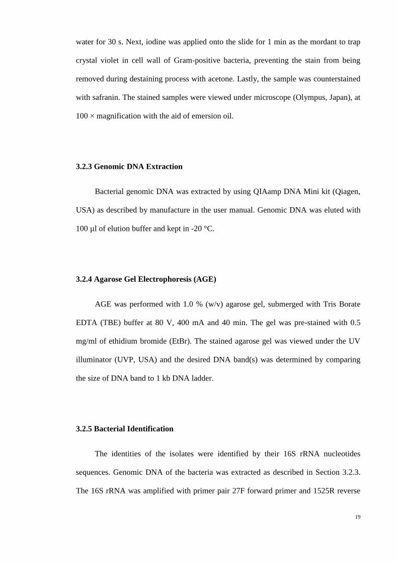
3.1.4 Biosensors and Strains
Table 3.1 : Biosensors, Positive Control, Negative Control and Host for Transformation
Used in This Project
Sample Description Reference
Chromobacterium
violaceum 026
A double mini Tn5 mutant derived from C.
violaceum ATCC 31532. It is a violacein-
(McClean et al.,
1997)

17
(CV026) negative, white mutant defective in the
production of the AHLs. Production of
purple colour pigment, i.e. violacein, is
inducible by the addition of exogeneous
short chain AHLs, range from C4-HSL to
C8-HSL.
Escherichia coli
DH5α
Host that carries no plasmid that yield high
quality and concentration of inserted
plasmid.
(Taylor et al.,
1993)
Bacillus cereus
Serve as positive control in the AHL
inactivation assay. It is capable of
degrading AHL by targeting the lactone
bond and amide linkage, via AHL
lactonase, encoded by aiiA homologues
(Chan et al., 2010)
Erwinia carotovora
GS101
Restrictionless, modificationless
derivatives that produces OC6HSL for the
regulation of its production of carbapenem.
(McGowan et al.,
1995)
Erwinia carotovora
PNP22
Mutants derive from GS101 with defective
carI gene which code for the production of
AHL synthase. It serve as the negative
control for the cross streak due to its
inability to synthesize OC6HSL.
(McGowan et al.,
1995)
Agrobacterium
tumefaciens NTL4
(pZLR4)
Biosensor that based on the traI/R to detect
a broad range of AHLs
(Farrand et al.,
2002)
18
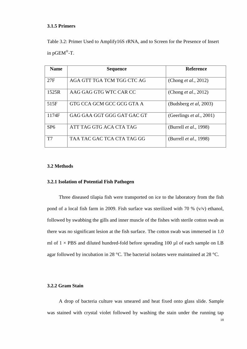
3.1.5 Primers
Table 3.2: Primer Used to Amplify16S rRNA, and to Screen for the Presence of Insert
in pGEM®-T.
Name Sequence Reference
27F AGA GTT TGA TCM TGG CTC AG (Chong et al., 2012)
1525R AAG GAG GTG WTC CAR CC (Chong et al., 2012)
515F GTG CCA GCM GCC GCG GTA A (Budsberg et al, 2003)
1174F GAG GAA GGT GGG GAT GAC GT (Geerlings et al., 2001)
SP6 ATT TAG GTG ACA CTA TAG (Burrell et al., 1998)
T7 TAA TAC GAC TCA CTA TAG GG (Burrell et al., 1998)
3.2 Methods
3.2.1 Isolation of Potential Fish Pathogen
Three diseased tilapia fish were transported on ice to the laboratory from the fish
pond of a local fish farm in 2009. Fish surface was sterilized with 70 % (v/v) ethanol,
followed by swabbing the gills and inner muscle of the fishes with sterile cotton swab as
there was no significant lesion at the fish surface. The cotton swab was immersed in 1.0
ml of 1 × PBS and diluted hundredfold before spreading 100 µl of each sample on LB
agar followed by incubation in 28 °C. The bacterial isolates were maintained at 28 °C.
3.2.2 Gram Stain
A drop of bacteria culture was smeared and heat fixed onto glass slide. Sample
was stained with crystal violet followed by washing the stain under the running tap
19
water for 30 s. Next, iodine was applied onto the slide for 1 min as the mordant to trap
crystal violet in cell wall of Gram-positive bacteria, preventing the stain from being
removed during destaining process with acetone. Lastly, the sample was counterstained
with safranin. The stained samples were viewed under microscope (Olympus, Japan), at
100 × magnification with the aid of emersion oil.
3.2.3 Genomic DNA Extraction
Bacterial genomic DNA was extracted by using QIAamp DNA Mini kit (Qiagen,
USA) as described by manufacture in the user manual. Genomic DNA was eluted with
100 µl of elution buffer and kept in -20 °C.
3.2.4 Agarose Gel Electrophoresis (AGE)
AGE was performed with 1.0 % (w/v) agarose gel, submerged with Tris Borate
EDTA (TBE) buffer at 80 V, 400 mA and 40 min. The gel was pre-stained with 0.5
mg/ml of ethidium bromide (EtBr). The stained agarose gel was viewed under the UV
illuminator (UVP, USA) and the desired DNA band(s) was determined by comparing
the size of DNA band to 1 kb DNA ladder.
3.2.5 Bacterial Identification
The identities of the isolates were identified by their 16S rRNA nucleotides
sequences. Genomic DNA of the bacteria was extracted as described in Section 3.2.3.
The 16S rRNA was amplified with primer pair 27F forward primer and 1525R reverse

20
primer (Chong et al., 2012). PCR was performed at the following condition where it
was programmed to be 1 cycle of denaturation at 94 °C for 5 min, followed by 30 cycles
of denaturation at 94 °C for 30 s, annealing at 63 °C for 30 s and extension at 72 °C for
90 s, with 1 cycle of final extension at 72 °C for 7 min. The PCR products of 16S rRNA
amplification were checked and purified from the gel via AGE as described at Section
3.2.4.
3.2.6 PCR Product Purification
After performing AGE as described in Section 3.2.4, the desired PCR product was
purified from agarose gel with QIAquick PCR Purification Kit (Qiagen, USA) as per
the manufacturer’s instruction. Purified DNA was eluted with 50 µl of sterile distilled
water and kept at -20 °C.
3.2.7 Competent Cell
Escherichia coli DH5α chemically competent cell was prepared according to
Russell Sambrook et al., (2001). First, 100 ml of sterile LB broth was seeded with 1 ml
of overnight E. coli DH5α culture and was incubated at 37 °C shaking incubator until
the OD600 reaches 0.5. The culture was placed on ice for 30 min to slow down the cell
activity and later it was pelleted at 8000 × g for 10 min. The cell pellet was washed
twice with 10 ml of 1 M calcium chloride (CaCl2). Finally, the cell pellet was
resuspended with 1 ml of 100 mM CaCl2 gently followed by incubation in 4 °C for 16
hr in order to increase the competency before adding 16 % (v/v) glycerol and stored at -
80 °C.
21
3.2.8 Ligation and Transformation
The purified PCR product was inserted into pGEM®-T (Promega, USA) as
descript in the manufacture protocol. This mixture was incubated overnight at 4 °C
without shaking before transforming into the host i.e E. coli DH5α competent cell.
Prior to transformation, competent cells were thawed on ice before used. It was
added with 2 µl of the ligation product and the tube was tapped gently to mix the
reaction. The mixture was placed on ice for 10 min followed by heat shock
transformation at 42 °C for 40 s. Next, the cell was immediately transferred to ice for 2
min.
For recovery, 500 µl of SOC was added to the transformant and it was
immediately incubated at 37 °C for 90 min before spreading on the LB agar plates
added with 100 µg/ml ampicillin and 20 µg/ml Xgal.
White colonies resulted from the disruption of lacZ gene during insertion will be
selected for the screening. The screening of transformant was performed by PCR using
primers SP6 and T7 (Burrell et al., 1998). The PCR condition was set at 1 cycle of
denaturation at 94 °C for 5 min, followed by 25 cycles of denaturation at 94 °C for 30 s,
annealing at 58 °C for 30 s and extension at 72 °C for 90 s, with 1 cycle of final
extension at 72 °C for 7 min. The PCR product was subjected to AGE as described at
Section 3.2.4.
22
3.2.9 Plasmid Extraction
Transformant was cultured in LB broth supplemented with 100 µg/ml ampicillin
at 37 °C for 18 hr prior to plasmid extraction. Plasmid extraction was performed as
described in the user manual of QIAprep Spin Miniprep kit (Qiagen, USA). Plasmid
was eluted with 50 µl of elution buffer.
The extracted plasmid was sent to First Base Laboratories for Sanger sequencing.
The 16S rRNA sequence was sequenced using primers SP6 and T7 and two internal
primers namely 515F and 1174F (Budsberg et al., 2003; Burrell et al., 1998).
3.2.10 Phylogenetic Analysis
DNA fragments were aligned with MEGA (version 5) with Cluster W algorithm
(Tamura et al., 2011). The aligned 16S rRNA sequence was compared against National
Center for Biotechnology Information (NCBI) 16S ribosomal RNA gene sequences
database via BLASTN, whereas the rest of the aligned sequences were compared with
the NCBI-nt database. Next, the phylogenetic trees were constructed with MEGA
(version 5) based on maximum-likelihood (Tamura-Nei model) analysis with bootstrap
value of 1000, expressed as percentages of 1000 replicates which are shown at branch
(Tamura et al., 2007; Tamura et al., 2011). The gene nucleotide sequences were then
deposited into GenBank.

23
3.2.11 Screening for Hemolytic Activity
Hemolytic activity of bacteria isolated from diseased tilapia fish was screened
with 5.0 % (v/v) sheep blood agar (BD, USA). The presence of hemolytic activity was
detected with the presence of halo-zone on the agar.
3.2.12 Screening for Proteinase Activity
Bacteria isolated from diseased tilapia fish was screened for proteinase activity
using skimmed milk agar (containing 0.05 % (w/v) yeast extract, 0.1 % (w/v) tryptone,
1.0 % (w/v) NaCl and 1.5 % (v/v) skimmed milk, solidified with 1.5 % (w/v) bacto agar.
The presence of proteinase activity was shown with the presence of halo zone on the
agar.
3.2.13 Screening of QS activity
Bacteria were screen for the presence of short chain AHLs using cross streaking
approach with the biosensor CV026. E. carotovora GS101 served as positive control
while E. carotovora PNP22 served as negative control. Purple pigment production by
CV026 after one day incubation at 28 °C indicates the production of short chain AHL
by the bacteria.
3.2.14 AHL Extraction
A loop full of bacteria was cultured overnight in 5.0 ml of LB broth buffered with
50 mM MOPS. A total of 1.0 ml of the bacterial culture was sub-cultured into 200 ml of

24
fresh LB-MOPS and incubated for 18 hr. The spent supernatant was extracted three
times with equal volume of acidified ethyl acetate (0.1 % (v/v) acetate acid). The extract
was airdried in fume hood and kept in -20 °C (Wong et al., 2012).
3.2.15 Thin Layer Chromatography (TLC)
The extracted AHL as described in Section 3.2.14 was resuspended in 100 µl of
acetonitrile (ACN). Characterization of long chain AHLs was performed by using
normal phase TLC plate, 20 cm × 20 cm, Silica gel 60, F254, (Merck, Germany). The
TLC plate was buffered by 5 % (v/v) KH2PO4 and was dried at 90 °C for an hour. After
drying, 10 µl of the AHL extracts were spotted on the normal phase TLC plate and 0.05
µg of synthetic N-dodecanoyl-L-homoserine lactone (C12-HSL) and 0.05 µg of N-(3-
oxohexadecanoyl)-L-homoserine lactone (OC16-HSL) were used as standard for this
assay. It was chromatographed with 95 % (v/v) dichloromethane: 5 % (v/v) methanol.
Two days before developing the TLC plate, A. tumefaciens NTL4 pZLR4 was cultured
in AB medium at 28 °C. The developed TLC plate was airdried before overlaying the
biosensor seeded AB agar supplied with 0.5 % (w/v) glucose and 150 µg/ml gentamycin
on the TLC plate (Chang et al., 2012; Yin et al., 2012).
3.2.16 Liquid Chromatography Mass Spectrometry/Mass Spectrometry
(LCMS/MS)
Further characterization of the QS signalling molecules produced by the bacteria
was carried out by LCMS. Before the LCMS analysis, AHLs extracted from 2 ml of
spent supernatant were dissolved in 150 µl of ACN. Next, 100 µl of the dissolved
extract was sent for LCMS analysis using Agilent RRLC 1200 system for separation

25
and Agilent 6500 Q-TOF MS/MS (Agilent, Germany) for high resolution mass
spectrometry. The column used for chemical analysis was Agilent ZORBAK Rapid
Resolution HT column (Agilent, Germany). The analysis was performed according to
the reported method by Ortori et al., (2011). In brief, the gradient elution was performed
with water and ACN constituted 0.1 % (v/v) formic acid as mobile phase. The analysis
started with an isocratic elution of 10 % (v/v) of ACN, followed by a linear gradient
elution which increases the 10 % of ACN to 50 % over 0.5 min. Next, a second gradient
elution was performed in which the mobile phase was increase from 50 % (v/v) ACN to
99 % (v/v) over 4 min. Lastly, an isocratic elution was performed with 99 % of ACN
over 1.5 min. After each round of analysis, the column was re-equilibrated for 2.9 min
before the next injection. This analysis was conducted at 45 °C with the flow rate of 0.4
ml/min.
3.2.17 Resting Cell Preparation
A loop of bacteria was cultured overnight at 28 °C except E. coli DH5α was
cultured at 37 °C. The absorbance OD600 of the overnight culture was adjusted to OD600
= 1.0 before pelleting the cell at 5000 × g. The bacteria cells were washed three times
with 200 µl of PBS. Lastly, it was resuspended in 100 µl of PBS.
3.2.18 Whole Cell Inactivation Assay
Synthetic C4-HSL and C6-HSL with the final concentration of 0.5 µg/µl was
airdried in a 1.5 ml tube. Next, 100 µl of resting cell was added to the tube followed by
incubation of 0 hr and 24 hr at 28 °C. Heat inactivation was carried out at 95 °C for 3

26
min. Lastly, 10 µl of each sample was spotted on agar seeded with CV026 and was
incubated at 28 °C for purple pigment formation.

27
CHAPTER 4.0
RESULTS
4.1 Bacteria Identification
Five bacteria were isolated from diseased tilapia fish using LB Agar. Strain W2.3
produced red pigment and possessed swarming characteristic; strain W3.1 produced
green pigment and possessed swimming characteristic; strain W3 appeared as white
round colony; strain W2.2 appeared to be white colony with uneven shape and edge,
with flat rough surface while strain W4.2 appeared to be smooth, raised round colony
on LB agar plates. Gram stain profile showed that strain W2.3, W3.1, W3 and W4.2 to
be Gram-negative bacteria while strain W2.2 is a Gram-positive bacterium.
The % of similarity of the isolates’ 16S rRNA genes nucleotides with the 16S
rRNA available in the database and the phylogenetic analysis (refer Figure 4.1) showed
that strain W2.3 was assigned to S. marcescens, strain W3.1 was assigned to P.
aeruginosa, strain W3 was assigned to Pseudomonas sp., strain W2.2 was assigned to
Bacillus sp. and strain W4.2 was assigned to Klebsiella oxytoca. The 16S rRNA
sequence of strain W2.3, W3.1, W3, W2.2, and W4.2 can be obtained from NCBI with
the accession number of JF317350.1, JF317349.1, JF487789.1, JF487790.1 and
JF317350.1.

28
Figure 4.1 Phylogenetic analysis of 16s rRNA sequences of strain W2.3: Serratia
marcescens, W3.1: Pseudomonas aeruginosa, W3: Pseudomonas sp., W2.2: Bacillus sp.
and W4.2: Klebsiella oxytoca. The evolutionary distance is 0.05 changes per nucleotide
position.
4.2 Screening for Virulence Factors Expression
Hemolysin and protease are important virulence factors that enable the bacteria to
infect its host. Thus, hemolysin production was screened using 5.0 % (v/v) sheep blood
agar. Similarly, proteinase activity was screened using 1.5 % (v/v) of skimmed milk
agar. The production of hemolysin and exoprotease productions were determined by the
presence of clear zone around the bacteria colony after one day of incubation.
Based on Figure 4.2 (A), Bacillus sp. strain W2.2, Pseudomonas sp. strain W3
and P. aeruginosa strain W3.1 showed the formation of a halo zone around its colony,
29
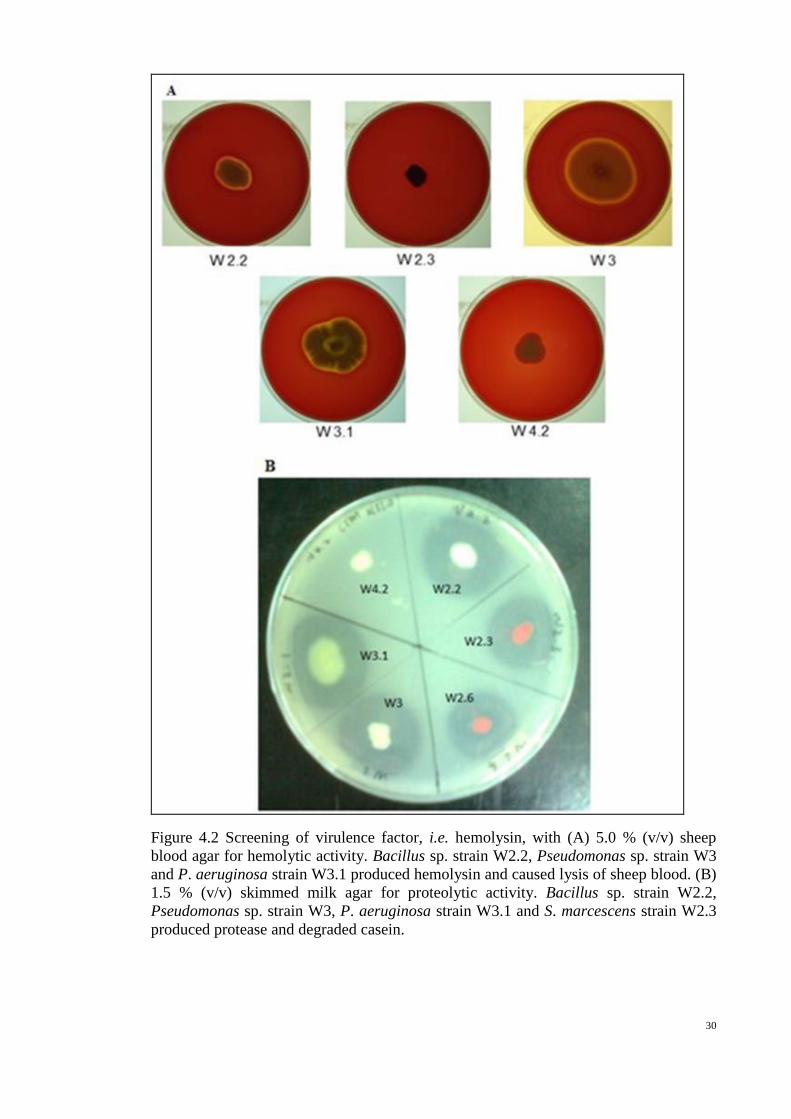
indicating the presence of hemolytic activity. Meanwhile almost all strains isolated from
diseased tilapia fish, except K. oxytoca strain W4.2 produces proteinase. This can be
observed from the formation of clear zone around the colony (Figure 4.2(B)).
4.3 Screening for Short Chain AHL Production by CV026
Production of purple colour pigment, i.e. violacein, was induced by the presence
of exogenous short chain AHLs (McClean et al., 1997). E. carotovora GS101 which
carries carI gene that responsible for the production of OC6HSL was used as the
positive control while the carI defect mutant E. carotovora PNP 22 served as negative
control (McGowan et al., 1995). Among the tested isolates, only P. aeruginosa strain
W3.1 triggered the purple pigment formation of CV026 (Figure 4.3).
30
Figure 4.2 Screening of virulence factor, i.e. hemolysin, with (A) 5.0 % (v/v) sheep
blood agar for hemolytic activity. Bacillus sp. strain W2.2, Pseudomonas sp. strain W3
and P. aeruginosa strain W3.1 produced hemolysin and caused lysis of sheep blood. (B)
1.5 % (v/v) skimmed milk agar for proteolytic activity. Bacillus sp. strain W2.2,
Pseudomonas sp. strain W3, P. aeruginosa strain W3.1 and S. marcescens strain W2.3
produced protease and degraded casein.

31
Figure 4.3 Screening of short chain AHL production with CV026. The AHL produced
by the bacteria will induced the purple pigment formation of CV026. E. carotovora GS
101 is the positive control whereas E. carotovora PNP22. Only P. aeruginosa strain
W3.1 induced the purple pigment formation.
4.4 Thin Layer Chromatography (TLC)
4.4.1 TLC Analysis of AHLs Extracts of the Isolates from Diseased Tilapia Fish
The chromatographed TLC was overlaid with AB agar lawn, seeded with
biosensor A. tumefaciens NTL4 pZLR4 as described in Section 3.2.15. Different types
of AHLs presence were separated according to their polarity. The cleavage of
chromophor of Xgal occurred when A. tumefaciens NTL4 pZLR4 was activated by the
presence of AHLs. Hence, blue pigment formation was observed (Adonizio et al., 2006).
The retardation factor (Rf) were calculated based on the formula:
From the TLC (Figure 4.4), the Rf value of synthetic C12HSL is 0.82 and 0.57
for synthetic OC16HSL. By comparing the chromatograms of AHL extracted from

32
strain W3.1 suggesting the production both OC12HSL and OC16HSL, but the Rf
value cannot be calculated as the blue spots for different types of AHLs were merge.
Technique with higher sensitivity and stronger separation power was required to fully
characterize the AHLs produced by this strain. Strain W3.1 was suspected as
OC12HSL producer instead of C12HSL as the blue spot in the chromatogram carries a
“comet” tail that resembled the characteristic of an AHL with 3oxo substituted site
chain. The Rf value of the AHL produced by S. marcescens strain W2.3 is 0.80. By
comparing the migration distance between the sample and standard it was detected to be
C12HSL.

33
Figure 4.4 Characterization of long chain AHL produced by P. aeruginosa strain W3.1
and S. marcescens strain W2.3 by A. tumefaciens NTL4 pZLR4 with thin layer
chromatography. C12HSL is mark by as “**” while OC16HSL is marked as “*”.P.
aeruginos strain W3.1 produces of both C12HSL and OC16HSL while S. marcescens
strain W2.3 produces C12-HSL.
4.5 LCMS/MS
AHL extracted as described in Section 3.2.14 were subjected to LCMS/MS
analysis in order to compensate the sensitivity of biosensor and also the low separation
resolution of TLC. The presence of different of QS signalling molecules were

34
confirmed by the detection of its precursor ion at certain retention time with the aid of
reference to the standard of each molecule.
4.5.1 Characterization of AHL Molecules Produces by the Isolates from Disease
Tilapia Fish using LCMS/MS
Only P. aeruginosa strain W3.1 produced short chain AHL while both P.
aeruginosa strain W3.1 and S. marcescens strain W2.3 produces long chain AHLs. The
mass-to-charge ration (m/z) of the precursor ion received from the chemist confirmed
the occurrence of C4HSL (m/z: 172), OC12HSL (m/z: 298) and OC16HSL (m/z:
354.26) (Figure 4.5 (A)) from strain W3.1. The precursor ion of C12HSL (m/z: 288.22)
was detected from the AHL extract confirmed the production of C12HSL by S.
marcescens strain W2.3 (Figure 4.5 (B)). In addition to AHLs, P. aeruginosa strain
W3.1 also produces 2-heptyl-3-hydroxy-4(1H)-quinolone (C7PQS) (m/z: 260), 2-
heptyl-4-hydroxyquinolone (C7AQ) (m/z: 244), 2-hydroxy-2-nonyl-4(1H)-quinolone
(C9PQS) (m/z: 288), 2-nonyl-4-hydroxyquinolone (C9AQ) (m/z: 272), 2-heptyl-4-
hydroxyquinoline N-oxide (C7NOxide) (m/z: 260), and 2-nonyl-4-hydroxyquinoline
N-oxide (C9NOxide) (m/z: 288).

35
Figure 4.5 LCMS spectrum (A) P. aeruginosa strain W3.1 which was detected to
produce OC16HSL (m/z 354.26) and (B) S. marcescens strain W2.3 which was
detected to produce C12HSL (m/z 284.2233).
In summary, the AHL molecules producer isolated from diseased tilapia are as
listed in Table 3. AHL molecules producers were P. aeruginosa strain W3.1 and S.
marcescens strain W2.3. P. aeruginosa strain W3.1 produced both short and long chain
AHLs, namely the C4HSL, OC12HSL and OC16HSL. On top of AHLs, P.
aeruginosa W3.1 also produced C7PQS, C7AQ, C9PQS, C9AQ, C7NOxide, and
C9NOxide. S. marcescens strain W2.3 only produced C12HSL.

36
Table 4.1: Summary of QS Molecules Produced by Isolates from Diseased Tilapia Fish
Strains Bacteria Identity Based
on 16S rRNA Gene
QS Signalling Molecules Detected by
LCMS (m/z ration)
W3.1 P. aeruginosa
C4-HSL (m/z:172)
OC12-HSL (m/z:298)
OC16-HSL (m/z: 354.26)
C7-PQS (m/z: 260)
C9-PQS (m/z:288)
C7-AQ (m/z:244)
C9-AQ (m/z: 272)
C7-N-Oxide (m/z: 260)
C9-N-Oxide (m/z: 288)
W2.3 S. marcescens C12-HSL (m/z :284.22)
W2.2 Bacillus sp. Not Detectable
W3 P. aeruginosa Not Detectable
W4.2 K. oxytoca Not Detectable
4.6 Screening for QQ Activity
The degradation of the AHL was determined by the decrease in pigmentation or
loss of pigment after 24 hr of incubation. B. cereus which was reported to be an AHL
degrader served as the positive control for this QQ assay (Chan et al., 2010). The
negative controls used in this assay involved the suspension buffer namely PBS buffer
and E. coli DH5α cells (Taylor et al., 1993).

37
S. marcescens strain W2.3, Bacillus sp. strain W2.2, K. oxytoca strain W4.2, and
P. aeruginosa strain W3.1 did not show any degradation on C4HSL (Figure 4.6(A))
and C6HSL (Figure 4.6 (B)) as no pigment loss was observed. Only Pseudomonas sp.
strain W3 was able to degrade C4HSL as shown in Figure 4.6 (A) where there was a
significant loss of pigment at 24 hr. In addition to C4HSL, C6 HSL was also observed
to be degraded by Pseudomonas sp. strain W3 as the purple pigmentation was reduced
after 24 hr incubation.
Figure 4.6 Residual of (A) C4HSL and (B) C6HSL was incubated with the bacteria for
0 and 24 hr. The AHL degradation was determined by either the decrease of pigment
size or loss of pigment for the residual of 24 hr incubation.
38
CHAPTER 5.0
DISCUSSION
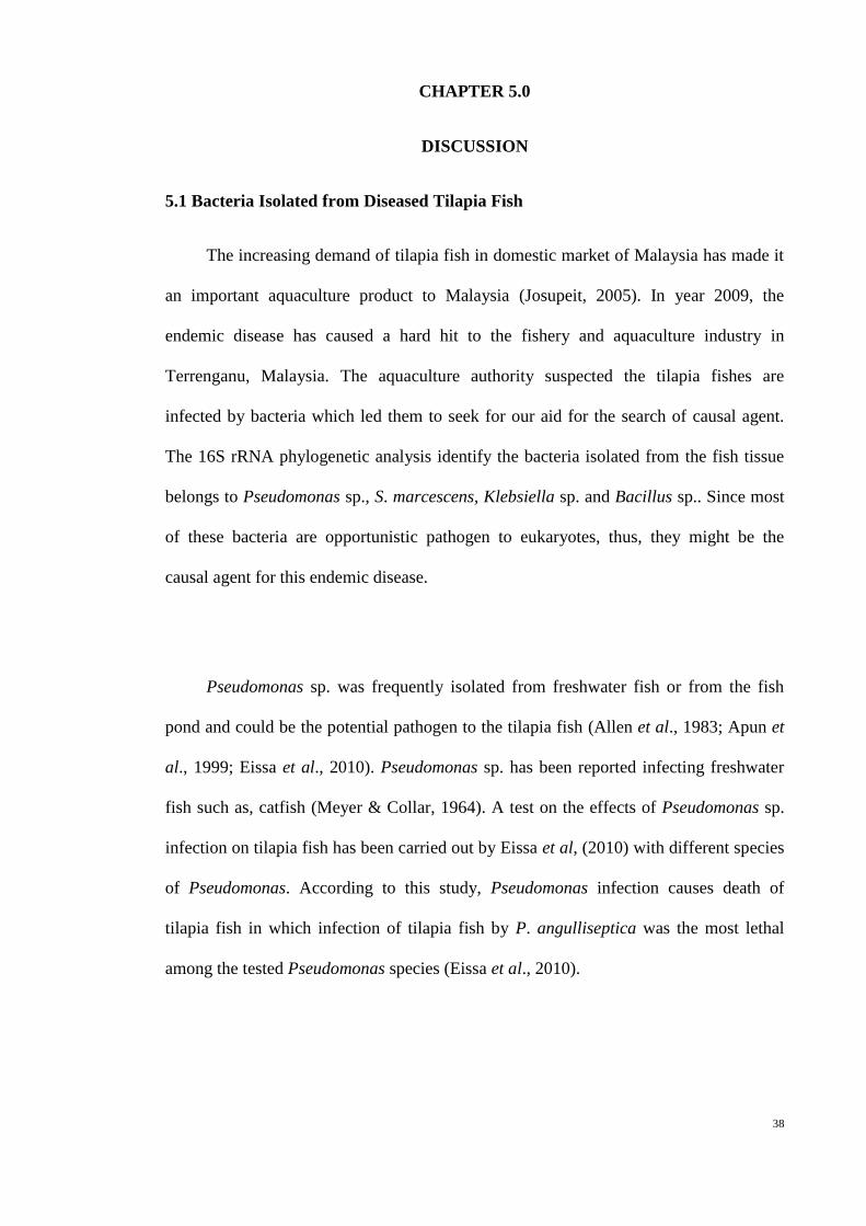
5.1 Bacteria Isolated from Diseased Tilapia Fish
The increasing demand of tilapia fish in domestic market of Malaysia has made it
an important aquaculture product to Malaysia (Josupeit, 2005). In year 2009, the
endemic disease has caused a hard hit to the fishery and aquaculture industry in
Terrenganu, Malaysia. The aquaculture authority suspected the tilapia fishes are
infected by bacteria which led them to seek for our aid for the search of causal agent.
The 16S rRNA phylogenetic analysis identify the bacteria isolated from the fish tissue
belongs to Pseudomonas sp., S. marcescens, Klebsiella sp. and Bacillus sp.. Since most
of these bacteria are opportunistic pathogen to eukaryotes, thus, they might be the
causal agent for this endemic disease.
Pseudomonas sp. was frequently isolated from freshwater fish or from the fish
pond and could be the potential pathogen to the tilapia fish (Allen et al., 1983; Apun et
al., 1999; Eissa et al., 2010). Pseudomonas sp. has been reported infecting freshwater
fish such as, catfish (Meyer & Collar, 1964). A test on the effects of Pseudomonas sp.
infection on tilapia fish has been carried out by Eissa et al, (2010) with different species
of Pseudomonas. According to this study, Pseudomonas infection causes death of
tilapia fish in which infection of tilapia fish by P. angulliseptica was the most lethal
among the tested Pseudomonas species (Eissa et al., 2010).

39
S. marcescens is a normal microflora in various environment including soil and
freshwater (Hejazi & Falkiner, 1997). It is well known as a human opportunistic
pathogen which often associated with nosocomial infection (Hejazi & Falkiner, 1997;
Kurz et al., 2003; Patterson et al., 2002). In addition to human, certain strain of Serratia
has evolved into highly pathogenic strains attacking both freshwater and marine fishes
as well as invertebrate (Aydin et al., 2001; Baya et al., 1992). However, to date, not
much information regarding the S. marcescens infecting of freshwater fish are available.
Bacillus sp. and Klebsiella sp. were naturally occurs in freshwater and were found
in the intestine of freshwater fish (Apun et al., 1999; Austin, 2002; Kunst et al., 1997).
Amylase producing Bacillus which aids in starch degradation has been isolated from the
intestine of freshwater fish (Apun et al., 1999; Sugita et al., 1997). This is important for
freshwater fish such as carps and tilapia where their foods are high in starch content
(Sugita et al., 1997). However, there is also a report stating Bacillus could serve as the
causal agent of freshwater fish septicemia condition in fish farm which leads to the
death of the fishes (Oladosu et al., 1994).
Unexpectedly, Streptococcus which is the common freshwater fish disease causal
agent was not found from the death infected fish. Streptococcus has causes several
outbreaks to the tilapia fish farm around the world possess high morbidity and mortality
rate of the fish (Sun et al., 2007; Weinstein et al., 1997). Streptococcus-infected tilapia
fish turns lethargic, swims erratically, suffer dorsal rigidity, and eventually die if left
without treatment (Weinstein et al., 1997). This infection is one of the most serious
threats to tilapia industry (Pretto‐Giordano et al., 2009). The effect of infected fish

40
towards public health gained more concern when the disease was passed from fish to
human who had physical contact with the fish (Sun et al., 2007; Yagoub, 2009).
5.2 Virulence Factors and AHLs Detection
All isolates were screened for exoprotease and hemolysin production. Titball et al.,
(1985), founds the relations between caseinase and protease whereby the inhibition of
caseinase from fish pathogen, Aeromonas salmonicida contributes to the suppression of
hemolysin production. Proteolytic enzyme production is directly correlated with
Pseudomonas pathogenicity as the concentration of the protease presence affects its
invasiveness (Pavlovskis & Wretlind, 1979). In this assays, S. marcescens strain W2.3
showed exoprotease production while P. aeruginosa strain W3.1, Pseudomonas sp.
strain W3, Bacillus sp. strain W2.2 were able to produce both exoprotease and
hemolysin. The production of hemolysin and exoproteases suggested that these bacteria
acquire nutrients from its host cells (Chan, 2009).
Since QS plays an important role in regulating the expression of virulence genes
in certain bacteria, all isolates were subjected to the screening for the AHL and
virulence factor production. In the studies, with the exception of Bacillus strain W2.2,
all isolates are taxonomically classified in to the phylum of Proteobacteria. The major
QS signalling molecules produced and utilized by Proteobacteria are AHLs (Case et al.,
2008), therefore this study focused on the AHL characterization.

41
As Pseudomonas sp., S.marcescens and Klebsiella sp. are mainly short chain
AHLs producer, a rapid detection of short chain AHL was hence conducted using
biosensor CV026 (Van Delden & Iglewski, 1998; Van Houdt et al., 2007; Wilder et al.,
2011). Only P. aeruginosa strain W3.1 triggered the purple pigment formation of
CV026, indicating the production of short AHL. On the other hand, long chain AHL
was detected from the spent supernatant of P. aeruginosa strain W3.1 via TLC with the
aid of biosensor A. tumefaciens NTL4 pZLR. However TLC failed is not an accurate
method to characterize the long chain AHL presence in the spent supernatant of P.
aeruginosa strain W3.1.
Similar to the previously reported finding, C4HSL and OC12HSL were found in
the spent supernatant extract of P. aeruginosa strain W3.1, as confirmed by LCMS/MS
(Diggle et al., 2003; Zhu et al., 2004). These AHLs were shown to regulate the
production of virulence factors, including hemolysin and proteases (elastase and
alkaline protease) of P. aeruginosa (Zhu et al., 2002). Aside from C4HSL and
OC12HSL, OC16HSL was also detected from the spent supernatant of P. aeruginosa
strain W3.1. This is the first report of P. aeruginosa producing OC16HSL. To my
knowledge, this is the AHL with the longest side chain synthesized by P. aeruginosa,
but further studies are required in order to further understand the function of OC16HSL
produced by this P. aeruginosa.
Although AHL is the most studied QS signalling molecules, the function and
production of long chain AHLs such as NhexadecanoylLhomoserine lactone
(C16HSL) are poorly understood. Most of the long chain AHLs was identified in

42
αProteobacteria. For instance, Rhodobacter capsulates and Paracoccus denitrificans
produces C16HSL, Acidithiobacillus ferroxidans produces
3hydroxyhexadecanoylLhomoserine lactone (OHC16HSL) and Sinorhizobium
meliloti produces OC16HSL and C16HSL (Farah et al., 2005; Marketon et al., 2002;
Rivas et al., 2005; Schaefer et al, 2002). In R. capsulates, C16HSL is responsible in
activating the production of gene transfer between Rhodobacter and its host (Schaefer et
al., 2002). Meanwhile, QS of Sinorhizobium meliloti plays a role in controlling its
symbiosis, and its long chain AHL might involve in nodulation efficiency i.e. the pink
nodule formation (Marketon et al., 2002).
In addition to AHLs, PQS, HHQ and its Noxide derivative were detected from
spent supernatant of P. aeruginosa strain W3.1. These three compounds are interrelated
where HHQ is the intermediate of PQS while Noxide is synthesed via the similar AHQ
synthesis pathway for HHQ synthesis (Déziel et al., 2004; Machan et al., 1992). Both
PQS and HHQ are QS signaling molecules that regulate secondary metabolites
production, virulence factor expression, and extracellular enzyme production (Dubern &
Diggle, 2008; Gallagher et al., 2002; Pesci et al., 1999). On the other hand, the
Noxides PQS produced by P. aeruginosa is not classified as QS signalling molecule. It
is an antibacterial compound against Gram positive bacteria. According MaChan et al,
(1992), the production of PQS Noxide by P. aeruginosa inhibits the growth of
Staphylococcus aureus and this result explains the absences of S. aureus in the sputum
of cystic fibrosis patient.

43
Production of virulence factors namely swarming ability, biofilm formation,
protease and nuclease production of Serratia are regulated by its AHLbased QS system
(Morohoshi et al., 2007; Wei & Lai, 2006; Wei et al., 2006; Williams, 1973). However,
instead of synthesizing C4 to C8HSL as previous reported, TLC and LCMS/MS results
showed that S. marcescens W2.3 produced C12HSL (Horng et al., 2002; Thomson et
al., 2002; Van Houdt et al., 2007). The utilization of C12HSL in regulating the
virulence factor expression has been reported in P. aeruginosa (Erickson et al., 2002).
However, more work is required to investigate the relationship between the C12HSL
produce by S. marcescens strain W2.3 and its regulation on pathogenicity.
5.3 QQ Activity
Bacteria degrade AHLs in the environment in order to acquire carbon and
nitrogen for survival (Chan et al., 2010; Hong et al., 2012). Besides, it also serves as
advantage towards survival in the competitive environment (Park et al., 2008). Since
members of Bacillus, Pseudomonas, and Klebsiella genera have been reported to be
degrade AHLs, hence in this study, C4HSL and C6HSL were used as the substrate of
AHL degradation assay.
P. aeruginosa PAO1, a human pathogen harbors both QS and QQ genes (Huang
et al., 2006; Latifi et al., 1995; Sio et al., 2006). It utilizes acylase as QQ enzyme
selectively cleaving the acyl side chain of certain AHL (Sio et al., 2006). In this study,
QQ activity was found in Pseudomonas sp. strain W3 but not P. aeruginosa strain W3.1.
The absence of QQ activity in P. aeruginosa strain W3.1 may be due to the substrates
used in the study. Previous studies showed that acylase of P. aeruginosa degrades

44
C7HSL and AHL with longer acyl side chain (Hong et al., 2012; Huang et al., 2006;
Sio et al., 2006). In this study, C4HSL and C6HSL were used as the substrates for
degradation, Hence, the selectivity of the QQ enzyme might be explained in this study.
Pseudomonas sp. strain W3 did not produce AHLs but it degraded both C4HSL
and C6HSL. It is speculated that this strain is a social cheater where it used the AHL
produced by other bacteria to coordinates its gene expression, thus, saving the cost of
producing QS signalling molecules (Wilder et al., 2011). Despite Pseudomonas strain
W3 and P. aeruginosa W3.1 are closely related, the differences of QS and QQ activities
exerted by both strains has suggested distinctive gene regulation in these two closely
related species.
Bacillus sp. degrades AHLs via the production of lactonase enzyme, interrupting
the QS by hydrolysing the lactone ring of AHL (Dong & Zhang, 2005; Dong et al.,
2002). B. cereus known to quench OC6HSL and N(3oxooctanoyl)Lhomoserine
lactone (OC8HSL) was used as the positive control for this study (Chan et al., 2010).
Contradictory to most of the reports, Bacillus sp. strain W2.2 did not shown to degrade
both C4HSL and C6HSL. This also applies to K. oxytoca strain W4.2 which does not
possesses any QQ activity on the tested AHLs, this finding is distinguishable from the
Klebsiella sp. isolated from ginger’s rhizosphere that was reported to degrade various
AHL via lactonase activity (Chan et al., 2011).
45
CHAPTER 6.0
CONCLUSION
As a summary, five bacteria were isolated from this study, namely P. aeruginosa
strain W3.1, Pseudomonas sp. strain W3, S. marcescens strain W2.3, Bacillus sp. strain
W2.2 and K. oxytoca strain W4.2. Except K. oxytoca strain W4.2, all isolates produced
either one or both virulence factors tested namely proteinase and hemolysin. This
suggests that they could be the causal agent of the infectious disease.
Aside from C4HSL, OC12HSL and PQS, an unexpected long chain AHL,
namely OC16HSL was detected from P. aeruginosa strain W3.1. Contradictory to the
reported findings, an unusual long chain AHL i.e. C12HSL also detected from spent
supernatant of S. marcescens. The detection of an unexpectedly long chain AHL from
these well-studied bacteria i.e. P. aeruginosa and S. marcescens suggested that bacteria
possess unknown QS mechanism. Therefore, more studies are required in order to
further characterize and understand the mechanism of these unusual long chainAHL in
these bacteria. Besides QS bacteria, Pseudomonas sp. strain W3 also degraded both
C4HSL and C6HSL.
For future work, it is suggested that whole genome sequencing can be used to
study the QS and QQ genes and the genes regulated by these systems.
46
REFERENCES
Adonizio, A.L., Downum, K., Bennett, B.C., & Mathee, K. (2006). Anti-quorum
sensing activity of medicinal plants in southern Florida. Journal of
Ethnopharmacology, 105(3), 427-435.
Aendekerk, S., Diggle, S.P., Song, Z., Høiby, N., Cornelis, P., Williams, P., & Cámara,
M. (2005). The MexGHI-OpmD multidrug efflux pump controls growth,
antibiotic susceptibility and virulence in Pseudomonas aeruginosa via 4-
quinolone-dependent cell-to-cell communication. Microbiology, 151(4), 1113-
1125.
Allen, DA., Austin, B., & Colwell, RR. (1983). Numerical taxonomy of bacterial
isolates associated with a freshwater fishery. Journal of General Microbiology,
129(7), 2043-2062.
Apun, K., Yusof, A.M., & Jugang, K. (1999). Distribution of bacteria in tropical
freshwater fish and ponds. International Journal of Environmental Health
Research, 9(4), 285-292.
Atkinson, S., & Williams, P. (2009). Quorum sensing and social networking in the
microbial world. Journal of The Royal Society Interface, 6(40), 959-978.
Austin, B. (2002). The bacterial microflora of fish. The Scientific World Journal, 2,
558-572.
Aydin, S., Erman, Z., & Bilgin, O.C. (2001). Investigations of Serratia liquefaciens
infection in rainbow trout (Oncorhynchus mykiss Walbaum). Turkish Journal of
Veterinary and Animal Sciences, 25(5), 643-650.
Bassler, B.L. (2002). Small talk: cell-to-cell communication in bacteria. Cell, 109(4),
421-424.
Baya, A.M., Toranzo, A.E., Lupiani, B., Santos, Y., & Hetrick, F.M. (1992). Serratia
marcescens: a potential pathogen for fish. Journal of Fish Diseases, 15(1), 15-
26.
Bertani, I., & Venturi, V. (2004). Regulation of the N-acyl homoserine lactone-
dependent quorum-sensing system in rhizosphere Pseudomonas putida WCS358
and cross-talk with the stationary-phase RpoS sigma factor and the global
regulator GacA. Applied and Environmental Microbiology, 70(9), 5493-5502.
Bostock, J., McAndrew, B., Richards, R., Jauncey, K., Telfer, T., Lorenzen, K., . . .
Gatward, I. (2010). Aquaculture: global status and trends. Philosophical
Transactions of the Royal Society B: Biological Sciences, 365(1554), 2897-2912.
Budsberg, K.J., Wimpee, C.F., & Braddock, J.F. (2003). Isolation and identification of
photobacterium phosphoreum from an unexpected niche: migrating salmon.
Applied and Environmental Microbiology, 69(11), 6938-6942. doi:
10.1128/aem.69.11.6938-6942.2003

47
Burrell, P.C., Keller, J., & Blackall, L.L. (1998). Microbiology of a nitrite-oxidizing
bioreactor. Applied and Environmental Microbiology, 64(5), 1878-1883.
Callahan, S.M., & Dunlap, P.V. (2000). LuxR and acyl-homoserine lactone controlled
non lux genes define a quorum sensing regulon in Vibrio fischeri. Journal of
Bacteriology, 182(10), 2811-2822.
Case, R.J., Labbate, M., & Kjelleberg, S. (2008). AHL-driven quorum-sensing circuits:
their frequency and function among the Proteobacteria. The ISME Journal, 2(4),
345-349.
Chan, K.G., Wong, C.S., Yin, W.F., Sam, C.K., & Koh, C.L. (2010). Rapid degradation
of N-3-oxo-acylhomoserine lactones by a Bacillus cereus isolate from Malaysian
rainforest soil. Antonie Van Leeuwenhoek, 98(3), 299-305. doi: 10.1007/s10482-
010-9438-0
Chan, K.G. (2009). Exochelin production in Mycobacterium neoaurum. International
Journal of Molecular Sciences, 10(1), 345-353.
Chan, K.G., Atkinson, S., Mathee, K., Sam, C.K., Chhabra, S.R., Cámara, M., . . .
Williams, P. (2011). Characterization of N-acylhomoserine lactone-degrading
bacteria associated with the Zingiber officinale (ginger) rhizosphere: Co-
existence of quorum quenching and quorum sensing in Acinetobacter and
Burkholderia. BMC Microbiology, 11(1), 51.
Chang, C.Y., Koh, C.L., Sam, C.K., Chan, X.Y., Yin, W.F., & Chan, K.G. (2012).
Unusual long-chain N-acyl homoserine lactone production by and presence of
quorum quenching activity in bacterial isolates from diseased tilapia fish. PloS
One, 7(8), e44034.
Chernin, L.S., Winson, M.K., Thompson, J.M., Haran, S., Bycroft, B.W., Chet, I., . . .
Stewart, G.S.A.B. (1998). Chitinolytic activity in Chromobacterium violaceum:
substrate analysis and regulation by quorum sensing. Journal of Bacteriology,
180(17), 4435-4441.
Chong, T.M., Koh, C.L., Sam, C.K., Choo, Y.M., Yin, W.F., & Chan, K.G. (2012).
Characterization of quorum sensing and quorum quenching soil bacteria isolated
from Malaysian tropical montane forest. Sensors (Basel), 12(4), 4846-4859. doi:
10.3390/s120404846
Chong, Y.M., Yin, W.F., Ho, C.Y., Mustafa, M.R., Hadi, A.H.A, Awang, K., . . . Chan,
K.G. (2011). Malabaricone C from Myristica cinnamomea exhibits anti-quorum
sensing activity. Journal of Natural Products, 74(10), 2261-2264.
Cvitkovitch, D.G., Li, Y.H., & Ellen, R.P. (2003). Quorum sensing and biofilm
formation in Streptococcal infections. Journal of Clinical Investigation, 112(11),
1626-1632.
Czajkowski, R., & Jafra, S. (2009). Quenching of acyl-homoserine lactone-dependent
quorum sensing by enzymatic disruption of signal molecules. Acta Biochim Pol,
56(1), 1-16.
48
De Kievit, T.R., Gillis, R., Marx, S., Brown, C., & Iglewski, B.H. (2001). Quorum-
sensing genes in Pseudomonas aeruginosa biofilms: their role and expression
patterns. Applied and Environmental Microbiology, 67(4), 1865-1873.
De Kievit, T.R., & Iglewski, B.H. (2000). Bacterial quorum sensing in pathogenic
relationships. Infection and Immunity, 68(9), 4839-4849.
Defoirdt, T., Boon, N., Sorgeloos, P., Verstraete, W., & Bossier, P. (2007). Quorum
sensing and quorum quenching in Vibrio harveyi: lessons learned from in vivo
work. The ISME Journal, 2(1), 19-26.
Déziel, E., Gopalan, S., Tampakaki, A.P., Lépine, F., Padfield, K.E., Saucier, M., . . .
Rahme, L.G. (2004). The contribution of MvfR to Pseudomonas aeruginosa
pathogenesis and quorum sensing circuitry regulation: multiple quorum
sensing‐regulated genes are modulated without affecting lasRI, rhlRI or the
production of N‐acyl‐l‐homoserine lactones. Molecular Microbiology, 55(4),
998-1014.
Déziel, E., Lépine, F., Milot, S., He, J.X., Mindrinos, M.N., Tompkins, R.G., & Rahme,
L.G. (2004). Analysis of Pseudomonas aeruginosa 4-hydroxy-2-alkylquinolines
(HAQs) reveals a role for 4-hydroxy-2-heptylquinoline in cell-to-cell
communication. Proceedings of the National Academy of Sciences of the United
States of America, 101(5), 1339-1344.
Diggle, S.P., Winzer, K., Chhabra, S.R., Worrall, K.E., Cámara, M., & Williams, P.
(2003). The Pseudomonas aeruginosa quinolone signal molecule overcomes the
cell density‐dependency of the quorum sensing hierarchy, regulates
rhl‐dependent genes at the onset of stationary phase and can be produced in the
absence of LasR. Molecular Microbiology, 50(1), 29-43.
Dong, Y.H., & Zhang, L.H. (2005). Quorum sensing and quorum-quenching enzymes.
Journal of Microbiology, 43(5), 101-109.
Dong, Y.H., Gusti, A.R., Zhang, Q., Xu, J.L., & Zhang, L.H.. (2002). Identification of
quorum-quenching N-acyl homoserine lactonases from Bacillus species. Applied
and Environmental Microbiology, 68(4), 1754-1759.
Dubern, J.F., & Diggle, S.P. (2008). Quorum sensing by 2-alkyl-4-quinolones in
Pseudomonas aeruginosa and other bacterial species. Molecular BioSystem, 4(9),
882-888.
Dunlap, P.V. (1999). Quorum regulation of luminescence in Vibrio fischeri. Journal of
Molecular Microbiology and Biotechnology, 1(1), 5-12.
Eissa, N.M.E., El-Ghiet, E.A., Shaheen, A.A., & Abbass, A. (2010). Characterization of
Pseudomonas species isolated from tilapia" Oreochromis niloticus" in Qaroun
and Wadi-El-Rayan Lakes, Egypt. Global Veterinaria Journal, 5, 116-121.
El-Sayed, A.F.M. (2006). Tilapia culture in salt water: environmental requirements,
nutritional implications and economic potentials. VIII Simposium Internacional

49
de Nutrición Acuícola. Universidad Autónoma de Nuevo León. Anais...
Monterrey. Monterrey.
Erickson, D. L, Endersby, R., Kirkham, A., Stuber, K., Vollman, D.D., Rabin, H.R., . . .
Storey, D.G. (2002). Pseudomonas aeruginosa quorum-sensing systems may
control virulence factor expression in the lungs of patients with cystic fibrosis.
Infection and Immunity, 70(4), 1783-1790.
Farah, C., Vera, M., Morin, D., Haras, D., Jerez, C.A., & Guiliani, N. (2005). Evidence
for a functional quorum-sensing type AI-1 system in the extremophilic
bacterium Acidithiobacillus ferrooxidans. Applied and Environmental
Microbiology, 71(11), 7033-7040.
Farrand, S.K., Qin, Y., & Oger, P. (2002). Quorum-sensing system of Agrobacterium
plasmids: Analysis and utility. Methods in Enzymology, 358, 452-484.
Fitzsimmons, K. (2000). Future trends of tilapia aquaculture in the Americas. Tilapia
Aquaculture in the Americas, 2, 252-264.
Fletcher, M.P., Diggle, S.P., Cámara, M., & Williams, P. (2007). Biosensor-based
assays for PQS, HHQ and related 2-alkyl-4-quinolone quorum sensing signal
molecules. Nature Protocols, 2(5), 1254-1262.
Fuqua, C., & Greenberg, E.P. (2002). Listening in on bacteria: acyl-homoserine lactone
signalling. Nature Reviews: Molecular Cell Biology, 3(9), 685-695.
Fuqua, W.C., Winans, S.C., & Greenberg, E.P. (1994). Quorum sensing in bacteria: the
LuxR-LuxI family of cell density-responsive transcriptional regulators. Journal
of Bacteriology, 176(2), 269.
Gallagher, L.A., McKnight, S.L., Kuznetsova, M.S., Pesci, E.C., & Manoil, C. (2002).
Functions required for extracellular quinolone signalling by Pseudomonas
aeruginosa. Journal of Bacteriology, 184(23), 6472-6480.
Geerlings, S.E., Brouwer, E.C., Gaastra, W., Stolk, R., Diepersloot, R.J.A., &
Hoepelman, A.I.M. (2001). Virulence factors of Escherichia coli isolated from
urine of diabetic women with asymptomatic bacteriuria: correlation with clinical
characteristics. Antonie Van Leeuwenhoek, 80(2), 119-127.
Gonzales, J.M., & Brown, P.B. (2006). Nile tilapia Oreochromis niloticus as a food
source in advanced life support systems: Initial considerations. Advances in
Space Research, 38(6), 1132-1137.
Hammer, B.K., & Bassler, B.L. (2003). Quorum sensing controls biofilm formation in
Vibrio cholerae. Molecular Microbiology, 50(1), 101-104.
Haudecoeur, E., Tannières, M., Cirou, A., Raffoux, A., Dessaux, Y., & Faure, D. (2009).
Different regulation and roles of lactonases AiiB and AttM in Agrobacterium
tumefaciens C58. Molecular Plant-Microbe Interactions, 22(5), 529-537.

50
Häussler, S., & Becker, T. (2008). The Pseudomonas quinolone signal (PQS) balances
life and death in Pseudomonas aeruginosa populations. PLoS Pathogens, 4(9),
e1000166.
Hejazi, A, & Falkiner, FR. (1997). Serratia marcescens. Journal of Medical
Microbiology, 46(11), 903-912.
Hong, K.W., Koh, C.L., Sam, C.K., Yin, W.F., & Chan, K.G. (2012). Quorum
quenching revisited—from signal decays to signalling confusion. Sensors, 12(4),
4661-4696.
Hong, K.W., Koh, C.L., Sam, C.K., Yin, W.F., & Chan, K.G. (2012). Complete
genome sequence of Burkholderia sp. strain GG4, a Betaproteobacterium that
reduces 3-Oxo-N-acylhomoserine lactones and produces different N-
acylhomoserine lactones. Journal of bacteriology, 194(22), 6317-6317.
Horng, Y.T., Deng, S.C., Daykin, M., Soo, P.C., Wei, J.R., Luh, K.T., . . . Williams, P.
(2002). The LuxR family protein SpnR functions as a negative regulator of
N‐acylhomoserine lactone‐dependent quorum sensing in Serratia marcescens.
Molecular M icrobiology, 45(6), 1655-1671.
Huang, J.J., Petersen, A., Whiteley, M., & Leadbetter, J.R. (2006). Identification of
QuiP, the product of gene PA1032, as the second acyl-homoserine lactone
acylase of Pseudomonas aeruginosa PAO1. Applied and Environmental
Microbiology, 72(2), 1190-1197.
Huber, B., Riedel, K., Hentzer, M., Heydorn, A., Gotschlich, A., Givskov, M., . . . Eberl,
L. (2001). The cep quorum-sensing system of Burkholderia cepacia H111
controls biofilm formation and swarming motility. Microbiology, 147(9), 2517-
2528.
Jayaraman, A., & Wood, T.K. (2008). Bacterial quorum sensing: signals, circuits, and
implications for biofilms and disease. Annual Reviel Biomedical Engineering, 10,
145-167.
Joint, I. (2006). Bacterial conversations: talking, listening and eavesdropping. A NERC
Discussion Meeting held at the Royal Society on 7 December 2005. Journal of
The Royal Society Interface, 3(8), 459-463.
Josupeit, Helga. (2005). World market of tilapia: FAO.
Juhas, M., Eberl, L., & Tümmler, B. (2005). Quorum sensing: the power of cooperation
in the world of Pseudomonas. Environmental Microbiology, 7(4), 459-471.
Juhas, M., Wiehlmann, L., Huber, B., Jordan, D., Lauber, J., Salunkhe, P., . . . Eberl,
Leo. (2004). Global regulation of quorum sensing and virulence by VqsR in
Pseudomonas aeruginosa. Microbiology, 150(4), 831-841.
Kim, K., Kim, Y.U., Koh, B.H., Hwang, S.S., Kim, S.H., Lépine, F., . . . Lee, G.R.
(2010). HHQ and PQS, two Pseudomonas aeruginosa quorum‐sensing

51
molecules, down‐regulate the innate immune responses through the nuclear
factor‐κB pathway. Immunology, 129(4), 578-588.
Köhler, T., Curty, L.K., Barja, F., Van Delden, C., & Pechère, J.C.. (2000). Swarming
of Pseudomonas aeruginosa is dependent on cell-to-cell signalling and requires
flagella and pili. Journal of Bacteriology, 182(21), 5990-5996.
Krishnan, T., Yin, W.F., & Chan, K.G. (2012). Inhibition of quorum sensing-controlled
virulence factor production in Pseudomonas aeruginosa PAO1 by Ayurveda
Spice Clove (Syzygium Aromaticum) bud extract. Sensors, 12(4), 4016-4030.
Kunst, F., Ogasawara, N., Moszer, I., Albertini, A.M., Alloni, G.O., Azevedo, V., . . .
Borchert, Sea. (1997). The complete genome sequence of the gram-positive
bacterium Bacillus subtilis. Nature, 390(6657), 249-256.
Kurz, C.L., Chauvet, S., Andrès, E., Aurouze, M., Vallet, I., Michel, G.P.F., . . . De B.S.
(2003). Virulence factors of the human opportunistic pathogen Serratia
marcescens identified by in vivo screening. The EMBO Journal, 22(7), 1451.
Labbate, M., Queck, S.Y., Koh, K.S., Rice, S.A., Givskov, M., & Kjelleberg, S. (2004).
Quorum sensing-controlled biofilm development in Serratia liquefaciens MG1.
Journal of Bacteriology, 186(3), 692-698.
Latifi, A., Winson, M.K., Foglino, M., Bycroft, B.W., Stewart, G.S., Lazdunski, A., &
Williams, P. (1995). Multiple homologues of LuxR and LuxI control expression
of virulence determinants and secondary metabolites through quorum sensing in
Pseudomonas aeruginosa PAO1. Molecular microbiology, 17(2), 333-343.
Leadbetter, J.R., & Greenberg, EP. (2000). Metabolism of acyl-homoserine lactone
quorum-sensing signals by Variovorax paradoxus. Journal of Bacteriology,
182(24), 6921-6926.
Lee, B.Y., Lee, W.J., Streelman, J.T., Carleton, K.L, Howe, A.E, Hulata, G., . . . Kocher,
T.D. (2005). A second-generation genetic linkage map of tilapia (Oreochromis
spp.). Genetics, 170(1), 237-244.
Lindum, P.W, Anthoni, U., Christophersen, C., Eberl, L., Molin, S., & Givskov, M.
(1998). N-Acyl-L-homoserine lactone autoinducers control production of an
extracellular lipopeptide biosurfactant required for swarming motility of
Serratia liquefaciens MG1. Journal of Bacteriology, 180(23), 6384-6388.
Livermore, D.M. (2002). Multiple mechanisms of antimicrobial resistance in
Pseudomonas aeruginosa: our worst nightmare? Clinical Infectious Diseases,
34(5), 634-640.
Llamas, I., Keshavan, N., & González, J.E. (2004). Use of Sinorhizobium meliloti as an
indicator for specific detection of long-chain N-acyl homoserine lactones.
Applied and Environmental Microbiology, 70(6), 3715-3723.

52
Lyon, G.J., & Novick, R.P. (2004). Peptide signalling in Staphylococcus aureus and
other Gram-positive bacteria. Peptides, 25(9), 1389-1403.
Machan, Z.A., Taylor, G.W., Pitt, T.L., Cole, P.J., & Wilson, R. (1992). 2-Heptyl-4-
hydroxyquinoline N-oxide, an antistaphylococcal agent produced by
Pseudomonas aeruginosa. Journal of Antimicrobial Chemotherapy, 30(5), 615-
623.
Marketon, M.M., Gronquist, M.R., Eberhard, A., & González, J.E. (2002).
Characterization of the Sinorhizobium meliloti sinR/sinI locus and the
production of novel N-acyl homoserine lactones. Journal of Bacteriology,
184(20), 5686-5695.
McClean, K.H., Winson, M.K., Fish, L., Taylor, A., Chhabra, S.R., Camara, M., . . .
Bycroft, B.W. (1997). Quorum sensing and Chromobacterium violaceum:
exploitation of violacein production and inhibition for the detection of N-
acylhomoserine lactones. Microbiology, 143(12), 3703-3711.
McGowan, S., Sebaihia, M., Jones, S., Yu, B., Bainton, N., Chan, PF, . . . Salmond,
GPC. (1995). Carbapenem antibiotic production in Erwinia carotovora is
regulated by carR, a homologue of the LuxR transcriptional activator.
Microbiology, 141(3), 541-550.
McGrath, S., Wade, D.S., & Pesci, E.C. (2006). Dueling quorum sensing systems in
Pseudomonas aeruginosa control the production of the Pseudomonas quinolone
signal (PQS). FEMS Microbiol Lett, 230(1), 27-34.
Meyer, F.P., & Collar, J.D., (1964). Description and treatment of a Pseudomonas
infection in white catfish. Applied Microbiology, 12(3), 201-203.
Morohoshi, T., Shiono, T., Takidouchi, K., Kato, M., Kato, N., Kato, J., & Ikeda, T.
(2007). Inhibition of quorum sensing in Serratia marcescens AS-1 by synthetic
analogs of N-acylhomoserine lactone. Applied and Environmental Microbiology,
73(20), 6339-6344.
O'Grady, E.P., Viteri, D.F., & Sokol, P.A. (2012). A unique regulator contributes to
quorum sensing and virulence in Burkholderia cenocepacia. PloS One, 7(5),
e37611.
Oladosu, G.A., Ayinla, O.A., & Ajiboye, M.O. (1994). Isolation and pathogenicity of a
Bacillus sp. associated with a septicaemic condition in some tropical freshwater
fish species. Journal of Applied Ichthyology, 10(1), 69-72.
Ortori, C.A., Dubern, J.F., Chhabra, S.R., Cámara, M., Hardie, K., Williams, P., &
Barrett, D.A. (2011). Simultaneous quantitative profiling of N-acyl-L-
homoserine lactone and 2-alkyl-4 (1H)-quinolone families of quorum-sensing
signalling molecules using LC-MS/MS. Analytical and Bioanalytical Chemistry,
399(2), 839-850.

53
Park, S.J., Park, S.Y., Ryu, C.M., Park, S.H., & Lee, J.K. (2008). The role of aiiA, a
quorum-quenching enzyme from Bacillus thuringiensis, on the rhizosphere
competence. Journal Microbiol Biotechnology,18(9), 1518-1521.
Park, S.Y., Lee, S.J., Oh, T.K., Oh, J.W., Koo, B.T., Yum, D.Y., & Lee, J.K. (2003).
AhlD, an N-acylhomoserine lactonase in Arthrobacter sp., and predicted
homologues in other bacteria. Microbiology, 149(6), 1541-1550.
Passador, L., Cook, J.M., Gambello, M.J., Rust, L., & Iglewski, B.H. (1993).
Expression of Pseudomonas aeruginosa virulence genes requires cell-to-cell
communication. Science (New York, NY), 260(5111), 1127.
Patterson, K.L., Porter, J.W., Ritchie, K.B., Polson, S.W., Mueller, E., Peters, E.C., . . .
Smith, G.W. (2002). The etiology of white pox, a lethal disease of the Caribbean
elkhorn coral, Acropora palmata. Proceedings of the National Academy of
Sciences, 99(13), 8725-8730.
Pavlovskis, O.R., & Wretlind, B. (1979). Assessment of protease (elastase) as a
Pseudomonas aeruginosa virulence factor in experimental mouse burn infection.
Infection and Immunity, 24(1), 181-187.
Pearson, J.P., Pesci, E.C., & Iglewski, B.H. (1997). Roles of Pseudomonas aeruginosa
las and rhl quorum-sensing systems in control of elastase and rhamnolipid
biosynthesis genes. Journal of Bacteriology, 179(18), 5756-5767.
Pesci, E.C, Milbank, J.B, Pearson, J.P., McKnight, S., Kende, A.S., Greenberg, E.P., &
Iglewski, B.H. (1999). Quinolone signalling in the cell-to-cell communication
system of Pseudomonas aeruginosa. Proceedings of the National Academy of
Sciences, 96(20), 11229-11234.
Pesci, E.C., Pearson, J.P., Seed, P.C., & Iglewski, B.H. (1997). Regulation of las and rhl
quorum sensing in Pseudomonas aeruginosa. Journal of Bacteriology, 179(10),
3127-3132.
Põllumaa, L., Alamäe, T., & Mäe, A. (2012). Quorum Sensing and Expression of
Virulence in Pectobacteria. Sensors, 12(3), 3327-3349.
Pretto‐Giordano, L.G., Müller, E.E., Klesius, P., & Da Silva, V. (2009). Efficacy of an
experimentally inactivated Streptococcus agalactiae vaccine in Nile tilapia
(Oreochromis niloticus) reared in Brazil. Aquaculture research, 41(10), 1539-
1544.
Reading, N.C., & Sperandio, V. (2005). Quorum sensing: the many languages of
bacteria. FEMS Microbiology Letter, 254(1), 1-11.
Rice, S.A., Koh, K.S., Queck, S.Y., Labbate, M., Lam, K.W., & Kjelleberg, S. (2005).
Biofilm formation and sloughing in Serratia marcescens are controlled by
quorum sensing and nutrient cues. Journal of Bacteriology, 187(10), 3477-3485.

54
Rivas, M., Seeger, M., Holmes, D.S., & Jedlicki, E. (2005). A Lux-like quorum sensing
system in the extreme acidophile, Acidithiobacillus ferrooxidans. Biological
Research, 38(2/3), 283.
Russell, D.W., & Sambrook, J. (2001). Molecular cloning. A laboratory manual.
Schaefer, A.L., Taylor, T.A., Beatty, J.T., & Greenberg, E.P. (2002). Long-chain acyl-
homoserine lactone quorum-sensing regulation of Rhodobacter capsulatus gene
transfer agent production. Journal of Bacteriology, 184(23), 6515-6521.
Sio, C.F., Otten, L.G., Cool, R.H., Diggle, S.P., Braun, P.G., Bos, R., . . . Quax, W.J..
(2006). Quorum quenching by an N-acyl-homoserine lactone acylase from
Pseudomonas aeruginosa PAO1. Infection and Immunity, 74(3), 1673-1682.
Smith, R.S., Harris, S.G., Phipps, R., & Iglewski, B. (2002). The Pseudomonas
aeruginosa quorum-sensing molecule N-(3-oxododecanoyl) homoserine lactone
contributes to virulence and induces inflammation in vivo. Journal of
Bacteriology, 184(4), 1132-1139.
Steindler, L., & Venturi, V. (2007). Detection of quorum-sensing N-acyl homoserine
lactone signal molecules by bacterial biosensors. FEMS Microbiology Letter,
266(1), 1-9. doi: 10.1111/j.1574-6968.2006.00501.x
Storey, D.G., Ujack, E.E, Rabin, H.R, & Mitchell, I. (1998). Pseudomonas aeruginosa
lasR transcription correlates with the transcription of lasA, lasB, and toxA in
chronic lung infections associated with cystic fibrosis. Infection and Immunity,
66(6), 2521-2528.
Stover, C.K., Pham, X.Q., Erwin, A.L., Mizoguchi, S.D., Warrener, P., Hickey, M.J., . . .
Lagrou, M. (2000). Complete genome sequence of Pseudomonas aeruginosa
PAO1, an opportunistic pathogen. Nature, 406(6799), 959-964.
Sugita, H., Kawasaki, J., & Deguchi, Y. (1997). Production of amylase by the intestinal
microflora in cultured freshwater fish. Letters in Applied Microbiology, 24(2),
105-108.
Sun, J.R., Yan, J.C., Yeh, C.Y., Lee, S.Y., & Lu, J.J. (2007). Invasive infection with
Streptococcus iniae in Taiwan. Journal of medical microbiology, 56(9), 1246-
1249.
Swift, S., Karlyshev, A.V., Fish, L., Durant, E.L., Winson, M.K., Chhabra, S.R., . . .
Stewart, GS. (1997). Quorum sensing in Aeromonas hydrophila and Aeromonas
salmonicida: identification of the LuxRI homologs AhyRI and AsaRI and their
cognate N-acylhomoserine lactone signal molecules. Journal of Bacteriology,
179(17), 5271-5281.
Tamura, K., Dudley, J., Nei, M., & Kumar, S. (2007). MEGA4: molecular evolutionary
genetics analysis (MEGA) software version 4.0. Molecular Biology and
Evolution, 24(8), 1596-1599.
55
Tamura, K., Peterson, D., Peterson, N., Stecher, G., Nei, M., & Kumar, S. (2011).
MEGA5: molecular evolutionary genetics analysis using maximum likelihood,
evolutionary distance, and maximum parsimony methods. Molecular Biology
and Evolution, 28(10), 2731-2739.
Tan, L.Y., Yin, W.F., & Chan, K.G. (2012). Silencing quorum sensing through extracts
of melicope lunu-ankenda. Sensors, 12(4), 4339-4351.
Taylor, R.G., Walker, D.C., & McInnes, RR. (1993). E. coli host strains significantly
affect the quality of small scale plasmid DNA preparations used for sequencing.
Nucleic Acids Research, 21(7), 1677.
Thomson, N.R., Crow, M.A., McGowan, S.J., Cox, A., & Salmond, G.P.C. (2002).
Biosynthesis of carbapenem antibiotic and prodigiosin pigment in Serratia is
under quorum sensing control. Molecular Microbiology, 36(3), 539-556.
Titball, R.W, Bell, A., & Munn, C.B. (1985). Role of caseinase from Aeromonas
salmonicida in activation of hemolysin. Infection and Immunity, 49(3), 756-759.
Uroz, S., Dessaux, Y., & Oger, P. (2008). Quorum sensing and quorum quenching: the
yin and yang of bacterial communication. ChemBioChem, 10(2), 205-216.
Van Delden, C., & Iglewski, B.H. (1998). Cell-to-cell signalling and Pseudomonas
aeruginosa infections. Emerging Infectious Diseases, 4(4), 551.
Van Houdt, R., Givskov, M., & Michiels, C.W. (2007). Quorum sensing in Serratia.
FEMS Microbiology Reviews, 31(4), 407-424.
Van Houdt, R., Moons, P., Buj, M.H., & Michiels, C.W. (2006). N-acyl-L-homoserine
lactone quorum sensing controls butanediol fermentation in Serratia plymuthica
RVH1 and Serratia marcescens MG1. Journal of Bacteriology, 188(12), 4570-
4572.
Wade, D.S., Calfee, M.W., Rocha, E.R., Ling, E.A., Engstrom, E., Coleman, J.P., &
Pesci, E.C. (2005). Regulation of Pseudomonas quinolone signal synthesis in
Pseudomonas aeruginosa. Journal of Bacteriology, 187(13), 4372-4380.
Wang, Y., Yuan, Y., Zhou, L., Su, Q., Fang, X., Li, T., . . . Xu, G.. (2012). Draft
genome sequence of Serratia marcescens strain LCT-SM213. Journal of
Bacteriology, 194(16), 4477-4478.
Waters, C.M., & Bassler, B.L. (2005). Quorum sensing: cell-to-cell communication in
bacteria. Annual Review Cell Developmental Biology, 21, 319-346.
Wei, J.R., & Lai, H.C. (2006). N-Acylhomoserine lactone-dependent cell-to-cell
communication and social behavior in the genus Serratia. International Journal
of Medical Microbiology, 296(2), 117-124.
Wei, J.R., Tsai, Y.H., Horng, Y.T., Soo, P.C., Hsieh, S.C., Hsueh, P.R., . . . Lai, H.C.
(2006). A mobile quorum-sensing system in Serratia marcescens. Journal of
Bacteriology, 188(4), 1518-1525.
56
Weinstein, M.R., Litt, M., Kertesz, D.A., Wyper, P., Rose, D., Coulter, Mark., . . .
Willey, B.M. (1997). Invasive infections due to a fish pathogen, Streptococcus
iniae. New England Journal of Medicine, 337(9), 589-594.
Whitehead, N.A., Barnard, A.M.L., Slater, H., Simpson, N.J.L., & Salmond, G.P.C.
(2006). Quorum‐sensing in Gram‐negative bacteria. FEMS Microbiology
Reviews, 25(4), 365-404.
Wilder, C.N., Diggle, S.P., & Schuster, M. (2011). Cooperation and cheating in
Pseudomonas aeruginosa: the roles of the las, rhl and pqs quorum-sensing
systems. The ISME Journal, 5(8), 1332-1343.
Williams, P. (2007). Quorum sensing, communication and cross-kingdom signalling in
the bacterial world. Microbiology, 153(12), 3923-3938.
Williams, P., Winzer, K., Chan, W.C., Camara, M., & Cámara, M. (2007). Look who's
talking: communication and quorum sensing in the bacterial world.
Philosophical Transactions of the Royal Society B: Biological Sciences,
362(1483), 1119-1134.
Williams, P., & Cámara, M. (2009). Quorum sensing and environmental adaptation in
Pseudomonas aeruginosa: a tale of regulatory networks and multifunctional
signal molecules. Current Opinion in Microbiology, 12(2), 182-191.
Williams, R.P. (1973). Biosynthesis of prodigiosin, a secondary metabolite of Serratia
marcescens. Applied Microbiology, 25(3), 396-402.
Winson, M.K., Swift, S., Fish, L., Throup, J.P., Jørgensen, F., Chhabra, S.R., . . .
Stewart, G.S. (2006). Construction and analysis of luxCDABE‐based plasmid
sensors for investigating N‐acyl homoserine lactone‐mediated quorum sensing.
FEMS Microbiology Letters, 163(2), 185-192.
Wong, C.S., Yin, W.F., Choo, Y.M., Sam, C.K., Koh, C.L., & Chan, K.G. (2012).
Coexistence of quorum-quenching and quorum-sensing in tropical marine
Pseudomonas aeruginosa strain MW3A. World Journal of Microbiology and
Biotechnology, 28(2), 453-461.
Yagoub, S.O. (2009). Isolation of Enterobacteriaceae and Pseudomonas spp. from raw
fish sold in fish market in Khartoum state. Journal of Bacteriology Research,
1(7), 085-088.
Yin, W.F., Purmal, K., Chin, S., Chan, X.Y., Koh, C.L., Sam, C.K., & Chan, K.G.
(2012). N-acyl homoserine lactone production by Klebsiella pneumoniae
isolated from human tongue surface. Sensors, 12(3), 3472-3483.
Young, J.A, & Muir, J.F. (2002). Tilapia: Both Fish and Fowl? Marine Resource
Economics, 17(2), 163-174.
Zhu, H., Bandara, R., Conibear, T.C., Thuruthyil, S.J., Rice, S.A., Kjelleberg, S., . . .
Willcox, M.D.P. (2004). Pseudomonas aeruginosa with lasI quorum-sensing

57
deficiency during corneal infection. Investigative Ophthalmology & Visual
Science, 45(6), 1897-1903.
Zhu, H., Thuruthyil, S.J., & Willcox, M.D. (2002). Determination of quorum-sensing
signal molecules and virulence factors of Pseudomonas aeruginosa isolates from
contact lens-induced microbial keratitis. Journal of Medical Microbiology,
51(12), 1063-1070.