quantitative assessment of carotid plaque surface irregularities and
TRANSCRIPT

CARDIOVASCULAR ULTRASOUND
Kanber et al. Cardiovascular Ultrasound 2013, 11:38http://www.cardiovascularultrasound.com/content/11/1/38
RESEARCH Open Access
Quantitative assessment of carotid plaque surfaceirregularities and correlation to cerebrovascularsymptomsBaris Kanber1, Timothy C Hartshorne1,2, Mark A Horsfield1, A Ross Naylor1,2, Thompson G Robinson1,3
and Kumar V Ramnarine4*
Abstract
Background: The purpose of this study was to determine whether surface irregularities measured from ultrasoundimages of carotid artery plaques and quantified using a novel method, correlate with the presence of ipsilateralhemispheric cerebrovascular symptoms.
Methods: A plaque surface irregularity index (SII) was measured in 47 carotid artery plaques (32 subjects, stenosisrange 10% -95%, 49% symptomatic) using ultrasound image sequences spanning several cardiac cycles. The differencesin the distribution of SII in plaques with ipsilateral hemispheric symptoms versus those without symptoms and thecorrelation between the SII of plaques and the degrees of stenosis of the corresponding arteries were assessed.Diagnostic performance of plaque SII was evaluated on its own and in combination with the degree of stenosis.
Results: The mean SII was significantly greater for plaques with ipsilateral hemispheric symptoms (1.89 radians/mm)than for asymptomatic plaques (1.67 radians/mm, p = 0.03). There was no statistically significant association betweenthe SII and the degree of stenosis (p = 0.30). SII predicted the presence of cerebrovascular symptoms with an accuracyof 66% (sensitivity 65%, specificity 67%) on its own and with an accuracy of 83% (sensitivity 96%, specificity 71%) incombination with the degree of stenosis.
Conclusions: Quantitative assessment of carotid plaque surface irregularities using a novel SII parameter correlates withthe presence ipsilateral hemispheric cerebrovascular symptoms and may increase diagnostic performance beyond thatprovided by the degree of stenosis.
Keywords: Carotid plaque, Surface irregularities, Surface irregularity index, Cerebrovascular symptoms
BackgroundThere is growing interest in using ultrasound images ofthe carotid artery to assess plaque surface irregularitiesand use this as a surrogate marker of carotid plaque ul-ceration and vulnerability. Previous studies have investi-gated plaque surface irregularities using qualitativeclassification schemes such as smooth vs. irregular or byusing specific criteria for classifying ulceration [1-5].Surface structure determined from ultrasound imageshas been found to correlate, to some extent, with surface
* Correspondence: [email protected] of Medical Physics, University Hospitals of Leicester NHS Trust,Sandringham Building, Leicester Royal Infirmary, Infirmary Square, LE1 5WW,Leicester, England, UKFull list of author information is available at the end of the article
© 2013 Kanber et al.; licensee BioMed CentralCommons Attribution License (http://creativecreproduction in any medium, provided the or
structure found on angiography [6,7], intra-plaque haemor-rhage [8], CT/MRI-determined cerebral infarctions [9-12]and the incidence and presence of cerebrovascular eventsand symptoms [6,8,9,12-16]. Ultrasound-determined sur-face structure agreed with that found on surgical/autopsyspecimens with varying degrees of success [1,3,5,17-26]. Alarge, prospective study found that the unadjusted, cumula-tive, 5-year risk of ischaemic stroke was 8.5% when ir-regular plaques were seen on ultrasound, comparedto 1.3% and 3.0% for no plaque and smooth plaques,respectively [27].Quantitative assessments of plaque surface irregular-
ities may have benefits over qualitative assessments,since they should be more operator-independent. Yet,even with quantitative analyses, manual delineation of
Ltd. This is an open access article distributed under the terms of the Creativeommons.org/licenses/by/2.0), which permits unrestricted use, distribution, andiginal work is properly cited.

Kanber et al. Cardiovascular Ultrasound 2013, 11:38 Page 2 of 8http://www.cardiovascularultrasound.com/content/11/1/38
the plaque-arterial lumen boundary on ultrasound im-ages introduces some subjectivity into the process and isless likely to capture small defects on the plaque surface.There have been only a few attempts to quantify the sur-face irregularities of carotid artery plaques. Tegos et al.[28] quantified surface irregularities by calculating thebending energy of the plaque surface. However, plaquesurfaces were manually outlined by the operator, and theauthors obtained similar bending energies for symptom-atic and asymptomatic plaques. More recent studiesquantified plaque surface irregularities by measuring theprincipal curvatures of plaque surfaces in 3 dimensions[29,30]; however, the underlying 3-dimensional ultra-sound techniques are still under development, and moredifficult to implement in the vascular clinic compared to2-dimensional techniques.We hypothesized that an objective, quantitative meas-
urement of carotid plaque surface irregularities using 2-dimensional, cross-sectional ultrasound imaging wouldcorrelate with the presence of ipsilateral hemisphericsymptoms. This study defined a novel surface irregular-ity index (SII) and investigated whether it enhances diag-nostic performance compared to the degree of stenosisof the carotid artery alone.
Methods32 consecutive patients (20 males and 12 females) whoattended the University Hospitals of Leicester NHSTrust’s Rapid Access Transient Ischaemic Attack (TIA)clinic were recruited. The study was approved by theNational Research Ethics Service (NRES) CommitteeEast Midlands - Northampton (reference 11/EM/0249),followed institutional guidelines, and each patient gaveinformed consent before participating in the study. Pa-tients who did not have carotid stenoses were excludedfrom the study. In total, surface irregularity indices of 47carotid artery plaques (stenosis range 10% -95%) weremeasured. Plaques were classified as either havingcaused ipsilateral hemispheric cerebrovascular symptoms(i.e. symptomatic) or asymptomatic following specialistmedical review. Symptoms included aphasia, transientmonocular blindness and hemimotor/sensory symptomsconsistent with transient ischaemic attack or stroke.
Data acquisitionLongitudinal cross-sections of the carotid plaque were ac-quired by experienced sonographers using a Philips iU22ultrasound scanner (Philips Healthcare, Eindhoven, TheNetherlands) with an L9-3 probe. B-Mode (greyscale) andColour Doppler image sequences were recorded asDICOM files over an average of 5 cardiac cycles (meanframe rate was 32 frames per second) using the vascularcarotid preset on the scanner (Vasc Car preset, persistencelow, XRES and SONOCT on). Colour Doppler image
sequences were used as a qualitative aid to identifying thelocation and extent of the plaques, and for qualitative as-sessments, while the greyscale data were used for thequantitative analyses of plaque surface irregularities.
AnalysisQuantitative analyses were carried out using MATLAB ver-sion 7.14, release 2012a (MathWorks, Natick, Massachusetts,USA) and employed a novel technique to track plaquesthroughout ultrasound image sequences [31]. We measuredplaque surface irregularities using a novel surface irregularityindex which was calculated by computationally summingthe angular deviations from a straight line, of the luminalplaque surface, and dividing this by the length of the plaquesurface. The measurements were made without a prioriknowledge of the patient symptomatic status. The surfaceirregularity index was also combined with the degree ofstenosis of the corresponding artery by taking their prod-uct, resulting in a combined risk indicator. Degrees of sten-osis were measured using criteria consistent with theNASCET method utilizing blood flow velocities in con-junction with the B-Mode and colour flow imaging [32-34]and plaque SII measurements were averaged across allimage frames. As Doppler velocity measurements are notable to reliably discriminate degrees of stenosis below 50%,we used B-Mode diameter measurements and colour flowimaging to grade the degree of stenosis into deciles forminor stenoses. We assessed the reproducibility of oursurface irregularity measurements by calculating the intra-observer and inter-frame variabilities. Intra-observervariabilities were determined by measuring the surfaceirregularity indices of nine selected plaques five times usingthe same carotid file-video for each plaque, respectively.The nine plaques were selected from the available datasetto give a wide range of stenosis severity and plaque echo-genicity for reproducibility analysis. Inter-frame variabil-ities, on the other hand, were assessed for all the plaquesincluded in the study, to give a measure of the magnitudeof variations seen in the surface irregularity indices acrossimage frames. A qualitative assessment of plaque surfaceirregularities was also performed by an experiencedvascular scientist, off-line and blinded to patient clinicalhistory, classifying plaque surfaces as either smooth orirregular using the greyscale and Colour Doppler images asa guide.
Statistical analysisStatistical analyses were carried out using SPSS version 20(IBM Corporation, Armonk, New York, USA). The non-parametric Mann–Whitney U-test was used to determinewhether the surface irregularity indices differed significantlybetween the symptomatic and asymptomatic plaque groupsand those plaques qualitatively classified as having an irregu-lar or smooth surface. Kendall’s tau was used to establish

Kanber et al. Cardiovascular Ultrasound 2013, 11:38 Page 3 of 8http://www.cardiovascularultrasound.com/content/11/1/38
whether the SII, the degree of stenosis, and the plaque areacould be regarded as statistically independent and ReceiverOperating Characteristic (ROC) curves were used to investi-gate the diagnostic performance of the plaque SII on itsown and in combination with the degree of stenosis. Thecorrelation between the symptomatic and asymptomaticplaque groups and the qualitative plaque surface assessmentwas performed using Pearson’s χ2.
ResultsTwenty-four of the 47 plaques investigated were foundto be free from symptoms, while the remaining 23 werefound to be symptomatic following expert specialiststroke physician assessment. The mean age of the symp-tomatic patients was 75.3 years compared with 77.8 yearsfor the asymptomatic (p > 0.05, Mann–Whitney test).None of the patient characteristics sex (20 males),current or past tobacco smoking (63%), hypertension(63%), hypercholesterolaemia (53%), diabetes mellitus(53%), ischaemic heart disease (38%), family history ofstroke (34%), previous TIA/stroke (44%), alcohol con-sumption (28%) and peripheral vascular disease (13%)had a statistically significant relationship to the presenceof symptoms (p > 0.05 for all, Pearson’s χ2).Examples of a symptomatic and an asymptomatic
plaque, with their corresponding surface irregularity
Figure 1 Two plaques of markedly different surface irregularity indicean asymptomatic plaque with an SII of 1.57 radians/mm. The plaque surfacthe pink and green dashed lines overlap). (a) is also a plaque qualitatively cqualitatively classified as having a smooth surface.
measurements, are shown in Figures 1 and 2. Across thefull data-set, the mean SII of symptomatic plaques was1.89 radians/mm compared with 1.67 radians/mm for theasymptomatic plaques. Plaque SII (p = 0.03), the degree ofstenosis (p < 0.01), and the product of the two (p < 0.01)were all significantly higher in symptomatic plaques com-pared with the asymptomatic (Figure 3). There was no sta-tistically significant relationship between the plaquesurface irregularity index and the degree of stenosis or theplaque area (p = 0.30 for both, Figure 4).Qualitatively, 27 of the 47 plaques were classified as hav-
ing an irregular surface, and 20 were classified as beingsmooth. Figure 1 illustrates examples of plaques qualitativelyclassified as having irregular and smooth surfaces. Therewere 11 smooth and 13 irregular plaques in the asymptom-atic group, and 9 smooth and 14 irregular plaques in thesymptomatic group. There was no statistically significant as-sociation between the qualitative assessment of surface ir-regularities and the symptomatic status (p = 0.64). However,the SII of the plaques qualitatively classified as having an ir-regular surface was significantly higher than those classifiedas having a smooth surface (p = 0.01, Figure 5).Receiver operating characteristic (ROC) curve analysis
showed that the SII could predict the presence of ipsilateralhemispheric cerebrovascular symptoms with an accuracy of66% (sensitivity 65%, specificity 67%) on its own and with an
s: (a) a symptomatic plaque with an SII of 2.25 radians/mm; and (b)e is the boundary between the plaque and the arterial lumen (wherelassified as having an irregular surface, while (b) is a plaque

Figure 2 Full-size ultrasound images corresponding to the close-up plaque views shown in Figure 1. (top row) the symptomatic plaque,(bottom row) the asymptomatic plaque.
Kanber et al. Cardiovascular Ultrasound 2013, 11:38 Page 4 of 8http://www.cardiovascularultrasound.com/content/11/1/38
accuracy of 83% (sensitivity 96%, specificity 71%) in combin-ation with the degree of stenosis (Figure 6). The area underthe ROC curve was largest for the product of the degree ofstenosis and the SII (0.866) compared to either the degree ofstenosis (0.832) or the SII on its own (0.687).Our study of plaque SII measurement reproducibility
showed a mean intra-observer coefficient of variation of
4.4%. The mean intra-observer, inter-frame coefficient ofvariation was 10.6%.
DiscussionThis study defined a novel ultrasound plaque surface ir-regularity index which was found to have potential clinicalvalue for improving the identification of the vulnerable
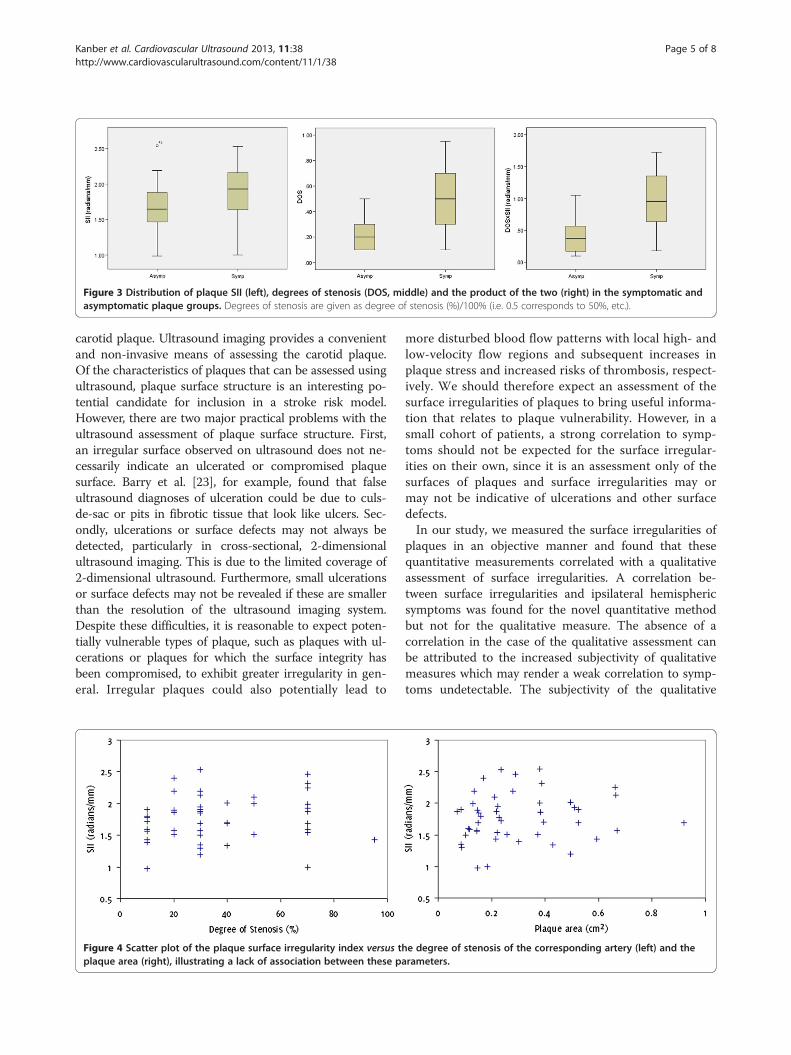
Figure 3 Distribution of plaque SII (left), degrees of stenosis (DOS, middle) and the product of the two (right) in the symptomatic andasymptomatic plaque groups. Degrees of stenosis are given as degree of stenosis (%)/100% (i.e. 0.5 corresponds to 50%, etc.).
Kanber et al. Cardiovascular Ultrasound 2013, 11:38 Page 5 of 8http://www.cardiovascularultrasound.com/content/11/1/38
carotid plaque. Ultrasound imaging provides a convenientand non-invasive means of assessing the carotid plaque.Of the characteristics of plaques that can be assessed usingultrasound, plaque surface structure is an interesting po-tential candidate for inclusion in a stroke risk model.However, there are two major practical problems with theultrasound assessment of plaque surface structure. First,an irregular surface observed on ultrasound does not ne-cessarily indicate an ulcerated or compromised plaquesurface. Barry et al. [23], for example, found that falseultrasound diagnoses of ulceration could be due to culs-de-sac or pits in fibrotic tissue that look like ulcers. Sec-ondly, ulcerations or surface defects may not always bedetected, particularly in cross-sectional, 2-dimensionalultrasound imaging. This is due to the limited coverage of2-dimensional ultrasound. Furthermore, small ulcerationsor surface defects may not be revealed if these are smallerthan the resolution of the ultrasound imaging system.Despite these difficulties, it is reasonable to expect poten-tially vulnerable types of plaque, such as plaques with ul-cerations or plaques for which the surface integrity hasbeen compromised, to exhibit greater irregularity in gen-eral. Irregular plaques could also potentially lead to
Figure 4 Scatter plot of the plaque surface irregularity index versus thplaque area (right), illustrating a lack of association between these pa
more disturbed blood flow patterns with local high- andlow-velocity flow regions and subsequent increases inplaque stress and increased risks of thrombosis, respect-ively. We should therefore expect an assessment of thesurface irregularities of plaques to bring useful informa-tion that relates to plaque vulnerability. However, in asmall cohort of patients, a strong correlation to symp-toms should not be expected for the surface irregular-ities on their own, since it is an assessment only of thesurfaces of plaques and surface irregularities may ormay not be indicative of ulcerations and other surfacedefects.In our study, we measured the surface irregularities of
plaques in an objective manner and found that thesequantitative measurements correlated with a qualitativeassessment of surface irregularities. A correlation be-tween surface irregularities and ipsilateral hemisphericsymptoms was found for the novel quantitative methodbut not for the qualitative measure. The absence of acorrelation in the case of the qualitative assessment canbe attributed to the increased subjectivity of qualitativemeasures which may render a weak correlation to symp-toms undetectable. The subjectivity of the qualitative
e degree of stenosis of the corresponding artery (left) and therameters.

Figure 5 Distribution of plaque SII across the plaque groups qualitatively classified as having an irregular or smooth surface.
Kanber et al. Cardiovascular Ultrasound 2013, 11:38 Page 6 of 8http://www.cardiovascularultrasound.com/content/11/1/38
assessment is most apparent with plaques that can notbe classified as smooth or irregular with any certainty.In such cases, the assessor may make a highly subjectivedecision to place the plaque in one or the other group.The alternative is to mark such plaques as having an
Figure 6 Comparison between ROC curves for plaque surface irregula(DOS × SII).
indeterminate surface characteristic and thereforeunclassified.We found that the combination of the plaque surface
irregularity index with the degree of stenosis of the cor-responding artery resulted in a more effective diagnostic
rity index (SII), the degree of stenosis (DOS) and their product
Kanber et al. Cardiovascular Ultrasound 2013, 11:38 Page 7 of 8http://www.cardiovascularultrasound.com/content/11/1/38
test compared to the degree of stenosis on its own. Thisindicates that the objective study of plaque surface ir-regularities may provide useful additional informationfor predicting the presence of cerebrovascular symp-toms. There was no significant correlation between theplaque SII and the degree of stenosis in our assessment,indicating that the former may provide information thatis complementary to the latter.Our surface irregularity index was combined with the de-
gree of stenosis of the corresponding artery as the latter isan established parameter widely used in clinical practice andassociated with an increased risk of cerebrovascular events.We took the product of the two parameters as the presenceof ipsilateral hemispheric symptoms was directly related toboth the degree of stenosis and the surface irregularityindex. Our study found that combining the surface irregu-larity index with the degree of stenosis results in a more ef-fective risk indicator than the degree of stenosis on its own.The measurement technique we used had good reprodu-
cibility. The intra-observer variations were due to the hu-man operator involvement required for the initial setup ofthe boundary detection procedure that resulted in the semi-automatic delineation of the plaque-arterial lumen boundar-ies, while the inter-frame variations were probably chieflydue to out-of-plane plaque, patient, and probe motion.Further work can be directed towards studying the sur-
face irregularities of plaques taking into account the echo-genicity characteristics local to the surface. This would beuseful as it may be more likely for surface irregularities tocorrespond to surface defects such as ulcerations or hae-morrhages if the plaque has a less echogenic pattern(e.g. a ruptured fibrous cap or a haemorrhage) comparedto being highly echogenic (e.g. fibrous or calcified). Thevariation of surface irregularities across plaque surfacesshould also be explored in a follow-up study since plaquesurfaces may contain both smooth and rough segmentsand their distribution may provide useful additional infor-mation that relates to plaque vulnerability.
ConclusionsOur study has shown that an objective assessment ofplaque surface irregularities using a novel surface irregu-larity index may correlate with the presence of ipsilateralhemispheric cerebrovascular symptoms. We found anincrease in diagnostic performance with the use of theplaque SII versus that provided by the degree of stenosisalone. Plaque SII may therefore be a valuable tool forimproving risk assessment, by means of helping identifythe vulnerable plaques in patients with carotid arterydisease. The potential clinical value of this parametershould be explored in follow-up studies.
Competing interestsThe authors declare that they have no competing interests.
Authors’ contributionsThe study was conceived by KVR and BK. Ultrasound data were collected byTCH. Algorithm development and analyses were carried out by BK. Allauthors contributed to the interpretation and presentation of the results andall authors read and approved the final manuscript.
AcknowledgementsThe research was funded by and took place at the National Institute forHealth Research (NIHR) Collaboration for Leadership in Applied HealthResearch and Care based at the University Hospitals of Leicester NHS Trust.The views expressed are those of the authors and not necessarily those ofthe NHS, the NIHR or the Department of Health.
Author details1Department of Cardiovascular Sciences, University of Leicester, Leicester,England, UK. 2Department of Surgery, University Hospitals of Leicester NHSTrust, Leicester, England, UK. 3NIHR Biomedical Research Unit forCardiovascular Sciences, University of Leicester, Leicester, England, UK.4Department of Medical Physics, University Hospitals of Leicester NHS Trust,Sandringham Building, Leicester Royal Infirmary, Infirmary Square, LE1 5WW,Leicester, England, UK.
Received: 14 August 2013 Accepted: 4 November 2013Published: 6 November 2013
References1. Leary DH, Holen J, Ricotta JJ, Roe S, Schenk EA: Carotid bifurcation disease:
prediction of ulceration with B-mode US. Radiology 1987, 162:523–525.2. De Bray JM, Baud JM, Dauzat M: Consensus Concerning the Morphology
and the Risk of Carotid Plaques. Cerebrovasc Dis 1997, 7:289–296.3. Muraki M, Mikami T, Yoshimoto T, Fujimoto S, Tokuda K, Kaneko S, et al:
New criteria for the sonographic diagnosis of a plaque ulcer in theextracranial carotid artery. Am J Roentgenol 2012, 198:1161–1166.
4. Schminke U, Motsch L, Hilker L, Kessler C: Three-dimensional ultrasoundobservation of carotid artery plaque ulceration. Stroke 2000, 31:1651–1655.
5. Sitzer M, Müller W, Rademacher J, Siebler M, Hort W, Kniemeyer HW, et al:Color-flow Doppler-assisted duplex imaging fails to detect ulceration inhigh-grade internal carotid artery stenosis. J Vasc Surg 1996, 23:461–465.
6. Steinke W, Hennerici M, Rautenberg W, Mohr JP: Symptomatic andasymptomatic high-grade carotid stenoses in Doppler color-flowimaging. Neurology 1992, 42:131–138.
7. Young N, Soo YS, Fischer P: Comparison of duplex ultrasound withangiography in assessment of carotid bifurcation disease. Australas Radiol1992, 36:54–58.
8. Aburahma AF, Kyer PD, Robinson PA, Hannay RS: The correlation ofultrasonic carotid plaque morphology and carotid plaque hemorrhage:clinical implications. Surgery 1998, 124:721–726.
9. Aburahma AF, Covelli MA, Robinson PA, Holt SM: The role of carotidduplex ultrasound in evaluating plaque morphology: potential use inselecting patients for carotid stenting. J Endovasc Surg 1999, 6:59–65.
10. Kessler C, Von Maravic M, Brückmann H, Kömpf D: Ultrasound for theassessment of the embolic risk of carotid plaques. Acta Neurol Scand1995, 92:231–234.
11. Manolio TA, Burke GL, Leary DH, Evans G, Beauchamp N, Knepper L, et al:Relationships of cerebral MRI findings to ultrasonographic carotidatherosclerosis in older adults : the Cardiovascular Health Study. CHSCollaborative Research Group. Arterioscler Thromb Vasc Biol 1999, 19:356–365.
12. Pedro LM, Pedro MM, Gonçalves I, Carneiro TF, Balsinha C, FernandesFernandes R, et al: Computer-assisted carotid plaque analysis:characteristics of plaques associated with cerebrovascular symptomsand cerebral infarction. Eur J Vasc Endovasc Surg 2000, 19:118–123.
13. Carra G, Visonà A, Bonanome A, Lusiani L, Pesavento R, Bortolon M, et al:Carotid plaque morphology and cerebrovascular events. Int Angiol 2003,22:284–289.
14. Ding S, Zhang M, Zhao Y, Chen W, Yao G, Zhang C, et al: The role ofcarotid plaque vulnerability and inflammation in the pathogenesis ofacute ischemic stroke. Am J Med Sci 2008, 336:27–31.
15. Golledge J, Cuming R, Ellis M, Davies AH, Greenhalgh RM: Carotid plaquecharacteristics and presenting symptom. Br J Surg 1998, 84:1697–1701.

Kanber et al. Cardiovascular Ultrasound 2013, 11:38 Page 8 of 8http://www.cardiovascularultrasound.com/content/11/1/38
16. Meairs S, Hennerici M: Four-dimensional ultrasonographiccharacterization of plaque surface motion in patients with symptomaticand asymptomatic carotid artery stenosis. Stroke 1999, 30:1807–1813.
17. Denzel C, Fellner F, Wutke R, Bazler K, Müller K, Lang W: Ultrasonographicanalysis of arteriosclerotic plaques in the internal carotid artery. Eur JUltrasound 2003, 16:161–167.
18. Gaunt ME, Brown L, Hartshorne T, Bell PR, Naylor AR: Unstable carotidplaques: preoperative identification and association with intraoperativeembolisation detected by transcranial Doppler. Eur J Vasc Endovasc Surg1996, 11:78–82.
19. Rubin JR, Bondi JA, Rhodes RS: Duplex scanning versus conventionalarteriography for the evaluation of carotid artery plaque morphology.Surgery 1987, 102:749–755.
20. Van Damme H, Vivario M: Pathologic aspects of carotid plaques: surgicaland clinical significance. Int Angiol 1993, 12:299–311.
21. Widder B, Paulat K, Hackspacher J, Hamann H, Hutschenreiter S, Kreutzer C,et al: Morphological characterization of carotid artery stenoses byultrasound duplex scanning. Ultrasound Med Biol 1990, 16:349–354.
22. Wolverson MK, Bashiti HM, Peterson GJ: Ultrasonic tissue characterizationof atheromatous plaques using a high resolution real time scanner.Ultrasound Med Biol 1983, 9:599–609.
23. Barry R, Pienaar C, Nel CJ: Accuracy of B-mode ultrasonography indetecting carotid plaque hemorrhage and ulceration. Ann Vasc Surg 1990,4:466–470.
24. Bluth EI, Mcvay LV, Merritt CR, Sullivan MA: The identification of ulcerativeplaque with high resolution duplex carotid scanning. J Ultrasound Med1988, 7:73–76.
25. Comerota AJ, Katz ML, White JV, Grosh JD: The preoperative diagnosis ofthe ulcerated carotid atheroma. J Vasc Surg 1990, 11:505–510.
26. European Carotid Plaque Study Group: Carotid artery plaquecomposition–relationship to clinical presentation and ultrasoundB-mode imaging. Eur J Vasc Endovasc Surg 1995, 10:23–30.
27. Prabhakaran S, Rundek T, Ramas R, Elkind MSV, Paik MC, Boden-albala B,et al: Carotid plaque surface irregularity predicts ischemic stroke: thenorthern Manhattan study. Stroke 2006, 37:2696–2701.
28. Tegos TJ, Kalomiris KJ, Sabetai MM, Kalodiki E, Nicolaides AN: Significanceof sonographic tissue and surface characteristics of carotid plaques.Am J Neuroradiol 2001, 22:1605–1612.
29. Chiu B, Beletsky V, Spence JD, Parraga G, Fenster A: Analysis of carotidlumen surface morphology using three-dimensional ultrasound imaging.Phys Med Biol 2009, 54:1149–1167.
30. Fenster A, Blake C, Gyacskov I, Landry A, Spence JD: 3D ultrasound analysisof carotid plaque volume and surface morphology. Ultrasonics 2006,44:E153–E157.
31. Kanber B, Hartshorne TC, Horsfield MA, Naylor AR, Robinson TG, RamnarineKV: Dynamic variations in the ultrasound greyscale median of carotidartery plaques. Cardiovasc Ultrasound 2013, 11:21.
32. North American Symptomatic Carotid Endarterectomy Trial Collaborators:Beneficial effect of carotid endarterectomy in symptomatic patients withhigh-grade carotid stenosis. N Engl J Med 1991, 325:445–453.
33. Grant EG, Benson CB, Moneta GL, Alexandrov AV, Baker JD, Bluth EI, et al:Carotid artery stenosis: gray-scale and Doppler US diagnosis–Society ofRadiologists in Ultrasound Consensus Conference. Radiology 2003,229:340–346.
34. Oates CP, Naylor AR, Hartshorne T, Charles SM, Fail T, Humphries K, et al:Joint recommendations for reporting carotid ultrasound investigationsin the United Kingdom. Eur J Vasc Endovasc Surg 2008, 37:251–261.
doi:10.1186/1476-7120-11-38Cite this article as: Kanber et al.: Quantitative assessment of carotidplaque surface irregularities and correlation to cerebrovascularsymptoms. Cardiovascular Ultrasound 2013 11:38.
Submit your next manuscript to BioMed Centraland take full advantage of:
• Convenient online submission
• Thorough peer review
• No space constraints or color figure charges
• Immediate publication on acceptance
• Inclusion in PubMed, CAS, Scopus and Google Scholar
• Research which is freely available for redistribution
Submit your manuscript at www.biomedcentral.com/submit