quantifying protein diffusion and capture on filaments · quantifying protein diffusion and...
TRANSCRIPT
Quantifying protein diffusion and capture onfilaments
Emanuel Reithmann, Louis Reese, and Erwin Frey ([email protected])
Arnold Sommerfeld Center for Theoretical Physics (ASC) and Center for NanoScience (CeNS),
Department of Physics, Ludwig-Maximilians-Universität München, Theresienstraße 37, D-80333
Munich, Germany, and Nanosystems Initiative Munich (NIM), Ludwig-Maximilians-Universität
München, Schellingstraße 4, D-80333 Munich, Germany
Abstract
The functional relevance of regulating proteins is often limited to specific binding sites such asthe ends of microtubules or actin-filaments. A localization of proteins on these functional sites isof great importance. We present a quantitative theory for a diffusion and capture process, whereproteins diffuse on a filament and stop diffusing when reaching the filament’s end. It is found thatend-association after one-dimensional diffusion is the main source for tip-localization of such pro-teins. As a consequence, diffusion and capture is highly efficient in enhancing the reaction velocityof enzymatic reactions, where proteins and filament ends are to each other as enzyme and substrate.We show that the reaction velocity can effectively be described within a Michaelis-Menten frame-work. Together one-dimensional diffusion and capture beats the (three-dimensional) Smoluchowskidiffusion limit for the rate of protein association to filament ends.
The catalytic activity of enzymes is often restricted to specific binding sites. The ends of microtubules
(MTs) for example are binding sites for a plethora of MT associated proteins (MAPs) [1]. At MT ends,
MAPs can catalyze biochemical processes [2], or serve as substrates for other enzymes. This makes an
efficient association of MAPs to MT tips important. Recent experiments suggest that one-dimensional
diffusion of MAPs on MTs facilitates tip-targeting [3, 4]. This idea goes back to the concept of “reduction
in dimensionality” suggested by Adam and Delbrück [5] and has been largely applied [6, 7]. However,
a quantitative understanding of tip-binding mediated by diffusion on the filament and subsequent capture
at the tip has remained elusive [3, 8–16].
Here we show that capturing at the tip is crucial for tip-localization of proteins. We present a the-
ory where diffusion and capture is accurately quantified with an effective association rate constant and
provide a result which depends only on experimentally accessible parameters. For proteins which are en-
zymatically active at filament ends, our theory predicts that diffusion and capture leads to an enhancement
of the enzymatic reaction velocity due to stronger tip-localization. We observe that the reaction veloc-
ity in dependence of the enzyme concentration closely follows a Michaelis-Menten curve and quantify
the contribution of one-dimensional diffusion to tip-localization and enzymatic processes downstream
thereof.
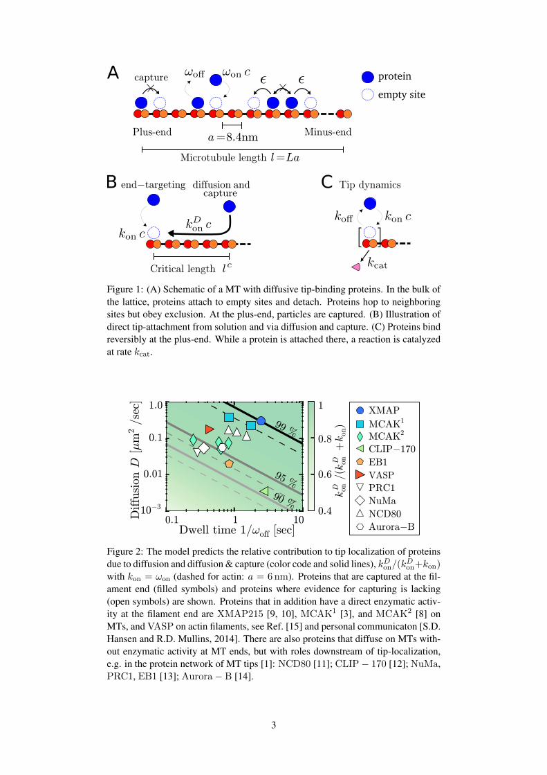
To model the diffusive motion of proteins on a filament we consider a one-dimensional lattice of
length l with lattice spacing a = 8.4 nm [Fig. 1A]. The lattice corresponds to a single protofilament of a
stabilized MT in the absence of dynamic instability. Proteins perform a random walk on the lattice with
a hopping rate ε; the diffusion constant is D = ε a2. Each site can be occupied by only one protein; the
system is an exclusion process [17]. Proteins attach to and detach from the lattice at rates ωonc and ωoff ,
respectively, where c is the concentration of proteins in solution. The tip of the MT is represented by
the first lattice site in our model. To account for its particular structure, different on- and off-rates are
1
arX
iv:1
503.
0087
8v1
[ph
ysic
s.bi
o-ph
] 3
Mar
201
5
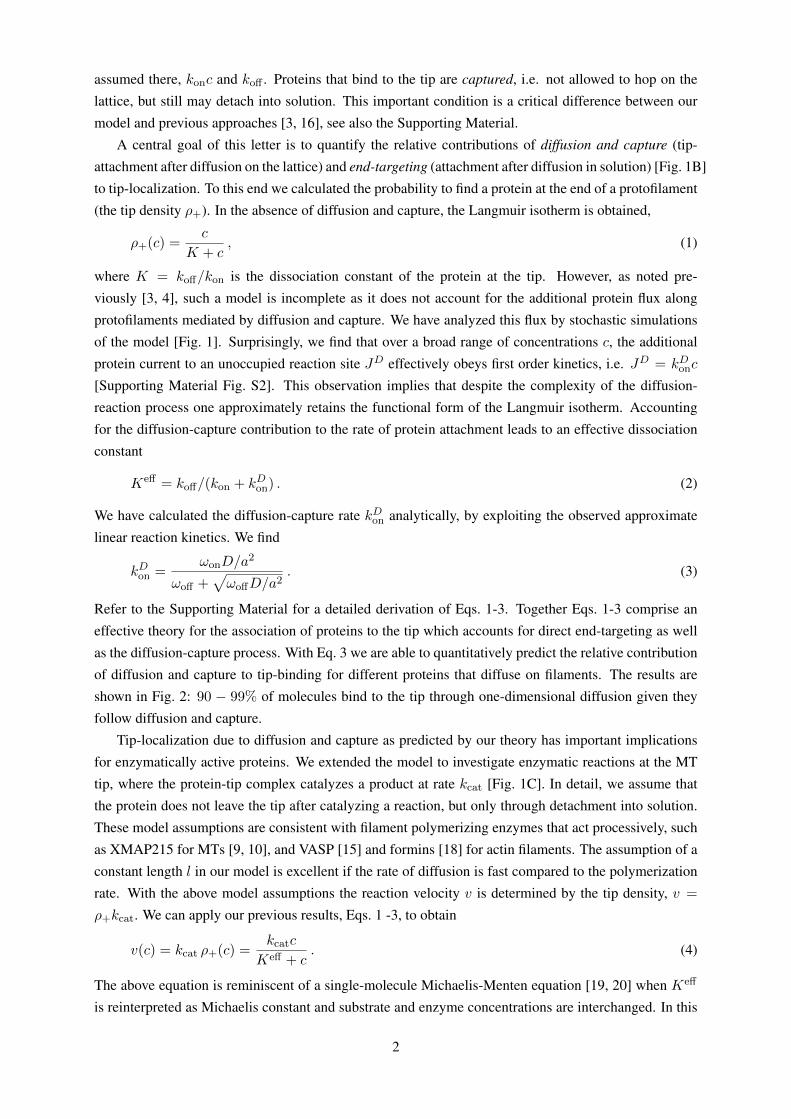
assumed there, konc and koff . Proteins that bind to the tip are captured, i.e. not allowed to hop on the
lattice, but still may detach into solution. This important condition is a critical difference between our
model and previous approaches [3, 16], see also the Supporting Material.
A central goal of this letter is to quantify the relative contributions of diffusion and capture (tip-
attachment after diffusion on the lattice) and end-targeting (attachment after diffusion in solution) [Fig. 1B]
to tip-localization. To this end we calculated the probability to find a protein at the end of a protofilament
(the tip density ρ+). In the absence of diffusion and capture, the Langmuir isotherm is obtained,
ρ+(c) =c
K + c, (1)
where K = koff/kon is the dissociation constant of the protein at the tip. However, as noted pre-
viously [3, 4], such a model is incomplete as it does not account for the additional protein flux along
protofilaments mediated by diffusion and capture. We have analyzed this flux by stochastic simulations
of the model [Fig. 1]. Surprisingly, we find that over a broad range of concentrations c, the additional
protein current to an unoccupied reaction site JD effectively obeys first order kinetics, i.e. JD = kDonc
[Supporting Material Fig. S2]. This observation implies that despite the complexity of the diffusion-
reaction process one approximately retains the functional form of the Langmuir isotherm. Accounting
for the diffusion-capture contribution to the rate of protein attachment leads to an effective dissociation
constant
Keff = koff/(kon + kDon) . (2)
We have calculated the diffusion-capture rate kDon analytically, by exploiting the observed approximate
linear reaction kinetics. We find
kDon =ωonD/a
2
ωoff +√ωoffD/a2
. (3)
Refer to the Supporting Material for a detailed derivation of Eqs. 1-3. Together Eqs. 1-3 comprise an
effective theory for the association of proteins to the tip which accounts for direct end-targeting as well
as the diffusion-capture process. With Eq. 3 we are able to quantitatively predict the relative contribution
of diffusion and capture to tip-binding for different proteins that diffuse on filaments. The results are
shown in Fig. 2: 90 − 99% of molecules bind to the tip through one-dimensional diffusion given they
follow diffusion and capture.
Tip-localization due to diffusion and capture as predicted by our theory has important implications
for enzymatically active proteins. We extended the model to investigate enzymatic reactions at the MT
tip, where the protein-tip complex catalyzes a product at rate kcat [Fig. 1C]. In detail, we assume that
the protein does not leave the tip after catalyzing a reaction, but only through detachment into solution.
These model assumptions are consistent with filament polymerizing enzymes that act processively, such
as XMAP215 for MTs [9, 10], and VASP [15] and formins [18] for actin filaments. The assumption of a
constant length l in our model is excellent if the rate of diffusion is fast compared to the polymerization
rate. With the above model assumptions the reaction velocity v is determined by the tip density, v =
ρ+kcat. We can apply our previous results, Eqs. 1 -3, to obtain
v(c) = kcat ρ+(c) =kcatc
Keff + c. (4)
The above equation is reminiscent of a single-molecule Michaelis-Menten equation [19, 20] when Keff
is reinterpreted as Michaelis constant and substrate and enzyme concentrations are interchanged. In this
2
protein
empty site
Figure 1: (A) Schematic of a MT with diffusive tip-binding proteins. In the bulk ofthe lattice, proteins attach to empty sites and detach. Proteins hop to neighboringsites but obey exclusion. At the plus-end, particles are captured. (B) Illustration ofdirect tip-attachment from solution and via diffusion and capture. (C) Proteins bindreversibly at the plus-end. While a protein is attached there, a reaction is catalyzedat rate kcat.
Figure 2: The model predicts the relative contribution to tip localization of proteinsdue to diffusion and diffusion & capture (color code and solid lines), kDon/(k
Don+kon)
with kon = ωon (dashed for actin: a = 6nm). Proteins that are captured at the fil-ament end (filled symbols) and proteins where evidence for capturing is lacking(open symbols) are shown. Proteins that in addition have a direct enzymatic activ-ity at the filament end are XMAP215 [9, 10], MCAK1 [3], and MCAK2 [8] onMTs, and VASP on actin filaments, see Ref. [15] and personal communicaton [S.D.Hansen and R.D. Mullins, 2014]. There are also proteins that diffuse on MTs with-out enzymatic activity at MT ends, but with roles downstream of tip-localization,e.g. in the protein network of MT tips [1]: NCD80 [11]; CLIP− 170 [12]; NuMa,PRC1, EB1 [13]; Aurora− B [14].
3
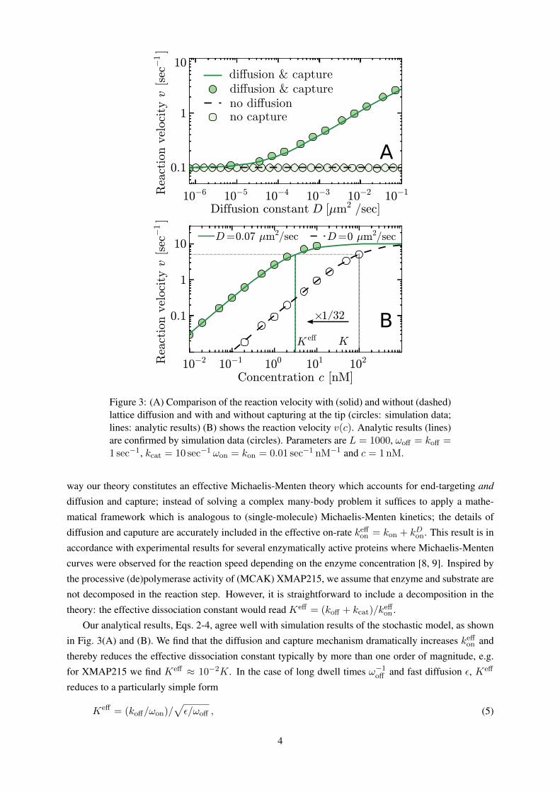
Figure 3: (A) Comparison of the reaction velocity with (solid) and without (dashed)lattice diffusion and with and without capturing at the tip (circles: simulation data;lines: analytic results) (B) shows the reaction velocity v(c). Analytic results (lines)are confirmed by simulation data (circles). Parameters are L = 1000, ωoff = koff =1 sec−1, kcat = 10 sec−1 ωon = kon = 0.01 sec−1 nM−1 and c = 1nM.
way our theory constitutes an effective Michaelis-Menten theory which accounts for end-targeting and
diffusion and capture; instead of solving a complex many-body problem it suffices to apply a mathe-
matical framework which is analogous to (single-molecule) Michaelis-Menten kinetics; the details of
diffusion and caputure are accurately included in the effective on-rate keffon = kon + kDon. This result is in
accordance with experimental results for several enzymatically active proteins where Michaelis-Menten
curves were observed for the reaction speed depending on the enzyme concentration [8, 9]. Inspired by
the processive (de)polymerase activity of (MCAK) XMAP215, we assume that enzyme and substrate are
not decomposed in the reaction step. However, it is straightforward to include a decomposition in the
theory: the effective dissociation constant would read Keff = (koff + kcat)/keffon .
Our analytical results, Eqs. 2-4, agree well with simulation results of the stochastic model, as shown
in Fig. 3(A) and (B). We find that the diffusion and capture mechanism dramatically increases keffon and
thereby reduces the effective dissociation constant typically by more than one order of magnitude, e.g.
for XMAP215 we find Keff ≈ 10−2K. In the case of long dwell times ω−1off and fast diffusion ε, Keff
reduces to a particularly simple form
Keff = (koff/ωon)/√ε/ωoff , (5)
4
where the denominator is the square root of the average number of diffusive steps a protein performs on
the filament. Note that one-dimensional diffusion without capturing [16] does not lead to a particle flux
on the filament [Supporting Material Fig. S4] and hence the reaction velocity is not increased [Fig. 3A].
Further, the particle flux might be limited by the length of the filament: Below a threshold length lc
(which is smaller than typical in vivo lengths of MTs) we observe a length dependent behavior of the
reaction velocity [Supporting Material Fig. S3] where our theory is not valid.
Our analysis reveals diffusion and capture as an efficient mechanism to circumvent the diffusion
limit for the rate of end-targeting: Smoluchowski’s theory of three-dimensional diffusion physically
limits the rate of direct tip-attachment from solution [21]. As shown here, one-dimensional diffusion
along a filament and subsequent capture at the filament end overcomes this limitation. This has been
shown experimentally for MCAK [3]. Our work provides an applicable theory for reaction kinetics
facilitated by diffusion and capture: specific parameter values for diffusion, tip-association and dwell
times can be accounted for, cf. Eqs. 3 and 4. On a broader perspective our results may also be applicable
to other systems where one-dimensional diffusion is important [6] including transcription factor binding
on DNA [22].
AcknowledgementThe authors thank Scott Hansen and Dyche Mullins for helpful correspondence on diffusing actin
binding proteins. This research was supported by the Deutsche Forschungsgemeinschaft (DFG) via
project B02 within the SFB 863.
References
[1] Akhmanova, A., and M. O. Steinmetz, 2008. Tracking the ends: a dynamic protein network controls
the fate of microtubule tips. Nat. Rev. Mol. Cell Biol. 9:309–322.
[2] Howard, J., and A. A. Hyman, 2007. Microtubule polymerases and depolymerases. Curr. Opin.
Cell Biol. 19:31–5.
[3] Helenius, J., G. J. Brouhard, Y. Kalaidzidis, S. Diez, and J. Howard, 2006. The depolymerizing
kinesin MCAK uses lattice diffusion to rapidly target microtubule ends. Nature 441:115–9.
[4] Cooper, J. R., and L. Wordeman, 2009. The diffusive interaction of microtubule binding proteins.
Curr. Opin. Cell Biol. 21:68–73.
[5] Adam, G., and M. Delbrück, 1968. Reduction of dimensionality in biological diffusion processes.
In Structural chemistry and molecular biology, Freeman, 198–215.
[6] von Hippel, P. H., and O. G. Berg, 1989. Facilitated Target Location in Biological Systems. J. Biol.
Chem. 264:675–678.
[7] Mirny, L., M. Slutsky, Z. Wunderlich, A. Tafvizi, J. Leith, and A. Kosmrlj, 2009. How a protein
searches for its site on DNA: the mechanism of facilitated diffusion. J. Phys. A: Math. Theor.
42:434013.
5
[8] Cooper, J. R., M. Wagenbach, C. L. Asbury, and L. Wordeman, 2010. Catalysis of the microtubule
on-rate is the major parameter regulating the depolymerase activity of MCAK. Nat. Struct. Mol.
Biol. 17:77–82.
[9] Brouhard, G. J., J. H. Stear, T. L. Noetzel, J. Al-Bassam, K. Kinoshita, S. C. Harrison, J. Howard,
and A. A. Hyman, 2008. XMAP215 is a processive microtubule polymerase. Cell 132:79–88.
[10] Widlund, P. O., J. H. Stear, A. Pozniakovsky, M. Zanic, S. Reber, G. J. Brouhard, A. A. Hyman, and
J. Howard, 2011. XMAP215 polymerase activity is built by combining multiple tubulin-binding
TOG domains and a basic lattice-binding region. Proc. Nat. Acad. Sci. USA 108:2741–6.
[11] Powers, A. F., A. D. Franck, D. R. Gestaut, J. Cooper, B. Gracyzk, R. R. Wei, L. Wordeman,
T. N. Davis, and C. L. Asbury, 2009. The Ndc80 Kinetochore Complex Forms Load-Bearing
Attachments to Dynamic Microtubule Tips via Biased Diffusion. Cell 136:865–875.
[12] Dixit, R., B. Barnett, J. E. Lazarus, M. Tokito, Y. E. Goldman, and E. L. F. Holzbaur, 2009. Micro-
tubule plus-end tracking by CLIP-170 requires EB1. Proc. Natl. Acad. Sci. USA 106:492–497.
[13] Forth, S., K.-C. Hsia, Y. Shimamoto, and T. M. Kapoor, 2014. Asymmetric friction of nonmotor
MAPs can lead to their directional motion in active microtubule networks. Cell 157:420 – 432.
[14] Noujaim, M., S. Bechstedt, M. Wieczorek, and G. J. Brouhard, 2014. Microtubules Accelerate the
Kinase Activity of Aurora-B by a Reduction in Dimensionality. PLoS ONE 9:e86786.
[15] Hansen, S. D., and R. D. Mullins, 2010. VASP is a processive actin polymerase that requires
monomeric actin for barbed end association. J. Cell Biol. 191:571–584. & private communication.
[16] Klein, G., K. Kruse, G. Cuniberti, and F. Jülicher, 2005. Filament Depolymerization by Motor
Molecules. Phys. Rev. Lett. 94:108102.
[17] Krapivsky, P. L., S. Redner, and E. Ben-Naim, 2010. A kinetic view of statistical physics. Cam-
bridge University Press.
[18] Vavylonis, D., D. R. Kovar, B. O’Shaughnessy, and T. D. Pollard, 2006. Model of formin-associated
actin filament elongation. Mol. Cell 21:455–66.
[19] Kou, S., B. J. Cherayil, W. Min, B. P. English, and X. X. Sunney, 2005. Single-molecule michaelis-
menten equations. J. Phys. Chem. B 109:19068–19081.
[20] Michaelis, L., and M. L. Menten, 1913. Die Kinetik der Invertinwirkung. Biochem. Z. 49:333–369.
[21] von Smoluchowski, M., 1917. Versuch einer mathematischen Theorie der Koagulationskinetik. Z.
Phys. Chem 92:129–168.
[22] Hammar, P., P. Leroy, A. Mahmutovic, E. G. Marklund, O. G. Berg, and J. Elf, 2012. The lac
repressor displays facilitated diffusion in living cells. Science 336:1595–1598.
6
Supporting Material “Quantifying proteindiffusion and capture on filaments”Emanuel Reithmann, Louis Reese, and Erwin Frey ([email protected])
Arnold Sommerfeld Center for Theoretical Physics (ASC) and Center for NanoScience (CeNS),
Department of Physics, Ludwig-Maximilians-Universität München, Theresienstraße 37, D-80333
Munich, Germany, and Nanosystems Initiative Munich (NIM), Ludwig-Maximilians-Universität
München, Schellingstraße 4, D-80333 Munich, Germany
A Derivation of Eq. (1), (2), and (4)
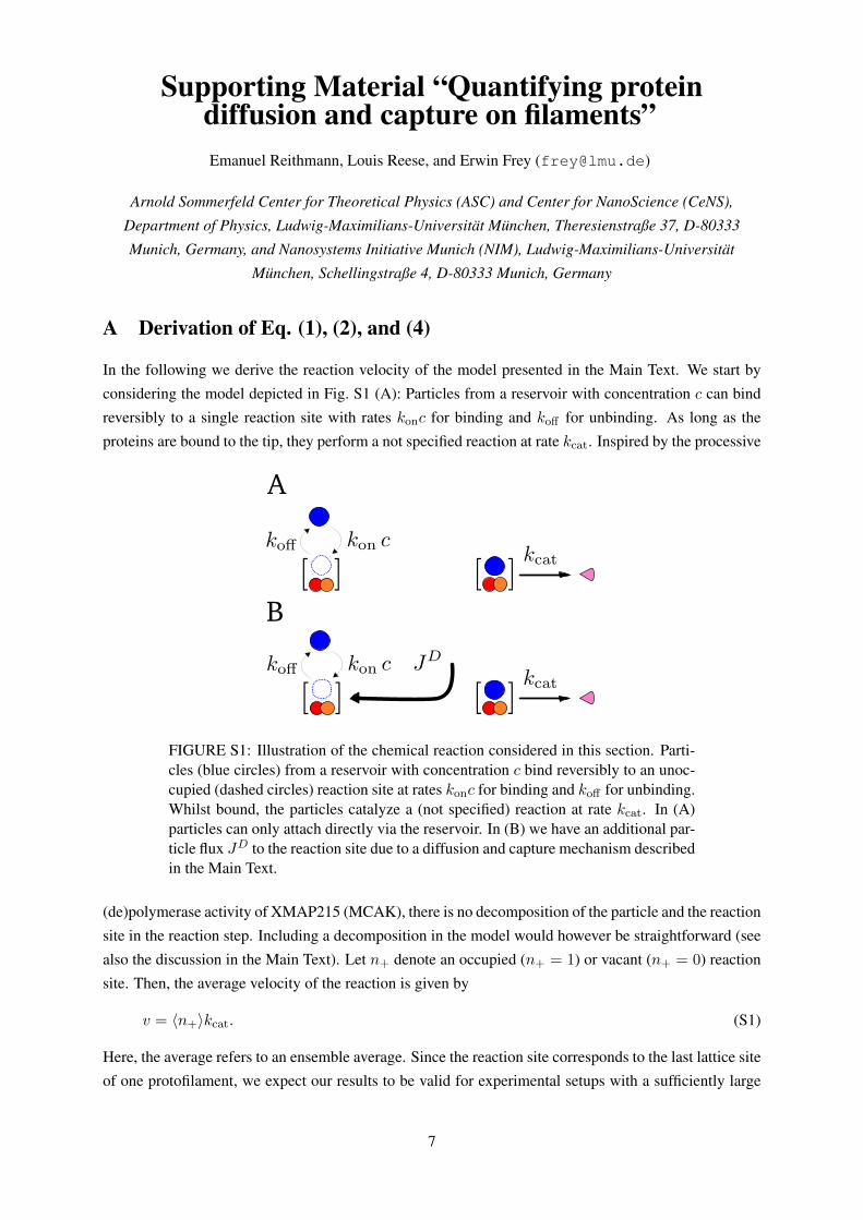
In the following we derive the reaction velocity of the model presented in the Main Text. We start by
considering the model depicted in Fig. S1 (A): Particles from a reservoir with concentration c can bind
reversibly to a single reaction site with rates konc for binding and koff for unbinding. As long as the
proteins are bound to the tip, they perform a not specified reaction at rate kcat. Inspired by the processive
A
B
FIGURE S1: Illustration of the chemical reaction considered in this section. Parti-cles (blue circles) from a reservoir with concentration c bind reversibly to an unoc-cupied (dashed circles) reaction site at rates konc for binding and koff for unbinding.Whilst bound, the particles catalyze a (not specified) reaction at rate kcat. In (A)particles can only attach directly via the reservoir. In (B) we have an additional par-ticle flux JD to the reaction site due to a diffusion and capture mechanism describedin the Main Text.
(de)polymerase activity of XMAP215 (MCAK), there is no decomposition of the particle and the reaction
site in the reaction step. Including a decomposition in the model would however be straightforward (see
also the discussion in the Main Text). Let n+ denote an occupied (n+ = 1) or vacant (n+ = 0) reaction
site. Then, the average velocity of the reaction is given by
v = 〈n+〉kcat. (S1)
Here, the average refers to an ensemble average. Since the reaction site corresponds to the last lattice site
of one protofilament, we expect our results to be valid for experimental setups with a sufficiently large
7
and constant number of protofilaments or (due to ergodicity) for the time average with respect to a single
protofilament. In the steady state the equation for the average occupation of the reaction site reads
0 =d
dt〈n+〉 = konc(1− 〈n+〉)− koff〈n+〉 . (S2)
Solving for 〈n+〉 leads to an equation for the reaction velocity which is analogous to a single molecule
Michaelis-Menten equation [1]:
v =kcatc
K + c(S3)
with K = koff/kon. Note that here c is the concentration of the enzyme, not the substrate.
If we now have an additional particle flux J to the reaction site via one-dimensional diffusion, Fig. S1
(B), the dynamics are changed. Including this flux in the equation for the average reaction site occupation
leads to
0 =d
dt〈n+〉 = konc(1− 〈n+〉) + J − koff〈n+〉 (S4)
=
(konc+
J
1− 〈n+〉
)(1− 〈n+〉)− koff〈n+〉 . (S5)
Note that the term JD := J/(1−〈n+〉) can be interpreted as the conditional particle flux to an unoccupied
tip. If the conditional diffusive current can be written as JD := kDonc we conserve the functional form
of Eq. S3 but kon is replaced by an effective on-rate for direct binding as well as diffusion and capture:
keff = kon + kDon.
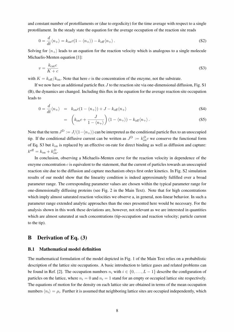
In conclusion, observing a Michaelis-Menten curve for the reaction velocity in dependence of the
enzyme concentration c is equivalent to the statement, that the current of particles towards an unoccupied
reaction site due to the diffusion and capture mechanism obeys first order kinetics. In Fig. S2 simulation
results of our model show that the linearity condition is indeed approximately fulfilled over a broad
parameter range. The corresponding parameter values are chosen within the typical parameter range for
one-dimensionally diffusing proteins (see Fig. 2 in the Main Text). Note that for high concentrations
which imply almost saturated reaction velocities we observe a, in general, non-linear behavior. In such a
parameter range extended analytic approaches than the ones presented here would be necessary. For the
analysis shown in this work these deviations are, however, not relevant as we are interested in quantities
which are almost saturated at such concentrations (tip-occupation and reaction velocity; particle current
to the tip).
B Derivation of Eq. (3)
B.1 Mathematical model definition
The mathematical formulation of the model depicted in Fig. 1 of the Main Text relies on a probabilistic
description of the lattice site occupations. A basic introduction to lattice gases and related problems can
be found in Ref. [2]. The occupation numbers ni with i ∈ {0, . . . , L − 1} describe the configuration of
particles on the lattice, where ni = 0 and ni = 1 stand for an empty or occupied lattice site respectively.
The equations of motion for the density on each lattice site are obtained in terms of the mean occupation
numbers 〈ni〉 = ρi. Further it is assumed that neighboring lattice sites are occupied independently, which
8
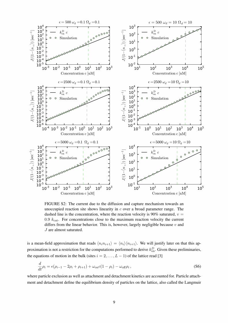
FIGURE S2: The current due to the diffusion and capture mechanism towards anunoccupied reaction site shows linearity in c over a broad parameter range. Thedashed line is the concentration, where the reaction velocity is 90% saturated, v =0.9 kcat. For concentrations close to the maximum reaction velocity the currentdiffers from the linear behavior. This is, however, largely negligible because v andJ are almost saturated.
is a mean-field approximation that reads 〈nini+1〉 = 〈ni〉〈ni+1〉. We will justify later on that this ap-
proximation is not a restriction for the computations performed to derive kDon. Given these preliminaries,
the equations of motion in the bulk (sites i = 2, . . . , L− 1) of the lattice read [3]
d
dtρi = ε(ρi−1 − 2ρi + ρi+1) + ωonc(1− ρi)− ωoffρi , (S6)
where particle exclusion as well as attachment and detachment kinetics are accounted for. Particle attach-
ment and detachment define the equilibrium density of particles on the lattice, also called the Langmuir
9
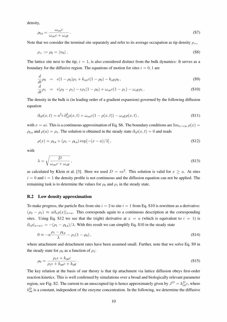
density,
ρLa =ωonc
ωonc+ ωoff. (S7)
Note that we consider the terminal site separately and refer to its average occupation as tip density ρ+,
ρ+ := ρ0 = 〈n0〉 . (S8)
The lattice site next to the tip, i = 1, is also considered distinct from the bulk dynamics: It serves as a
boundary for the diffusive region. The equations of motion for sites i = 0, 1 are
d
dtρ0 = ε(1− ρ0)ρ1 + konc(1− ρ0)− koffρ0 , (S9)
d
dtρ1 = ε(ρ2 − ρ1)− ερ1(1− ρ0) + ωonc(1− ρ1)− ωoffρ1 . (S10)
The density in the bulk is (in leading order of a gradient expansion) governed by the following diffusion
equation
∂tρ(x, t) = a2ε ∂2xρ(x, t) + ωonc(1− ρ(x, t))− ωoffρ(x, t) , (S11)
with x = ai. This is a continuous approximation of Eq. S6. The boundary conditions are limx→∞ ρ(x) =
ρLa and ρ(a) = ρ1. The solution is obtained in the steady state ∂tρ(x, t) = 0 and reads
ρ(x) = ρLa + (ρ1 − ρLa) exp[−(x− a)/λ] , (S12)
with
λ =
√D
ωonc+ ωoff, (S13)
as calculated by Klein et al. [3]. Here we used D = εa2. This solution is valid for x ≥ a. At sites
i = 0 and i = 1 the density profile is not continuous and the diffusion equation can not be applied. The
remaining task is to determine the values for ρ0 and ρ1 in the steady state.
B.2 Low density approximation
To make progress, the particle flux from site i = 2 to site i = 1 from Eq. S10 is rewritten as a derivative:
(ρ2 − ρ1) ≈ a∂xρ(x)|x=a. This corresponds again to a continuous description at the corresponding
sites. Using Eq. S12 we see that the (right) derivative at x = a (which is equivalent to i = 1) is
∂xρ|x=a+ = −(ρ1 − ρLa)/λ. With this result we can simplify Eq. S10 in the steady state
0 ≈ −aρ1 − ρLa
λ− ρ1(1− ρ0) , (S14)
where attachment and detachment rates have been assumed small. Further, note that we solve Eq. S9 in
the steady state for ρ0 as a function of ρ1:
ρ0 =ρ1ε+ konc
ρ1ε+ konc+ koff. (S15)
The key relation at the basis of our theory is that tip attachment via lattice diffusion obeys first-order
reaction kinetics. This is well confirmed by simulations over a broad and biologically relevant parameter
region, see Fig. S2. The current to an unoccupied tip is hence approximately given by JD = kDonc, where
kDon is a constant, independent of the enzyme concentration. In the following, we determine the diffusive
10
current for infinitesimally low enzyme concentrations and thereby determine kDon. In this parameter
region correlations become negligible such that the mean-field assumption becomes valid. Further, we
assume ρ1 to be small. This is well justified for low concentrations. Note also that the density along the
lattice is minimal at i = 1. Up to first order in ρ1 Eq. S14 reduces to
0 = − a ε ωonc√ε (ωoff + ωonc)
+(a√ε (ωoff + ωonc) +
ε koff
konc+ koff
)ρ1 . (S16)
The solution of the above equation determines the tip density via Eq. S15. In our low-density approxi-
mation and up to first order in c we obtain
ρlow-c0 =
kon + (ωon ε)/(ωoff +√ε ωoff)
koffc . (S17)
B.3 Site attachment due to lattice diffusion
The solution of ρ+ = ρ0 allows us to determine the diffusive current. In Eq. S17 there is an additional
term that adds to the direct attachment rate kon which vanishes for ε = 0. Identifying this term as the
diffusive on-rate the final result reads
kDon =
ε ωon
ωoff +√ε ωoff
. (S18)
C XMAP215 parameter values
Model parametersε ωon ωoff kcat koff kon
s−1 (nM s)−1 s−1 s−1 s−1 (nM s)−1
XMAP215 4.7 ×10³ 6 ×10⁻⁵ 4.1 ×10⁻¹ 1.3 ×10² 2.6 ×10⁻¹ 6 ×10⁻⁵
TABLE S1: Parameters used in our simulation for XMAP215. The numbers werederived from experimental data [5, 6].
D Length dependent behavior for short filaments
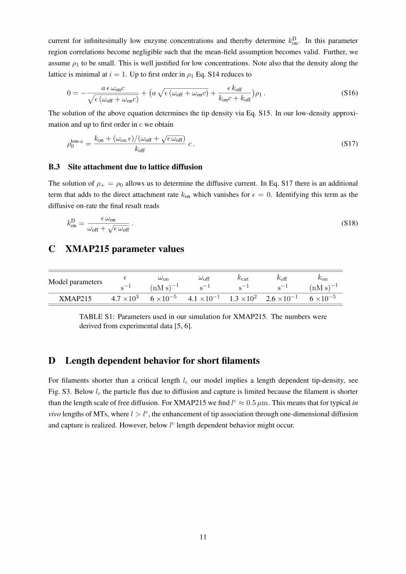
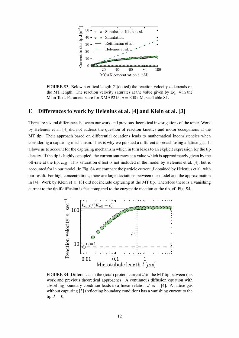
For filaments shorter than a critical length lc our model implies a length dependent tip-density, see
Fig. S3. Below lc the particle flux due to diffusion and capture is limited because the filament is shorter
than the length scale of free diffusion. For XMAP215 we find lc ≈ 0.5µm. This means that for typical in
vivo lengths of MTs, where l > lc, the enhancement of tip association through one-dimensional diffusion
and capture is realized. However, below lc length dependent behavior might occur.
11
20 40 60 80 100
MCAK concentration c [nM]
0
10
20
30
40
50
Curr
entto
the
tip
J[s−
1]
Simulation Klein et al.
Simulation
Reithmann et al.
Helenius et al.
FIGURE S3: Below a critical length lc (dotted) the reaction velocity v depends onthe MT length. The reaction velocity saturates at the value given by Eq. 4 in theMain Text. Parameters are for XMAP215, c = 300 nM, see Table S1.
E Differences to work by Helenius et al. [4] and Klein et al. [3]
There are several differences between our work and previous theoretical investigations of the topic. Work
by Helenius et al. [4] did not address the question of reaction kinetics and motor occupations at the
MT tip. Their approach based on differential equations leads to mathematical inconsistencies when
considering a capturing mechanism. This is why we pursued a different approach using a lattice gas. It
allows us to account for the capturing mechanism which in turn leads to an explicit expression for the tip
density. If the tip is highly occupied, the current saturates at a value which is approximately given by the
off-rate at the tip, koff . This saturation effect is not included in the model by Helenius et al. [4], but is
accounted for in our model. In Fig. S4 we compare the particle current J obtained by Helenius et al. with
our result. For high concentrations, there are large deviations between our model and the approximation
in [4]. Work by Klein et al. [3] did not include capturing at the MT tip. Therefore there is a vanishing
current to the tip if diffusion is fast compared to the enzymatic reaction at the tip, cf. Fig. S4.
FIGURE S4: Differences in the (total) protein current J to the MT tip between thiswork and previous theoretical approaches. A continuous diffusion equation withabsorbing boundary condition leads to a linear relation J ∝ c [4]. A lattice gaswithout capturing [3] (reflecting boundary condition) has a vanishing current to thetip J = 0.
12
References
[1] Kou, S., B. J. Cherayil, W. Min, B. P. English, and X. X. Sunney, 2005. Single-molecule michaelis-
menten equations. J. Phys. Chem. B 109:19068–19081.
[2] Krapivsky, P. L., S. Redner, and E. Ben-Naim, 2010. A kinetic view of statistical physics. Cambridge
University Press.
[3] Klein, G., K. Kruse, G. Cuniberti, and F. Jülicher, 2005. Filament Depolymerization by Motor
Molecules. Phys. Rev. Lett. 94:108102.
[4] Helenius, J., G. J. Brouhard, Y. Kalaidzidis, S. Diez, and J. Howard, 2006. The depolymerizing
kinesin MCAK uses lattice diffusion to rapidly target microtubule ends. Nature 441:115–9.
[5] Brouhard, G. J., J. H. Stear, T. L. Noetzel, J. Al-Bassam, K. Kinoshita, S. C. Harrison, J. Howard,
and A. A. Hyman, 2008. XMAP215 is a processive microtubule polymerase. Cell 132:79–88.
[6] Widlund, P. O., J. H. Stear, A. Pozniakovsky, M. Zanic, S. Reber, G. J. Brouhard, A. a. Hyman,
and J. Howard, 2011. XMAP215 polymerase activity is built by combining multiple tubulin-binding
TOG domains and a basic lattice-binding region. Proc. Nat. Acad. Sci. USA 108:2741–6.
13