ptolemaiida, a orderofmammalia-with ptolemaia - pnas.org · ptolemaiida,...
TRANSCRIPT

Proc. Natl. Acad. Sci. USAVol. 92, pp. 3269-3273, April 1995Evolution
Ptolemaiida, a new order of Mammalia-with description of thecranium of Ptolemaia grangeriELWYN L. SIMONS* AND THOMAS M. BOWNt*Duke University Primate Center, Durham, NC 27705; and tU.S. Geological Survey, Paleontology and Stratigraphy Branch, Denver, CO 80225
Contributed by Elwyn L. Simons, November 23, 1994
ABSTRACT All records of the exotic mammalian familyPtolemaiidae are known from 182 m of section in the lower tomiddle parts of the upper Eocene and lower Oligocene JebelQatrani Formation, Fayum Depression, Egypt. Previous ten-tative assignments of ptolemaiid affinity have suggested thatthese animals are allied with the primitive suborder Panto-lesta (currently placed in the order Cimolesta). Thoughperhaps ultimately derived from an unknown member of thatgroup, the likelihood that ptolemaiids constitute a distinctgroup is considered, and analysis of all known materials ofPtolemaia, Qarunavus, and Cleopatrodon demonstrates thatthese genera belong in their own order, the Ptolemaiida,described here. The morphologically unique dentition andonly known ptolemaiid cranium, that ofPtolemaia grangeri, isdescribed. Although Qarunavus and Ckopatrodon show somesimilarities in primitive characters to European merialineParoxyclaenidae (suborder Pantolesta), their affinities clearlylie with Ptolemaia and the Ptolemaiida.
Paleontolo'gical expeditions to the lower Tertiary badlands ofthe Fayum Depression of Egypt under the direction of thesenior author have greatly expanded knowledge of PaleogeneNorth African mammals. At present, 19 families are recog-nized. These families fall into two groups-one of which hadits origin outside Africa, and the other, a smaller and perhapsmore intriguing set, had an apparent African endemic origin.Among the latter groups are at least some of the variousfamilies of early primates, the macroscelidid and tenrecoidinsectivores, the proboscideans, the arsinoitheres, and, possi-bly, members of the aquatic order Sirenia. With these alsobelongs the group that forms the subject of this contribution,the new order Ptolemaiida.The Ptolemaiida is one of the most restricted of mammalian
orders known in terms of both its temporal and its geographicdistribution. It is confined to an occurrence in or near threefossil vertebrate quarries between the 62- and 244-m levels (1)of the Jebel Qatrani Formation, and fossils of Ptolemaiidahave been found only in a small area of about 3.0 x 1.5 km inthe Fayum Depression of Egypt. No postcranial bones havebeen positively identified to date, and all known specimenstotal only eight mandibles, two maxillary fragments, a skullfragment bearing the frontal bones, one skull, and 36 isolatedteeth.The oldest known members of this order come from in or
near quarry A in the lower sequence of the Jebel QatraniFormation, a site actively worked by the American Museum ofNatural History expedition of 1907. There, the first ptolemaiidspecimen, the type ofPtolemaia lyonsi, was collected, and it waslater described by Osborn (2). Early collectors also securedfrom quarryA right and left juvenile mandibles of a somewhatlarger ptolemaiid. These specimens became part of the col-lections of the British Museum of Natural History and theStuttgart Natural History Museum. Simons and Gingerich (3)
assigned these specimens to a new genus and species, Qa-runavus meyeri. A specimen located on the desert surface northof quarry A in 1961 also belongs to Osborn's type species, P.lyonsi.
In 1978, after heavy rain eroded deeply into Fayum badlandchannels, fossil bones were exposed at what became vertebratefossil quarry V, at the 166-n level of the Jebel QatraniFormation. Since then, collecting at quarry V has yielded allother specimens of ptolemaiids (the new species Ptolemaiagrangeri and the new genus and species Cleopatrodon ayeshae),except for a single mandible and two isolated teeth from quarryI (244-m level). The quarry I specimens were placed in the newspecies Cleopatrodon robusta (4). The very restricted distribu-tion, even for Fayum sites and faunas, hints that ptolemaiidsmust have had a lifestyle that significantly constrained theirpaleoenvironmental distribution, an inference bolstered bytheir highly aberrant dental morphology. The Ptolemaiida arepresently known from 48 specimens that record three generaand five species. No certain record of the group exists else-where.
SYSTEMATIC PALEONTOLOGY
Ptolemaiida, New Order.
Holotype. Ptolemaia lyonsi Osborn, 1908. Diagnosis. Amongall mammals, the Ptolemaiida most closely resemble the Me-rialinae (Cimolesta, Pantolesta, Paroxyclaenidae) of the Eo-cene of England and France. From merialines (and thereforefrom Pantolesta and Order Cimolesta and all other orders ofmammals), the Ptolemaiida differ in the unique combinationof the following characters: (i) dental formula = I3/?3, Cl/i,P2-4/2-4, M3/3 where I, C, P, and M refer to numberedupper incisor, canine, premolar, and molar teeth (i, c, p, andm = lower); (ii) incisors small and slightly procumbent; (iii)upper canine tall, straight, and piercing, with striated enamel;(iv) lower canine short and broadly curved; (v) premolarsbunodont, high-crowned, and increasing in size posteriorly,greatly hypertrophied in Ptolemaia; (vi) P2/2-P3/3 elongate;(vii) P4/4 semimolariform or molariform with at least hypo-conid and either metaconid or lingually expanded protoconid(p4 of P. grangeri possesses all principal trigonid and talonidcusps); (viii) molars bunodont, somewhat high-crowned togreatly hypertrophied and decreasing in size from Mi/i toM3/3 (excepting M3/3 of Cleopatrodon; M3/3 is unknown inQarunavus); (ix) lower molar trigonids greatly compressedanteroposteriorly with large lingual paraconids closely ap-pressed to metaconids; (x) lower molar talonids with ento-conids weak or absent and talonid notch open; (xi) premolarsand molars lacking stylar shelves and lacking lingual and labialcingulae; and (xii) lower molars vespiform in occlusal view,with pronounced internal constriction at the hypoflexid andtalonid notch.
Abbreviations: CGM, Cairo Geological Museum; DPC, Duke Uni-versity Primate Center; I/C/P/M and i/c/p/m, numbered incisor/canine/premolar/molar upper and lower teeth respectively.
The publication costs of this article were defrayed in part by page chargepayment. This article must therefore be hereby marked "advertisement" inaccordance with 18 U.S.C. §1734 solely to indicate this fact.
3269
3270 Evolution: Simons and Bown
A14
B
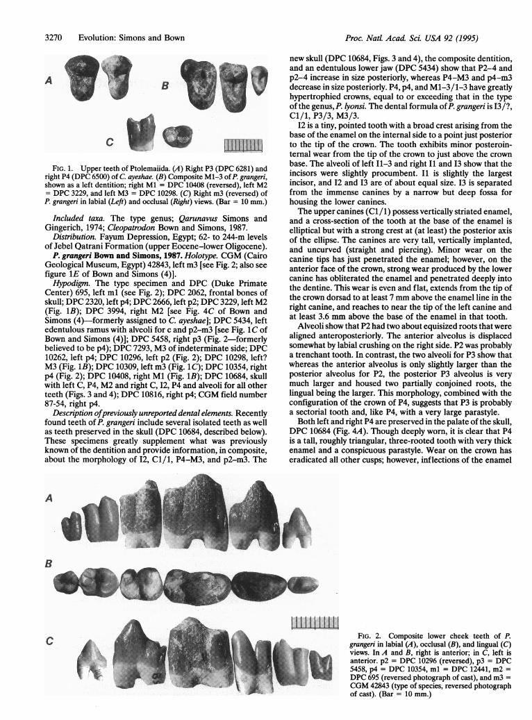
IUII,1FIG. 1. Upper teeth of Ptolemaiida. (A) Right P3 (DPC 6281) and
right P4 (DPC 6500) of C. ayeshae. (B) Composite M1-3 of P. grangeri,shown as a left dentition; right Ml = DPC 10408 (reversed), left M2= DPC 3229, and left M3 = DPC 10298. (C) Right m3 (reversed) ofP. grangeri in labial (Left) and occlusal (Right) views. (Bar = 10 mm.)
Included taxa. The type genus; Qarunavus Simons andGingerich, 1974; Cleopatrodon Bown and Simons, 1987.
Distribution. Fayum Depression, Egypt; 62- to 244-m levelsof Jebel Qatrani Formation (upper Eocene-lower Oligocene).
P. grangeri Bown and Simons, 1987. Holotype. CGM (CairoGeological Museum, Egypt) 42843, left m3 [see Fig. 2; also seefigure 1E of Bown and Simons (4)].Hypodigm. The type specimen and DPC (Duke Primate
Center) 695, left ml (see Fig. 2); DPC 2062, frontal bones ofskull; DPC 2320, left p4; DPC 2666, left p2; DPC 3229, left M2(Fig. 1B); DPC 3994, right M2 [see Fig. 4C of Bown andSimons (4)-formerly assigned to C. ayeshae]; DPC 5434, leftedentulous ramus with alveoli for c and p2-m3 [see Fig. 1C ofBown and Simons (4)]; DPC 5458, right p3 (Fig. 2-formerlybelieved to be p4); DPC 7293, M3 of indeterminate side; DPC10262, left p4; DPC 10296, left p2 (Fig. 2); DPC 10298, left?M3 (Fig. 1B); DPC 10309, left m3 (Fig. 1C); DPC 10354, rightp4 (Fig. 2); DPC 10408, right Ml (Fig. 1B); DPC 10684, skullwith left C, P4, M2 and right C, 12, P4 and alveoli for all otherteeth (Figs. 3 and 4); DPC 10816, right p4; CGM field number87-54, right p4.
Description ofpreviously unreported dental elements. Recentlyfound teeth of P. grangeri include several isolated teeth as wellas teeth preserved in the skull (DPC 10684, described below).These specimens greatly supplement what was previouslyknown of the dentition and provide information, in composite,about the morphology of 12, Cl/1, P4-M3, and p2-m3. The
A
new skull (DPC 10684, Figs. 3 and 4), the composite dentition,and an edentulous lower jaw (DPC 5434) show that P2-4 andp2-4 increase in size posteriorly, whereas P4-M3 and p4-m3decrease in size posteriorly. P4, p4, and M1-3/1-3 have greatlyhypertrophied crowns, equal to or exceeding that in the typeof the genus,P. lyonsi. The dental formula ofP. grangeri is 13/?,Cl/i, P3/3, M3/3.
I2 is a tiny, pointed tooth with a broad crest arising from thebase of the enamel on the internal side to a point just posteriorto the tip of the crown. The tooth exhibits minor posteroin-ternal wear from the tip of the crown to just above the crownbase. The alveoli of left 11-3 and right Ii and 13 show that theincisors were slightly procumbent. I1 is slightly the largestincisor, and 12 and I3 are of about equal size. I3 is separatedfrom the immense canines by a narrow but deep fossa forhousing the lower canines.The upper canines (C1 /1) possess vertically striated enamel,
and a cross-section of the tooth at the base of the enamel iselliptical but with a strong crest at (at least) the posterior axisof the ellipse. The canines are very tall, vertically implanted,and uncurved (straight and piercing). Minor wear on thecanine tips has just penetrated the enamel; however, on theanterior face of the crown, strong wear produced by the lowercanine has obliterated the enamel and penetrated deeply intothe dentine. This wear is even and flat, extends from the tip ofthe crown dorsad to at least 7 mm above the enamel line in theright canine, and reaches to near the tip of the left canine andat least 3.6 mm above the base of the enamel in that tooth.
Alveoli show that P2 had two about equisized roots that werealigned anteroposteriorly. The anterior alveolus is displacedsomewhat by labial crushing on the right side. P2 was probablya trenchant tooth. In contrast, the two alveoli for P3 show thatwhereas the anterior alveolus is only slightly larger than theposterior alveolus for P2, the posterior P3 alveolus is verymuch larger and housed two partially conjoined roots, thelingual being the larger. This morphology, combined with theconfiguration of the crown of P4, suggests that P3 is probablya sectorial tooth and, like P4, with a very large parastyle.Both left and right P4 are preserved in the palate of the skull,
DPC 10684 (Fig. 4A). Though deeply worn, it is clear that P4is a tall, roughly triangular, three-rooted tooth with very thickenamel and a conspicuous parastyle. Wear on the crown haseradicated all other cusps; however, inflections of the enamel
a
B
.. MIL.
_, i_
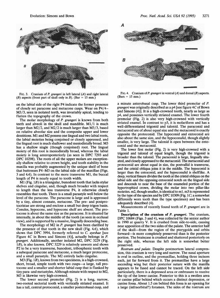
FIG. 2. Composite lower cheek teeth of P.grangeri in labial (A), occlusal (B), and lingual (C)views. In A and B, right is anterior; in C, left isanterior. p2 = DPC 10296 (reversed), p3 = DPC5458, p4 = DPC 10354, ml = DPC 12441, m2 =DPC 695 (reversed photograph of cast), and m3 =CGM 42843 (type of species, reversed photographof cast). (Bar = 10 mm.)
Proc. Natl. Acad Sci. USA 92 (1995)
c
c
Proc. Natl. Acad Sci USA 92 (1995) 3271
A
B
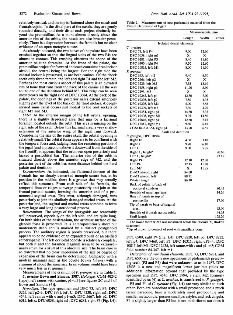
FIG. 3. Cranium of P. grangeri in left lateral (A) and right lateral(B) aspects (front part of skull only in B). (Bar = 15 mm.)
on the labial side of the right P4 indicate the former presenceof closely set paracone and metacone cusps. Wear on P4/4-M3/3, seen in isolated teeth, was invariably apical, tending toflatten the topography of the crown.The molar morphology of P. grangeri is known from both
teeth and alveoli in the skull and mandible. Mi/i is muchlarger than M2/2, and M2/2 is much larger than M3/3, basedon relative alveolar size and the composite upper and lowerdentitions. Ml and M2 possess one lingual and two labial roots,the labial moieties being conjoined or closely appressed, andthe lingual root is much shallower and mesiodistally broad. M3has a shallow single (though conjoined) root. The lingualmoiety of this root is mesiodistally broad, whereas the labialmoiety is long anteroposteriorly (as seen in DPC 7293 andDPC 10298). The roots of all the upper molars are exception-ally shallow relative to crown height, and tooth stability in themaxilla was probably augmented by the unique shelf of bonethat buttresses P4-M3 on the labial side of the maxillae (Figs.3 and 4A). In contrast to the more transverse Ml, the buccallength of P4 is nearly equal to tooth width.Ml (Fig. 1B) is a triangular, bunodont tooth that lacks stylar
shelves and cingulae, and, though much broader with respectto length than the less transverse P4, it otherwise closelyresembles that tooth. There is a small parastyle well below thelevel of the paracone. The paracone is large and is appressedby a tiny, almost connate, metacone. The pre- and postpro-tocristae are strong and enclose a small but deep trigon basin.Conules, hypocone, and hypocone shelf are absent. The pro-tocone is about the same size as the paracone. It is situated farinternally, in about the middle of the tooth (as seen in occlusalview), and is supported by a massive, lingually descending base.The morphology of M2 in P. grangeri has been clarified by
the presence of that tooth in the new skull (Fig. 4A), whichshows that DPC 3994, formerly referred to C. ayeshae [seeFigure 4C in Bown and Simons (4)], actually belongs in P.grangeri. Additionally, another isolated M2, DPC 3229 (Fig.1B), is also known. DPC 3229 is relatively unworn and showsP2 to be a very transverse, bunodont, tribosphenic tooth witha subequal paracone and metacone, a much larger protocone,and a small parastyle. The M2 entirely lacks cingulae.M3 (Fig. 1B), known from two specimens, is a high-crowned,
short, broad, simple tooth with two cusps-a large, tall pro-tocone and a smaller and shorter labial cusp that is flanked bytiny para- and metastyles. Although minute with respect to M2,M3 is likewise very high-crowned.The lower second premolar (Fig. 2) is a long, narrow,
two-rooted sectorial tooth with vertically striated enamel. Ithas a tall, central protoconid, a smaller posterobasal cusp, and
FIG. 4. Cranium ofP. grangeri in ventral (A) and dorsal (B) aspects.(Bars = 15 mm.)
a minute anterobasal cusp. The lower third premolar of P.grangeri was originally described as a p4 [see figure 4C of Bownand Simons (4)]. It is a high-crowned tooth, nearly as large asp4, and possesses vertically striated enamel. The lower fourthpremolar (Fig. 2) is also very high-crowned with verticallystriated enamel. In contrast to p3, it is molariform and has awell-differentiated trigonid and talonid. The paraconid andmetaconid are of about equal size and the metaconid is exactlyopposite the protoconid. The hypoconid and entoconid arealso about the same size, and the hypoconulid, though slightlysmaller, is very large. The talonid is open between the ento-conid and the metaconid.The lower first molar (Fig. 2) is very high-crowned with a
trigonid and talonid of equal length, though the trigonid isbroader than the talonid. The paraconid is large, lingually situ-ated, and closely appressed to the metaconid. The metaconid andprotoconid are about equal in size, the postvallid is transverse,and the cristid obliqua joins it in the middle. The hypoconid islarger than the entoconid, and the hypoconulid is shelflike. Adeep, vertical fissure divides the tooth at the cristid obliqua on thelabial side and the opposing entoconid notch on the lingual sideand descends to near the base of the enameled portion of thehypertrophied crown, dividing the molar into two pillar-likemoieties. m2, though smaller, is identical to ml. m3 is representedby the type of the species and byDPC 10309 (Fig. 1C; a somewhatdifferently worn tooth than the type specimen) and has beenadequately described (4).Measurements of recently found teeth of P. grangeri are in
Table 1.Description of the cranium of P. grangeri. The cranium,
DPC 10684 (Figs. 3 and 4), was collected by the senior authorin 1990 at quarry V. It is crushed from side-to-side, causingnear apposition of the two sides of the palate. The anterior halfof the skull-from the region of the pterygoids and orbitsforward-is more completely preserved than is the posteriorportion. The braincase is crushed and shattered posteriorly onthe right side, whereas the left side is somewhat betterpreserved.Rostrum and palate. Despite postmortem lateral compres-
sion, this cranium is very long and narrow. The nasal apertureis oval in outline, and the premaxillae, holding three incisorseach, jut far forward from it. The premaxillae have a largeascending wing but their posterior suture with the maxillaappears to be fused and cannot be traced. On the left sideparticularly, there is a depressed area or embrasure to receivethe tip of the lower canine. Posterior to this is a swollen areaor canine pillar behind which occurs a distinct depression, orcanine fossa. About 1.5 cm behind this fossa is an opening fora large (infraorbital?) foramen. The sides of the rostrum are
Evolution: Simons and Bown
3272 Evolution: Simons and Bown
relatively vertical, and the top is flattened where the nasals andfrontals cojoin. In the distal part of the nasals, they are gentlyrounded dorsally, and their distal ends project distinctly be-yond the premaxillae. At a point almost directly above theanterior rim of the orbits, the nasals are also broader poste-riorly. There is a depression between the frontals but no clearevidence of an open metopic suture.As already indicated, the two halves of the palate have been
crushed together so that the lingual sides of the two P4s arealmost in contact. This crushing obscures the shape of theanterior palatine foramina. At the front of the palate, thepremaxillae project far forward and each contains three incisoralveoli, the central being the largest. For the right side thecentral incisor is preserved, as are both canines. Of the cheekteeth only three remain, the left and right P4 and the left M2.Perhaps the most curious aspect of this palate is an elevatedrim of bone that runs from the back of the canine all the wayto the end of the dentition behind M3. This ridge can be seenmost clearly on the right side of DPC 10684. At the back of thepalate, the openings of the posterior nares extend anteriorlyslightly past the level of the back of the third molars. A deeplyincised sinus canal occurs just medial to the root sockets ofright M2 and M3.
Orbit. At the anterior margin of the left orbital opening,there is a slightly depressed area that may be a lacrimalforamen located outside the orbit. This area is missing on theright side of the skull. Below this lacrimal area, a very shallowextension of the anterior wing of the jugal runs forward.Considering the size of the entire skull, the orbital opening isrelatively small. The orbital fossa appears to be confluent withthe temporal fossa and, judging from the remaining portion ofthe jugal (and a projection above it downward from the side ofthe frontal), it appears that the orbit was open posteriorly andlacked a postorbital bar. The anterior rim of the orbit issituated directly above the anterior edge of M2, and theposterior part of the orbit lies some distance behind the hardpalate and dentition.Dorsacranium. As indicated, the flattened dorsum of the
frontals has no clearly demarked metopic suture but, at itsposition in the midline, there is a groove that may actuallyreflect this structure. From each side of the frontals, thetemporal lines or ridges converge posteriorly and join at thefrontal-parietal suture, forming the anterior end of a pro-nounced sagittal crest. This crest, although damaged, runsposteriorly to join the similarly damaged nuchal crests. At theposterior end, the sagittal and nuchal crests combine to forma very large and long posterodorsal process.
Basicranium. The wings of the pterygoids are reasonablywell preserved, especially on the left side, and are quite long.On both sides of the basicranium, the articular surface of theglenoid fossa is preserved. It is anteroposteriorly short butmoderately deep and is marked by a distinct postglenoidprocess. The auditory region is poorly preserved, but thereappears to be no evidence of an expanded bulla or an ossifiedectotympanic. The left occipital condyle is relatively complete,but both it and the foramen magnum seem to be extraordi-narily small for a skull of this absolute size. The brain case isso distorted that no clear impression of the size or degree ofexpansion of the brain can be determined. Compared with amodern mammal such as the coyote (Canis latrans) with acranium of about the same size, brain volume would have beenvery much less in P. grangeri.
Measurements of the cranium of P. grangeri are in Table 1.C. ayeshae Bown and Simons, 1987. Holotype. CGM 40242
(type), left ramus with canine, pl-m3 [see figures 2C and 3 ofBown and Simons (4)].Hypodigm. The type specimen and DPC 73, left P4; DPC
3265, left p2-3; DPC 3991, left C; DPC 4330, right ml; DPC4545, left ramus with c and pl-m3; DPC 5667, left p2; DPC6015, left c; DPC 6058, right ml; DPC 6281, right P3 (Fig. 1A);
Table 1. Measurements of new ptolemaiid material from theFayum Depression of Egypt
Measurements, mm
Length Width Other
Isolated dental elementsC. ayeshaeDPC 73, left P4 9.00DPC 6058, right ml XDPC 6281, right P3 8.40DPC 6500, right P4 9.50DPC 10815, left Ml 8.00
P. grangeriDPC 695, left m2 9.40DPC 2666, left p2 XDPC 3229, left M2 7.55DPC 5458, right p3 11.70DPC 7293, M3 XDPC 10262, left p4 12.30DPC 10296, left p2 7.80DPC 10298, left M3 5.00DPC 10309, left m3 7.30DPC 10354, right p4 14.30DPC 10408, right Ml 9.05DPC 10816, right p4 12.60DPC 12441, right ml 9.60CGM field 87-54, right p4 13.20
Skull and dentitionP. grangeri, DPC 10684
Right 12Right CLeft CRight C, height*Left C, height*Right P4Left P4Left M2I1-M3 alveoli, rightI1-M3 alveoli, leftPalatal lengthBack of palate to back of
occipital condylesBreadth of nasal apertureBase of nasals to top of
premaxilla
12.60x
11.8012.6011.50
6.50x
13.105.90x7.004.357.854.707.35
14.507.156.456.55
4.30 3.109.20 6.109.00 5.85
12.1012.15x
84.6088.7186.70
12.3011.7011.85
20.6023.18
90.4514.20
17.00Tip of nasals to base of saggital
crest 93.00Breadth of frontals across orbits 44.05Skull length 178.20
The lower tooth width was measured across the talonid. X, Brokenspecimen.*Tip of crown to contact of root with maxillary bone.
DPC 6500, right P4 (Fig. 1A); DPC 8220, left p3; DPC 8222,left p4; DPC 9468, left P3; DPC 10311, right dP3-4; DPC10815, left Ml; DPC 12435, left ramus with c and pl-m3; CGMfield number 84-347, left ml.
Description ofnew dental elements. DPC 73, DPC 6281, andDPC 6500 are the only new specimens of ptolemaiids preserv-ing teeth (P3 and P4) that were unknown to us in 1987. DPC12435 is a new and magnificent lower jaw but yields noadditional information beyond that provided by the typespecimen and DPC 4545. DPC 3994, a right M2, formerlyidentified by us (4) as C. ayeshae, is transferred to P. grangeri.P3 and P4 of C. ayeshae (Fig. 1A) are very similar to each
other. Both are bunodont with a small protocone and a muchlarger paracone, have a moderately sized protoconule andsmaller metaconule, possess small parastyles, and lack cingula.P4 is slightly larger than P3 but is not molariform nor does it
Proc. Natl. Acad Sci. USA 92 (1995)

Proc. Natl. Acad. Sci. USA 92 (1995) 3273
possess the large parastyle seen in P. grangeri. Measurementsof P3 and P4 of C. ayeshae are in Table 1.
DISCUSSION AND CONCLUSIONSThe history of study of the possible relationships of ptolemaiidmammals was reviewed most recently by Bown and Simons (4).Osborn (2) suggested that the dental peculiarities of P. lyonsiperhaps warranted its placement in a new order. However,most recent workers have consistently linked the ptolemaiidsto and compared them with various members of what nowstands as McKenna's (5) cimolestan suborder Pantolesta (3, 4,6-14). Bown and Simons (4) believed Qarunavus to be themost generalized ptolemaiid and that there was a major butpoorly understood radiation of these mammals in the earlyTertiary of Africa, with monotypic Qarunavus and the twospecies of Cleopatrodon constituting a conservative line, and P.lyonsi and P. grangeri belonging to another, dentally moreadvanced lineage. The new specimens of C. ayeshae andespecially of P. grangeri described above, together with infor-mation about new specimens of paroxyclaenid pantolestansfrom the Eocene of Europe, materially alter only our conclu-sions about the ordinal affinities of the Ptolemaiidae. Ptole-maia, Qarunavus, and Cleopatrodon, though perhaps ulti-mately of pantolestan derivation, possess sufficient gnathic andcranial specializations to warrant their recognition as a neworder of mammals, here named the Ptolemaiida.
Russell and Godinot (14) described in 1988 two new generaof paroxyclaenid pantolestans (Euhookeria and Merialus) fromupper Eocene rocks of England and lower Eocene rocks ofFrance, respectively, and placed them in a new subfamily of thepantolestan Paroxyclaenidae, the Merialinae. With regard toPtolemaia, Russell and Godinot (p. 328 of ref. 14) noted that"..perhaps a particular relationship to the merialine paroxy-claenids is discernable." Merialines differ from paroxycla-enines in (i) having lingually situated molar paraconids that areclose to the metaconids, (ii) possessing simple premolariformpremolars, and (iii) lacking a metaconid on p4. It is in thesethree merialine characters (of which at least the second andthird are surely plesiomorphic) that the merialines vaguelyresemble Cleopatrodon and Qarunavus; however, we recordseveral qualifications. First, the permanent p4 is unknown inQarunavus; second, the p4 in Cleopatrodon is semimolariformand possesses two talonid cusps and has either a metaconid ora greatly expanded protoconid; and last, the p4 in P. grangeri(and possibly P. lyonsi as well) is fully molariform and has notonly distinct but also large, paraconid, metaconid, hypoconu-lid, entoconid, and hypoconid cusps.No other mammalian order shares the combination of
dental characters seen in ptolemaiids. Whereas it may ulti-mately be proven that the Ptolemaiida were derived from theOrder Cimolesta, and even from the merialine paroxyclaenids,no meaningful diagnosis of either group is broad enough toinclude Ptolemaia and its bizarre dental specializations. Ptole-maia, Qarunavus, and Cleopatrodon are morphologically verydistant from Merialus and Euhookeria, their closest mamma-lian counterparts, and they are, correspondingly, equally dis-tant from the order and suborder containing the Merialinae.A hallmark of the Pantolesta (and the order Cimolesta ingeneral) is the lack of pronounced dental specializations-acircumstance that has caused members of that group (undervarious taxonomic appellations) to have been referred to-gether with many other relatively generalized (and unrelated)mammals in the "Insectivora" or "Proteutheria" for half acentury. Both Merialus and Euhookeria are very poorly known,and additional material (especially of Euhookeria) may showthat both are perhaps best regarded to be members of early,
Qarunavus and Cleopatrodon and the recognition of theirrelationship to Ptolemaia. Those two genera are clearly themost generalized members of the Ptolemaiida and, quixoti-cally, appear to have survived the latest (Cleopatrodon robustafrom quarry I, 244-m level of Jebel Qatrani Formation). Whatthe Fayum ptolemaiids clearly constitute is a small sample ofspecimens taken from a largely unknown but significant radi-ation of a unique group-all collected from rocks recording abrief interval of time, and all taken from a tiny area in theFayum Depression of Egypt.When assuming that an animal dentally similar to Cleopatro-
don or Qarunavus formed part of the ancestry of Ptolemaia,trends in the evolution of the ptolemaiid dentition included: (i)increase in proportionate size and height of P3/3-Mi/i; (ii)decrease in proportionate size and height of P2/2 and M2/2-M3/3; (iii) hypertrophy of P3/3-M3/3; (iv) decrease in sizeor loss of P1/1-P2/2; and (v) loss of transverse shear capacityin all teeth. Wear on ptolemaiid teeth is usually extensive andon P3/3-M2/2, even in the early stages, generally records noshear (i.e., it is rapidly flattening the topography of both theupper and lower cheek teeth). In Fig. 2, it is seen that p2-m3of P. grangeri form a convex-upward arcuate pattern, the mosthypsodont (and most worn) teeth occurring at the apex of thearc. In contrast, the upper cheek teeth (Fig. 1B), with theexception of the massive P4 and Ml, are diminutive andexceptionally shallow-rooted, apparently having acted as apestle against which worked the center of the massive lowerdental battery.A combination of morphology and the wear on teeth of P.
grangeri suggests that the function of such a dental array waslargely vertical crushing with virtually no shear capacity. Theextreme hypsodonty and wear on p3-m2 supports this hypoth-esis and indicates that the food utilized consisted principally ofa resilient, abrasive material. Dorr (11) fancifully conjecturedthat the pantolestine pantolestan Palaeosinopa simpsoni, whichpossesses a pattern of similar flat, apical molar wear, but lacksthe hypertrophy of the central cheek teeth seen in Ptolemaia,might have incorporated insects and mollusks as the majorportion of its food. Whereas a similar mode of life is possiblefor the ptolemaiids, there is as yet insufficient evidence todetermine what might have been the principal food prefer-ences of Ptolemaiida.
We thank the Egyptian Geological Survey and Mining Authorityand the Cairo Geological Museum for its sponsorship of our Egyptianfield program. Field work was funded by National Science FoundationGrant BNS 8809776 to E.L.S. The specimens described here wereprepared by E.L.S. and P. S. Chatrath; P. S. Chatrath also assisted withphotography. We are grateful to F. Ankel-Simons, E. M. Brouwers,and P. A. Holroyd for comments and assistance with the manuscript,and M. C. Maas and D. T. Rasmussen for technical review.
1.
2.3.
4.5.
6.7.
8.9.
10.11.12.
13.
14.
very generalized branches of the Order Ptolemaiida.The ordinal status of the Ptolemaiida is rendered more
interesting, but none the less secure, by the descriptions of
Bown, T. M. & Kraus, M. J. (1988) U.S. Geol. Surv. Prof Pap. 1452,1-64.Osborn, H. F. (1908) Am. Mus. Natl. Hist. Bull. 24, 265-272.Simons, E. L. & Gingerich, P. D. (1974) Ann. Geol. Surv. Egypt 4,157-166.Bown, T. M. & Simons, E. L. (1987) J. Vert. Paleontol. 7, 311-324.McKenna, M. C. (1975) in Phylogeny of the Primates, eds. Luckett,P. W. & Szalay, F. S. (Plenum, New York), pp. 21-46.Schlosser, M. (1910) Zool. Anz. 35, 500-508.Schlosser, M. (1922) in Grundzuege der Paldeontologie II Abteilung:Vertebrata, ed. von Zittel, K. A. (Oldenbourg, Munich), pp. 402-689.Matthew, W. D. (1918) Am. Mus. Nati. Hist. Bull. 38, 565-657.Van Valen, L. (1966) Am. Mus. Natl. Hist. Bull. 132, 1-126.Coombs, M. C. (1971) Am. Mus. Novit. 2455, 1-41.Dorr, J. A. (1977) Contrib. Mus. Paleontol. Univ. Mich. 24, 281-307.Butler, P. M. (1978) in Evolution of African Mammals, ed. Maglio,V. J. (Harvard Univ., Cambridge, MA), pp. 56-68.Savage, D. E. & Russell, D. E. (1983) Mammalian Paleofaunas of theWorld (Addison-Wesley, London).Russell, D. E. & Godinot, M. (1988) Palaontol. Zeitschr. 62, 319-331.
Evolution: Simons and Bown