protocol for postoperative epidural analgesia · protocol for postoperative epidural analgesia | 7...
TRANSCRIPT
Protocol for Postoperative Epidural Analgesia | 1
Protocol forPostoperative
Epidural AnalgesiaFor reference by
anaesthetic staff, nursing staff in theatres and ACCU
Wishaw General Hospital

2 | Protocol for Postoperative Epidural Analgesia
Protocol for Postoperative Epidural Analgesia | 3
Contents Page
Basic Requirements for Use of Epidural Analgesia 5
Epidural Drugs 8
Nurse administered epidural “Top ups” 9
Inadequate Analgesia 10
Manual Top Up of Epidural Analgesia 11
Hypotension 13
Excessive Motor or Sensory Block 14
Spinal/Epidural Haematoma/Abscess 14
Respiratory Depression 15
Postoperative Sedation, Nausea and Vomiting, Pruritis 16
Catheter Disconnection 17
Discontinuing Epidural Analgesia and Step Down Analgesia 17
Medical On Call Arrangements 18
Appendix 1: Guidelines for Neuraxial Block and Coagulation 19
Key Words: Epidural; Analgesia; Neuraxial; Local Anaesthetic; Spinal abscess; Epidural haematoma; Coagulation
Staff Groups: Anaesthetics; Surgeons; ACCU staff; Surgical and Theatre Nursing Staff Author: C. Slorach

4 | Protocol for Postoperative Epidural Analgesia
Protocol for Postoperative Epidural Analgesia | 5
General Considerations
Provision of analgesia by the infusion of drugs into the epidural space is intended to provide patient comfort even in situations such as mobilising and physiotherapy where parenteral opioids may not be as effective. Other benefits include patients who are generally more alert and the possibility of an earlier return of gut function. There is now evidence that return to normal activity is accelerated after major bowel surgery. Conversely the technique is more invasive and the possibility of severe and potentially fatal complications exists. The implementation of this protocol and the involvement of suitably trained staff should reduce this risk to an acceptable minimum.
BasiC requirements for use of epidural analGesia.The Anaesthetist inserting the epidural catheter should take the following points into consideration:
Indication - this would usually include major lower limb, pelvic, abdominal, major vascular or thoracic surgery, especially in patients with pre-operative respiratory insufficiency.
Contra-indications - patient refusal, local infection at the insertion site, septicaemia, uncorrected coagulopathy and current antiplatelet therapy with the exception of aspirin (see appendix 1), raised intracranial pressure and lack of suitable monitoring and nursing care are normally absolute contra-indications. Spinal deformity, diabetic or other neuropathy and cardiac conditions with fixed output might be regarded as relative contra-indications. As with other invasive procedures, informed consent should normally be obtained at the pre-operative visit and documented.
An intravenous canula must be inserted before the epidural catheter is inserted and remain functional throughout the time the catheter stays in place.
The epidural catheter should be inserted under sterile conditions according to local guidelines for infection control. The inserting anaesthetist should use an aseptic technique including Gown Mask Gloves and Hat. All staff within 3 metres of the patient and trolley should wear a facemask during trolley preparation and epidural insertion. Skin should be prepared with 0.5% Chlorhexidine in 70% alcohol solution (e.g. Hydrex) which should be applied separately and allowed to dry before application of sterile drapes and commencement of procedure. In cases where there is an increased risk of catheter infection (e.g. diabetics, the elderly) it would seem prudent to commence any surgically indicated prophylactic antibiotics prior to epidural insertion where possible.
The epidural catheter should be inserted at the appropriate level taking into consideration the position of the incision and surgery i.e. the catheter tip should be placed approximately at the mid-point of the dermatomes involved.
An adequate length of catheter should be left in the epidural space. 4 to 5 cm are recommended to decrease the risk of failure due to catheter dislodgement.
he catheter should be secured appropriately so as to minimise movement into or out of the epidural space. A proprietary “lock it” fixation device covered with a suitable transparent dressing should be applied to allow inspection of the insertion site. A Tegaderm with Mefix around are appropriate.
The site should be visually inspected every 12 hours without removing the clear dressing and this documented on the epidural chart or nursing notes. The transparent dressing should only be removed and replaced if loose or if excessive leak of blood/fluid beneath skin. This should be done in an aseptic manner. An anaesthetist should be notified to review the site if this is the case.
The epidural giving set and epidural infusion bag should be changed every 48 hours. The filter should remain in situ.
6 | Protocol for Postoperative Epidural Analgesia
Test Dose: An Anaesthetist should give the first dose of local anaesthetic through the epidural catheter and satisfy themself that the epidural tip is not placed intrathecally or intravenously. Where a combined spinal/epidural technique has been used this rule still applies. The use of adrenaline to detect intravascular catheter placement is controversial, but is thought by some experts to enhance safety. 3 ml of levobupivacaine 5mg/ml or lidocaine 2% is recommended as the test dose to detect an intrathecal or intravenous tip position. This dose if administered into the epidural space should not cause a significant lower limb motor block after 5 minutes, whereas with an inadvertently placed intrathecal catheter the test dose would give an easily appreciable lower limb block within 5 minutes. The addition to the local anaesthetic test dose of adrenaline 1:200,000 (5 micrograms/ml) has been recommended to detect an inadvertent intravascular placement of an epidural catheter. In the case of the intravascular placement of the epidural catheter tip, one would expect to see a rise in pulse rate of some 20 – 30 /min within a minute of administering the test dose. This may not be the case in the elderly or those patients taking beta receptor blockers. The anaesthetist should decide on an individual basis whether to use adrenaline in the test dose. Ampoules of lidocaine 2% with 1:200,000 adrenaline sterile wrapped are available. The above dose of the newer, less toxic local anaesthetics may not cause the usual symptoms of toxicity as compared to lidocaine 2% or bupivacaine 0.5%. The Anaesthetist should be familiar with the current 2010 AAGBI guidelines for management of severe local anaesthetic toxicity and aware of the location of the nearest bag of lipid emulsion. The Anaesthetist should be sure that the epidural catheter tip is in the epidural space.
An Anaesthetist should prescribe a suitable epidural prescription and check that the infusion device has been suitably programmed. The anaesthetist should also discuss with the surgeon and indicate the intended duration of epidural infusion (usually 48 to 72 hours) and an absolute maximum duration of epidural therapy on the epidural chart, which should not exceed 5 days.
The inserting anaesthetist should document on the epidural chart a minimum lower limit for Systolic blood pressure, after agreement with the Consultant Surgeon at time of operation.
Solutions for continuous epidural analgesia should be kept to the lowest possible effective concentrations of local anaesthetic/opioid. The Anaesthetist should record the drug(s) and their concentration(s) to be infused, the volume of any bolus dose with lockout time, an initial rate of infusion and a range of permissible infusion rates that may be used. Our default settings are 3ml bolus and 15 minute lockout for nurse/doctor pump administered bolus. An hourly dose limit ensures that at the maximal prescribed infusion rate, only 2 pump delivered boluses can be administered over and above in any one hour (e.g. rate 0 to 14ml/hour, hourly limit 20ml, that is: 14 + 3 + 3). A suggested top up in event of ineffective pump bolus is helpful.
When an opioid is included in the epidural infusion, parenteral opioids should normally be avoided.
The use of sedative drugs should generally be minimised. Consider the sedative effect of some of the older types of antiemetic, especially in the elderly.
The patient should have an epidural block “ice test” documented using either Ethyl Chloride BP [if not contra-indicated] or ice and satisfactory analgesia in the recovery area before being discharged to ACCU. On handover to ACCU care the recovery nurse and receiving ACCU nurse should together check the epidural prescription sheet, bag content labels and epidural pump settings to minimise risk of drug error.
The patient will be monitored in the ACCU by members of the nursing staff who have been instructed in the care of patients with epidurals and also in the use of the epidural infusion device.
Protocol for Postoperative Epidural Analgesia | 7
Minimum monitoring should include ECG, oxygen saturation and blood pressure using NIBP or invasive device. Measurements should be taken and recorded at no less than 2 hourly intervals. In addition sedation score, pain scores on movement and block height by “ice test” or Ethyl Chloride BP should be monitored. Presence of motor block using Bromage score and nausea/vomiting should also be assessed and recorded. Continuous monitoring is mandatory when a top-up of high concentration local anaesthetic is required i.e. more than 0.1% (1.0mg/ml) concentration of long acting solution or 2% lidocaine with adrenaline.
A member of the Anaesthetic staff should be present in the hospital at all times who has received instruction in epidural analgesia. There should be a minimum of 1 visit per 24 hours by a member of the Anaesthetic Staff/Acute Pain Nurse Specialist to assess patients who are receiving epidural analgesia. In addition, an Anaesthetist will be on call to give advice and assistance should the need arise. This is usually the ACCU duty doctor on pager 003.
Members of the Surgical staff should refer to a member of the Anaesthetic staff should they wish to prescribe anti-coagulant therapy (including DVT prophylaxis) and all staff should be vigilant to the signs and symptoms of bleeding in the epidural space (back pain, progressive lower limb weakness and/or incontinence). The use of anti-platelet drugs in addition to low dose heparin is an unquantifiable additional risk.
Protocols covering the areas of inadequate pain relief, side effects and complications, step-down pain relief, anticoagulant use, infusion bag mixtures and catheter removal are included.
Audit of patient numbers, pain scores, and complications will be kept.
• Remember that epidural analgesia is not a “set and forget” or “low maintenance” technique. The Anaesthetist who establishes an epidural catheter for postoperative analgesia is responsible for ensuring that the appropriate nursing and medical care is available. This is to ensure that regular checks are made of the patient’s analgesia and any side effects or complications of the treatment are detected.
8 | Protocol for Postoperative Epidural Analgesia
infusion BaG mixtures Levobupivacaine 0.1% (1mg/ml) with Fentanyl 2 micrograms/ml
The standard mixed bag is an infusion of Levobupivacaine 0.1% (1mg/ml) with Fentanyl 2 micrograms/ml. This is presented in a 250ml or 500ml volume; pre-mixed in a licensed pharmacy unit. Plain bags of local anaesthetic solution are also available and exceptionally may have opioid added by an Anaesthetist for epidural infusion. Consultants have the discretion to use an alternative solution if thought advantageous for an individual patient, however where possible a licensed infusion bag should be used.
Levobupivacaine (Chirocaine)
This has equivalent potency and duration of action to bupivacaine but levobupivacaine is less toxic in the case of intravascular injection, for this reason Levobupivicaine has replaced Bupivicaine in NHS Lanarkshire; with the exception of the 4ml sterile wrapped “heavy” 0.5% Bupivicaine used for intrathecal anaesthesia.
Levobupivicaine is used to induce and maintain local and regional anaesthesia and analgesia. Typically 2.5mg/ml - 7.5mg/ml is used to establish an epidural block. 0.625 - 1.25mg/ml concentration is then used as a continuous infusion. Infusion rates are usually set at around 6 - 10 ml/hour but may vary between 4 - 15 ml/hour depending on the insertion level of the catheter, concentration of any opioid in the infusion mixture, nature and extent of surgery, the analgesic effect obtained and the incidence of side effects. Maximum recommended dose is 2mg/Kg /6 hour period (in the fit and healthy)
Ropivacaine (Naropin)
This long acting local anaesthetic may be used to induce and maintain anaesthesia. It is slightly less potent than bupivacaine and may produce a lower incidence of unwanted motor block. It is reported to be less toxic than bupivacaine in the event of inadvertent intravascular injection. 7.5mg/ml and 10mg/ml concentrations are used for surgical epidural anaesthesia and the 2mg/ml concentration used for postoperative epidural infusion. It has the advantage of being packaged in a 200ml Polybag. Infusing the drug alone may be sufficient for lower limb and some abdominal surgery; however its use with upper abdominal incisions does not always provide adequate analgesia without fairly frequent top-ups. In this circumstance it is better to add diamorphine or fentanyl. The addition of 10mg of diamorphine to the 200ml bag has worked satisfactorily for patients under 70 years. 5mg should be added for use with the old or frail patient. Infusion rate may range from 4 – 15 ml/hour, but are usually set at 5 – 6ml/hour for lower limb operations. 6 - 10 ml/hr usually suffices for abdominal operations where the epidural catheter has been placed in the thoracic spine. The data sheet is slightly vague regarding the upper dose range but appears to be 3 – 4mg/Kg as a single dose with a recommended infusion rate of 6 – 14ml/hour of the 2mg/ml concentration. Fentanyl at a concentration of 2 – 5 micrograms/ml may be used.
Protocol for Postoperative Epidural Analgesia | 9
nurse administered epidural “top ups”In the case of inadequate epidural analgesia a nurse with appropriate training on ACCU can deliver up to 2 x 3ml boluses per hour via epidural pump as per the epidural prescription sheet using the bolus button. Inadequate epidural analgesia is defined as a documented pain score of more than or equal to 3 on movement (or at rest) on the 0-4 scale. Time of bolus should be also be recorded on the epidural chart.
The latency (time to start taking effect) of both levobupivacaine and ropivacaine is in the 8 - 10 minute range, and for this reason a 15 minute lockout period is the default setting for pump delivered bolus administration. A 3ml pump bolus dose is the default setting: this is mainly to prevent drug toxicity in the event the catheter tip has migrated into a blood vessel, but is also to help detect an intrathecal or “spinal” injection at an early stage should the catheter have migrated into the subarachnoid space as evidenced by a very short onset time of profound analgesia with a pump bolus with or without motor block or reduction in blood pressure.
Epidural catheters can without warning migrate into a blood vessel or into the CSF space. Either situation may put the patient in danger if undetected. In any case should there be any doubt about catheter tip position, the infusion must be stopped, and no further injections given through it until advice has been sought from an Anaesthetist.
Manual top-ups are only administered by an appropriately trained Anaesthetist as below.
Requests for Advice: In addition to the rounds to monitor patients with epidurals, the nursing staff will call an Anaesthetist to see patients where a problem has been detected. They should perform observations including ice test. The following advice should assist in dealing with various scenarios. If in any doubt at all as to how to proceed, the resident anaesthetist must contact the on-call Consultant Anaesthetist.
10 | Protocol for Postoperative Epidural Analgesia
inadequate analGesia
If the pain scores are 3 or over on movement (0 - 4 scale), analgesia should be improved.
Poor analgesia is usually caused by:
1) A displaced epidural catheter. Check the catheter dressing is intact and no excessive leakage apparent at insertion site through clear dressing. A small amount of serosanguinous fluid is often seen under the transparent dressing this is normal. Epidural insertion site must only be covered by a clear dressing for this reason. If in doubt whether epidural catheter is still in epidural space or is lying subcutaneously or in the ligaments consider a single top up of strong local anaesthetic solution before abandoning the epidural.
2) Inadequate block: not covering operative site. The block may show signs on “ice test” of being insufficient to cover operative site. The nurses may have given up to 2 bolus doses of infusion mixture within the preceding hour. If there is some evidence of a block give these pump top ups now (2 x 3ml with 15 minute lockout). However if the patient is obviously in severe pain and no block is evident consider manual top up of strong local anaesthetic solution. See below for manual top up.
3) Unilateral Block. Evidenced by a report of unilateral pain and no block on ice test on one side of the abdomen but analgesia and block evident on the other side. In the case of a unilateral block withdrawing the epidural catheter to leave 3cm in the epidural space followed by a top up with the patient in the lateral position, sore side down, will often remedy the situation. The anaesthetic chart should indicate the depth of the epidural space and the length of catheter originally inserted. The patient’s back should be examined to determine which mark on the catheter is at skin level, as it may have moved since insertion. Care will be needed in removing the dressing not to pull out the epidural catheter, and sterile gloves should be worn when manipulating it. A fresh Tegaderm dressing and repair of the Mefix will be required. A note of this procedure should be made in the medical record including the aseptic precautions used. A stronger concentration of local anaesthetic may be required for a manual top up in this instance, but usually not more than 0.25% (2.5mg/ml) Levobupivacaine should be needed.
4) Inadequate drug dosing/mixture. Weak local anaesthetic solutions alone are unlikely to give adequate analgesia. If a plain solution is in use check the anaesthetic chart to ascertain if epidural or intrathecal opioid has been given or not. Check the patient for signs of any residual sedation or respiratory depression and if neither is present a loading dose of epidural opioid (see below) can be given followed by a 3ml pump bolus to flush in before switching to a standard 2microgram/ml fentanyl pre-mixed bag. If a standard pre-mixed bag is already in use consider a further opioid bolus manual top up with or without switching to a stronger fentanyl or daimorphine mixed bag. Switching to specially made mixed bags should be discussed with the on call Consultant Anaesthetist as an individual risk/benefit decision is required for individual patients as these bags require to be made up by anaesthetic staff.
5) Intrabdominal catastrophe:
Rare within period of time of an epidural infusion. Consider if sudden severe abdominal pain with previously good functioning epidural and no evidence of catheter displacement or any disruption of epidural infusion administration with a block still evident on “ice test” especially if abdominal signs and patient appears systemically unwell. Seek senior anaesthetic and surgical advice. Consider opioid only top up pending urgent surgical review.
Protocol for Postoperative Epidural Analgesia | 11
manual top up of epidural analGesia
Bolus dosing of a strong local anaesthetic solution is performed by an Anaesthetist only. The Anaesthetist should be familiar with the current 2010 AAGBI guidelines for management of severe local anaesthetic toxicity and aware of the location of the nearest bag of lipid emulsion.
A manual Top Up of Epidural Analgesia is appropriate in the event of inadequate epidural analgesia with no, or inadequate, block on “ice test” and after 1 or 2 pump boluses in the past hour have been ineffective or the patient is in very severe pain with no apparent block. Proceed as below guideline:
a) Check patient: sedation level, sensory and motor block. Check that the blood pressure is adequate (over 110mmHg) and ensure the patient is in the bed (rather than sitting out), also that there is a free flowing intravenous canula in place. A supply of ephedrine should be readily available in case of hypotension.
b) Check the epidural and anaesthetic chart: note infusion mixture and rate and any suggested top up. Check a test dose has been documented as given on the on the anaesthetic chart. If not draw up 10 ml of preservative free (double sterile wrapped) 2% lidocaine with adrenaline 1:200,000 otherwise 10ml of 0.25% Levobupivicaine and connect this to the epidural filter. Be careful not to contaminate the end of the infusion pump set tubing. Aspirate gently. You should not be able to aspirate more than 1ml of fluid and certainly no blood. If you are able to aspirate clear fluid freely withdraw a small quantity in a 2ml syringe and test for glucose, the presence of glucose will confirm the catheter is in the CSF or if frank blood can be aspirated, obtain consultant advice. Do not inject further local anaesthetic.
c) Slowly inject 3ml of local anaesthetic and watch for signs of local anaesthetic toxicity e.g. slurred speech, altered conscious state and ask the patient if they feel any different e.g. light headed. If adrenaline containing solution is used watch the ECG monitor for a transient rise in pulse rate some 20 - 40 seconds following the injection. Treat this is as if it were a test dose. The catheter tip may still be intrathecal or in a blood vessel even although CSF or blood could not be aspirated.
d) Check the blood pressure every 2 minutes for 10 minutes. If the epidural catheter is in the correct position, there should be some improvement in analgesia in the area of the epidural with or without some fall in blood pressure and leg weakness. These changes should evolve over 5 to 15 minutes; however sudden profound analgesia and loss of lower limb sensation and leg weakness should raise suspicion of intrathecal catheter migration.
e) If there is no evidence of intravascular or intrathecal placement then continue topping up as needed until the patient is comfortable. Up to a total of 10ml of this strong local anaesthetic solution may be necessary, especially where the catheter has been placed in the lumbar area to rescue an inadequate block. In other cases the weaker infusion mixture should be suitable once some analgesia and a block is seen to be evolving; and will help avoid significant motor block. The infusion device should be re-connected to effect this, and then the programme changed to increase the infusion rate - usually by 2ml/hour. Close monitoring of blood pressure and pulse should be maintained for at least 30 minutes.

12 | Protocol for Postoperative Epidural Analgesia
f) If no block develops at all with above regime, before abandoning the epidural, cautiously and slowly administer a further 10ml of stronger 0.5% Levobupivicaine. After this if there is no block or improvement in analgesia then it is reasonably safe to conclude that the catheter tip is misplaced and can be removed. As the catheter tip is no longer in the epidural space, there is no longer any need to observe Low Molecular Weight Heparin (LMWH) dosing times prior to removal of catheter and dressings. However as the time that the catheter spontaneously migrated from the epidural space is not known then delay any dose of LMWH till 4 hours have elapsed from removal of catheter and dressings from the back.
The patient should be monitored over the next 48 hours for any signs of intraspinal bleeding if the patient is on LMWH by continuation of the epidural chart’s sensory and motor observations.
g) Where an epidural has failed as at f) above consider what analgesia options are:
1. Insert another epidural catheter The patient should be kept informed and consent obtained if the epidural is to be re-inserted.
2. Convert to PCA. Titration of intravenous opioid should be used to restore analgesia and opioid Patient Controlled Analgesia [PCA] or subcutaneous morphine by protocol instituted.
3. Oral analgesia. If the G.I. tract is working, oral opioid and/or Non-steroidal Anti-Inflammatory Drug [NSAID] in addition to regular paracetamol may be appropriate if not contraindicated for the patient at the time.
h) If the catheter is correctly positioned but analgesia is still inadequate at the maximum recommended infusion rate, usually 15ml/hour, the drug concentration or drugs themselves may need to be changed e.g. local anaesthetic alone is rarely satisfactory, especially where a lumbar placed catheter is used and there is a thoracic or abdominal incision. Addition of opioid is recommended as above e.g. Fentanyl 2 to 5 microgram/ml or diamorphine at 20 - 50 micrograms /ml. When used with opioids, ropivacaine is effective down to about 1mg/ml concentration. Levobupivacaine is usually effective down to 0.625mg/ml concentration, but occasionally a stronger solution is required e.g. 1.25mg/ml. If opioid is required a bolus dose will provide a more rapid effect. Epidural diamorphine 2mg diluted in 0.9% saline for the frail and elderly and up to 5mg for the young fit patient is appropriate. 2.5mg for all but the frail will often suffice however. Respiratory depression and sedation usually presents within the first 30 minutes with diamorphine, although delayed respiratory depression is possible. Fentanyl in a dose of 50 – 100 micrograms can be used. Novel epidural adjuncts such as bolus clonidine or infusions of preservative free Ketamine are not covered here and should not be used unless the anaesthetist is both familiar with their use and has discussed the case with a Consultant Anaesthetist. If after optimising the epidural: position, rate, infusion content with bolus opioid and boluses of 0.5% L Levobupivacaine, there is still unsatisfactory analgesia but there is clearly some epidural effect only then consider a plain local anaesthetic epidural and opioid PCA.
Protocol for Postoperative Epidural Analgesia | 13
Hypotension
Depending on the extent of sympathetic block, the cardio-vascular status of the patient and the adequacy of hydration, the blood pressure will often remain at a level lower than the normal for that patient. If the pressure drops to below 90mmHg systolic for normotensives however, it is probably reasonable to take action to raise the blood pressure as the patient is liable to have difficulty when mobilising due to orthostatic hypotension. A higher minimum pressure may be indicated in patients following surgery with bowel anastomosis, and instructions should be charted by the anaesthetist who inserted the epidural, or the Surgeon, regarding this.
Assess the patient’s ABCs including cerebration, urine output, state of peripheral perfusion and CVP (if available) as well as the abdomen and motor and sensory block.
Consider other causes of hypotension such as hypovolaemia, bleeding, concurrent drug administration, low cardiac output, myocardial ischaemia or infarction, sepsis, bowel anastamotic breakdown/leak or intrathecal epidural catheter migration. Considering these:
* Ensure the patient is lying supine and has a satisfactory oxygen saturation – at least 93%.
* If the patient is known to have good cardiac function, give a fluid challenge of 250ml of colloid solution e.g. Gelofusine. Should the blood pressure be below 80mmHg Systolic particularly if the patient is symptomatic, it may be necessary to use vasopressor and inotrope to restore adequate pressure more rapidly and to temporarily stop the epidural until this is accomplished. Ephedrine 3 – 6mg intravenous bolus is usually appropriate although adrenaline 1:100,000 or even 1:10,000 titrated may be used if the systolic pressure is below 60mmHg and/or the patient has lost consciousness. In the presence of a thoracic epidural blockade unopposed vagal activity may be present and some patients may have a bradycardia that is contributing to their hypotension. Atropine (or Glycopyrolate) 0.3mg intravenously or more may be required.
* If LA/opioid infusion is used and the patient is comfortable, with mild hypotension, consider reducing the epidural infusion rate to 75% of the original rate. Treat the hypotension as indicated in addition.
* If using 0.1% bupivacaine or 0.2% ropivacaine with opioid and there is significant hypotension and the patient is comfortable, consider stopping the infusion for 30 minutes and restarting at 1/2 - 2/3 of the infusion rate. Give fentanyl 50 – 100 micrograms epidurally if the patient has pain in the interim. Remember to treat the hypotension immediately as indicated above.
* If using bupivacaine or ropivacaine without opioid and the patient is uncomfortable and hypotensive, consider switching to a mixed bag to enable a reduction in the rate and dose of local anaesthetic if volume loading does not restore the blood pressure. Depending on the sedation level and the degree of pain, a small bolus (2mg) of diamorphine and an infusion of the opioid alone may suffice to give adequate analgesia, given at around 0.5mg per hour for diamorphine or 30 – 40 micrograms/hr for fentanyl. Remember to treat the hypotension first. If using an epidural opioid infusion alone, it is unlikely to be contributing to the hypotension and should not be discontinued or the rate decreased for this reason alone. Another cause for the hypotension should be sought.
* Take care with patients who have poor myocardial function – they may rapidly develop pulmonary oedema with over-enthusiastic fluid infusion.
* Consider the use of a low dose Noradrenaline infusion (1 - 5 micrograms/minute: that is up to 4ml/hr of 8mg in 100ml) earlier rather than later where it would be unwise to administer significant volumes of fluid and the patient is euvolaemic if central line in situ. Peripheral Metaraminol diluted 0.5mg per ml by infusion or Phenylephrine infusion (10mg in 500ml Saline at a rate up to 100ml/hour) can be used when central access is not available. There are monographs available for these in ACCU.
* A Consultant Anaesthetist should be contacted in any situation that is not rapidly resolved.
* If in spite of adjusting infusion rate/mixture of epidural there is still merely troublesome postural hypotension preventing mobilisation consider prescribing as prn 15 – 30mg of neat ephedrine subcutaneously to be given 15 to 30mins prior to mobilisation.
14 | Protocol for Postoperative Epidural Analgesia
exCessive motor or sensory BloCk
Excessive motor block may result in the inability of the patient to ambulate or even cough adequately. Excessive sensory block may in addition result in pressure sores on the heels or buttocks. The peroneal nerve is also vulnerable to damage from cot sides it being superficial and lying laterally below the knee on the head of the fibula.
High block. Any motor or sensory block affecting the upper limbs must prompt the infusion to be suspended with urgent anaesthetic review. Otherwise this is defined as being above the level required by the surgery or wounds and rate should be decreased with input from the anaesthetist.
Early postoperative motor block may be caused by the residual effect of intraoperative local anaesthetic. This should have receded after some 8 hours at most on an epidural infusion unless top-ups of more concentrated LA have been given.
• If by 6 hours postoperatively the patient has a significant motor block with little or no leg movement, stop the infusion until motor function returns, and recommence.
• If motor block appears to be increasing over a period, reduce the infusion rate by 2 - 3ml/hr.
• If this results in poor analgesia, decrease the concentration of local anaesthetic e.g. from 1mg/ml levobupivacaine or 0.2% ropivacaine to 0.625mg/ml bupivacaine or 1.0 mg/ml ropivacaine.
• Ensure that there is adequate opioid of a suitable type. Fentanyl by bolus as below with consideration of stronger 4micrograms/ml solution if already on 2micrograms/ml bag.
• Add a non-opioid adjuvant analgesic e.g. paracetamol by iv or oral route. An NSAID could also be considered if not contra-indicated (e.g. diclofenac intravenous infusion, Per rectum [PR] or oral) NSAIDs may not be advisable however within 24 hours of major surgery due to the risk of oliguria or renal failure, bleeding and GI tract problems.
• As a last resort consider using an epidural opioid without local anaesthetic.
spinal/epidural Haematoma/aBsCess
If excessive motor/sensory block does not resolve within 2 - 3 hours of taking action to resolve it, the possibility of an epidural haematoma, abscess or similar neural problem must be considered. These are neurosurgical emergencies.
The following symptoms and signs should be sought:
• sharp back pain with radiating character
• sensory and motor deficit, usually progressive, outlasting the expected duration of the spinal or epidural blockade.
• bowel and/or bladder dysfunction.
• Notify the Pain Team or on call Anaesthetist. If in doubt, obtain Consultant advice immediately.
• If spinal haematoma is suspected, radiological confirmation must be obtained without delay, especially if paraplegia is present. MRI is the investigation of choice, CT scanning may suffice if MRI is not available. The advice of a Consultant Radiologist should be obtained. Complete recovery of neurological function is possible if decompressive surgery is performed within 8 hours of the onset of the paraplegia, however this is by no means assured.
Protocol for Postoperative Epidural Analgesia | 15
respiratory depression
The incidence of significant respiratory depression in patients with epidural opioid infusions in the postoperative period has been stated to be 0.5% or less. There are several recognised risk factors: 1) Drug factors – large doses, repeated doses, concurrent parenteral opioid or other CNS depressant. 2) Patient factors – elderly, debilitated, coexisting respiratory disease, and sensitivity to opioids. Even without these factors, respiratory depression may occur. Ways to reduce the risk of this are:
Epidural catheter placement at the level appropriate for the surgical procedure.
Reduction of the dose of opioid where patient risk factors are identified, even to the extent of using local anaesthetic alone.
Note that respiratory depression is usually accompanied by excessive sedation.
If called to see a patient with respiratory depression (resp. rate of under 10/min) and/or excessive sedation, you should do the following:
• Intubate patients who are deeply unconscious and apnoeic, following basic resuscitation with bag, mask and oxygen.
• Consider the causes of respiratory depression other than opioids e.g. evolving sepsis, pneumonia, CCF with pulmonary oedema, acute CNS event (CVA) or the effects of other sedatives or hypnotics.
• Severe hypercapnea may cause unconsciousness and respiratory depression, even if the oxygen saturation is satisfactory when supplementary oxygen is given.
• Consider naloxone in 100 - 400 microgram increments up to 2mg intravenously for severe respiratory depression.
• For less severe depression where respiratory arrest is not imminent, give naloxone in 40 microgram increments intravenously.
• As naloxone has a short half-life, consider starting a naloxone infusion at 0.4 – 1.0 micrograms/Kg/hour if the patient has significant respiratory depression in addition. Add 2mg naloxone to 500ml of 0.9% saline and infuse at 8 – 20 ml/hour as required.
• Check the arterial blood gas status if the situation is not resolving rapidly.
• Depending on the degree of depression and the cause, the epidural infusion should be stopped until the patient has a satisfactory level of sedation (2 or less) and maintains a respiratory rate of 10 or more breaths/minute without naloxone. It may then be restarted at 1/2 -
2/3 of the original rate. If the patient again tends to develop respiratory depression, the concentration of opioid should be reduced or opioid omitted.
• All cases where respiratory depression has required treatment should be noted on the record sheet with a record of what action was taken. A record should be made in the Acute Pain Service computer database. Severe cases require Datix and Anaesthetic Department critical incident reporting.
Consider that cases of respiratory depression where there are complicating circumstances e.g. pneumonia may need admission to the level 3 care. If in doubt, contact the Duty Consultant.
16 | Protocol for Postoperative Epidural Analgesia
postoperative sedation
This may accompany respiratory depression and result from the central effects of epidural opioid. Consider also residual general anaesthetic effects, concurrent drug administration (especially antiemetics), neurological pathology, sepsis, hypoxaemia and hypercarbia. Neurological examination and a blood gas analysis may be useful.
• If the patient is comfortable but moderately sedated, consider reducing the infusion rate to 1/2 - 2/3
the original rate.
• If the patient is comfortable but excessively drowsy, stop the infusion for 1 – 2 hours and restart at 1/2 the original rate.
• Consider reducing the opioid concentration and maintaining the infusion rate if the above action results in poor analgesia.
• Naloxone by bolus or infusion may rarely be necessary, if so discontinue opioid. An infusion rate of 20 – 40 micrograms/hour should allow an appropriate sedation level without antagonising the analgesia significantly if diamorphine has been used.
nausea and vomitinG
See PONV (Post Operative Nausea & Vomiting) Wishaw Acute Pain Service Guideline on intranet.
Consider that may be due to ileus.
pruritis
This feature of neuraxial opioids may sometimes be troublesome. It usually becomes less severe after some 24 – 36 hours and seems to be dose dependant. Treatment includes reassurance that it is only a transient irritating side effect of the opioid, rather than due an allergy. Antihistamines e.g. chlorpheniramine are not proven to be efficacious and may cause sedation. If the patient is distressed by the itch then low dose naloxone is effective. Titrated intravenous 40microgram increments or 200micrograms subcutaneously work but warn patients their pain may not be so well controlled. Then an infusion of naloxone could be considered 2mg of naloxone in 500ml 0.9% saline administered at 5 – 10ml/hour (20 – 40 micrograms/hour) may counteract pruritis without causing a significant decrease in analgesia.
postoperative Confusion
The effect of epidural infusion of local anaesthetic and opioids on postoperative delirium is controversial. Risk factors are pre-existing dementia, old age, fractures and postoperative metabolic disturbances, also sepsis, pain, hypoxaemia and the use of psychoactive drugs. In elderly patients, a small dose of haloperidol (0.5 – 1.0mg iv) may be effective. If you consider that there may be local anaesthetic toxicity, you may stop the infusion for 1 – 2 hours while looking for other reasons for the delirium. If the confusion clears, you may start the infusion at a slower rate or with a lower drug concentration. This cause is more likely in the frail/elderly on the second or more day of infusion, or if dose/concentration of LA is at the higher end of the spectrum.
Protocol for Postoperative Epidural Analgesia | 17
CatHeter disConneCtion
Should the epidural catheter become detached from the filter unit, it should be assumed that there will be bacterial contamination of the catheter end and lumen and you should remove the epidural catheter unless this event is witnessed in which case a careful risk/benefit analysis of continuing by the Anaesthetist trimming as long a length of catheter as possible under aseptic precautions and reinserting it into a clean filter may be considered after discussion with a consultant anaesthetist. Alternative analgesia must be instituted, e.g. replacement epidural, PCA or Subcutaneous morphine.
disContinuinG epidural analGesia
When you consider that the patient will be comfortable using analgesics suitable for mild to moderate pain, the infusion rate should be reduced and then stopped. Normally this will be at least 48 hours and not longer than 5 days after abdominal surgery. Unless there are constraints on the time a patient may stay in the HDU, it is preferable not to substitute parenteral opioids for epidural analgesia when the patient has the potential for moderate to severe pain. Establish the patient on step down analgesics and warn them their pain may not initially be so well controlled before stopping an epidural.
Protocol for Epidural Catheter removal – see appendix 1, page 20
The epidural chart observations for motor and sensory block require to be continued for 24 hours after catheter removal as part of surveillance for the rare but potentially devastating complication of spinal haematoma/abscess. Once the epidural infusion is discontinued the motor/sensory block should resolve within 2 - 3 hours. The patient should be counselled to report any new onset back pain, sensory loss or weakness of limbs, or bowel or blabber disturbance. The acute pain team or anaesthetist on call must be notified immediately in the event of residual or new onset back pain or sensory or motor disturbance.
step down analGesia
Decisions regarding further analgesia depend on various factors:
1. the degree of pain likely to be experienced after the epidural is discontinued.
2. whether the G.I. tract is working or not.
3. whether there are any contraindications to the commonly used analgesics.
Unless epidural infusion analgesia has had to be discontinued due to technical failure or premature discharge from the ACCU, PCA opioid should not be routinely prescribed as stepdown analgesia.
If the oral route is not available, consider:
• intravenous paracetamol
• intravenous NSAID e.g. diclofenac.
if the oral route is available, consider:
• Regular paracetamol: this may be better than codeine/paracetamol compound after GI surgery as codeine may cause problems with constipation.
• Regular NSAID: if not contraindicated due to fears over GI bleeding, renal failure or decreased platelet function should be used regularly. e.g. diclofenac, Ibuprofen, naproxen. Consider PPI cover.
• Oral Morphine Sulphate Tablet (modified release) 20 to 30mg 12 hourly (b.d.) with oral Morphine 10 to 20mg 2 hourly as required for breakthrough pain. Alternatively Oxycodone Modified Release 10 to 20mg bd with Oxycodone 5 to 10mg 2 hourly prn can be used. Ensure that there is no ileus present if these drugs are prescribed.
Tramadol may be suitable in parenteral or oral form when it is wished to avoid constipation.
Regular dosing of NSAIDs and paracetamol are advisable where significant movement pain is expected.
The Adult Inpatient Post-operative Analgesia Guideline is available as a guide (see intranet).
18 | Protocol for Postoperative Epidural Analgesia
on Call arranGements
There should be at least one pain round on ACCU per day, seven days per week.
The Acute Pain Nurse Specialist will normally make a morning round on weekdays which will take in ACCU and the surgical wards (13 to 18). When she is not available on planned leave, the Acute Pain Nurse Specialist will inform the wards, theatres and the Anaesthetic Secretary by email that a pain round will not occur. The anaesthetic weekly rota will reflect this. The ACCU medical team should see the pain patients on ACCU or delegate if they are too busy.
The Acute Pain Nurse Specialist will leave the pain pager (021) with the ACCU resident (page 003) on her departure from the hospital and pick it up on her return. This Out of Hours arrangement will provide for handover of any problem cases, ensure that the pain pager is answered and make it explicit that when the 021 page is not picked up on a weekday morning then the Acute Pain Nurse Specialist is unavailable (unplanned leave) prompting the ACCU anaesthetic resident to perform a morning pain round in ACCU.
At evenings and week-ends the on-call ACCU resident (Page 003) is responsible for making morning and evening pain rounds on ACCU. They will be supported by the on-call Consultant Anaesthetist.
The Acute Pain Nurse Specialist will normally take calls for assistance from 9am - 5pm Monday - Friday, unless the patient’s nurse judges that urgent Medical attention is required, in which case the ACCU staff should be called.
Should the Acute Pain Nurse Specialist not be available or if she requires medical assistance, the ACCU team should assist.
Should the ACCU team be too busy to attend, they should call the CEPOD Anaesthetic team. Depending on availability and workload, the Obstetric team may be able to assist.
In the event that all of the above people are too busy to assist on ACCU the on-call Consultant Anaesthetist should be informed without delay.
ACCU should receive a copy of the weekly Department rota to enable them to identify the Anaesthetic staff covering the pain service.
Protocol for Postoperative Epidural Analgesia | 19
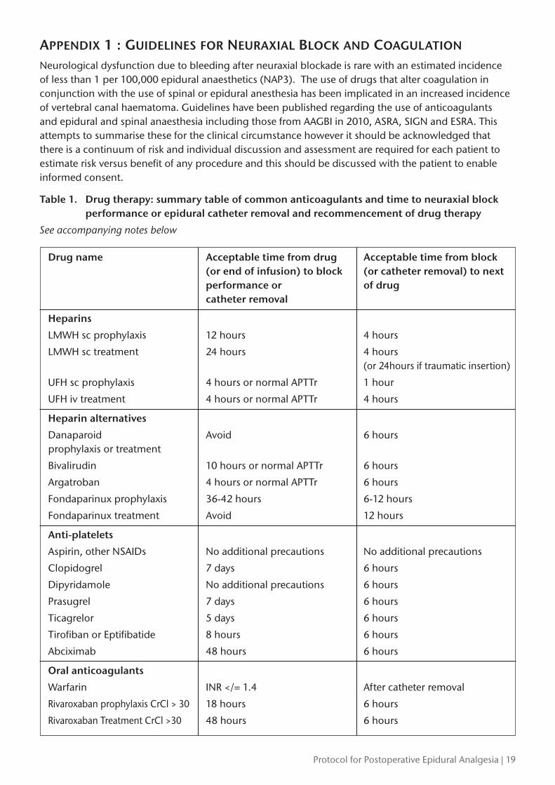
appendix 1 : Guidelines for neuraxial BloCk and CoaGulation
Neurological dysfunction due to bleeding after neuraxial blockade is rare with an estimated incidence of less than 1 per 100,000 epidural anaesthetics (NAP3). The use of drugs that alter coagulation in conjunction with the use of spinal or epidural anesthesia has been implicated in an increased incidence of vertebral canal haematoma. Guidelines have been published regarding the use of anticoagulants and epidural and spinal anaesthesia including those from AAGBI in 2010, ASRA, SIGN and ESRA. This attempts to summarise these for the clinical circumstance however it should be acknowledged that there is a continuum of risk and individual discussion and assessment are required for each patient to estimate risk versus benefit of any procedure and this should be discussed with the patient to enable informed consent.
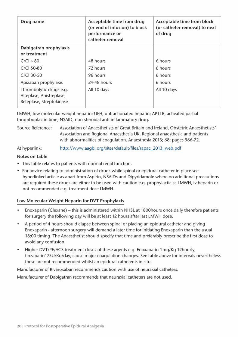
Table 1. Drug therapy: summary table of common anticoagulants and time to neuraxial block performance or epidural catheter removal and recommencement of drug therapy
See accompanying notes below
Drug name Acceptable time from drug Acceptable time from block (or end of infusion) to block (or catheter removal) to next performance or of drug catheter removal
Heparins
LMWH sc prophylaxis 12 hours 4 hours
LMWH sc treatment 24 hours 4 hours (or 24hours if traumatic insertion)
UFH sc prophylaxis 4 hours or normal APTTr 1 hour
UFH iv treatment 4 hours or normal APTTr 4 hours
Heparin alternatives
Danaparoid Avoid 6 hoursprophylaxis or treatment
Bivalirudin 10 hours or normal APTTr 6 hours
Argatroban 4 hours or normal APTTr 6 hours
Fondaparinux prophylaxis 36-42 hours 6-12 hours
Fondaparinux treatment Avoid 12 hours
Anti-platelets
Aspirin, other NSAIDs No additional precautions No additional precautions
Clopidogrel 7 days 6 hours
Dipyridamole No additional precautions 6 hours
Prasugrel 7 days 6 hours
Ticagrelor 5 days 6 hours
Tirofiban or Eptifibatide 8 hours 6 hours
Abciximab 48 hours 6 hours
Oral anticoagulants
Warfarin INR </= 1.4 After catheter removal
Rivaroxaban prophylaxis CrCl > 30 18 hours 6 hours
Rivaroxaban Treatment CrCl >30 48 hours 6 hours
20 | Protocol for Postoperative Epidural Analgesia
Drug name Acceptable time from drug Acceptable time from block (or end of infusion) to block (or catheter removal) to next performance or of drug catheter removal
Dabigatran prophylaxis or treatment
CrCl > 80 48 hours 6 hours
CrCl 50-80 72 hours 6 hours
CrCl 30-50 96 hours 6 hours
Apixaban prophylaxis 24-48 hours 6 hours
Thrombolytic drugs e.g. All 10 days All 10 daysAlteplase, Anistreplase,Reteplase, Streptokinase
LMWH, low molecular weight heparin; UFH, unfractionated heparin; APTTR, activated partial thromboplastin time; NSAID, non-steroidal anti-inflammatory drug.
Source Reference: Association of Anaesthetists of Great Britain and Ireland, Obstetric Anaesthetists’ Association and Regional Anaesthesia UK. Regional anaesthesia and patients with abnormalities of coagulation. Anaesthesia 2013; 68: pages 966-72.
At hyperlink: http://www.aagbi.org/sites/default/files/rapac_2013_web.pdf
Notes on table
• This table relates to patients with normal renal function.
• For advice relating to administration of drugs while spinal or epidural catheter in place see hyperlinked article as apart from Aspirin, NSAIDs and Dipyridamole where no additional precautions are required these drugs are either to be used with caution e.g. prophylactic sc LMWH, iv heparin or not recommended e.g. treatment dose LMWH.
Low Molecular Weight Heparin for DVT Prophylaxis
• Enoxaparin (Clexane) – this is administered within NHSL at 1800hours once daily therefore patients for surgery the following day will be at least 12 hours after last LMWH dose.
• A period of 4 hours should elapse between spinal or placing an epidural catheter and giving Enoxaparin - afternoon surgery will demand a later time for initiating Enoxaparin than the usual 18:00 timing. The Anaesthetist should specify that time and preferably prescribe the first dose to avoid any confusion.
• Higher DVT/PE/ACS treatment doses of these agents e.g. Enoxaparin 1mg/Kg 12hourly, tinzaparin175U/Kg/day, cause major coagulation changes. See table above for intervals nevertheless these are not recommended whilst an epidural catheter is in situ.
Manufacturer of Rivaroxaban recommends caution with use of neuraxial catheters.
Manufacturer of Dabigatran recommends that neuraxial catheters are not used.
Protocol for Postoperative Epidural Analgesia | 21
removal of epidural CatHeters
When it has been decided that epidural analgesia is no longer required, the epidural catheter should be removed as soon as possible provided that a coagulopathy is unlikely. The risk of infection in the epidural space increases with time, especially after 4 days. The majority of epidural catheters should have been removed by this time. The following points should be considered:
• Suitable alternative or “step-down” analgesia should have been prescribed.
• The coagulation status of the patient should be assessed. A coagulation screen is indicated only when this may be abnormal due to use of oral anticoagulants, unfractionated heparin infusion, DIC, major haemorrhage or renal/hepatic dysfunction. If in doubt it is safer to check platelet and coagulation profile. Practitioners should remember however, that some agents (eg LMWH) do not cause any changes in routine coagulation indices.
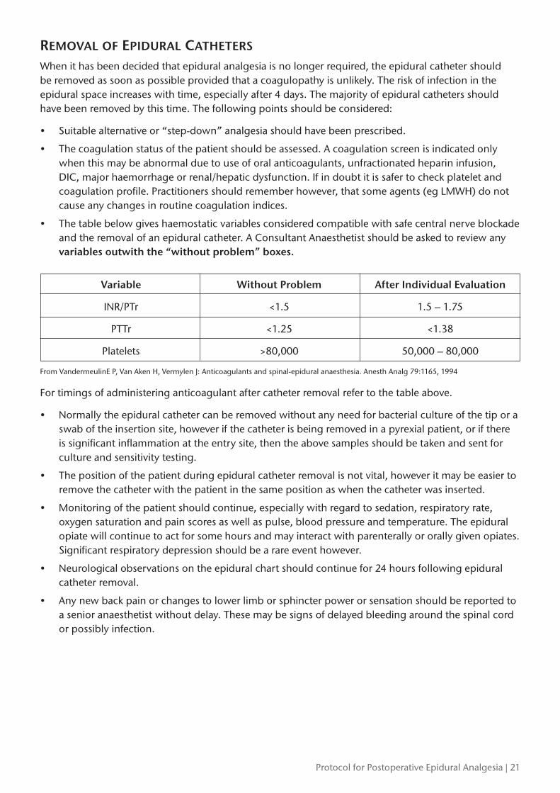
• The table below gives haemostatic variables considered compatible with safe central nerve blockade and the removal of an epidural catheter. A Consultant Anaesthetist should be asked to review any variables outwith the “without problem” boxes.
Variable Without Problem After Individual Evaluation
INR/PTr <1.5 1.5 – 1.75
PTTr <1.25 <1.38
Platelets >80,000 50,000 – 80,000
From VandermeulinE P, Van Aken H, Vermylen J: Anticoagulants and spinal-epidural anaesthesia. Anesth Analg 79:1165, 1994
For timings of administering anticoagulant after catheter removal refer to the table above.
• Normally the epidural catheter can be removed without any need for bacterial culture of the tip or a swab of the insertion site, however if the catheter is being removed in a pyrexial patient, or if there is significant inflammation at the entry site, then the above samples should be taken and sent for culture and sensitivity testing.
• The position of the patient during epidural catheter removal is not vital, however it may be easier to remove the catheter with the patient in the same position as when the catheter was inserted.
• Monitoring of the patient should continue, especially with regard to sedation, respiratory rate, oxygen saturation and pain scores as well as pulse, blood pressure and temperature. The epidural opiate will continue to act for some hours and may interact with parenterally or orally given opiates. Significant respiratory depression should be a rare event however.
• Neurological observations on the epidural chart should continue for 24 hours following epidural catheter removal.
• Any new back pain or changes to lower limb or sphincter power or sensation should be reported to a senior anaesthetist without delay. These may be signs of delayed bleeding around the spinal cord or possibly infection.

22 | Protocol for Postoperative Epidural Analgesia
Revised August 2017 by M Herron
For review August 2020
Des
ign
- Med
ical
Illu
stra
tion,
NH
S La
nark
shire
MLT.PROTOC.17_17237.W