programmed cell death (apoptosis) · programmed cell death ! (apoptosis)! stereotypic death process...
TRANSCRIPT
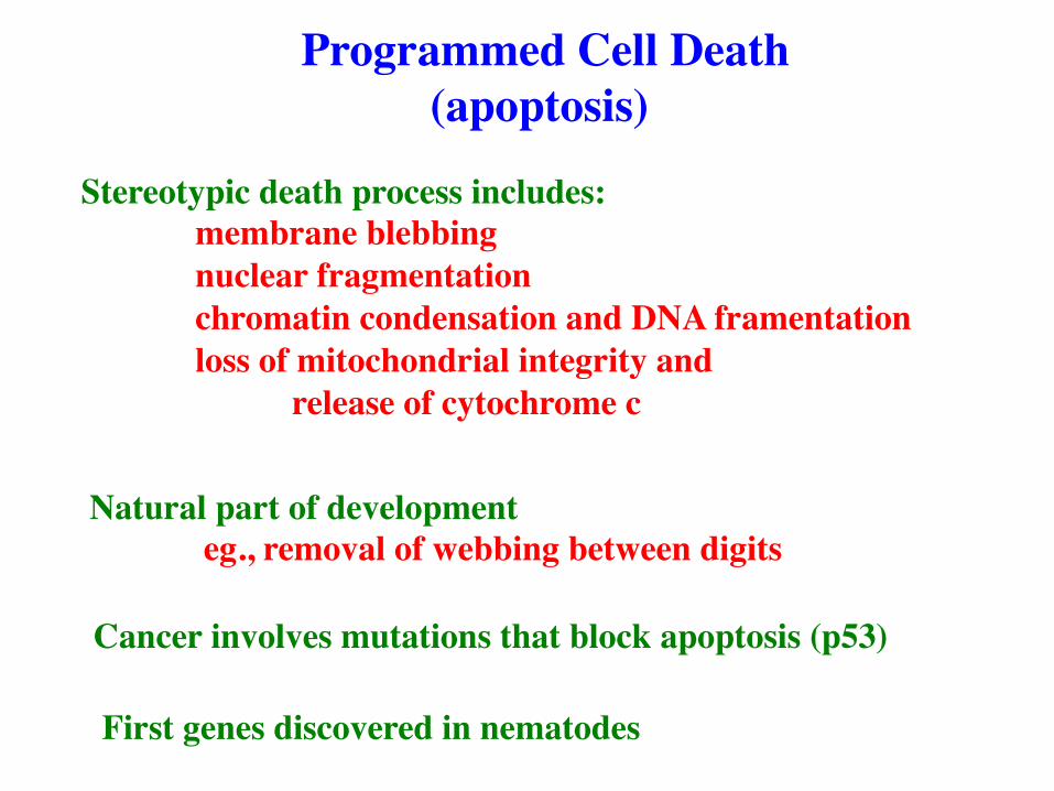
Programmed Cell Death (apoptosis)
Stereotypic death process includes: membrane blebbing nuclear fragmentation chromatin condensation and DNA framentation loss of mitochondrial integrity and release of cytochrome c
Natural part of development eg., removal of webbing between digits
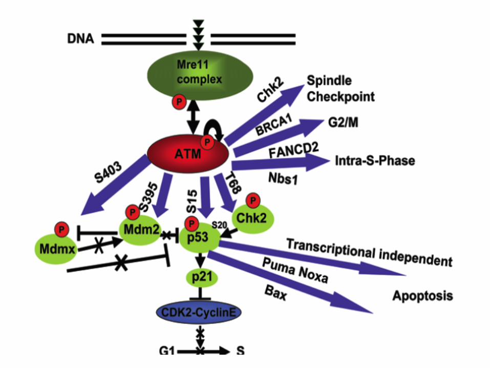
Cancer involves mutations that block apoptosis (p53)
First genes discovered in nematodes
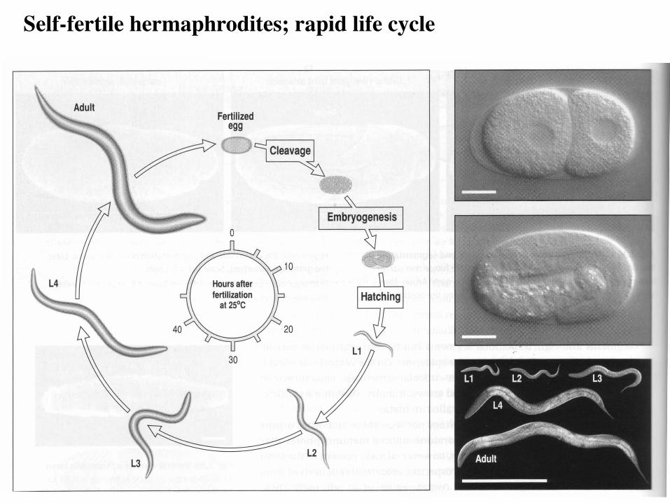
Self-fertile hermaphrodites; rapid life cycle
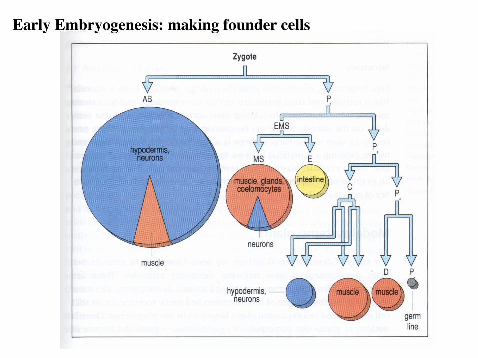
Early Embryogenesis: making founder cells
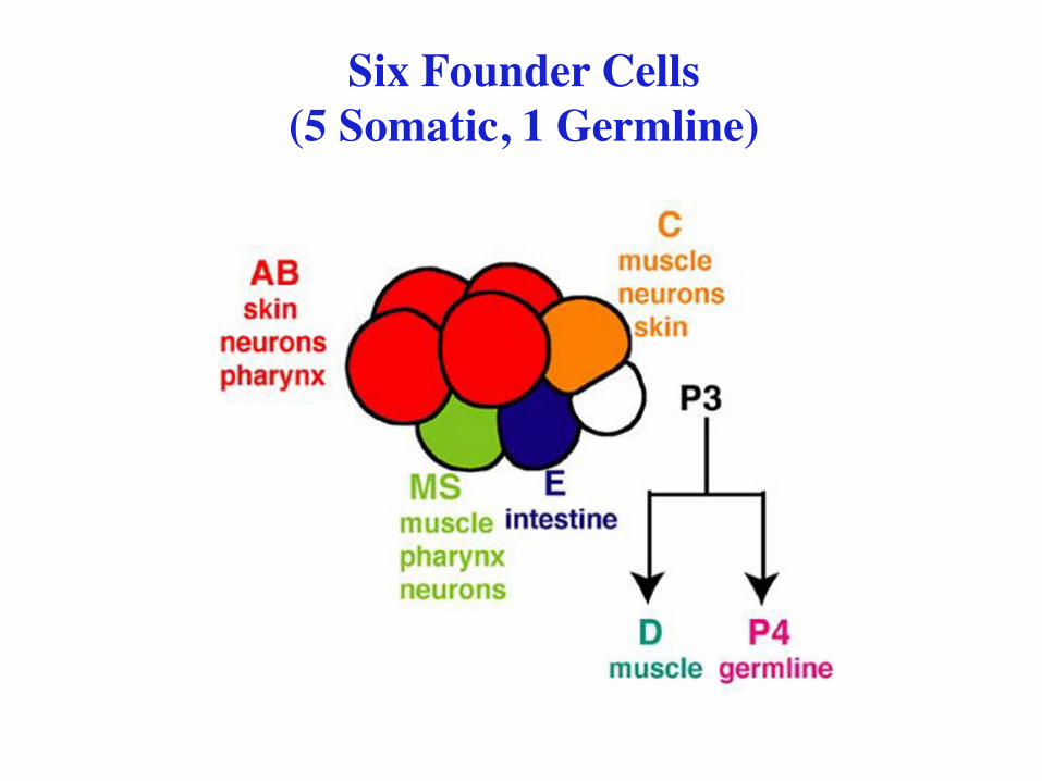
Six Founder Cells (5 Somatic, 1 Germline)
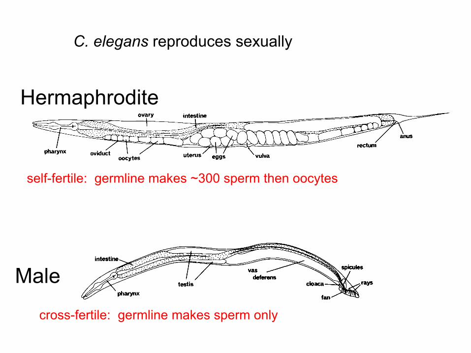
C. elegans reproduces sexually
Hermaphrodite
Male
self-fertile: germline makes ~300 sperm then oocytes
cross-fertile: germline makes sperm only

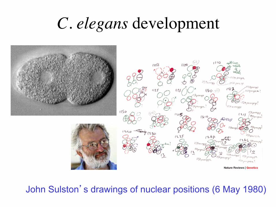
C. elegans development
John Sulston’s drawings of nuclear positions (6 May 1980)
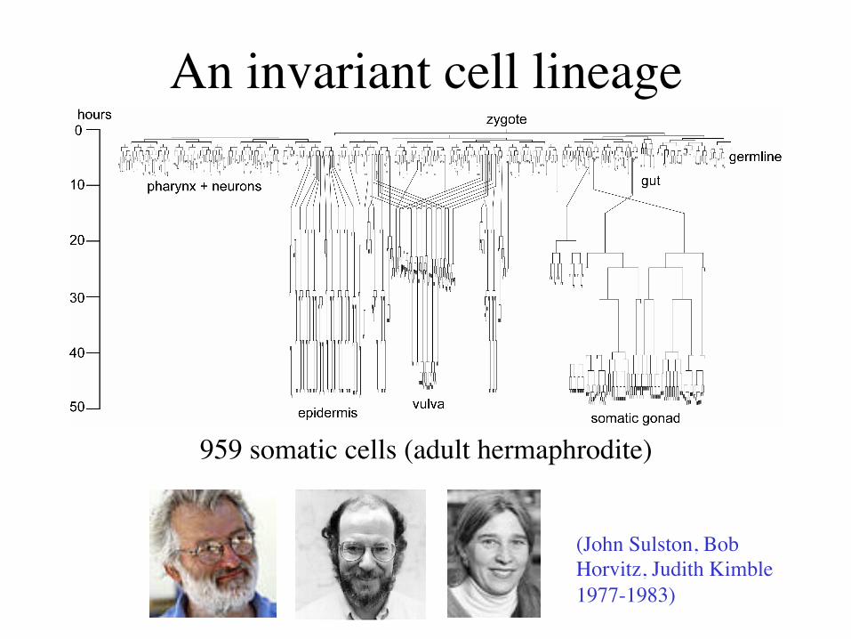
An invariant cell lineage
959 somatic cells (adult hermaphrodite)
(John Sulston, Bob Horvitz, Judith Kimble 1977-1983)
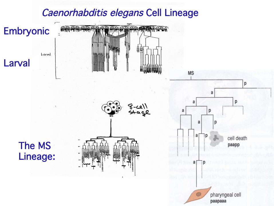
Caenorhabditis elegans Cell Lineage!
Embryonic!
Larval!
The MS !Lineage:!
Programmed Cell Death in C. elegans: Embryogenesis produces a hatched larva with
--558 living cells --131 cells eliminated by programmed cell death (shortly after their births).
Additional cell deaths occur during larval development. Most cell deaths are in neuronal lineages. Cell death (apoptosis) conserved in most animals.
Cancer connection.
ced mutants: programmed cell death-defective
Ed Hedgecock: unbiased Nomarski screen for mutants with defects in cellular anatomy (F2 screens)
Identified two mutants, ced-1 and ced-2, with persistent
cell death corpses.
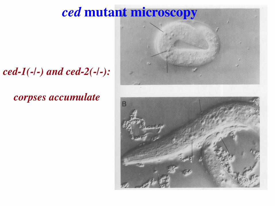
ced-1(-/-) and ced-2(-/-): corpses accumulate
ced mutant microscopy
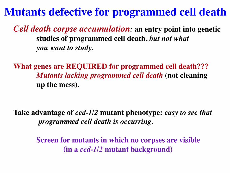
Cell death corpse accumulation: an entry point into genetic studies of programmed cell death, but not what you want to study.
What genes are REQUIRED for programmed cell death???
Mutants lacking programmed cell death (not cleaning up the mess).
Take advantage of ced-1/2 mutant phenotype: easy to see that
programmed cell death is occurring.
Screen for mutants in which no corpses are visible (in a ced-1/2 mutant background)
Mutants defective for programmed cell death

ced-1(-/-)
ced-1(-/-); ced-3(-/-)
Horvitz lab rides again. Ellis et al, Cell 44, 817- 829 (1986)
ced3 mutants fail to accumulate corpses
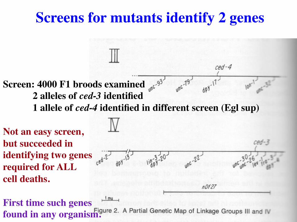
Screen: 4000 F1 broods examined 2 alleles of ced-3 identified 1 allele of ced-4 identified in different screen (Egl sup)
Not an easy screen, but succeeded in identifying two genes required for ALL cell deaths. First time such genes found in any organism.
Screens for mutants identify 2 genes
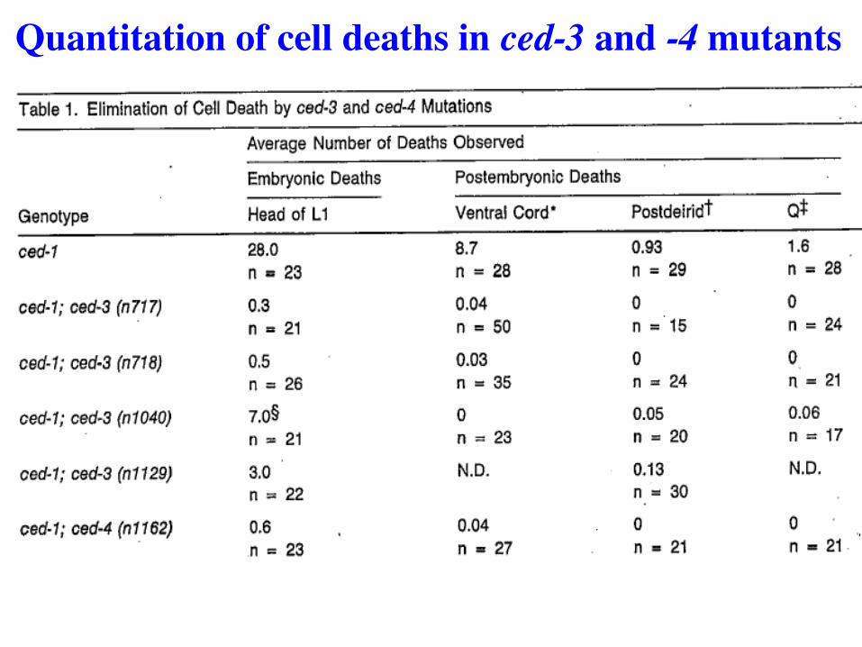
Quantitation of cell deaths in ced-3 and -4 mutants
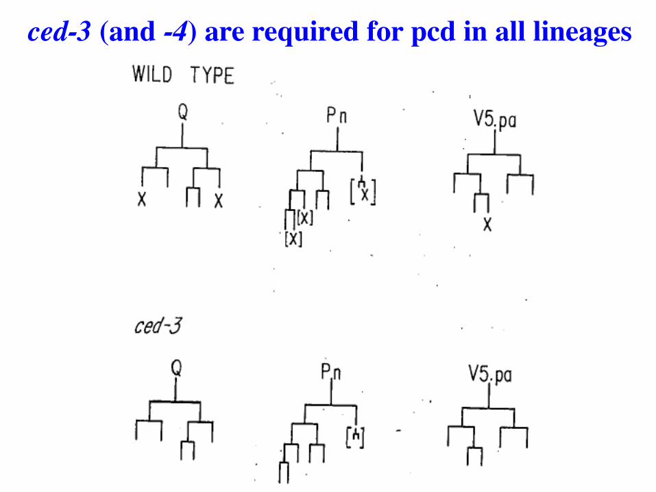
ced-3 (and -4) are required for pcd in all lineages
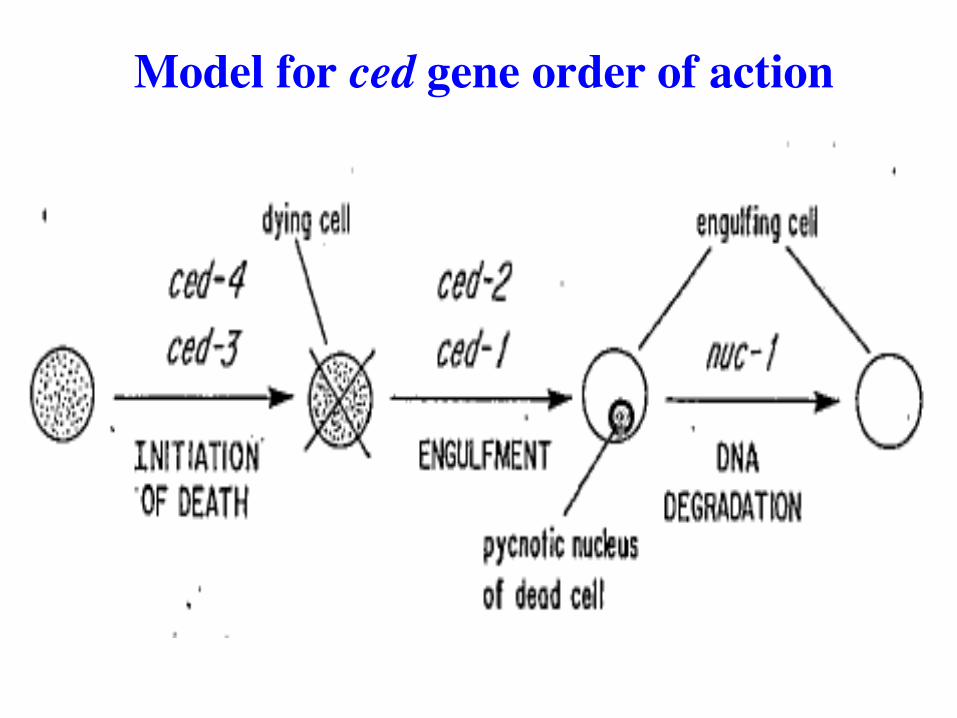
Model for ced gene order of action
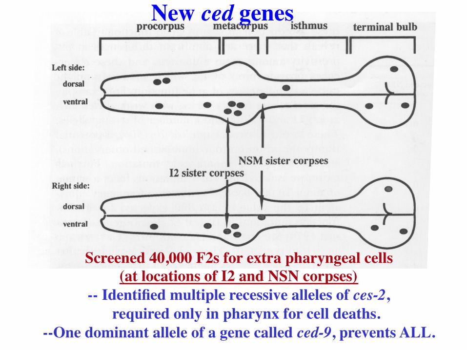
Screened 40,000 F2s for extra pharyngeal cells (at locations of I2 and NSN corpses)
-- Identified multiple recessive alleles of ces-2, required only in pharynx for cell deaths.
--One dominant allele of a gene called ced-9, prevents ALL.
New ced genes
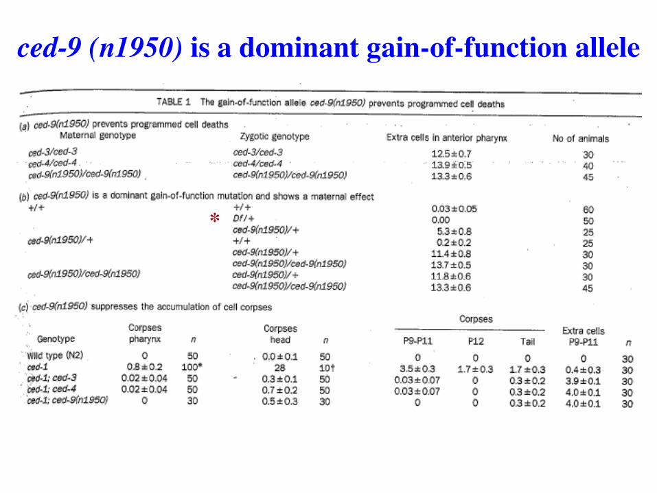
ced-9 (n1950) is a dominant gain-of-function allele
*
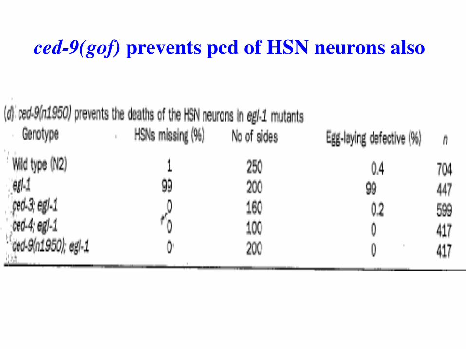
ced-9(gof) prevents pcd of HSN neurons also
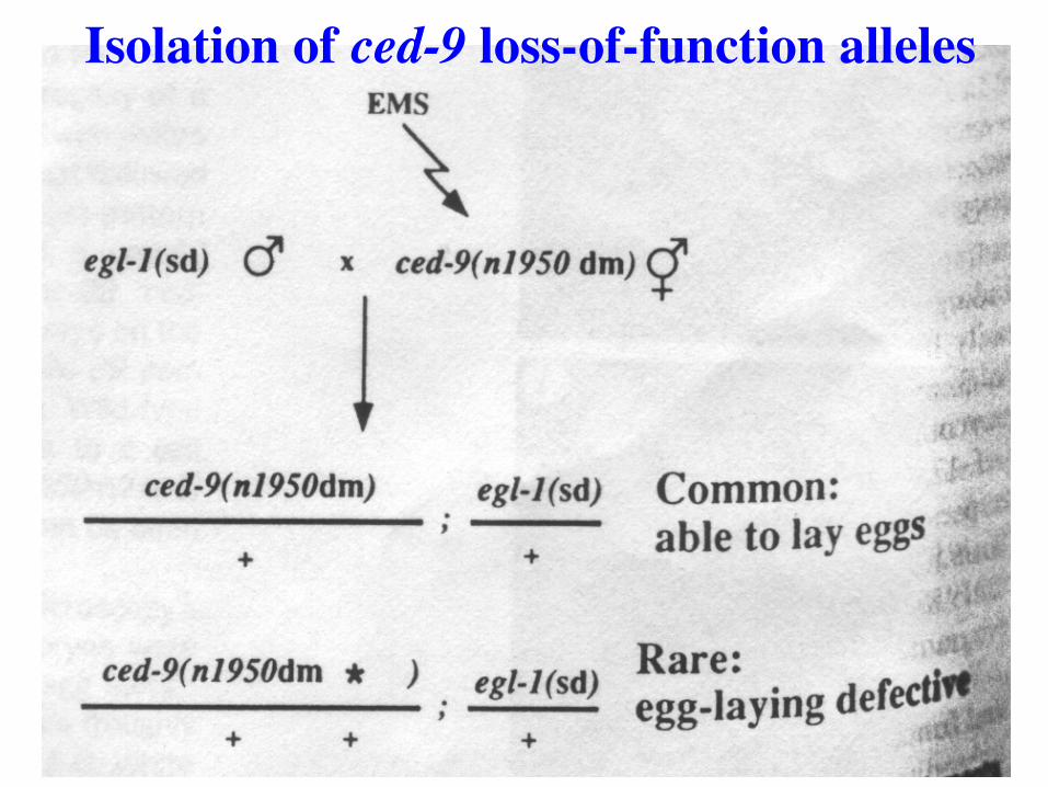
Isolation of ced-9 loss-of-function alleles
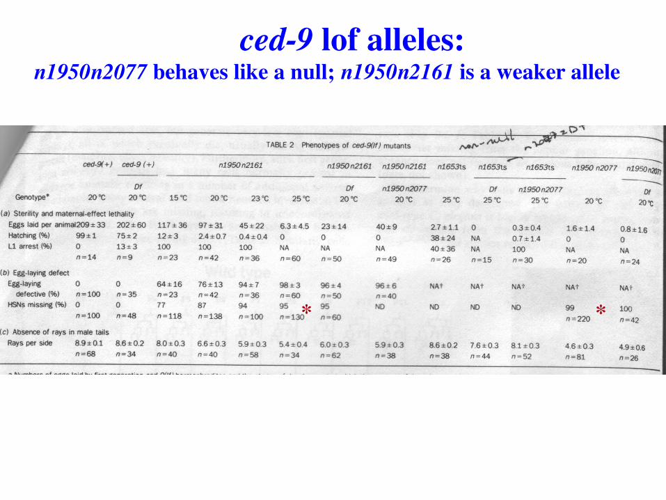
ced-9 lof alleles: n1950n2077 behaves like a null; n1950n2161 is a weaker allele
* *
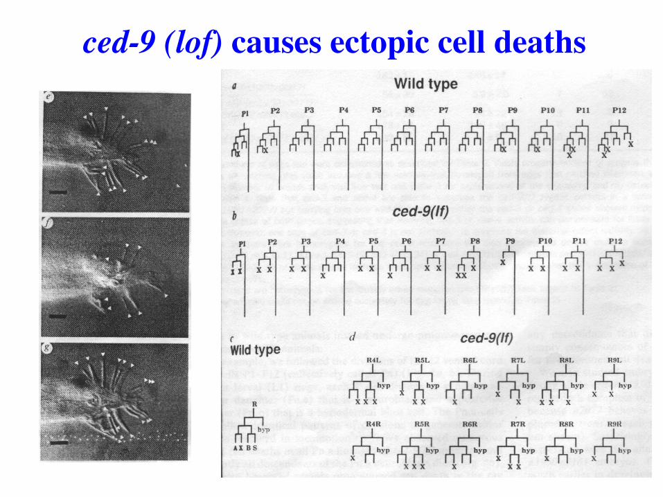
ced-9 (lof) causes ectopic cell deaths
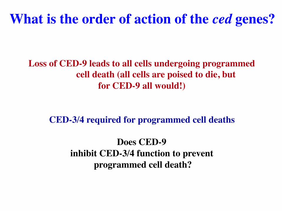
Loss of CED-9 leads to all cells undergoing programmed cell death (all cells are poised to die, but
for CED-9 all would!)
CED-3/4 required for programmed cell deaths
Does CED-9 inhibit CED-3/4 function to prevent
programmed cell death?
What is the order of action of the ced genes?
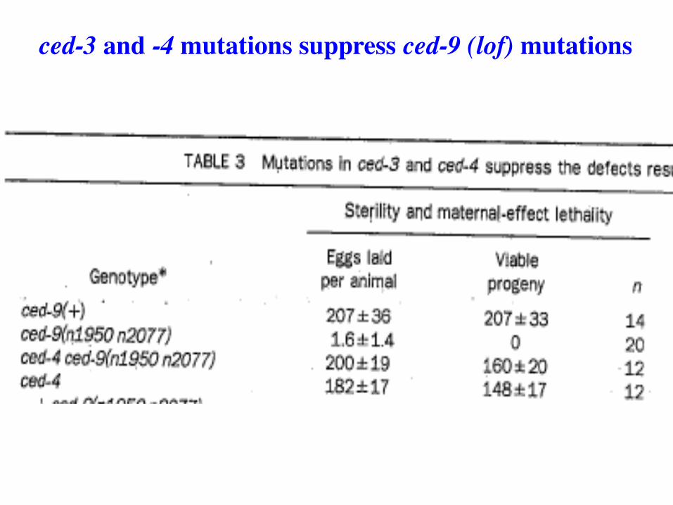
ced-3 and -4 mutations suppress ced-9 (lof) mutations
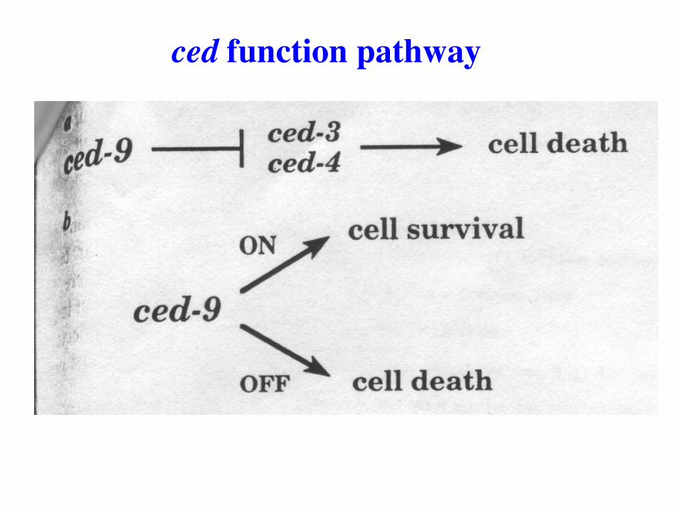
ced function pathway

Overexpression of Ced-3 and Ced-4 causes ectopic cell death
Enables another genetic test of ced-9 relationship to ced-3 and -4
Also lets us test the relationship between ced-3 and ced-4
Adding more relationships to the pathway

Overexpression of Ced-3 and Ced-4 causes ectopic cell death
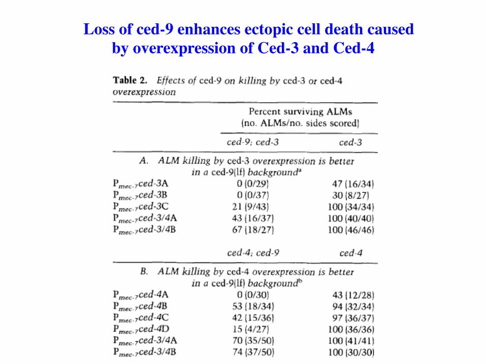
Loss of ced-9 enhances ectopic cell death caused by overexpression of Ced-3 and Ced-4

Ced-3 is required for ectopic killing caused by overexpression of Ced-4, but not vice versa
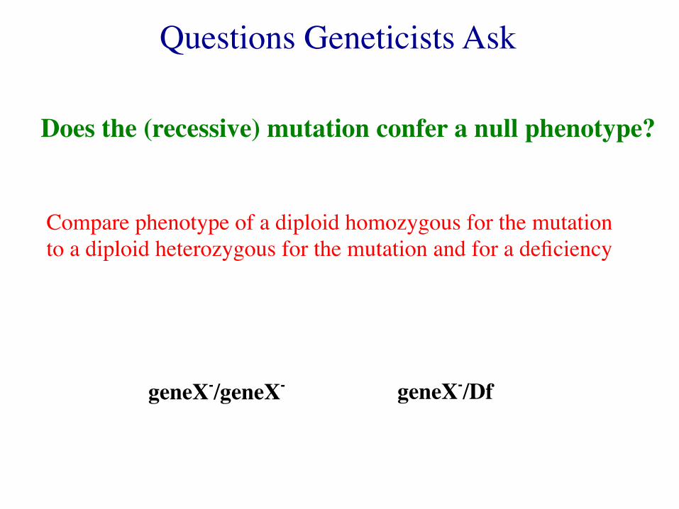
Questions Geneticists Ask
Does the (recessive) mutation confer a null phenotype?
Compare phenotype of a diploid homozygous for the mutation to a diploid heterozygous for the mutation and for a deficiency
geneX-/geneX- geneX-/Df
Questions Geneticists Ask
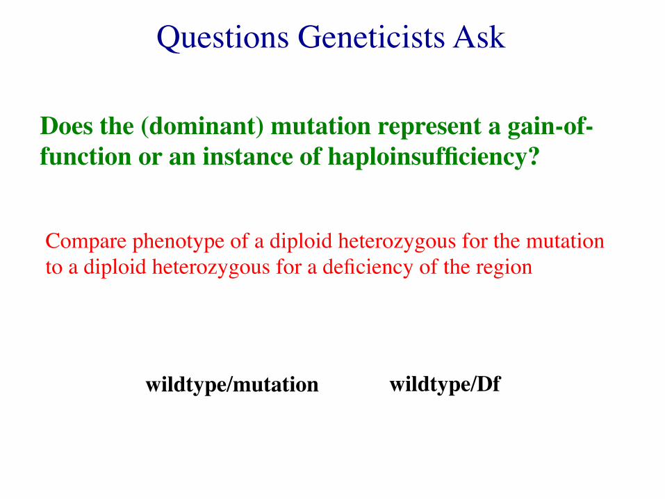
Does the (dominant) mutation represent a gain-of-function or an instance of haploinsufficiency?
Compare phenotype of a diploid heterozygous for the mutation to a diploid heterozygous for a deficiency of the region
wildtype/mutation wildtype/Df
Questions Geneticists Ask
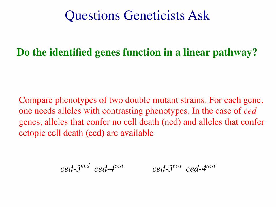
Do the identified genes function in a linear pathway?
Compare phenotypes of two double mutant strains. For each gene, one needs alleles with contrasting phenotypes. In the case of ced genes, alleles that confer no cell death (ncd) and alleles that confer ectopic cell death (ecd) are available
ced-3ncd ced-4ecd ced-3ecd ced-4ncd
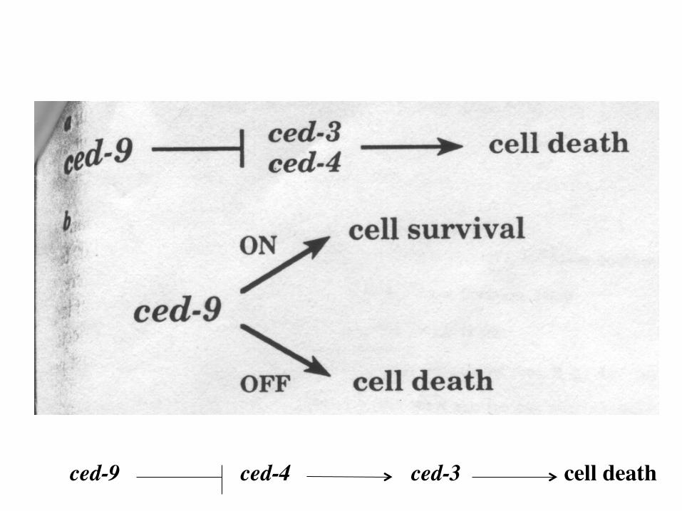
ced-9 ced-4 ced-3 cell death
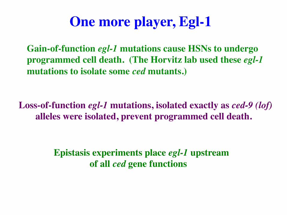
One more player, Egl-1
Gain-of-function egl-1 mutations cause HSNs to undergo programmed cell death. (The Horvitz lab used these egl-1 mutations to isolate some ced mutants.)
Loss-of-function egl-1 mutations, isolated exactly as ced-9 (lof) alleles were isolated, prevent programmed cell death.
Epistasis experiments place egl-1 upstream of all ced gene functions
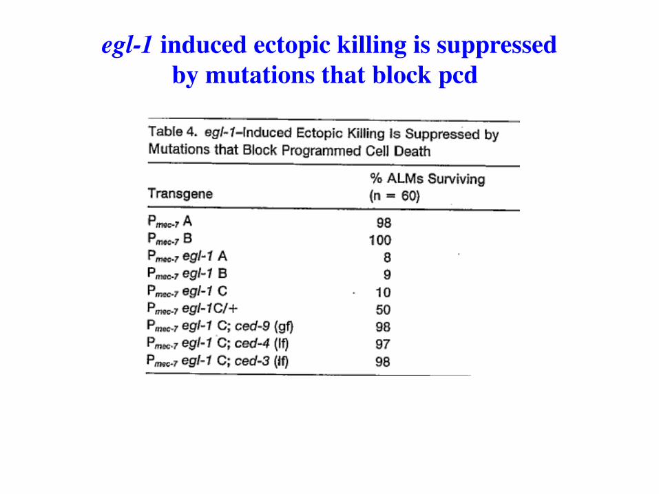
egl-1 induced ectopic killing is suppressed by mutations that block pcd
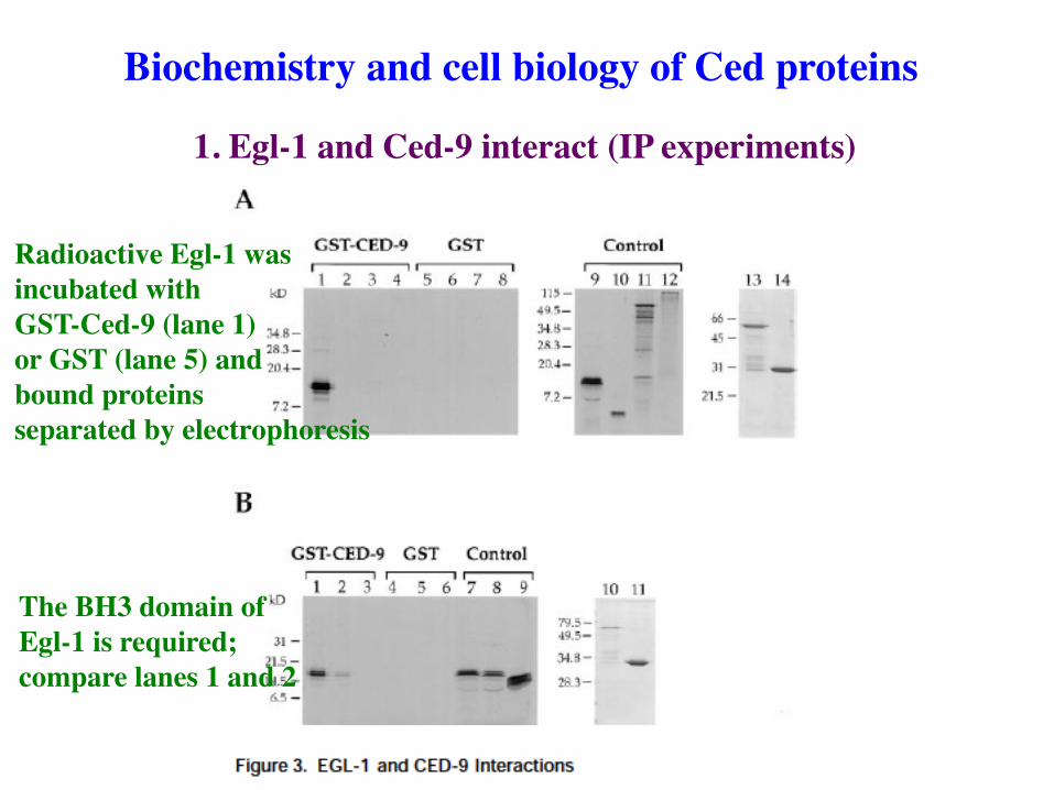
1. Egl-1 and Ced-9 interact (IP experiments)
Biochemistry and cell biology of Ced proteins
Radioactive Egl-1 was incubated with GST-Ced-9 (lane 1) or GST (lane 5) and bound proteins separated by electrophoresis
The BH3 domain of Egl-1 is required; compare lanes 1 and 2
3. Ced-9 is localized to the mitochondrial outer membrane and recruits Ced-4
4. Induction of programmed cell death induces Ced-4 translocation to the nuclear membrane but not in gof Ced-9 mutants
2. Bunches of IP experiments demonstrate interaction between Egl-1 and Ced-9, between Ced-9 and Ced-4, and between Ced-3 and Ced-4
Biochem and cell biology, continued
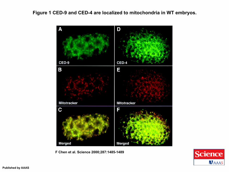
Figure 1 CED-9 and CED-4 are localized to mitochondria in WT embryos.
F Chen et al. Science 2000;287:1485-1489
Published by AAAS

Figure 2 CED-9 is required for the localization of CED-4 to mitochondria.
F Chen et al. Science 2000;287:1485-1489
Published by AAAS
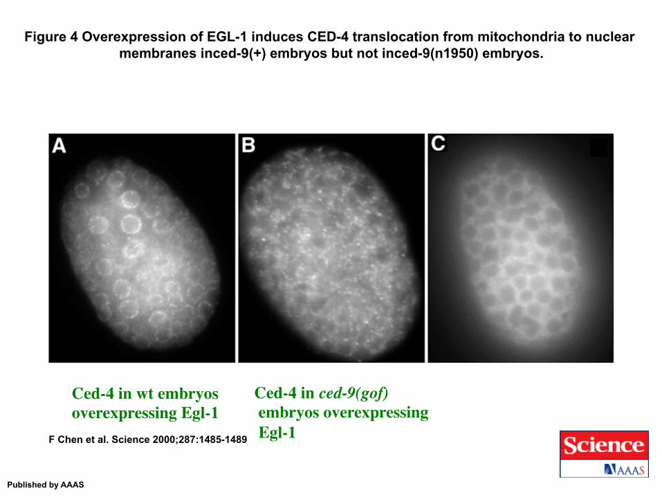
Figure 4 Overexpression of EGL-1 induces CED-4 translocation from mitochondria to nuclear membranes inced-9(+) embryos but not inced-9(n1950) embryos.
F Chen et al. Science 2000;287:1485-1489
Published by AAAS
Ced-4 in wt embryos overexpressing Egl-1
Ced-4 in ced-9(gof) embryos overexpressing Egl-1
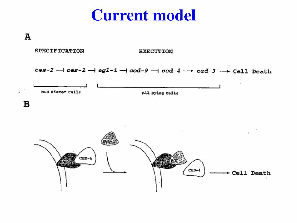
Current model
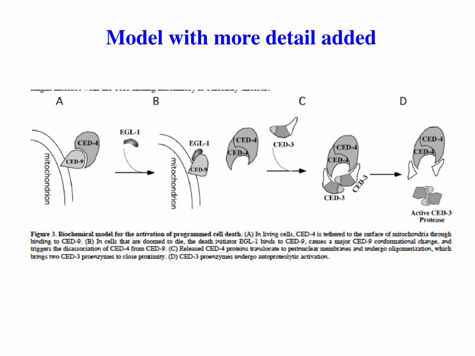
Model with more detail added
Cloning ced genes revealed similarities to mammalian proteins
Ced-3 is similar to mammalian interleukin converting enzyme, a cysteine protease
Ced-3 is therefore proposed to be a cysteine protease
Ced-9 is a homolog of Bcl-2, a mammalian oncogene
In vitro substrates for Ced-3 include actin, tubulin, and proteins involved in ATP synthesis and in DNA synthesis

Comparison of apoptopic pathways
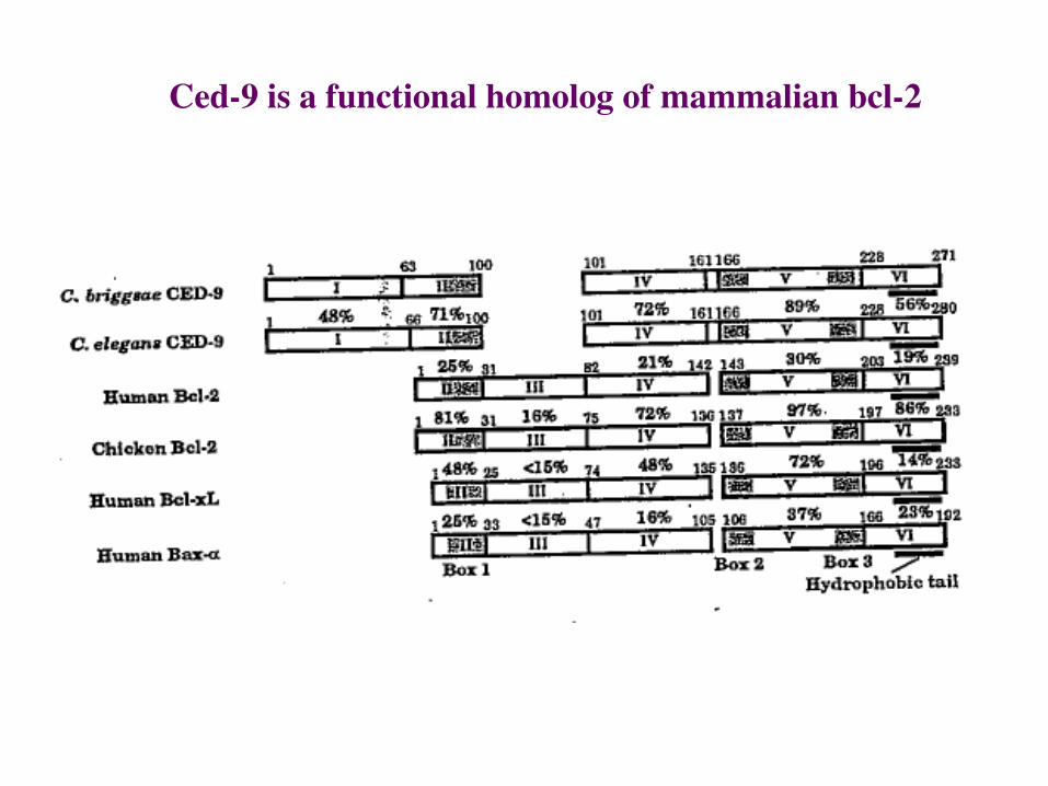
Ced-9 is a functional homolog of mammalian bcl-2
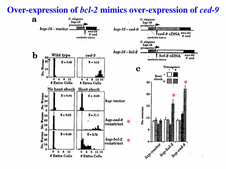
Over-expression of bcl-2 mimics over-expression of ced-9
*
*
*
*
Parallels of CED-9 in worms and Bcl-2 in humans.
Bcl-2 is a human oncogene with properties similar to CED-9
Over-expression of Bcl-2 prevents or delays cell death in B-cell and T-cell lineages.
Bcl-2 expressed at high levels in blood stem cell lineages;
loss of expression correlates with appearance of cell death
In cancer, chromosome translocations activate
Bcl-2 expression, preventing cell death in hematopoietic lineages. This results in a leukemia due to
over-proliferation of some blood cell lineages. No LOF alleles.
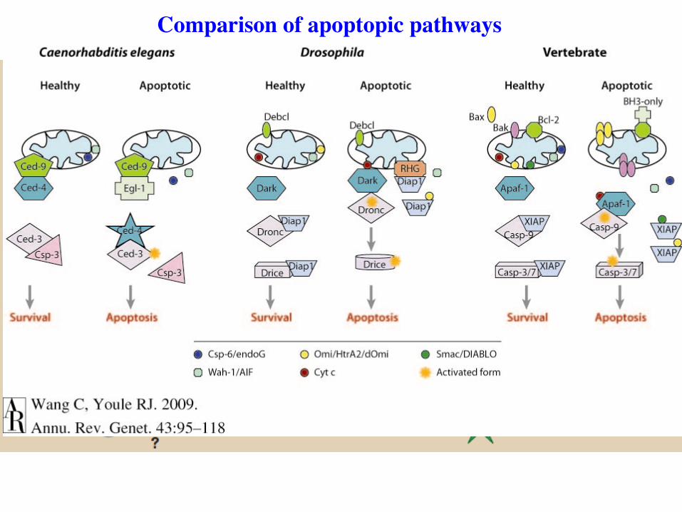
Comparison of apoptopic pathways
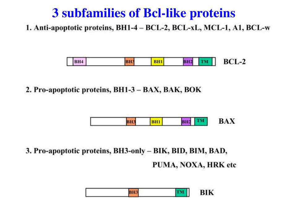
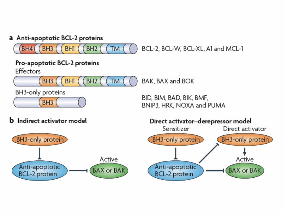
3 subfamilies of Bcl-like proteins
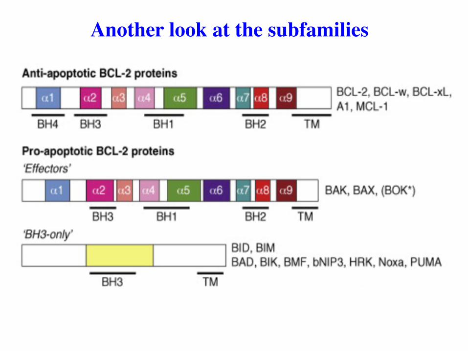
Another look at the subfamilies
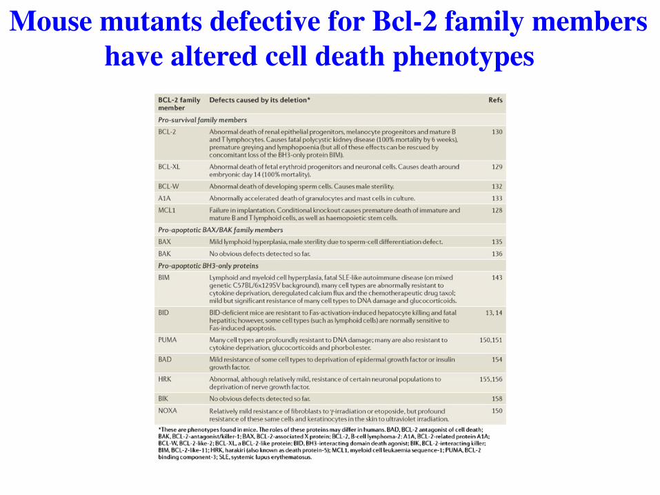
Mouse mutants defective for Bcl-2 family members have altered cell death phenotypes
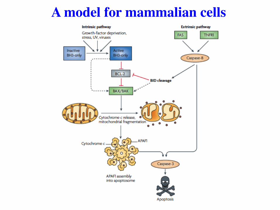
A model for mammalian cells

Same model, a little more detail
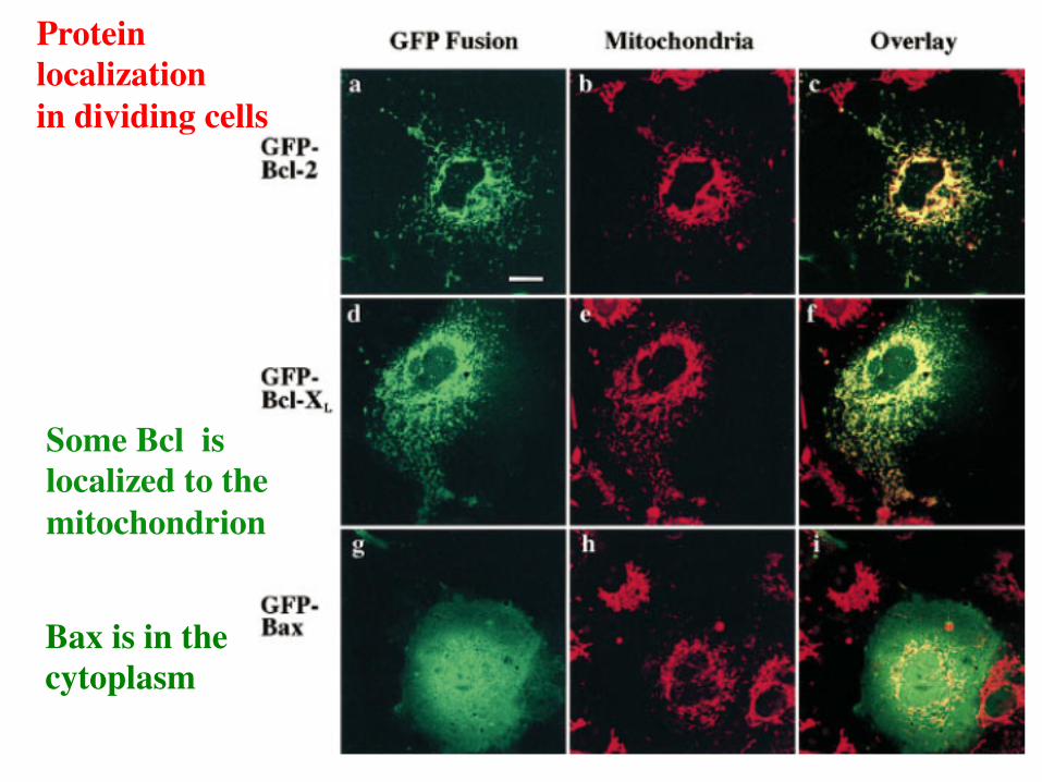
Protein localization in dividing cells
Some Bcl is localized to the mitochondrion
Bax is in the cytoplasm
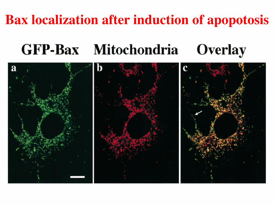
Bax localization after induction of apopotosis
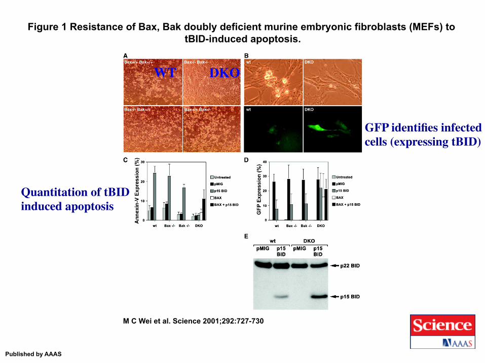
Figure 1 Resistance of Bax, Bak doubly deficient murine embryonic fibroblasts (MEFs) to tBID-induced apoptosis.
M C Wei et al. Science 2001;292:727-730
Published by AAAS
WT DKO
GFP identifies infected cells (expressing tBID)
Quantitation of tBID induced apoptosis
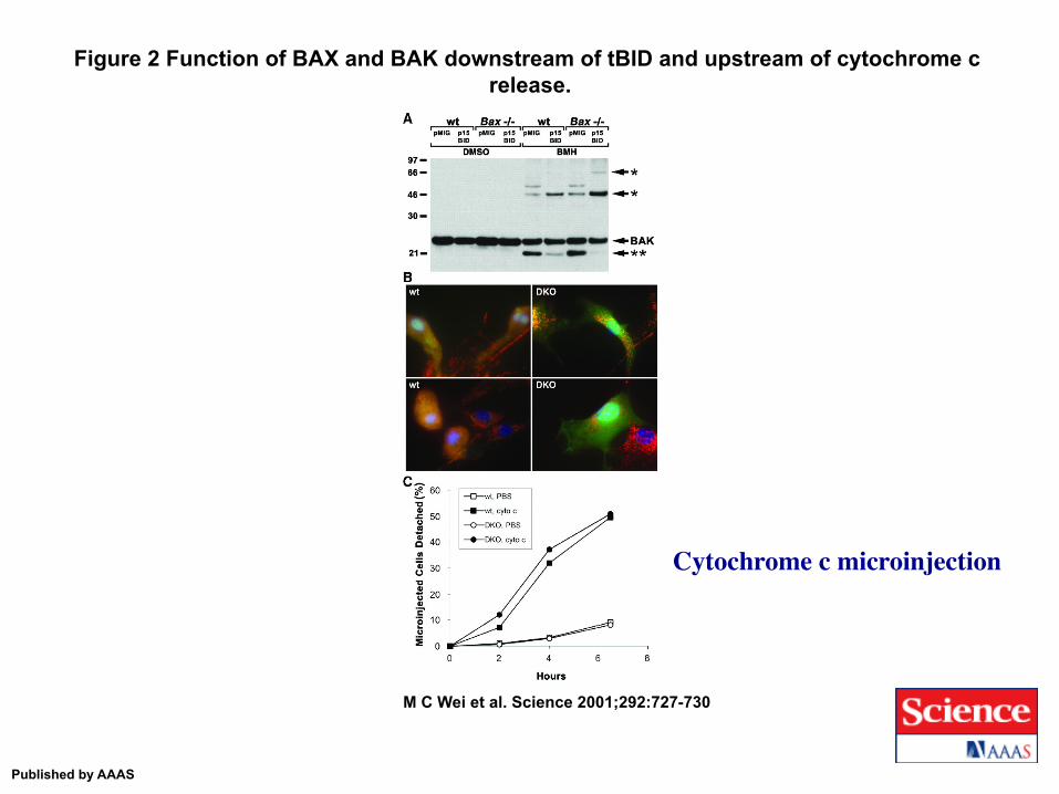
Figure 2 Function of BAX and BAK downstream of tBID and upstream of cytochrome c release.
M C Wei et al. Science 2001;292:727-730
Published by AAAS
Cytochrome c microinjection
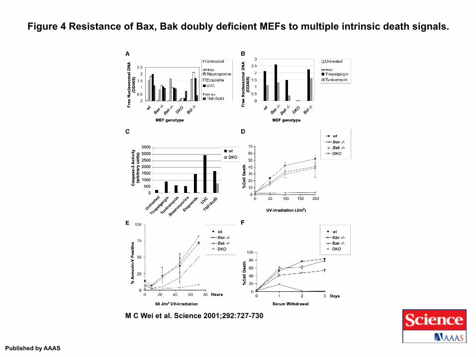
Figure 4 Resistance of Bax, Bak doubly deficient MEFs to multiple intrinsic death signals.
M C Wei et al. Science 2001;292:727-730
Published by AAAS
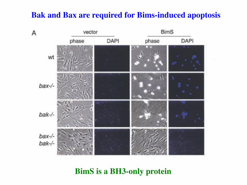
BimS is a BH3-only protein
Bak and Bax are required for Bims-induced apoptosis
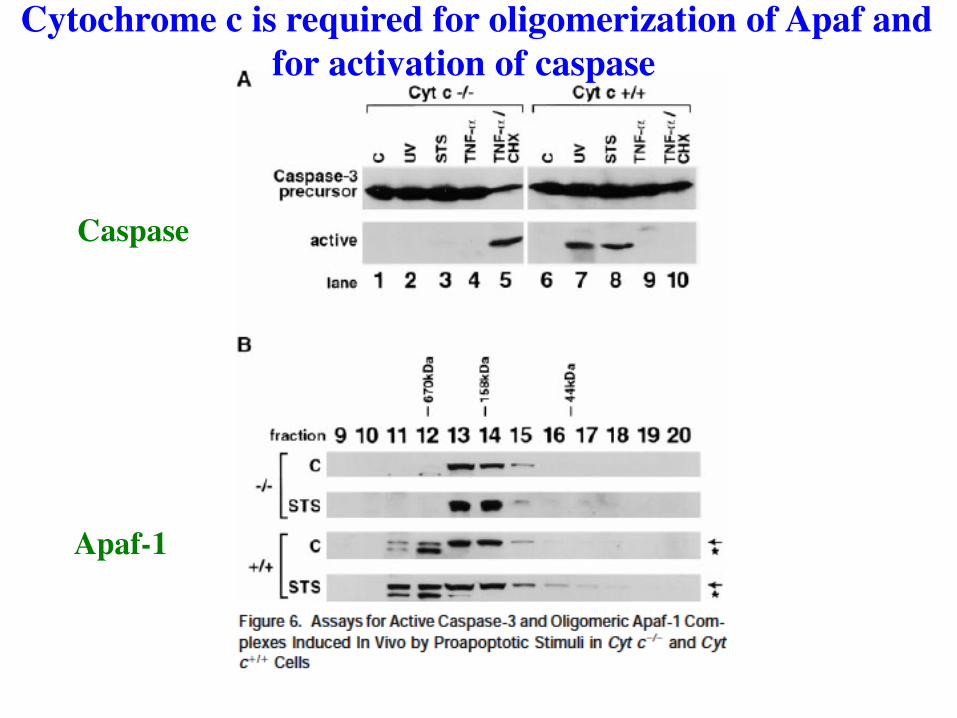
Cytochrome c is required for oligomerization of Apaf and for activation of caspase
Caspase
Apaf-1
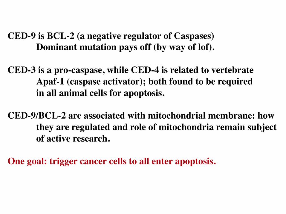
CED-9 is BCL-2 (a negative regulator of Caspases) Dominant mutation pays off (by way of lof).
CED-3 is a pro-caspase, while CED-4 is related to vertebrate
Apaf-1 (caspase activator); both found to be required in all animal cells for apoptosis.
CED-9/BCL-2 are associated with mitochondrial membrane: how
they are regulated and role of mitochondria remain subject of active research.
One goal: trigger cancer cells to all enter apoptosis.
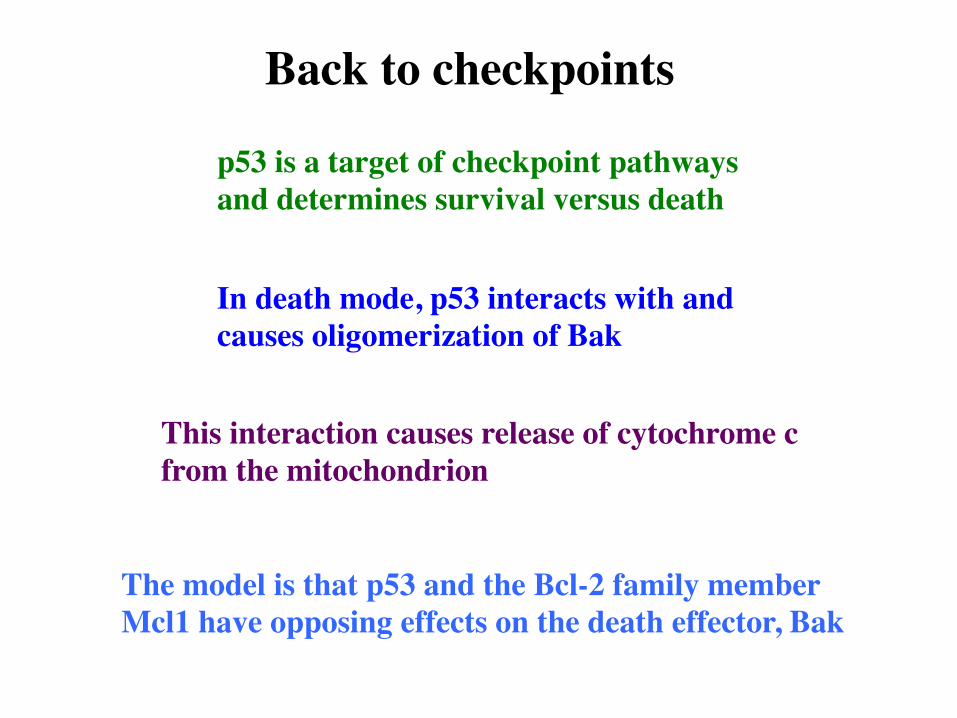
Back to checkpoints
p53 is a target of checkpoint pathways and determines survival versus death
In death mode, p53 interacts with and causes oligomerization of Bak
This interaction causes release of cytochrome c from the mitochondrion
The model is that p53 and the Bcl-2 family member Mcl1 have opposing effects on the death effector, Bak