program - citometria.org program_lisbon.pdf · alexandre salvador antónio medina almeida artur...
TRANSCRIPT

PROGRAM
n
25 to 27 MAY 2017LISBON, PORTUGALCentro Cultural de Belém
th th

PRESIDENTE José Enrique O'Connor
VICEPRESIDENTE Maria Jorge Arroz
SECRETARIO-TESORERO Julía Almeida Parra
VOCALESRui GardnerMaría Teresa Molero LabartaMartín Pérez AndrésLola Martínez
ORGANIZING COMITTEECatarina Martins
Alexandre SalvadorAntónio Medina AlmeidaArtur PaivaJoão LoureiroLola MartinezLuís Miguel BorregoMargarida Lima Margarida SaraivaNatacha SantosPaulo Rodrigues-SantosRui Gardner
SCIENTIFIC COMITEECatarina Martins
Alexandre SalvadorAntónio Medina AlmeidaArtur PaivaBruno Costa Silva João Farela NevesJoão LoureiroJosé Enrique O'ConnorJuana CiudadJulía Almeida ParraLola MartinezLuís Miguel BorregoMargarida Lima Margarida SaraivaMaria Jorge ArrozMaría Teresa Molero LabartaMariona PascalMartín Pérez AndrésNatacha SantosPaulo Rodrigues-SantosRui Gardner
JUNTA DIRECTIVA DE LA SOCIEDAD IBÉRICA DE CITOMETRÍA | SIC
Dear Colleagues and friends,
The real adventure of the XV SIC Congress started about one year ago for us, Organizing and Scientific Committees. It was in fact a great challenge that the SIC Board gave us!Therefore, we settled a multidisciplinary Iberian network to build from scratch this important event for flow cytometrists, particularly in Portugal and Spain. The members of the Organizing and Scientific Committees worked very hard to choose the more relevant themes, to bring brilliant experts and researchers in diverse fields of Flow Cytometry, and at the same time, to address an interesting social program that honors the beautiful Lisbon.We believe that we have put together all the ingredients for this to be a memorable Congress! We truly wish it will have future scientific repercussions in the professional pathways of all participants (clinicians, researchers, speakers, students, sponsors).For now, it is wonderful to finally see you in the skyline of the Portuguese capital. We would really like to thank you for your presence in the XV SIC Congress, and we truly hope you enjoy attending it as much as we did organizing this meeting. Welcome, and let's have Flow Cytometry shining in the sunny skies of Lisbon!On behalf of the Organizing and Scientific Committeesof the XV SIC Congress,
WELCOME MESSAGE
XV SIC |003
n
Catarina Martins
Dear Colleagues:
It is with great pleasure that I welcome you to the XV Congress of the Iberian Society of Cytometry (SIC). This scientific event, held in the splendid city of Lisbon, represents a new milestone for the SIC in its desire of being the home of Iberian cytometrists. I want to acknowledge all the friends in the Organization that have made possible this Congress, bringing together educational activities, state-of-the-art cytometry and an attractive social program.I want to give a special welcome to our distinguished invited speakers, among them the two Presidents of the International Society for the Advance of Cytometry, Paul Wallace and Andrea Cossarizza. Their presence highlights the links of SIC with fraternal societies in the field of Cytometry.Last but not least, my special welcome to our young attendants. The educational and scientific programs of the Congress, carefully elaborated to cover established and emerging applications, will offer to them a panoramic view of modern cytometry. But the best value of this Congress for the younger should be, undoubtedly, the interaction with their expert colleagues.Thus, on behalf of the Junta Directiva, I wish to all of you a productive and pleasant time in Lisbon.
WELCOME MESSAGE FROM THE SIC PRESIDENT
n
José-Enrique O'ConnorPresident of SIC
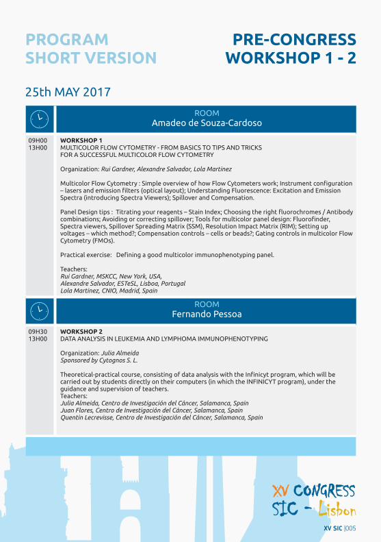
PRE-CONGRESS WORKSHOP 1 - 2
25th MAY 2017
ROOM Amadeo de Souza-Cardoso
09H00 13H00
WORKSHOP 1 MULTICOLOR FLOW CYTOMETRY - FROM BASICS TO TIPS AND TRICKS FOR A SUCCESSFUL MULTICOLOR FLOW CYTOMETRY
Organization: Rui Gardner, Alexandre Salvador, Lola Martinez
Multicolor Flow Cytometry : Simple overview of how Flow Cytometers work; Instrument configuration – lasers and emission filters (optical layout); Understanding Fluorescence: Excitation and Emission Spectra (introducing Spectra Viewers); Spillover and Compensation.
Panel Design tips : Titrating your reagents – Stain Index; Choosing the right fluorochromes / Antibody combinations; Avoiding or correcting spillover; Tools for multicolor panel design: Fluorofinder, Spectra viewers, Spillover Spreading Matrix (SSM), Resolution Impact Matrix (RIM); Setting up voltages – which method?; Compensation controls – cells or beads?; Gating controls in multicolor Flow Cytometry (FMOs).
Practical exercise: Defining a good multicolor immunophenotyping panel.
Teachers: Rui Gardner, MSKCC, New York, USA, Alexandre Salvador, ESTeSL, Lisboa, Portugal Lola Martinez, CNIO, Madrid, Spain
ROOM Fernando Pessoa
09H30 13H00
WORKSHOP 2DATA ANALYSIS IN LEUKEMIA AND LYMPHOMA IMMUNOPHENOTYPING
Organization: Julia Almeida Sponsored by Cytognos S. L.
Theoretical-practical course, consisting of data analysis with the Infinicyt program, which will be carried out by students directly on their computers (in which the INFINICYT program), under the guidance and supervision of teachers.Teachers: Julia Almeida, Centro de Investigación del Cáncer, Salamanca, SpainJuan Flores, Centro de Investigación del Cáncer, Salamanca, Spain Quentin Lecrevisse, Centro de Investigación del Cáncer, Salamanca, Spain
PROGRAM SHORT VERSION
XV SIC |005
n
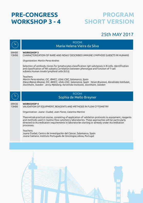
PRE-CONGRESS WORKSHOP 3 - 4
25th MAY 2017
ROOM Maria Helena Vieira da Silva
09H00 13H00
WORKSHOP 3 CHARACTERIZATION OF RARE AND NEWLY DESCRIBED IMMUNE LYMPHOID SUBSETS IN HUMANS
Organization: Martin Perez-Andres
Selection of antibody clones for lymphocytes classification: IgH subclasses in B-cells. Identification and classification of NK subsets.Correlation between phenotype and function of T-cell subsets.Human Innate lymphoid cells (ILCs).
Teachers: Martin Perez-Andres, CIC, IBMCC, USAL-CSIC, Salamanca, SpainElena Blanco Álvarez, CIC, IBMCC, USAL-CSIC, Salamanca, Spain Yenan Bryceson, Karolinska Institutet, Stockholm, Sweden Jenny Mjösberg, Karolinska Institutet, Stockholm, Sweden
ROOM Sophia de Mello Breyner
09H30 13H00
WORKSHOP 4VALIDATION OF EQUIPMENT, REAGENTS AND METHODS IN FLOW CYTOMETRY
Organization: Juana Ciudad, Juan Flores, Catarina Martins
Theoretical-practical course, consisting of application of validation protocols to equipment, reagents and methods used in routine flow cytometry laboratories. These approaches will be particularly directed to Accreditation requirements in laboratories starting or already under Accreditation processes.
Teachers: Juana Ciudad, Centro de Investigación del Cáncer, Salamanca, SpainJoana Caetano, Instituto Português de Oncologia,Lisboa, Portugal
PROGRAM SHORT VERSION
ROOM Sophia de Mello Breyner
ROOMFernando Pessoa
09H00 13H00
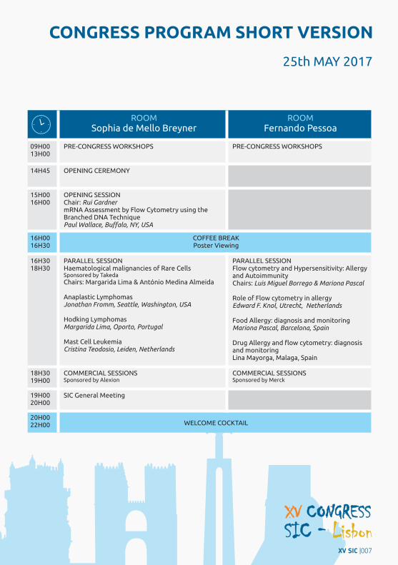
PRE-CONGRESS WORKSHOPS PRE-CONGRESS WORKSHOPS
14H45 OPENING CEREMONY
15H00 16H00
OPENING SESSION Chair: Rui GardnermRNA Assessment by Flow Cytometry using the Branched DNA TechniquePaul Wallace, Buffalo, NY, USA
16H00 16H30
COFFEE BREAK Poster Viewing
16H30 18H30
PARALLEL SESSIONHaematological malignancies of Rare CellsSponsored by Takeda
Chairs: Margarida Lima & António Medina Almeida
Anaplastic Lymphomas Jonathan Fromm, Seattle, Washington, USA
Hodking LymphomasMargarida Lima, Oporto, Portugal
Mast Cell LeukemiaCristina Teodosio, Leiden, Netherlands
PARALLEL SESSIONFlow cytometry and Hypersensitivity: Allergy and Autoimmunity Chairs: Luis Miguel Borrego & Mariona Pascal
Role of Flow cytometry in allergyEdward F. Knol, Utrecht, Netherlands
Food Allergy: diagnosis and monitoring Mariona Pascal, Barcelona, Spain
Drug Allergy and flow cytometry: diagnosis and monitoringLina Mayorga, Malaga, Spain
18H30 19H00
COMMERCIAL SESSIONS Sponsored by Alexion
COMMERCIAL SESSIONSSponsored by Merck
19H00 20H00
SIC General Meeting
20H00 22H00 WELCOME COCKTAIL
25th MAY 2017
CONGRESS PROGRAM SHORT VERSION
XV SIC |007
n
ROOM Sophia de Mello Breyner
ROOMFernando Pessoa
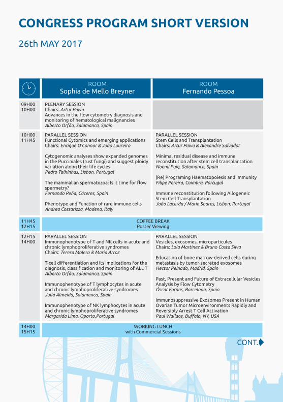
09H00 10H00
PLENARY SESSIONChairs: Artur PaivaAdvances in the flow cytometry diagnosis and monitoring of hematological malignanciesAlberto Orfão, Salamanca, Spain
10H00 11H45
PARALLEL SESSIONFunctional Cytomics and emerging applications Chairs: Enrique O'Connor & João Loureiro
Cytogenomic analyses show expanded genomes in the Pucciniales (rust fungi) and suggest ploidy variation along their life cyclesPedro Talhinhas, Lisbon, Portugal
The mammalian spermatozoa: Is it time for flow spermetry?Fernando Peña, Cáceres, Spain
Phenotype and Function of rare immune cells Andrea Cossarizza, Modena, Italy
PARALLEL SESSIONStem Cells and Transplantation Chairs: Artur Paiva & Alexandre Salvador
Minimal residual disease and immune reconstitution after stem cell transplantationNoemi Puig, Salamanca, Spain
(Re) Programing Haematopoiesis and Immunity Filipe Pereira, Coimbra, Portugal Immune reconstitution following Allogeneic Stem Cell Transplantation João Lacerda / Maria Soares, Lisbon, Portugal
11H45 12H15
COFFEE BREAK Poster Viewing
12H15 14H00
PARALLEL SESSIONImmunophenotype of T and NK cells in acute and chronic lymphoproliferative syndromes Chairs: Teresa Molero & Maria Arroz
T-cell differentiation and its implications for the diagnosis, classification and monitoring of ALL T Alberto Orfão, Salamanca, Spain
Immunophenotype of T lymphocytes in acute and chronic lymphoproliferative syndromes Julia Almeida, Salamanca, Spain
Immunophenotype of NK lymphocytes in acute and chronic lymphoproliferative syndromes Margarida Lima, Oporto,Portugal
PARALLEL SESSIONVesicles, exosomes, microparticulesChairs: Lola Martinez & Bruno Costa Silva
Education of bone marrow-derived cells during metastasis by tumor-secreted exosomesHector Peinado, Madrid, Spain
Past, Present and Future of Extracellular Vesicles Analysis by Flow Cytometry Òscar Fornas, Barcelona, Spain
Immunosuppressive Exosomes Present in Human Ovarian Tumor Microenvironments Rapidly and Reversibly Arrest T Cell ActivationPaul Wallace, Buffalo, NY, USA
14H00 15H15
WORKING LUNCH with Commercial Sessions
26th MAY 2017
CONT.
CONGRESS PROGRAM SHORT VERSION

ROOM Sophia de Mello Breyner
ROOMFernando Pessoa
15H15 17H00
PARALLEL SESSIONAdvances in primmary immunodeficiencies screening and diagnosis by flow cytometryChairs: Martin Perez-Andrés & João Farela Neves
New standardized strategies in the orientation of lymphoid defects by flow cytometry: Combined Immunodeficiencies and Primary Antibody Deficiences analysis by FCMMartín Perez Andrés, Salamanca, Spain Flow cytometry in the identification of innate citotoxic dysfunctionYenan Bryceson, Stockholm, Sweden Mendelian Susceptibility to Mycobacterial Disease - Strategies for Diagnosis by Flow Cytometry Júlia Vasconcelos, Oporto, Portugal
PARALLEL SESSIONMonitoring Immunotherapy for CancerSession with the collaboration of SPO & SEOM
Chairs: Gabriela Sousa, Luis De la Cruz, Paulo Rodrigues-Santos
Introduction to Immunotherapy Bruno Silva-Santos, Lisbon, Portugal
Immunodynamics assays for Cancer Immunotherapy Paulo Rodrigues-Santos, Coimbra Portugal
Monitoring NK cell-based Immunotherapy in the elderlyRafael Solana, Córdoba, Spain
17H00 17H30
COFFEE BREAK Poster Viewing
17H30 18H00
COMMERCIAL SESSIONSponsored by Beckman Coulter
COMMERCIAL SESSION
18H00 18H30
COMMERCIAL SESSION
18H00 18H30
SIC Groups presentations GECLID presentations
20H30 24H00 CONGRESS DINNER
26th MAY 2017
| CONT.
CONGRESS PROGRAM SHORT VERSION
XV SIC |009
n

ROOM Sophia de Mello Breyner
ROOMFernando Pessoa
09H00 10H00
PLENARY SESSIONChairs: Lola Martinez
Minimal Residual Disease detection in hemato logical malignancies: recent advances and future perspectivesJulia Almeida, Salamanca, Spain
10H00 11H00
PLENARY SESSION Accreditation of Flow Cytometry LaboratoriesChairs: Catarina Martins & Juana Ciudad
ISO 15189 and ISO 17025, standards for (clinical) laboratoriesCatarina Martins, Lisbon, Portugal IPAC / ENAC
Undergoing Accreditation in Flow Cytometry Labs: Steps to Follow and Critical PointsUlrich Sack, Leipzig, Germany
Flexible scope in flow cytometry: Experience of a laboratory accredited by UNE-EN-ISO 15189Juana Gil, Madrid, Spain
11H0011H30
COFFEE BREAK
Poster Viewing
11H30 13H00
PLENARY SESSION Immunology (SPI)Chairs: Margarida Saraiva & Manuel Vilanova Analysing the impact of T cell subsets on brain cognitive functionJulie C. Ribot, Lisboa, Portugal
Choreographing Immunity and Tolerance Induction in the ThymusNuno Alves, Porto, Portugal
Metabolic cues implicated on monocyte biologyRicardo Silvestre, Braga, Portugal.
Cell competition in the thymusVera Martins, Lisboa, Portugal
13H00 13H15 CLOSING CEREMONY
27th MAY 2017
CONGRESS PROGRAM SHORT VERSION
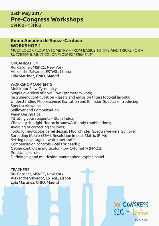
25th May 2017
Pre-Congress Workshops 09H00 - 13H00
Room Amadeo de Souza-CardosoWORKSHOP 1 MULTICOLOR FLOW CYTOMETRY – FROM BASICS TO TIPS AND TRICKS FOR A SUCCESSFUL MULTICOLOR FLOW EXPERIMENT"
ORGANIZATION Rui Gardner, MSKCC, New YorkAlexandre Salvador, ESTeSL, LisboaLola Martinez, CNIO, Madrid
WORKSHOP CONTENTSMulticolor Flow Cytometry:Simple overview of how Flow Cytometers work;Instrument configuration – lasers and emission filters (optical layout); Understanding Fluorescence: Excitation and Emission Spectra (introducing Spectra Viewers); Spillover and Compensation.Panel Design tips: Titrating your reagents – Stain Index; Choosing the right fluorochromes/Antibody combinations; Avoiding or correcting spillover; Tools for multicolor panel design: Fluorofinder, Spectra viewers, Spillover Spreading Matrix (SSM), Resolution Impact Matrix (RIM);Setting up voltages – which method?; Compensation controls – cells or beads?; Gating controls in multicolor Flow Cytometry (FMOs).Practical exercise: Defining a good multicolor immunophenotyping panel.
TEACHERSRui Gardner, MSKCC, New YorkAlexandre Salvador, ESTeSL, LisboaLola Martinez, CNIO, Madrid
XV SIC |011
n

Room Fernando PessoaWORKSHOP 2"DATA ANALYSIS IN LEUKEMIA AND LYMPHOMA IMMUNOPHENOTYPING"
ORGANIZATION: Julia Almeida, Centro de Investigación del Cáncer, Salamanca
WORKSHOP CONTENTS:Theoretical-practical course, consisting of data analysis with the Infinicyt program, which will be carried out by students directly on their computers (in which the INFINICYT program), under the guidance and supervision of teachers.
TEACHERSJulia Almeida, Centro de Investigación del Cáncer, SalamancaJuan Flores, Centro de Investigación del Cáncer, SalamancaQuentin Lecrevisse, Centro de Investigación del Cáncer, Salamanca
SPONSORSHIPCytognos S.L.
Room Maria Helena Vieira da SilvaWORKSHOP 3“CHARACTERIZATION OF RARE AND NEWLY DESCRIBED IMMUNE LYMPHOID SUBSETS IN HUMANS"
ORGANIZATIONMartin Perez-Andres, IBMCC, USAL-CSIC, Salamanca, Spain
WORKSHOP CONTENTSSelection of antibody clones for lymphocytes classification: IgH subclasses in B-cells.Identification and classification of NK subsets.Correlation between phenotype and function of T-cell subsets.Human Innate lymphoid cells (ILCs).
TEACHERSMartin Perez-Andres, CIC, IBMCC, USAL-CSIC, Salamanca, SpainElena Blanco Álvarez, CIC, IBMCC, USAL-CSIC, Salamanca, SpainYenan Bryceson, Karolinska Institutet, Stockholm, SwedenJenny Mjösberg, Karolinska Institutet, Stockholm, Sweden
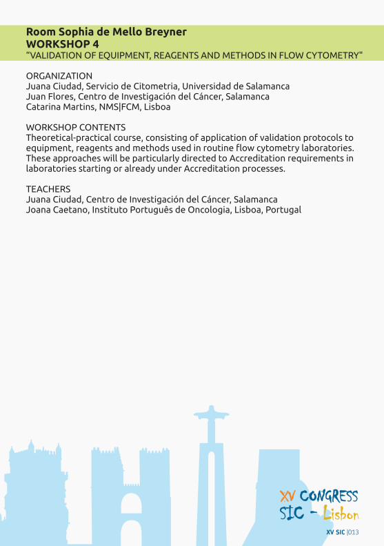
Room Sophia de Mello BreynerWORKSHOP 4“VALIDATION OF EQUIPMENT, REAGENTS AND METHODS IN FLOW CYTOMETRY"
ORGANIZATIONJuana Ciudad, Servicio de Citometria, Universidad de SalamancaJuan Flores, Centro de Investigación del Cáncer, SalamancaCatarina Martins, NMS|FCM, Lisboa
WORKSHOP CONTENTSTheoretical-practical course, consisting of application of validation protocols to equipment, reagents and methods used in routine flow cytometry laboratories. These approaches will be particularly directed to Accreditation requirements in laboratories starting or already under Accreditation processes.
TEACHERS Juana Ciudad, Centro de Investigación del Cáncer, SalamancaJoana Caetano, Instituto Português de Oncologia, Lisboa, Portugal
XV SIC |013
n
CONGRESS PROGRAM
25th May 2017
14H45 - 15H00 OPENING CEREMONY | Room Sophia de Mello Breyner
15H00 - 16H00 OPENING SESSION | Room Sophia de Mello Breyner
Chair: Rui Gardner
mRNA ASSESSMENT BY FLOW CYTOMETRY USING THE BRANCHED DNATECHNIQUEISAC LECTURE
Abstract: Flow cytometry permits the simultaneous measurements of many biomarkers in individual cells from bulk populations. Until now analysis has been limited to primarily analysis of proteins and total DNA or highly abundant DNA sequences. Since most mRNA gene transcripts are present at very low quantities our ability to detect these by flow cytometry has been limited. The branched DNA technique amplifies the signal from a single mRNA species several thousand fold. This procedure allows for the determination of low copy number mRNA expression within a mixed population of cells and is compatible with antibody-based targeting of both extracellular and intracellular antigens. To show proof of concept, we used the technique to detect BCR and ABL transcripts in cell lines positive for the t(9;22) translocation. While, as expected, all cells had detectable BCR and ABL transcripts, cells with the t(9;22) translocation had quantitatively more BCR and ABL mRNA than those that were negative for the translocation. These results correlated with data obtained by RT-PCR. We have extended these investigations to the evaluation of human peripheral blood and the expression of CD8. We detected CD8 mRNA expression in both T cells and NK cells which correlated with protein expression. After stimulation with phorbol 12-myristate 13-acetate and ionomycin for up to 4 h, an increase in CD8 mRNA was detectable 30 minutes before increased CD8 protein was detected. These data were correlated with EOMES and T-bet protein expression to identify distinct subset of CD8 positive T and NK cells. Collectively, these results exemplify how the branched RNA flow cytometry labeling procedure can be applied to simultaneously assess mRNA and protein dynamics to gain insight into the regulation of gene transcription and translation in individual cells.
16H00 - 16H30 COFFEE BREAK and Poster Viewing
16H30 - 18H30 PARALLEL SESSION | Room Sophia de Mello Breyner
Haematological malignancies of Rare CellsChairs: Margarida Lima & António Medina Almeida
Paul WallaceDepartment of Flow & Image Cytometry, Roswell Park Cancer Institute, Buffalo, NY

ANAPLASTIC LYMPHOMAS
Abstract: Anaplastic Large Cell Lymphoma (ALCL) is a large cell CD30+ lymphoma that is often systemic (usually nodal). However, a recently described subset of these lymphomas is associated with breast implants (breast implant ALCL, or BI-ALCL). The flow cytometric findings in systemic ALCL and BI-ALCL will be described. Systemic ALCL can be characterized by the presence (ALK+ ALCL) or absence (ALK- ALCL) of anaplastic lymphoma kinase (ALK), a tyrosine kinase aberrantly expressed by these large cell lymphomas that harbors a translocation of ALK (on chromosome 2) with one of various partners. ALK+ ALCL makes up 70% of systemic ALCL cases and both lymphomas involve nodal and extranodal sites.A number of studies have examined the flow cytometric findings in systemic ALCL (Juco et al (2003); Muzzafar et al. (2009); Kesler et al. (2007)). In general, the neoplastic cells of ALCL could frequently be detected and immunophenotyped by flow cytometry; specifically, the neoplastic population could be detected in 25 of 29 cases (86%) and 19 of 23 cases (83%) (Kesler et al and Muzzafar et al, respectively). Immunohistochemistry was frequently used to complement the flow cytometric results, including detection of the ALK protein by immunohistochemistry. The immunophenotype observed in the ALCL cases (composite data from 3 above papers) is shown in the Table 1 (below). These lymphomas expressed CD30 and CD45. Interestingly, although the neoplastic cells are of T cell origin, T cell antigen expression (CD2, CD3, CD4, CD5, CD7, and CD8) was often aberrantly lost, with only a subset of cases showing expression of a given T cell antigen. Expression of myeloid antigens (CD13 and CD15) was occasionally seen and B cell antigens (CD19 and CD20) were not expressed.Our laboratory at the University of Washington often evaluates systemic ALCL with a standard T cell tube (for CD2, CD3, CD4, CD5, CD7, CD8, CD30, CD34, CD45, and CD56) and a tube for classical Hodgkin lymphoma (CHL; CD5, CD15, CD20, CD30, CD40, CD45, CD64, CD71, and CD95). Expression of CD30 and CD71 in the CHL tube is often quite helpful in identifying the neoplastic population of ALCL. The neoplastic cells of systemic ALCL lack expression of CD40 (or express very weak CD40), while the neoplastic Hodgkin and Reed-Sternberg cells of CHL always express intermediate to bright CD40 (Carbone A et al. (1995); Fromm et al. (2009)), a feature that allows these CD30+ lymphomas to be distinguished.Breast implant ALCL (BI-ALCL) is a newly recognized type of CD30+, ALK- T cell lymphoma. These lymphoma are particularly uncommon with an incidence of 0.1-0.3 BI-ALCL cases/100,000 women with prostheses (Laurent et al. (2016)). As its name would imply, these lymphomas are associated with prior placement of breast prostheses; prognosis of these lymphomas differed depending on whether the neoplastic cells are invasive/infiltrative or not.Our group published a characterization of the immunophenotype of BI-ALCL by flow cytometry. Surprisingly, the neoplastic cells showed immunophenotypic features more in keeping with CHL than ALCL (loss of T cell antigens) and expression of CD15, CD30, CD40, CD71, and CD95). After this report, Montgomery-Goecker and co-workers (Montgomery-Goecker et al. (2015)) published their flow cytometric findings of two additional BI-ALCL cases. These two cases also showed loss of T cell antigens (no expression of CD3, CD5, CD7, CD8) except CD4, and uniform expression of CD30. Both cases were mainly negative for CD45. One of two of these cases showed expression of CD13 and CD33. Neoplastic cells represented 48% and 6.5% of the total events. Despite the similarities, CHL and BI-ALCL can typically be distinguished as: 1) a diagnosis of CHL in a breast capsule tissue would be extremely uncommon; 2) while some CHL cases may have expression of CD40 at the level seen in these BI-ALCL cases, the majority of CHL cases have significantly brighter levels of CD40 expression; 3) in CHL, a plot of CD45 versus CD5 demonstrates either two populations (one with lower CD45 and no CD5 and a second with brighter CD45 and CD5 expression) or a diagonal relationship for CD45 and CD5 due to T cell rosetting (Fromm and Wood (2012); and 4) reactive CD4+ T cell populations with increased expression of CD7 and CD45 are seen in greater than 80% of CHL cases (Fromm et al (2010)). These immunophenotypic features will not be seen in ALCL.
Jonathan Fromm, MD, PhDAssociate Professor, Laboratory Medicine and Associate Director, Hematopathology Laboratory, University of Washington Medical Center (UWMC); Seattle, Washington, USA
XV SIC |015
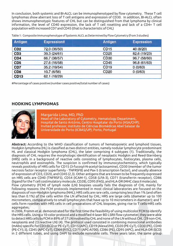
In conclusion, both systemic and BI-ALCL can be immunophenotyped by flow cytometry. These T cell lymphomas show aberrant loss of T cell antigens and expression of CD30. In addition, BI-ALCL often shows immunophenotypic features of CHL but can be distinguished from that lymphoma by clinical information, the level of CD40 expression, the lack of T cell rosetting and lack of a CD4+ T cell population with increased CD7 and CD45 (that is characteristic of CHL)
Table 1: Composite Immunophenotype of Systemic ALCL as Determined by Flow Cytometry (from 3 studies)
#Percentage of cases positive (number cases positive/total number of cases)
HODKING LYMPHOMAS
Margarida Lima, MD, PhDHead of the Laboratory of Cytometry, Hematology Department, Hospital de Santo António, Centro Hospitalar do Porto (HSA/CHP); Invited professor, Instituto de Ciências Biomédicas Abel Salazar da Universidade do Porto (ICBAS/UP); Porto, Portugal
Abstract: According to the WHO classification of tumors of hematopoietic and lymphoid tissues, Hodgkin lymphoma (HL) is classified as two distinct entities, namely nodular lymphocyte predominant HL and classical Hodgkin lymphoma (CHL), the later comprising 4 subtypes (1). Traditionally, the diagnosis of CHL requires the morphologic identification of neoplastic Hodgkin and Reed-Sternberg (HRS) cells in a background of reactive cells consisting of lymphocytes, histiocytes, plasma cells, neutrophils and eosinophils. The suspicion is confirmed by immunocytochemistry, which typically reveals positivity of HRS cells for CD15 (3-fucosyl-N-acetyl-lactosamine), CD30 (member of the tumor necrosis factor receptor superfamily - TNFRSF8) and Pax-5 (transcription factor), and usually absence of expression of CD3, CD20, and CD45 (2,3). Other antigens that are known to be frequently expressed on HRS cells are CD40 (TNFRSF5), CD54 (ICAM-1), CD58 (LFA-3), CD71 (transferrin receptor), CD86 (ligand for the T cell costimulatory molecule, CD28), CD95 (FAS), and HLA-DR (MHC class II molecule).Flow cytometry (FCM) of lymph node (LN) biopsies usually fails the diagnosis of CHL mainly for following reasons: the FCM protocols implemented in most clinical laboratories are focused on the diagnosis of non-Hodgkin lymphoma (NHL); HRS cells are rare cells, comprising less than 1% (and often less than 0.1%) of the cells within the LN affected by CHL; HRS are large cells (diameter up to 100 micrometers, comparatively to small lymphocytes that have up to 10 micrometers in diameter); and T cells form rosettes with HRS cells in cell preparations of CHL biopsies, giving rise to T cells-HRS cells aggregates.In 2006, Fromm et al. demonstrated for the first time the feasibility of using multicolor FCM to identify the HRS cells. Using a 10-color protocol and a modified 4-laser BD LSRII flow cytometer, they were able to detect HRS cells by FCM in 89% of 27 LN involved by CHL and none of the LN without CHL (29 non-CHL neoplasms and 23 reactive LN) (4). The protocol used consisted in combining monoclonal antibodies (mAbs) specific for CD3 (ECD or PE-Cy7), CD15 (APC), CD19 (Alexa594), CD20 (PE-Cy7), CD30 (PE), CD40 (PE-CY5.5), CD45 (APC-Cy7), CD64 (FITC), CD71 (APC-A700), CD86 (PE), CD95 (APC), and HLA-DR (ECD) in 2 different tubes, and using DAPI to exclude nonviable cells. Three years later, the same group
proposed a single 9-color tube to identify HRS cells, using the same cytometer: CD5 (APC-Cy7), CD15 (APC), CD20 (PE-Cy7), CD30 (PE), CD40 (PE-Cy5.5), CD45 (PE-Texas red), CD64 (FITC), CD71 (APC-A700), and CD95 (PB) (5). They used this tube to study 279 blindly identified and 141 selected (suggestive of CHL) LN biopsies. Of the 53 CHL cases identified by histopathology (10 in the unselected group; 43 in the selected group), the FC sensitivity and specificity were of 89% and 100%, respectively. Latter on (2014), the same group validated a 6-color tube for the same purpose, using a similar cytometer: CD3 (APC-Cy7), CD20 (PE-Cy7), CD30 (PE), CD40 (PE-Cy5.5), CD64 (FITC), and CD95 (APC) (6). By analyzing 408 biopsies (including 55 CHL) they obtained a sensitivity and specificity of 85.4% and 99.7%, respectively. In general, the most common immunophenotype of the HRS cells was found to be CD45+low, CD15+, CD30+, CD40+, CD54+, CD58+, CD71+, CD86+, CD95+, CD123+, HLA-DR+, CD3-, CD5-, CD19-, CD20-, and CD64- (5-8). HRS-T cell rosettes give rise to a composite (CD15+, CD30+, CD40+, CD95+, CD3+, CD5+) immunophenotype, and interactions between HRS cells and T cells can be disrupted with blocking antibodies (e.g. anti-CD2 and anti-CD58) (5). The methods used for the preparation of LN cells and immunophenotyping of HRS cells were previously described in detail (7). To the best of our knowledge, to date, no other group has devoted itself to the study of the HRS cells by FCM. However, several studies have focused on the FCM analysis of the background of inflammatory cells observed in HL (9-15). Relevant alterations described included increased percentages of neutrophils and eosinophils, normal or increased CD4/CD8 ratios (9,10), increased expression of CD7 and other T cell antigens (11,12), increased proportions of CD4+, CD25+, CD152+, and FoxP3+ regulatory T cells (13), and higher fractions of CD4+CD26- T cells (14,15). Considering all these parameters together makes FCM even more useful of FCM for the diagnosis of HL (16).Taking advantage of our large experience in the study of tissue biopsies by FCM (around 2.900 tissue biopsies studied from 2009 to 2016, mainly for the diagnosis of NHL), we have recently invested in the study of HL by FCM. First, biopsy samples are screening for NHL using three 8-color tubes (BD FACSCanto I I , 200.000 events/tube): Lymphocyte screening tube (CD8+IgLambda-FITC/CD56+IgKappa-PE/CD5-PC5.5/CD10-PC7/CD3-APC/CD19-APC-H7/CD4+CD20-V450/CD45-KrO); B-cell tube (BCL2cyt-FITC/CD10-PE/CD38-PC5.5/HLA-DR-PC7/CD81-APC/CD19-APC-H7/CD20-V450/CD45-KrO); and T-cell tube (CD2-FITC/CD7-PE/TCR-GD-PC5.5/CD28-PC7/CD5-APC/CD3-APC-H7/CD4-V450/CD8-KrO). If no evidence of NHL is found, and there is sample available, additional tubes are performed to identify HRS cells, with at least 400.000 events being collected in each tube. After a period of testing of several combinations of mAbs, and based on the reports of Fromm et al., we are now using a HL protocol, consisting of two 8-color tubes in order to identify HRS cells: CD15-FITC/CD95-PE/CD40-PERCP-Cy5.5/HLA-DR-PC7/CD30-APC/CD3-APC-H7/CD20-V450/CD45-KrO and CD14-FITC/CD86-PE/CD40-PERCP-Cy5.5/HLA-DR-PC7/CD30-APC/CD3-APC-H7/CD81-V450/CD45-KrO.Until now, our experience is based on the results obtained in 57 biopsies from patients suspected of having lymphoma. Fifty seven surgical biopsies (47 cases with negative screening for NHL and 10 cases with the diagnosis of NHL) from 57 patients (31 males and 25 females, median age of 47 years, ranging from 14 to 89 years) were evaluated for the presence of HRS cells using the HL protocol. Biopsy sites were as follows: LN – 52 cases (25 cervical, 10 inguinal, 7 axillary, 3 mediastinal, 2 intra-parotid, 5 non-specified); amygdala – 2 cases; and oropharynx or nasopharynx – 3 cases. Subsequent review of the histopathology reports revealed that these included biopsies from tissues affected by CHL (15 cases, all corresponding to LN), NHL (10 cases - B cell NHL 9 cases; T cell NHL 1 case), non-hematological tumors (9 cases), histiocytic disorders (2 cases - Langerhans cell histiocytosis and histiocytic lymphadenitis. All 15 HL consisted on CHL subtypes: nodular sclerosis (10 cases), mixed cellularity (1 case), lymphocyte rich (1 case) and lymphocyte depleted (0 cases); 3 cases are still waiting for sub-classification. The remaining 21 cases corresponded to reactive tissues.As previously reported by Fromm et al. (5-8), we observed that HRS cells with a distinctive phenotype (CD45+low, CD15+, CD30+, CD40+high, CD86+, CD95+, and HLA-DR+high in most cases) can be readily identified in CHL samples (14/15 cases, 93.3%). The question is whether FCM is specific enough to distinguish CHL from reactive immunoblastic proliferations arising in the setting of infections, drug reactions, vaccines, malignancies, and autoimmune diseases. Indeed, in our experience, as in the experience of other authors (17), HRS-like cells were present in some reactive LN, especially those containing T and B cell derived immunoblasts. These cells often show CD30 expression and they may morphologically mimic HRS cells, sometimes leading to erroneous diagnoses of HL by histology. Positivity of the immunoblastic cells for B (e.g. CD20) or T (e.g. CD3) antigens, and negativity for CD15 is usually useful to distinguish them from true HRS cells (17). Finally, as also stated by Fromm et al., in cases where FCM is consistent with CHL, other related lymphoid neoplasms where HRS-like cells can be found still remain on the differential diagnosis; these include, among others, the so-called grey zone lymphomas with features intermediate between CHL and diffuse large B cell NHL (16) (18). However, in these situations, routine FCM panels are usually sufficient to establish the diagnosis of NHL.
XV SIC |017
Cristina Teodósio, PhDPostDoc, Department of Immunohematology and Blood Transfusion, Leiden University Medical Centre (LUMI); Leiden, Netherlands
MAST CELL LEUKEMIA
1 2 3 4 4Authors: C Teodosio , A Mayado , M Jara-Acevedo , L Sanchez-Muñoz , I Alvarez-Twose , A 1 4 1 4 1 1García-Montero , A Matito , C Caldas , AF Henriques , A Lopez , JI Muñoz-González , N
1 1 1 1Dasilva-Freire , JI Sanchéz-Gallego , L Escribano , A Orfao , on behalf of the Spanish Network on Mastocytosis (REMA)
Institutions: Department of Immunohematology and Blood Transfusion, Leiden University Medical Centre, Leiden, The Netherlands.2Servicio General de Citometría (NUCLEUS), Centro de Investigación del Cáncer (IBMCC-CSIC/USAL and IBSAL) and Departamento de Medicina, Universidad de Salamanca, Salamanca, Spain.3DNA Sequencing Service (NUCLEUS), University of Salamanca, Research Biomedical Institute of Salamanca (IBSAL), Salamanca, Spain.4Instituto de Estudios de Mastocitosis de Castilla La Mancha (CLMast), Hospital Virgen del Valle, Toledo, Spain.
Abstract: Mast cell leukemia is a rare disorder which represents the leukemic manifestation of Systemic Mastocytosis (SM). Therefore, MCL patients fulfil the diagnostic criteria for SM, including the detection of prominent multifocal clustering of mast cells (MCs) in the bone marrow (BM), which represents a major diagnostic criterion, and four minor criteria, including an atypical MC morphology, expression of CD2 and/or CD25 in MCs, presence of an activating mutation at codon 816 of KIT and serum tryptase 20ng/mL. As a SM variant, MCL patients fulfil one major and a minor or at least three minor criteria. MCs in MCL are usually immature, sometimes bi- or multi-lobed (promastocytes), however in some patients more mature forms may be predominant. From a immunophenotypic point of view, MCs in MCL usually express CD117, with low tryptase and FcRI levels, often expressing CD25 and CD30 but not CD34. Detection of CD2 is only restricted to limited number of cases and lack of both CD2 and CD25 can also be observed in some patients. Expression of CD52, HLA-DR and CD123 may also be observed.Despite the rarity of the disease, MCL is known to have heterogeneous presentation. Based on the World Health Organization (WHO) criteria, MCL can be divided in a “leukemic” variant, with >10% MCs detected in peripheral blood (PB) and an “aleukemic” variant, with <10% PB infiltration. In both groups of patients MCs represent at least 20% of all nucleated cells in the BM, the primary diagnostic criterion for MCL, and the prognosis is poor. In addition to these variants, recent reports suggest that MCL may develop either as a de novo disease or secondary to e.g. aggressive systemic mastocytosis. Furthermore, in a few cases, the course of the disease may be less aggressive and more chronic, without signs and symptoms of organ damage (“C-findings”), showing a better overall prognosis than the acute variant of the disease. These observations have led to the recent proposal to discriminate between chronic MCL (without “C-findings”) and acute MCL with “C-findings”. Differential diagnosis of MCL may represent a challenge, not only due to its heterogeneous presentation, but also because e.g. it may or not present with an associated haematological neoplasm
+(AHN), CD25 atypical MCs can also be found in BM of patients with chronic eosinophilic leukemia (CEL), discrimination between blasts maturing to MCs vs. basophil might be difficult, and blast with maturation to MC can be detected in 13% of AML. In this presentation we will review the diagnostic criteria for MCL, as well as the recently defined variants of the disease and the differential diagnosis from other myeloid neoplasms, mainly focusing on the utility of multiparameter flow cytometry in the diagnostic work-up of the disease.

16H30 - 18H30 PARALLEL SESSION II | Room Fernando Pessoa
Flow cytometry and Hypersensitivity: Allergy and AutoimmunityChairs: Luis Miguel Borrego & Mariona Pascal
ROLE OF FLOW CYTOMETRY IN ALLERGY
Abstract: Development of flow cytometry, discovery of activation markers such as CD63 and unique markers identifying basophil granulocytes have led to the introduction of the so-called basophil activation test (BAT) in allergy research and diagnostics. BAT measures basophil responses to IgE-dependent and –independent stimuli on between 150 and 1000 basophil granulocytes in less than 0.1 ml fresh blood. CD63 is binding a non-basophil-specific epitope inside the granular membrane. Upon degranulation the CD63 epitope is exposed on the outside of basophils. The CD63 increase is related to release of histamine of basophils. In contrast to the measurement of histamine, the BAT can be easily introduced in research and diagnostic settings. BAT can be part of the diagnostic evaluation of patients with food-, insect venom-, and drug allergy and chronic urticaria. It may be helpful in determining the clinically relevant allergen. Basophil sensitivity may be used to monitor patients on allergen immunotherapy, anti-IgE treatment or in the natural resolution of allergy. BAT uses fewer resources and is more reproducible than challenge testing, especially in the diagnostic procedure of food allergy, in which double-blind placebo-controlled challenge tests are often still used. As it is less stressful for the patient and avoids severe allergic reactions, BAT ought to precede challenge testing. The nature of basophil activation as an ex vivo challenge makes it a multifaceted and promising tool not only for the allergist, but only in a research setting. BAT is implemented to screen for allergenic activity of (modified) allergens. In addition, also in the basic research of basophil activation BAT has been helpful. In this presentation, an overview of the characteristics of the CD63 in basophils and its implementation in BAT will be discussed.
Edward F. Knol, PhD, associate professorUniversity Medical Center Utrecht, Depts. Immunology and Dermatology/Allergology, Huispost F03.821Heidelberglaan 100, 3584 CX Utrecht, The Netherlands
SELECTED ORAL COMMUNICATIONS PRESENTATIONSCO_2428 BLASTIC PLASMACYTOID DENDRITIC CELL NEOPLASM. A CASE WITH DIFFERENT INMUNOPHENOTYPE AND CHROMOSOMAL ABERRATIONSCO_2433 THE PROLIFERATION INDEX OF BONE MARROW CELLS MAY ASSIST THE DIFFERENTIAL DIAGNOSIS OF CHRONIC MYELOMONOCYTIC LEUKEMIA VS. MONOBLASTIC/ MONOCYTIC LEUKEMIAS
Session Sponsored by Takeda
XV SIC |019
FOOD ALLERGY: DIAGNOSIS AND MONITORING
Abstract: Basophil activation test (BAT) is an in vitro test used for the diagnosis and monitoring of allergy to allergens such as foods, drugs, hymenoptera venoms and inhalants. Briefly, basophils of the patient are incubated with the suspected allergen, and if the patient is sensitised, they become activated. This activation can be assessed by the measurement of mediators released, but also with the activation markers expressed in the cell membrane. In food allergy diagnosis, Oral food challenge (OFC) is still the gold-standard; however, it is time- and resource-consuming with the risk of inducing an allergic reaction, indeed potentially severe. In several published studies, BAT has shown higher specificity than conventional specific IgE testing in the skin or in serum for diagnosing certain food allergies. Thus, BAT has emerged as a new diagnostic test for food allergy. Nevertheless, it is very important to note that the diagnostic utility of BAT is allergen-specific and needs to be validated for different allergen preparations and patient populations, as well as, the methodology adopted and the flow cytometry analysis, since all these can have a significant impact on the accuracy of the results obtained. Besides discrimination between food allergic and tolerant subjects, it can provide valuable information about the characteristics of food-induced reactions, p.e. the severity of the allergic symptoms and the dose of peanut protein at which patients may react during OFC have been reported, but further studies are required for other food allergies. Moreover, BAT can be useful in assessing the natural resolution of food allergies that are commonly outgrown over time (cow's milk and egg allergy), but also to distinguish different phenotypes of patients with these allergies (for instance, tolerance to extensively heated or unheated forms of these foods). In research studies, BAT has also been used to monitor the clinical response to immunomodulatory treatments for food allergy (peanut, cow's milk and egg), observing decreased basophil reactivity and sensitivity to the respective food allergens. In conclusion, BAT is a promising tool as a clinically useful test. However, further studies are required in order to enable a wider use of BAT in clinical practice.
DRUG ALLERGY AND FLOW CYTOMETRY: DIAGNOSIS AND MONITORING
Abstract: Drug hypersensitivity reactions (DHRs) are those adverse reactions produced after the exposure to a drug at doses normally tolerated by non-hypersensitive subjects. They constitute an important drug safety problem as a result of their severity and unpredictability. DHR diagnosis can lead to the withdrawal of important and often non-substitutable treatments, the prescription of alternative drugs that can be more expensive and potentially toxic. All this can be an issue for the public health system, especially when the diagnosis is not fully confirmed. Diagnosis of DHRs is complex, generally based on a detailed clinical history, skin testing (ST) and drug provocation testing (DPT). However, clinical history can be unreliable and ST have not optimal sensitivity for some drugs. This leaves DPT as the gold standard for diagnosing DHR, however, it is not risk-free, needs to be done in a specialised setting by trained personnel, is time consuming and cannot be performed for severe reactions. Therefore, there is a need to use in vitro testing to ensure accurate diagnosis, both to improve patient safety and to reduce health system costs.The methods used for the diagnosis of DHRs depend on the mechanism involved and the kinetic of the reaction. IgE- or T-cell-mediated.
Cristobalina Mayorga, PhDResearch Laboratory- Allergy Unit, University Hospital of Málaga, IBIMA, Málaga, Spain
Mariona Pascal, PhDImmunology Department, CDB, Hospital Clínic de Barcelona, Barcelona, Spain
In immediate or IgE-mediated DHRs, the determination of drug-specific IgE antibodies has been performed by immunoassays. However, it is available for a limited number of drugs and with a low sensitivity. In the last decades, basophil activation tests (BAT) using flow cytometry has allowed the detection of activated basophils using specific markers, despite the low percentage of peripheral basophils (<1% of total leukocytes). Moreover, BAT does not need to use drugs conjugated with a carrier molecule, allowing its use in a wide variety of drugs and is particularly useful for evaluating hypersensitivity reactions to drugs for which there is no other in vitro test available.The evaluation of cell-mediated DHR is more complex, as they include heterogeneous clinical symptoms and mechanisms. These reactions can be analysed in the acute phase by assessing different inflammatory markers on peripheral blood using flow cytometry or at the resolution phase of the reaction mainly aimed at identifying the responsible drug. For this last purpose lymphocyte transformation test (LTT) has been the most frequently used and is based on the capacity of lymphocytes to proliferate after stimulation with a specific drug generally evaluated by the incorporation of either 3H-thymidine. In the last years, the possibility of evaluating the proliferative response using carboxyfluorescein diacetate succinimidyl ester and flow cytometry has improve the LTT capacity since enables one to phenotype the proliferating cells in terms of population structure and production of inflammatory markers. This technique will be key to fully characterising the effector mechanism triggered by the suspected drug.Taken all together we can observe that flow cytometry helps the evaluation of DHRs giving the opportunity to perform more functional in vitro tests that mimic the effector mechanism involved in the DHRs. Multidisciplinary collaborations are needed to identify biomarkers and increasing our knowledge of the drug metabolites involved in the immunological mechanisms of DHRs that will improve the sensitivity and specificity of in vitro tests.
SELECTED ORAL COMMUNICATIONS PRESENTATIONSCO_2454 INTRAEPITHELIAL LYMPHOCYTES SUBSETS IN CELIAC DISEASE DIAGNOSED PATIENTSCO_2457 CHILDREN WITH COW´S MILK ALLERGY: THE IMPACT OF ORAL IMMUNOTHERAPY (OIT) IN THEIR IMMUNE PROFILE
18H30 - 19H00 COMMERCIAL SESSION by ALEXION | Room Sophia de Mello Breyner
Avances en el diagnóstico de la HPNChair: Amparo Sempere - Hospital La Fe, ValenciaSpeakers: Alberto Orfão, Centro de Investigación del Cáncer, Universidad de Salamanca (CSIC), Salamanca
Teresa Caballero, Hospital Universitario Virgen del Rocío, Sevilla
Antonio Almeida, Instituto Portugués de Oncología, Lisboa
XV SIC |021
18H30 - 19H00 COMMERCIAL SESSION by MERCK | Room Fernando Pessoa
STUDY OF IMMUNOLOGICAL SYNAPSE FORMATION USING FLOWSIGHT® AND IMAGESTREAM® IMAGING FLOW CYTOMETERS.
Abstract: During adaptive immune response, the activation of T lymphocytes by antigen-presenting cells (APC) results in the formation of an immunological synapse. Following contact, the formation of immune synapses requires multiple rearrangements in the actin cytoskeleton and the recruitment of adhesive and signaling molecules to the T cell-APC interface. During the past decades, the immunological synapse was studied using fluorescence microscopy. However, the interpretation of microscopic imagery could be subjective and laborious to analyze statistically, especially for the study of rare events like immunological synapse. In order to overcome these problems, Amnis® imaging flow cytometers were used. ImageStream®X and FlowSight® are multispectral imaging flow cytometers able to generate high resolution microscope images of cells in suspension. In particular, the ImageStream®X and FlowSight® systems allow to register multiple parameters for each cells, including brightfield, darkfield (SSC) and up to 10 fluorescent markers at high speed, thanks to the CCD camera and the time-delay integration (TDI) technologies. Due to these characteristics, ImageStream®X and FlowSight® combine the speed and sensitivity of flow cytometry with the resolution and detailed imagery of microscopy, overcoming the limitations of both techniques. Using the IDEAS® analysis software and its optimized wizards, we were able to demonstrate that both FlowSight® and ImageStream®X can distinguish the cells conjugates with an organized immunological synapse and assess their frequency. Moreover, the ImageStream®X can furtherly discriminate the specific location of adhesion and signaling molecules within the interface of the immunological synapse. In conclusion, the innovative design of Amnis® cytometers and the ease of use of IDEAS® software bring outstanding performances, opening the door to the study of an extensive range of applications, like the immunological synapse formation.
19H00 - 20H00 SIC GENERAL MEETING | Room Sophia de Mello Breyner
20H00 - 22H00 WELCOME COCKTAIL at Centro Cultural de Belém
Clara Cigni, Merck S.p.A., Vimodrone (MI), Italy
CYTOGENOMIC ANALYSES SHOW EXPANDED GENOMES IN THE PUCCINIALES (RUST FUNGI) AND SUGGEST PLOIDY VARIATION ALONG THEIR LIFE CYCLES
Abstract: Rust fungi (Basidiomycota, Pucciniales) are biotrophic plant pathogens with complex life cycles (up to five spore types). The urediniosporic infection cycle is frequently the most important in disease dissemination as the only stage capable of repeating itself. The cell nuclear content of rust fungi is thought to follow that of other Basidiomycota, with haploid nuclei throughout the life cycle, only becoming diploid upon karyogamy in telia and immediately returning to the haploid state as meiosis takes place leading to the formation of basidiospores. Recently, using genome size quantification techniques, the presence of 1C, 2C and a low proportion of 4C nuclei was detected in different stages of the urediniosporic cycle of several rust fungi. These results suggest the presence of diploid nuclei that supposedly only occur in teliospores, and compatible with the occurrence of karyogamy and meiosis prior to urediniospore formation, although endopolyplody or other
Pedro Talhinhas, PhDLEAF - Linking Landscape, Environment, Agriculture and Food, Instituto Superior de Agronomia, Universidade de Lisboa, Tapada da Ajuda, Lisboa, Portugal. de Lisboa, Tapada da Ajuda, Lisboa, Portugal.
10H00 - 11H45 PARALLEL SESSION III | Room Sophia de Mello Breyner
26th May 2017
09H00 - 10H00 PLENARY SESSION | Room Sophia de Mello Breyner
Chair: Artur PaivaADVANCES IN THE FLOW CYTOMETRY DIAGNOSIS AND MONITORING OF HEMATOLOGICAL MALIGNANCIES
Abstract: Flow cytometry immunophenotyping has long proven to be of great clinical utility in the diagnosis classification and monitoring of haematological malignancies. This is mainly due to the fact that flow cytometry allows for objective and robust multiparametric analysis of thousands of cells per second. However, along the years, both the protocols used for instrument set up and sample preparation, and the specific panels of reagents applied have varied substantially among laboratories. This is mostly due to continuous introduction of new technological advances in the equipment and reagents (e.g. potential for the measurement of more colors and more cells, broad availability of compatible fluorochrome and fluorochrome-conjugated antibody reagents and an increased availability of antibodies clones per CD marker), together with co-existence of multiple, only partially overlapping, consensus-based antibody panels.Despite such consensus guidelines have been proposed to standardize the antibody panels, they have been only partially successful. Since 2012, the EuroFlow consortium has introduced a new concept a strategy about how to apply flow cytometry for the immnunophenotypic diagnosis of haematological malignancies based on the use of fully standardized procedures and panels of reagents in order to allow clinical comparisons of immunophenotypic data obtained for individual samples against data bases of normal and leukemic/lymphoma patient samples. This permits automated gating and alarming for any altered cell population in the sample, identification of relevant technical problems, and a software-guided diagnostic help. Together with the introduction of new informative markers and software tools immunophenotyping this has paved the way for more robust, sensitive and reproducible diagnosis of haematological malignancies. In addition, the new tools and strategies have also proven useful in other fields such as the diagnostic screening and classification of primary immunodeficiencies and immune monitoring.
Alberto Orfão, MD, PhDDepartamento de Medicina and Servicio General de Citometria
XV SIC |023
PHENOTYPE AND FUNCTION OF RARE IMMUNE CELLS
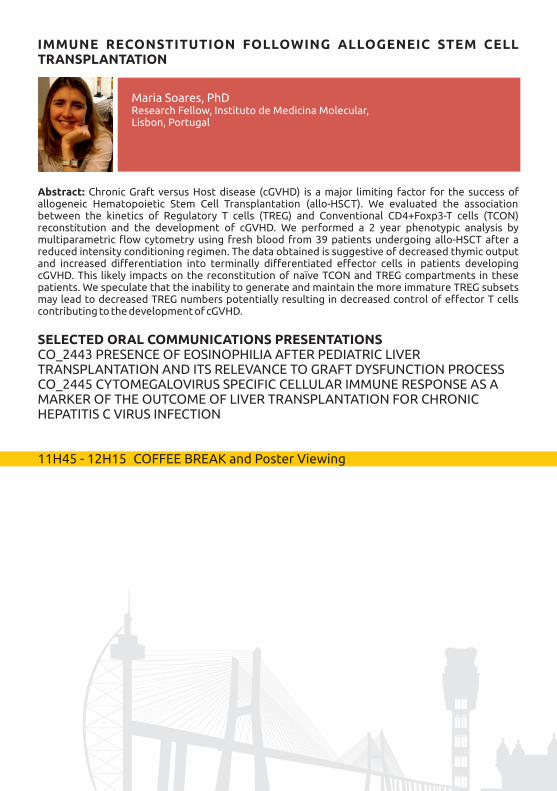
Abstract: The possibility to detect, count and perform functional analysis of rare cells is opening a new field in the world of cell analysis, and it is giving relevant information either to the basic scientist or to the clinician. The term “rare” is typically referred to those events that have a frequency of 0.01% or less. In the last year, my group has focussed on the development of new methodologies and techniques for studying rare events, and studies were performed on several different types of cells and different pathologies. Applications of rare cell analysis include, among others, the detection in blood of tumours such as metastatic breast cancer cells or neuroblastoma cells infiltrating the bone marrow, monitoring of minimal residual disease, detection of stem cells and rare HIV-infected cells in peripheral blood, identification of antigen (Ag)-specific T cells, invariant natural killer T cells, and analysis of mutation frequencies in genetic toxicology. In my talk, I will present and discuss the methods for the detection of some rare cell populations of interest for immunologists, along with some crucial technical issues, and with the requirements to isolate and enumerate such cells in peripheral blood.
SELECTED ORAL COMMUNICATIONS PRESENTATIONSCO_2429 EVALUATION OF THE ETHANOL TOLERANCE BY FLOW CYTOMETRY FOR WILD AND MUTANT SYNECHOCYSTIS STRAINSCO_2431 DESIGN OF MASS CYTOMETRY PANELS FOR CLINICAL STUDIES OF PATIENTS WITH SYSTEMIC AUTOIMMUNE DISEASES
Andrea Cossarizza, MD, PhDUniversity of Modena and Reggio Emilia School of Medicine, Modena, Italy
parasexuality phenomena cannot be ruled out. This unexpected phenomenon seems to be transversal to the Pucciniales. Moreover, the estimation of genome size ca. 60 rust species sets the average Pucciniales genome at ca. 380 Mbp, ranging from 70 to 2489 Mbp (the average fungal genome is ca. 44 Mbp), with no apparent phylogenetic structuration. Such genome size variations may be due to polyploidy phenomena, and may be linked to the nuclear content variation along rust life cycles.
THE MAMMALIAN SPERMATOZOA: IS IT TIME FOR FLOW SPERMETRY?
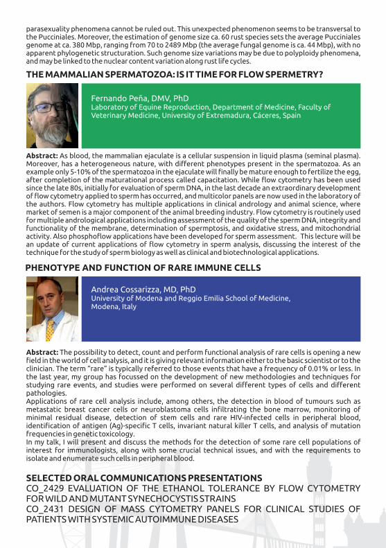
Abstract: As blood, the mammalian ejaculate is a cellular suspension in liquid plasma (seminal plasma). Moreover, has a heterogeneous nature, with different phenotypes present in the spermatozoa. As an example only 5-10% of the spermatozoa in the ejaculate will finally be mature enough to fertilize the egg, after completion of the maturational process called capacitation. While flow cytometry has been used since the late 80s, initially for evaluation of sperm DNA, in the last decade an extraordinary development of flow cytometry applied to sperm has occurred, and multicolor panels are now used in the laboratory of the authors. Flow cytometry has multiple applications in clinical andrology and animal science, where market of semen is a major component of the animal breeding industry. Flow cytometry is routinely used for multiple andrological applications including assessment of the quality of the sperm DNA, integrity and functionality of the membrane, determination of spermptosis, and oxidative stress, and mitochondrial activity. Also phosphoflow applications have been developed for sperm assessment. This lecture will be an update of current applications of flow cytometry in sperm analysis, discussing the interest of the technique for the study of sperm biology as well as clinical and biotechnological applications.
Fernando Peña, DMV, PhDLaboratory of Equine Reproduction, Department of Medicine, Faculty of Veterinary Medicine, University of Extremadura, Cáceres, Spain

(RE) PROGRAMING HAEMATOPOIESIS AND IMMUNITY
Abstract: Once committed, the differentiated state of a cell is normally stable and can be inherited through cell division. Under certain conditions cell fate can, however, be modified or reversed. Mouse and human differentiated cells can be reprogrammed to pluripotency or directly converted to unrelated somatic cell fates by combinations of transcription factors (TFs). Here I will discuss the requirements to reprogram fibroblasts to Hematopoietic Stem Cells (HSCs) and Dendritic Cells (DCs) using combinatorial TF approaches. I hypothesize that direct cell fate reprogramming can be employed for hematopoietic regeneration as well as to induce immunity.We have recently demonstrated the direct reprogramming of mouse fibroblasts into clonogenic hematopoietic progenitors with the combination of TFs, Gata2, Gfi1b, cFos and Etv6 (Pereira et al, Cell Stem Cell 2013). These four TFs induce a dynamic, multi-stage hemogenic process that progresses through an endothelial-like intermediate. As such, it recapitulates developmental hematopoiesis in vitro (Pereira et al, Dev Cell 2016). We now show that the same blood forming process can be induced in human skin-derived fibroblasts with a similar combination of TFs. Reprogrammed fibroblasts undergo morphological and transcriptional changes, display cell surface phenotypes of human HSCs, and repopulate immunodeficient mice. Interestingly, we have mapped the genomic targets of hemogenic TFs uncovering unexpected mechanisms underlying hematopoietic reprogramming. In addition to a regenerative strategy we are applying cellular reprogramming to induce immunity. We are employing direct reprogramming to induce DCs from mouse fibroblasts. Induced DCs express the main components of the antigen presenting machinery at the cell surface, are able to engulf particles, secrete inflammatory cytokines and present antigens to T-cells.Collectively this work has provided insight into HSC and DC transcriptional networks as well as remarkable information on the specification on both cell-types. This knowledge may be applied for patient-specific reprogrammed cell generation without transiting through pluripotency. Our work may have important implications for hematopoietic development, disease modeling and generation of cells for replacement therapies as well as the development of more efficient cancer immunotherapies.
Filipe Pereira, PhDCenter for Neuroscience and Cell Biology (CNC), Universidade de Coimbra, Coimbra, Portugal
10H00- 11H45 PARALLEL SESSION IV | Room Fernando Pessoa
Stem Cells and TransplantationChairs: Artur Paiva & Alexandre Salvador
MINIMAL RESIDUAL DISEASE AND IMMUNE RECONSTITUTION AFTER STEM CELL TRANSPLANTATION
Noemi Puig, Salamanca, Spain
XV SIC |025
IMMUNE RECONSTITUTION FOLLOWING ALLOGENEIC STEM CELL TRANSPLANTATION
Abstract: Chronic Graft versus Host disease (cGVHD) is a major limiting factor for the success of allogeneic Hematopoietic Stem Cell Transplantation (allo-HSCT). We evaluated the association between the kinetics of Regulatory T cells (TREG) and Conventional CD4+Foxp3-T cells (TCON) reconstitution and the development of cGVHD. We performed a 2 year phenotypic analysis by multiparametric flow cytometry using fresh blood from 39 patients undergoing allo-HSCT after a reduced intensity conditioning regimen. The data obtained is suggestive of decreased thymic output and increased differentiation into terminally differentiated effector cells in patients developing cGVHD. This likely impacts on the reconstitution of naïve TCON and TREG compartments in these patients. We speculate that the inability to generate and maintain the more immature TREG subsets may lead to decreased TREG numbers potentially resulting in decreased control of effector T cells contributing to the development of cGVHD.
SELECTED ORAL COMMUNICATIONS PRESENTATIONSCO_2443 PRESENCE OF EOSINOPHILIA AFTER PEDIATRIC LIVER TRANSPLANTATION AND ITS RELEVANCE TO GRAFT DYSFUNCTION PROCESSCO_2445 CYTOMEGALOVIRUS SPECIFIC CELLULAR IMMUNE RESPONSE AS A MARKER OF THE OUTCOME OF LIVER TRANSPLANTATION FOR CHRONIC HEPATITIS C VIRUS INFECTION
11H45 - 12H15 COFFEE BREAK and Poster Viewing
Maria Soares, PhDResearch Fellow, Instituto de Medicina Molecular, Lisbon, Portugal
12H15 - 14H00 PARALLEL SESSION V | Room Sophia de Mello Breyner
Immunophenotype of T and NK cells in acute and chronic lymphoproliferative SyndromesChairs: Teresa Molero & Maria Arroz
T-CELL DIFFERENTIATION AND ITS IMPLICATIONS FOR THE DIAGNOSIS, CLASSIFICATION AND MONITORING OF ALL T
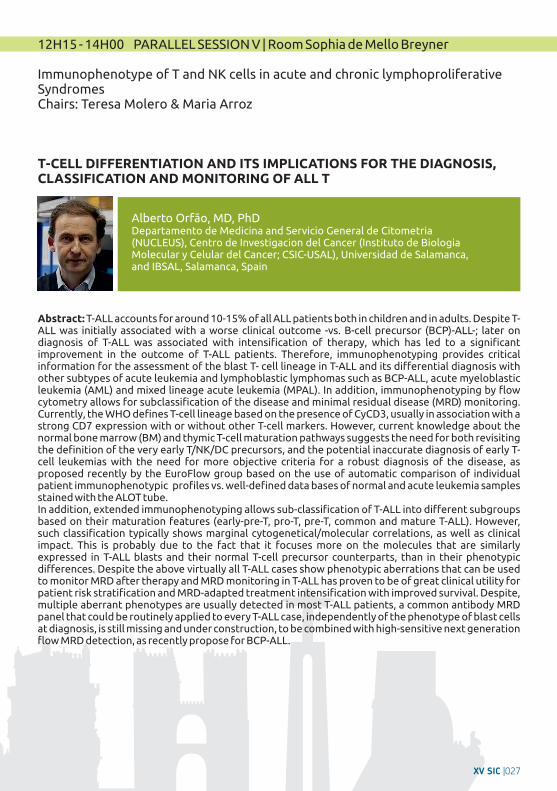
Alberto Orfão, MD, PhDDepartamento de Medicina and Servicio General de Citometria (NUCLEUS), Centro de Investigacion del Cancer (Instituto de Biologia Molecular y Celular del Cancer; CSIC-USAL), Universidad de Salamanca, and IBSAL, Salamanca, Spain
Abstract: T-ALL accounts for around 10-15% of all ALL patients both in children and in adults. Despite T-ALL was initially associated with a worse clinical outcome -vs. B-cell precursor (BCP)-ALL-; later on diagnosis of T-ALL was associated with intensification of therapy, which has led to a significant improvement in the outcome of T-ALL patients. Therefore, immunophenotyping provides critical information for the assessment of the blast T- cell lineage in T-ALL and its differential diagnosis with other subtypes of acute leukemia and lymphoblastic lymphomas such as BCP-ALL, acute myeloblastic leukemia (AML) and mixed lineage acute leukemia (MPAL). In addition, immunophenotyping by flow cytometry allows for subclassification of the disease and minimal residual disease (MRD) monitoring. Currently, the WHO defines T-cell lineage based on the presence of CyCD3, usually in association with a strong CD7 expression with or without other T-cell markers. However, current knowledge about the normal bone marrow (BM) and thymic T-cell maturation pathways suggests the need for both revisiting the definition of the very early T/NK/DC precursors, and the potential inaccurate diagnosis of early T-cell leukemias with the need for more objective criteria for a robust diagnosis of the disease, as proposed recently by the EuroFlow group based on the use of automatic comparison of individual patient immunophenotypic profiles vs. well-defined data bases of normal and acute leukemia samples stained with the ALOT tube.In addition, extended immunophenotyping allows sub-classification of T-ALL into different subgroups based on their maturation features (early-pre-T, pro-T, pre-T, common and mature T-ALL). However, such classification typically shows marginal cytogenetical/molecular correlations, as well as clinical impact. This is probably due to the fact that it focuses more on the molecules that are similarly expressed in T-ALL blasts and their normal T-cell precursor counterparts, than in their phenotypic differences. Despite the above virtually all T-ALL cases show phenotypic aberrations that can be used to monitor MRD after therapy and MRD monitoring in T-ALL has proven to be of great clinical utility for patient risk stratification and MRD-adapted treatment intensification with improved survival. Despite, multiple aberrant phenotypes are usually detected in most T-ALL patients, a common antibody MRD panel that could be routinely applied to every T-ALL case, independently of the phenotype of blast cells at diagnosis, is still missing and under construction, to be combined with high-sensitive next generation flow MRD detection, as recently propose for BCP-ALL.
XV SIC |027

Julia Almeida, MD, PhDDepartamento de Medicina and Servicio General de Citometria (NUCLEUS), Centro de Investigacion del Cancer (Instituto de Biologia Molecular y Celular del Cancer and IBSAL; CSIC-USAL), Universidad de Salamanca, Salamanca, Spain
Abstract: Mature T-cell malignancies are a biologically and clinically heterogeneous group of disorders that result from clonal proliferation of mature post-thymic lymphocytes. This wide group of chronic lymphoid malignancies is still much less understood than their B-cell counterpart, owing to their rarity and biologic heterogeneity, and to the fact that most categories lack distinctive genetic markers or recurrent biological abnormalities. Nonetheless, major advances on molecular characterization have been reached in some categories (WHO2016) in the last years; also, great efforts have been recently performed, as regards the characterization of tumor cells and the identification of their normal counterpart (i.e. naïve CD4+ T cells, central memory CD4+ T cells, follicular helper T cells or regulatory T cells have been claimed to be the normal counterpart of T-cell prolymphocytic leukemia, Sezary syndrome, angioimmunoblastic T-cell lymphoma of adult T-cell leukemia/lymphoma). Despite this, challenges remain in the subclassification of major disease groups within peripheral T-cell neoplasms; actually, the most common WHO “category” of mature T-cell neoplasms is peripheral T-cell lymphoma not otherwise specified –composed of a kind of “wastebasket” subtype defined by exclusion criteria–, which accounts for approximately one third of all these disorders. Based on this background, the EuroFlow Consortium (to which our group belongs) aimed to design a diagnostic strategy by 8-color flow cytometry to better classify T-CLPD into specific WHO categories. Our results clearly showed further contribution of the marker combinations used to distinguish between normal/reactive and aberrant/malignant T-cells -except for a few CD8+ T-LGL cases- and at the same time it allowed a more precise classification of T-CLPD into most WHO entities, particularly within the CD4+ T-CLPD.
1 1 1 1Authors: Julia ALMEIDA , Juan FLORES-MONTERO , Antonio LÓPEZ , Susana BARRENA , 1 1 1 2 3Paloma BÁRCENA , María JARA-ACEVEDO , Noemí MUÑOZ , Noemí PUIG , Ton LANGERAK ,
4 5 1Margarida LIMA , Jacques J. VAN DONGEN , Alberto ORFAO , on behalf of EuroFlow Consortium.
1Institutions: Departamento de Medicina and Servicio General de Citometria (NUCLEUS), Centro de Investigacion del Cancer (Instituto de Biologia Molecular y Celular del Cancer and IBSAL; CSIC-USAL),
2Universidad de Salamanca, Salamanca, Spain, and IBSAL; Department of Hematology, University 3Hospital, Salamanca, Spain; Department of Immunology, Erasmus MC, University Medical Center,
4Rotterdam, The Netherland; Department of Hematology, Santo António Hospital, Porto, Portugal; Department of Immunohematology and Blood Transfusion, Leiden University Medical Center, Leiden, The Netherlands
IMMUNOPHENOTYPE OF T LYMPHOCYTES IN CHRONIC LYMPHOPROLIFERATIVE SYNDROMES
IMMUNOPHENOTYPE OF NK LYMPHOCYTES IN ACUTE AND CHRONIC LYMPHOPROLIFERATIVE SYNDROMES
Margarida Lima, MD, PhDHead of the Laboratory of Cytometry, Hematology Department, Hospital de Santo António, Centro Hospitalar do Porto (HSA/CHP); Invited professor, Instituto de Ciências Biomédicas Abel Salazar da Universidade do Porto (ICBAS/UP); Porto, Portugal
SELECTED ORAL COMMUNICATIONS PRESENTATIONS
CO_2452 FLOW CYTOMETRY: MEASUREMENT OF PERIPHERAL BLOOD EXPRESSION/ LYMPHOCYTES OF CUTANEOUS T LYMPHOMASCO_2453 PERIPHERAL BLOOD VΔ1 T CELLS IN B-CLL EXHIBIT A CYTOTOXIC PROFILE, LIKE THE OTHER T CELL SUBPOPULATIONS
XV SIC |029
EDUCATION OF BONE MARROW-DERIVED CELLS DURING METASTASIS BY TUMOR-SECRETED EXOSOMES
Abstract: Cancer is a systemic disease, while most of the research effort has been focused on analyzing the primary tumor, there is a lack of information on how the tumor microenvironment influences metastasis. The importance of the microenvironment in metastasis is now fully acknowledged. Exosomes are secreted vesicles carrying lipids, proteins, RNA and DNA molecules. By carrying these molecules, and facilitating their cell-to-cell transfer, exosomes can modulate the behavior of resident cells that can impact disease progression. Our studies demonstrated that tumor exosomes are a major tumor-derived factor that acts systemically to promote bone marrow-derived cells (BMDCs) recruitment to the tumor and metastatic microenvironments. We showed that exosome secretion by melanoma cells influences BMDC mobilization and recruitment to pre-metastatic and metastatic niches, thus promoting metastasis in a process that we have termed “education”. We have recently analyzed the role of tumor-secreted exosomes in lymph node pre-metastatic niche formation. We found that melanoma-derived exosomes promote lympho(angio)genesis and enrichment of molecular adhesion-, autoimmune- and cytokine-related expression signatures in lymphatic endothelial cells (LECs). Our results support a role of tumor-secreted exosomes in promoting cellular and molecular alterations in the lymph node microenvironment fostering metastasis. In addition, we started the characterization of secreted exosomes by flow cytometry and particle fractionation to determine the characteristics of secreted vesicles. Analysis of exosome content in human lymphatic fluid suggest that proteomic cargo and particles are increased in melanoma patients opening the possibility of the use of circulating vesicles in lymphatic fluid as biomarkers and as a source of novel biomarkers to predict metastatic potential.
PAST, PRESENT AND FUTURE OF EXTRACELLULAR VESICLES ANALYSIS BY FLOW CYTOMETRY
Abstract: Extracellular vesicles (EVs) are circular fragments of membrane released by the cells with specific lipid, protein, and RNA contents reflective of parent cell status. This characteristic makes them to be increasingly recognized for their role in intercellular communication and as biomarkers of diseases. The size-range of the EVs secreted by cells is between 70-1000 nm, being the EVs with a size below 200 nm the most abundant. The challenge in the EVs field for diagnostic purposes in a clinical setting include the time required for EV isolation and analysis, as well as the lack of suitable methodology that has been the main challenge of EVs analysis during last decade. We developed a new methodology to identify and isolate EVs by Flow Cytometry with a resolution of 30-40 nm. This novel
Hector Peinado, PhDLaboratory of Microenvironment and Metastasis at CNIO, CNIO Universidad Autonoma de Madrid, Madrid, Spain
Òscar Fornas, PhDFlow Cytometry Unit. Pompeu Fabra University (UPF) and Centre for Genomic Regulation (CRG), The Barcelona Institute of Science and Technology (BIST). Barcelona, Spain.
12H15 - 14H00 PARALLEL SESSION VI | Room Fernando Pessoa
Vesicles, exosomes, microparticules
Chairs: Lola Martinez & Bruno Costa Silva
IMMUNOSUPPRESSIVE EXOSOMES PRESENT IN HUMAN OVARIAN TUMOR MICROENVIRONMENTS RAPIDLY AND REVERSIBLY ARREST T CELL ACTIVATION
Abstract: Virus size membrane bound extracellular vesicles isolated from ascites fluid from ovarian cancer patients have biophysical and compositional characteristics similar to vesicles called exosomes. The tumor-associated exosomes inhibit an early activation endpoint (translocation of NFAT from the cytosol into the nucleus) of a significant portion of virus (EBV and CMV) specific CD8+ T-cells that are stimulated with viral peptides presented in the context of Class I MHC. Early and late activation endpoints of peripheral blood CD4+ and CD8+ T-cells (of unknown specificity) stimulated with immobilized antibodies to CD3 and CD28 are also significantly inhibited by the exosomes. The inhibition of the T-cells is induced directly and rapidly (after just a 2h pulse with the exosomes), and occurs coincidentally with the exosomes binding to and internalization by the T-cells. The early arrest in the activation occurs without a loss of viability in the T-cells. The immune suppressive exosomes in the tumor microenvironment and the ability to block their T-cell inhibitory activity represent a potential therapeutic target to enhance the anti-tumor immunity of quiescent tumor-associated T cells, and to prevent the functional arrest of endogenous or adoptively transferred T-cells upon their entry into the tumor.
SELECTED ORAL COMMUNICATIONS PRESENTATIONSCO_2432 MULTICENTER HARMONIZATION OF FLOW CYTOMETERS IN THE CONTEXT OF THE MULTICENTRIC PRECISESADS PROJECTCO_2439 NEXT GENERATION FLOW CYTOMETRY IN THE MONITORING OF M O N O C Y T I C - L I N E A G E A LT E R AT I O N S D U R I N G F O L L O W - U P O F MYELODYSPLASTIC SYNDROMES AND ACUTE MONOBLASTIC/MONOCYTIC LEUKEMIA
14H00 - 15H15 LUNCH
Paul WallaceDepartment of Flow & Image Cytometry, Roswell Park Cancer Institute, Buffalo, NY
methodology provides an easy, reproducible and reliable analysis of EVs improving all existing ones. With this novel approach is not necessary to concentrate the EVs, avoiding inconsistent results that are obtained by using different isolation protocols and complex settings for the cytometer. We dramatically reduced the time consuming from 2 days to 1 hour. This is the first time where sorting of exosomes is successfully performed because has been confirmed and validated by complementary technologies such as confocal and electron microscopy. Our new method resolves the lack of robustness and reliability in exosomes analysis in the current flow cytometry approaches.
XV SIC |031
Institutions: Dep. Medicine-Serv. Cytometry, Cancer Research Center (IBMCC-CSIC/USAL) and Univ. of 2Salamanca, Salamanca, Spain; Dept. of Immunology, Erasmus MC, Rotterdam, The Netherlands;
3Department of Paediatric Haematology and Oncology, Second Faculty of Medicine, Charles University 4and University Hospital Motol, Prague, Czech Republic; Department Pediatrics and Allergology,
5University Hospital of Salamanca, Salamanca, Spain; Immunology, Universitario La Paz, Madrid, Spain; 6 7Department of Laboratory Medicine, University Hospital Ghent, Ghent, Belgium; Servicio
8Hematologia. Fundacion Jimenez Diaz. Madrid, Spain; Institute of Clinical Immunology and
9Allergology, St Anne`s University, Brno, Czech Republic; BRC-Translational Immunology Lab, 10University, Oxford, UK; Dept. of Immunology and Pathology, Central Clinical School, Monash
11University, Melbourne, Australia, Dept. of Immunohematology and Blood Transfusion (IHB), Leiden University Medical Center (LUMC), Leiden, The Netherlands
Abstract: Primary immunodeficiencies (PID) are inherited disorders of the immune system, generally presenting with recurrent, severe and sometimes life-threatening infections. To date, more than 300 genes have been identified that can be mutated in PID patients, that affect the functional abilities and/or numbers of one part of the immune system or one cell type. Since most of PID patients (60-65%) have a defect in the lymphoid system, involving B- and/or T-cells alone or in combination with other cells, flow cytometric immunophenotyping of lymphoid cells plays a central role in the stepwise diagnostic workup of patients suspected of PID. An accurate immunophenotypic diagnosis is of utmost importance for guiding genetic testing, whether targeted to specific genes or whole genome sequencing. It also contributes to understand the clinical heterogeneity in disease presentation and outcome, even within genetically homogeneous disease entities. Finally, immunophenotyping supports treatment decisions and patient monitoring such in case of immunoglobulin replacement therapy and in hematopoietic stem cell transplantation or gene therapy.Besides “genetics-first” approach using whole exome sequencing (WES) or whole genome sequencing (WGS) has been postulated for PID diagnosis by some investigators , flow cytometry offer considerable advantages for the clinical routine: 1) WES or WGS approaches are too slow and too demanding for routine first-choice diagnostics, 2) different genetic defects might show a different clinical course as compared to those previously reported for the affected gene (e.g. hypomorphic defects), 3) even when the genetic alteration has bene fully defined, immunological studies are required to identify the consequences of the defect for the immune system.Because of the central role of immunophenotyping, many PID centers have developed multi-color flow
1 2 3 1Authors: Martin Perez-Andres, Mirjam van der Burg, Tomas Kalina, Elena Blanco, Sonia de 4 5 5 6 6 Arriba, Juan Torres-Cañizales, Eduardo Lopez-Granados, Jan Philippé, Carolien Bonroy,
7 8 9 2 Cristina Serrano, Marcela Vlkova, Anne-Kathrin Kienzler, Marjolein Wentink, Ester 3 10 1 11Mejstříková, Menno van Zelm, Alberto Orfao, Jacques J.M. van Dongen on behalf of the
EuroFlow PID consortium
15H15 - 17H00 PARALLEL SESSION VII Room Sophia de Mello BreynerAdvances in primmary immunodeficiencies screening and diagnosis by flow CytometryChairs: Martin Perez-Andrés & João Farela Neves
NEW STANDARDIZED STRATEGIES IN THE ORIENTATION OF LYMPHOID DEFECTS BY FLOW CYTOMETRY: COMBINED IMMUNODEFICIENCIES AND PRIMARY ANTIBODY DEFICIENCIES ANALYSIS BY FCM
Martín Perez Andrés, PhDDep. Medicine-Serv. Cytometry, Cancer Research Center (IBMCC-CSIC/USAL) and University of Salamanca, Salamanca, Spain
Abstract: Cytotoxic lymphocytes, encompassing T cell and NK cell subsets, can kill target cells through directed release of specialized lysosomes, so called cytotoxic granules. They can identify and eradicate infected and malignant cells. Lymphocyte cytotoxicity also contributes to immune homeostasis, through killing of activated immune cells. Congenital defects in lymphocyte cytotoxicity are associated with a life-threatening hyperinflammatory syndrome, hemophagocytic lymphohistiocytosis (HLH), typically presenting in infancy or childhood. Individuals with more subtle impairments in lymphocyte cytotoxicity may also present with or related disorders later in life. In this talk I will provide a background to the mechanisms of lymphocyte cytotoxicity and discuss the clinical manifestations of impaired lymphocyte cytotoxicity. Contemporary diagnostic strategies for the identification of primary defects in lymphocyte cytotoxicity, largely based on flow cytometry, will be presented.
MENDELIAN SUSCEPTIBILITY TO MYCOBACTERIAL DISEASE - STRATEGIES FOR DIAGNOSIS BY FLOW CYTOMETRY
Yenan Bryceson, PhDKarolinska Institutet, Stockholm, Sweden
FLOW CYTOMETRY IN THE IDENTIFICATION OF INNATE CYTOTOXIC DYSFUNCTION
cytometric protocols and antibody panels for diagnosis and classification of PID. However, variability among these single center initiatives (sample processing, antibody panels, immunostaining procedures, instrument setup, sample measurement and data analysis) in combination with the the low incidence and the clinical-immunological heterogeneity of PID, hamper prospective validation in large patient series and age-matched healthy controls, and limit the exchange of data between PID centers.Accordingly, we aimed to develop a tool that would be universal for all lymphoid PIDs and offer high sensitivity to disclose a lymphoid defect. This approach takes advantage of the recently designed and validated EuroFlow procedures and tools resulting in standardized pre-analytical and analytical EuroFlow procedures, PID-specific ≥8-color antibody panels, and software tools for multidimensional data analysis. This strategy has been validated in >400 PID patients, and immunological alterations have been defined using a group of >200 healthy controls of different age-groups (from cord-bood to 80 years-old) as a reference. Together with this, new lymphoid subpopulations has been identified that might be of interest for diagnosis and monitoring particularly for primary antibody deficiencies (like dissection of memory B-cells and plasma cells compartments according to IgH subclasses). Together with strategy, an algorithm was designed for guiding immunophenotypic PID studies, based on available clinical and laboratory information. In summary, this strategy enables a sensitive screening, followed by in-depth analysis as cost-effective step-wise strategy that has been validated for a broad applicability for many different PID of the lymphoid system (not targeted on a few types of PID), based on the broad overview of all relevant subsets in a single analysis.
Júlia VasconcelosCentro Hospitalar do Porto, Oporto, Portugal
Abstract: Mendelian susceptibility to mycobacteria disease (MSMD) is a group of rather rare primary immunodeficiencies, etiologically and clinically heterogenous regarding severity. Patients are prone to develop disseminated infections by many environmental nontuberculosis mycobacteria, several
XV SIC |033
Mycobacterium bovis (BacilleCalmete-Guérin,BCG) vaccine substrains, Mycobacterium tuberculosis and other intramacrophagic pathogens, mostly Salmonella. Patients with MSMD may have mutations in 7 genes that encode for the following proteins involved in the IL-12 dependent, IFN-γ mediated immunity: IFN-γ Receptor 1, IFN-γ Receptor 2, IL-12 Receptor β1, IL-12p40, signal transducer and activator of transcription 1 (STAT1), IFN-γ regulation factor 8 (IRF8), for IL-12 production by myeloid dentritic cells CD1c+ and interferon-stimulated gene 15 (ISG15). Deficiency in other proteins that protect from Mycobacteria infections may also cause MSMD: gp91PHOX, involved in the oxidative burst; GATA2, implicated on early hematopoiesis; nuclear factor kB essential modulator (NEMO) causing transcription of several cytokines; TyK2, associated with hiperIgE. Some of these proteins can be evaluated by FCM (flow cytometry), allowing a rapid and skilful diagnosis that may guide the molecular diagnosis and also the imperative treatment. We show our experience on evaluation, in whole blood, of some proteins of the IL-12/IFN-γ axis: surface expression of IFNγR1 on monocytes, surface expression of IL-12Rβ1 on CD25+ blasts T cells, intracellular expression of IL-12p40 in monocytes and intracellular expression of IFN-γ in T cells. If a GATA2 deficiency is suspect, the characteristic severe monocytopenia and B and NK lymphopenia is easily recognized by FCM. An algorithm for diagnosis of MSMD, guided by the frequency of the defect and the clinical features, is showed, and the results of some cases reports are presented. About 20-50% of patients with MSMD still don't have an etiological cause, so new genes or gene products are yet to be discovered.
References:1. Steven M. Holland and Jean-Laurent Casanova: Inherited Disorders of the Interleukin-12-Interleukin-23/Interferon-g Circuit- Primary Immunodeficiency Diseases- A Molecular and Genetic Approach, Chapter 35, 2014: 450-466, 2. Waleed Al-Hertz, Aziz Bousfiha, and al. Primary immunodeficiency diseases: an update on the classification from the International Union of Immunological Societies Expert Committee for Primary Imnunodeficiency –Frontiers in Immunology, 20143. Isabel Caragol, Miguel Raspall et al: Clinical Tuberculosis in 2 of 3 siblings with Interleukin-12 Receptor b1. Clinical Infeccious Diseases, 2003: 302-306
SELECTED ORAL COMMUNICATIONS PRESENTATIONSCO_2441 NATURAL KILLER CELLS IN ACUTE MYELOID LEUKAEMIA PATIENTS: PHENOTYPIC AND FUNCTIONAL ANALYSISCO_2442 IDENTIFICATION AND ENUMERATION OF DISTINCT SUBSETS OF MONOCYTE-MACROPHAGES IN DIFFERENT HUMAN TISSUES THROUGHOUT LIFE USING 9-COLOR FLOW CYTOMETRY
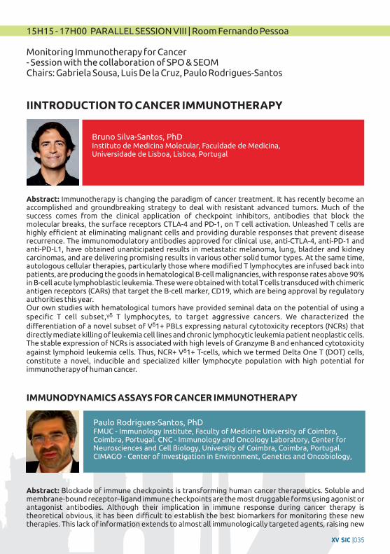
IMMUNODYNAMICS ASSAYS FOR CANCER IMMUNOTHERAPY
Abstract: Blockade of immune checkpoints is transforming human cancer therapeutics. Soluble and membrane-bound receptor–ligand immune checkpoints are the most druggable forms using agonist or antagonist antibodies. Although their implication in immune response during cancer therapy is theoretical obvious, it has been difficult to establish the best biomarkers for monitoring these new therapies. This lack of information extends to almost all immunologically targeted agents, raising new
Paulo Rodrigues-Santos, PhDFMUC - Immunology Institute, Faculty of Medicine University of Coimbra, Coimbra, Portugal. CNC - Immunology and Oncology Laboratory, Center for Neurosciences and Cell Biology, University of Coimbra, Coimbra, Portugal. CIMAGO - Center of Investigation in Environment, Genetics and Oncobiology,
15H15 - 17H00 PARALLEL SESSION VIII | Room Fernando Pessoa
Monitoring Immunotherapy for Cancer - Session with the collaboration of SPO & SEOMChairs: Gabriela Sousa, Luis De la Cruz, Paulo Rodrigues-Santos
IINTRODUCTION TO CANCER IMMUNOTHERAPY
Abstract: Immunotherapy is changing the paradigm of cancer treatment. It has recently become an accomplished and groundbreaking strategy to deal with resistant advanced tumors. Much of the success comes from the clinical application of checkpoint inhibitors, antibodies that block the molecular breaks, the surface receptors CTLA-4 and PD-1, on T cell activation. Unleashed T cells are highly efficient at eliminating malignant cells and providing durable responses that prevent disease recurrence. The immunomodulatory antibodies approved for clinical use, anti-CTLA-4, anti-PD-1 and anti-PD-L1, have obtained unanticipated results in metastatic melanoma, lung, bladder and kidney carcinomas, and are delivering promising results in various other solid tumor types. At the same time, autologous cellular therapies, particularly those where modified T lymphocytes are infused back into patients, are producing the goods in hematological B-cell malignancies, with response rates above 90% in B-cell acute lymphoblastic leukemia. These were obtained with total T cells transduced with chimeric antigen receptors (CARs) that target the B-cell marker, CD19, which are being approval by regulatory authorities this year.Our own studies with hematological tumors have provided seminal data on the potential of using a specific T cell subset,ᵞᵟ T lymphocytes, to target aggressive cancers. We characterized the
differentiation of a novel subset of Vᵟ1+ PBLs expressing natural cytotoxicity receptors (NCRs) that directly mediate killing of leukemia cell lines and chronic lymphocytic leukemia patient neoplastic cells. The stable expression of NCRs is associated with high levels of Granzyme B and enhanced cytotoxicity against lymphoid leukemia cells. Thus, NCR+ Vᵟ1+ T-cells, which we termed Delta One T (DOT) cells, constitute a novel, inducible and specialized killer lymphocyte population with high potential for immunotherapy of human cancer.
Bruno Silva-Santos, PhDInstituto de Medicina Molecular, Faculdade de Medicina, Universidade de Lisboa, Lisboa, Portugal
XV SIC |035

challenges for developing immune endpoints that correlate systemic and intratumoral immune effects with systemic dose, toxicity, and efficacy for cytotoxic therapies. Refining immune endpoints to better inform clinical trial design represents a high priority challenge. Immunodynamics was defined as a way to evaluate the impact of a drug or a therapy on the immune system. Assessment of the presence, functionality, and phenotype of infused and persisting adoptive cells, together with the impact of the infused cells on patient immunobiology and tumor microenvironment is one of the goals of immunodynamics assays. Complementary approaches including molecular, cellular and tissular assays are being used to accomplish this task. The quest for biomarker discovery results from the combination of pre-clinical studies using animal models and human in vitro results from patients included in clinical trials. In our lab we use a multi-parametric flow cytometry strategy for the analysis of the expression of several immune checkpoint inhibitors by different T, B, NK, monocyte and dendritic cell subsets. We recently added the analysis of xMAP (Luminex®) in a 59-plex panel of soluble molecules (14 immune checkpoints and 45 cytokines, chemokines and growth factors). Gene expression profiles of cell sorted populations and miRNA-mediated immune regulation is also under investigation. Functional studies complete our portfolio of laboratory investigations aiming at the immune monitoring in cancer therapy.
MONITORING NK CELL-BASED IMMUNOTHERAPY IN THE ELDERLY
Abstract: Natural killer (NK) cells are innate lymphocytes with a central role in the defence against tumour and virus-infected cells. NK cell activity is finely tuned by the balance between the signals from activating and inhibitory receptors. Activating receptors, like NCRs (NKp30, NKp46, NKp44) and NKG2D, sense the altered expression of their ligands on the cell surface of cancer cells. On the contrary Killer-cell immunoglobulin-like receptor (KIR) and CD94/ NKG2A are inhibitory receptors that recognize HLA class I molecules whose expression is often altered on tumour cells. Different therapies have been designed to enhance the antitumor response of NK cells including stimulation with cytokines adoptive therapy with autologous or allogeneic expanded NK cells. Checkpoint blockade of inhibitory receptors and the use of agonist antibodies to stimulate activating receptors are emerging areas of research. Altered expression of NK cells activating receptors has also been described in cancer patients, probably as a consequence of chronic exposure to ligands on tumour cells. The effect of NK cell-based cancer immunotherapy aiming to reverse the tumour-induced NK cell dysfunction observed in cancer patients should be evaluated by monitoring their phenotype and function. Cancer is primarily considered a disease of old age, and it is well known that ageing is associated to alterations in NK cells including a diminished expression of several activating receptors. Thus age and cancer may synergistically contribute to alterations in NK cell-mediated tumour immunosurveillance supporting that special caution should be taken into account in the monitoring of NK cell reconstitution after immunotherapy when elderly patient are considered.
SELECTED ORAL COMMUNICATIONS PRESENTATIONSCO_2427 DNA FLOW CYTOMETRY AS A CLINICALLY USEFUL PROGNOSTIC TOOL IN FEMALE BREAST CANCER. A LONG-STANDING 25-YEAR EXPERIENCECO_2455 EVALUATION OF T CELL-MEDIATED ANTITUMOR IMMUNE RESPONSE IN HEPATOCELLULAR AND CHOLANGIOCARCINOMA AT TUMOR AND PERIPHERAL LEVEL
17H00 - 17H30 COFFEE BREAK and Poster Viewing
Rafael Solana, MD, PhDUniversity of Extremadura and IMIBIC - Reina Sofia University Hospital - University of Cordoba, Spain.
17H30 - 18H00 COMMERCIAL SESSION by Beckman Coulter | Room Sophia de Mello Breyner
ClearLLabLS: Is it the definitive solution for the lymphoid neoplasm screening?Speaker Dr. Jose María Raya SánchezHead of Section of the Hematology Laboratory, Hospital Universitario de Canarias-Universidad de La Laguna
18H30 - 19H30 – SIC Groups Presentations | Room Sophia de Mello Breyner
18H30 - 19H30 – GECLID presentations | Room Fernando Pessoa
20H30 - 24H00 – CONGRESS DINNER at Navio Opera
XV SIC |037
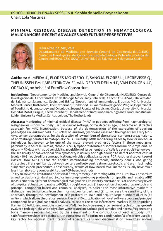
09H00 - 10H00 PLENARY SESSION II | Sophia de Mello Breyner RoomChair: Lola Martinez
MINIMAL RESIDUAL DISEA SE DETEC TION IN HEMATOLOGIC AL MALIGNANCIES: RECENT ADVANCES AND FUTURE PERSPECTIVES
1Institutions: Departamento de Medicina and Servicio General de Citometria (NUCLEUS), Centro de Investigacion del Cancer (Instituto de Biologia Molecular y Celular del Cancer; CSIC-USAL), Universidad
2de Salamanca, Salamanca, Spain, and IBSAL; Department of Immunology, Erasmus MC, University 3Medical Center, Rotterdam, The Netherland; Childhood Leukaemia Investigation Prague, Department
of Paediatric Haematology and Oncology, Second Faculty of Medicine, Charles University, University 4 Hospital Motol, Prague, Czech Republic; Department of Immunohematology and Blood Transfusion,
Leiden University Medical Center, Leiden, The Netherlands
1 1 1 1 Authors: ALMEIDA J , FLORES-MONTERO J , SANOJA-FLORES L , LECREVISSE Q ,2 3 2 4THEUNISSEN PMJ ,MEJSTRIKOVA E , VAN DER VELDEN VHJ , VAN DONGEN JJ ,
1ORFAO A , on behalf of EuroFlow Consortium.
Abstract: Monitoring of minimal residual disease (MRD) in patients suffering from haematological malignancies is now routinely used in clinical settings. Some decades ago, it became an attractive approach for MRD investigation, because of the demonstration of the expression of aberrant phenotypes in leukemic cells in >85-90% of leukemia/lymphoma cases and the higher sensitivity (<10-4) vs. conventional methods, for the detection of low numbers of aberrant cells among a great majority of normal/regenerative hematopoietic cells. Currently, MRD monitoring either by flow or molecular techniques has proven to be one of the most relevant prognostic factors in these neoplasms, particularly in acute leukemias, chronic-B-cell lymphoproliferative disorders and multiple myeloma. To obtain MRD data with good sensitivity, acquisition of large numbers of cells is a prerequisite; however, the sensitivity of conventional flow cytometry is usually not high enough to detect aberrant cells at levels <10-5, required for some applications, which is at disadvantage vs. PCR. Another disadvantage of classical flow MRD is that the applied immunostaining protocols, antibody panels, and gating strategies differ significantly between centers and between treatment protocols, and are in fact highly subjective expert procedures. Consequently, results of flow-based MRD methods usually have much less interlaboratory comparability than PCR-based methods. To try to solve the limitations of classical flow cytometry in detecting MRD, the EuroFlow Consortium aimed to design standardized 8-color immunophenotyping protocols for specific and reliable MRD measurement in different hematological malignancies, to identify aberrant cells at a sensitivity levels of tiv−p (i.e. at least comparable to PCR), through two steps: (1) application of novel software tools and principal component-based and canonical analyses, to select the most informative markers in distinguishing tumor cells from their normal counterpart; and (2) to increase the sensitivity of the approach, through the development of a protocol to stain and acquire large numbers of cells (>4 million), which allows theoretical sensitivities of ≤10−p. pplication of novel software tools and principal component-based and canonical analyses, to select the most informative markers in distinguishing tkemia (BCP-ALL) and multiple myeloma (MM). For both diseases, after several cycles of design-test-evaluate-redesign, the antibody tubes were tested on pathological samples and normal/regenerating bone marrow samples, followed by the assessment of the contribution of each antibody, until satisfactory results were obtained. Although the specific optimized combination(s) of markers used is a key factor for optimal identification of aberrant cells and discrimination from their normal
Julia Almeida, MD, PhDDepartamento de Medicina and Servicio General de Citometria (NUCLEUS), Centro de Investigacion del Cancer (Instituto de Biologia Molecular y Celular del Cancer and IBSAL; CSIC-USAL), Universidad de Salamanca, Salamanca, Spain

counterparts, another critical factor is the number of cells analyzed. Therefore, a new protocol was developed (The Euroflow bulk lysis protocol), allowing a 10-fold increase in the number of cells evaluated, in the context of fully standardized laboratory protocols. The novel protocol ("New Generation Flow”, NGF) is faster, standardized and reproducible, vs. classical cytometry, and even in MM it does not require a diagnostic sample or patient-specific probes. Then, the EuroFlow-NGF approach developed (with a sensitivity close to 10−N) was validated in a multicenter manner, and it showed similar (MM) or higher (BCP-ALL) applicability vs. conventional flow-MRD, together with a significantly increased sensitivity for MRD detection.
XV SIC |039
10H00 - 11h00 PLENARY SESSION III | Sophia de Mello Breyner RoomAccreditation of Flow Cytometry LaboratoriesChairs: Catarina Martins & Juana Ciudad
ISO 15189 AND ISO/IEC 17025, STANDARDS FOR (CLINICAL) LABORATORIES
Abstract: All over the world, accreditation by the standard ISO/IEC 17025 - General requirements for the competence of testing and calibration laboratories has been a reality for a long time in several fields of laboratory science, being even obligatory in several ones. The major goal of such a standard is to achieve harmonization of good laboratory practices. Biomedical diagnostic laboratories have resisted the implementation of ISO/IEC 17025, eventually due to the aseptic language of the standard, which must respond to a diversity of applications. Though ISO/IEC 17025 can also be applied to clinical fields, different countries developed individual codes and guidelines aiming to respond to the needs of medical laboratories, in a way better than ISO/IEC 17025.More recently, ISO (International Organization for Standardization) developed ISO 15189 - Medical laboratories - Particular requirements for quality and competence. A panel of international experts was called upon, in order to answer the requests for a more clinically driven standard, adapting the regular accreditation nomenclature and processes to the medical laboratories. Recognizing the importance of such a document, countries like Australia, Latvia, or more recently France, The Netherlands, and The United Kingdom are bringing this standard mandatory to all medical laboratories, which now have to adapt and respond adequately to its requirements. Although the expansion of the standard has been rather slow, efforts are being made, concretely in Europe, to reinforce its application, for instance in countries such as Portugal and Spain. With disadvantages, but mainly several advantages, ISO 15189 is undoubtedly the most common global reference for quality in medical laboratories, including clinical flow cytometry facilities. Auditors and accreditation bodies believe it is crucial to increase the knowledge on this standard amongst medical laboratories personnel, and clinical staff. Not only to achieve a solid implementation of ISO 15189, which can respond globally to the harmonization needs in the medical laboratory, but also in order to continue improving the standard and its applicability to the daily routine.
Catarina Martins, PhDNOVA Medica School|Faculdade de Ciências Médicas, Universidade NOVA de Lisboa, Lisboa, Portugal Quality Auditor, IPAC – Portuguese Institute for Accreditation, Caparica, Portugal
UNDERGOING ACCREDITATION IN FLOW CYTOMETRY LABS: STEPS TO FOLLOW AND CRITICAL POINTS
Abstract: Flow cytometric methods are well established in patients’ diagnostics and preparation of cellular therapeutics. These topics are subject to regulatory systems that have been as far as possible harmonized within the European community. By growing implementation of ISO 15189 based standardization in European laboratory diagnostics, flow cytometry laboratories are more and more challenged to introduce compliant quality management systems. Although in most countries accreditation of such laboratories is not yet compulsory, proof of following these rules is widely
Ulrich Sack, MD, PhDInstitute of Clinical Immunology, Medical Faculty at the University of Leipzig, Leipzig, Germany

FLEXIBLE SCOPE IN FLOW CYTOMETRY: EXPERIENCE OF A LABORATORY ACCREDITED BY UNE-EN-ISO 15189
Abstract: The Cellular Immunology Laboratory at the Hospital General Universitario “Gregorio Marañon” in Madrid achieved the Spanish national accreditation agency (Entidad Nacional de Acreditación, ENAC) first accreditation for flow cytometry in Immunology according to ISO 15189 standard in 2008. We got a fixed accreditation scope for lymphocyte counting including T (CD3+) cells along with T helper (CD3+CD4+) and T cytotoxic/suppressor (CD3+CD8+) subsets, B (CD19+) and NK (CD3-CD16/CD56+) cells. From that time on, we have developed several improvements and modifications that allowed us to progress and switch into a flexible scope during the last renewal audit in October 2016. Our current scope will let us include the quantitation of additional lymphocyte memory subsets, which are very useful in the clinical setting of immunodeficiency.
11H00 - 11H30 COFFEE BREAK
Juana Gil - Herrera, MD, PhDHospital General Universitario Gregorio Maragor, Madrid, Spain
requested. In general, adherence to quality management systems is considered to be very hard for cytometry. Therefore, we analyzed consequences of accreditation process for cytometry laboratories and investigated flow cytometrists’attitudes and misgivings according these requirements. As major challenges, staff qualification, adaptation of multicolor antibody panels, and quality assessment has been identified.
References ISO 15189:2012. Medical Laboratories — particular requirements for quality and competence, ISO, Geneva.Sack U, Barnett D, Demirel GY, Fossat C, Fricke S, Kafassi N, et al. Accreditation of flow cytometry in Europe. Cytometry B Clin Cytom 2013 May;84(3):135-42.Dorn-Beineke A, Sack U: Quality control and validation in flow cytometry. Lab. Med. 2016;40(2):65-79.
XV SIC |041
11H30 – 13H00 PLENARY SESSION IV | Sophia de Mello Breyner RoomImmunology (SPI)Chairs: Margarida Saraiva & Manuel Vilanova
ANALYSING THE IMPACT OF T CELL SUBSETS ON BRAIN COGNITIVE FUNCTION
Abstract: γδ T cells are known to populate multiple tissues, such as the skin, gut or lung, where they make major contributions to local physiology. Here we investigated whether γδ T cells could play a role in brain cognitive function, given that ab T cells were recently shown to be involved in learning behavior of mice: whereas IFN-γ producing subsets were detrimental, their IL-4-producing counterparts played a pro-cognitive role. We found that γδ T cells infiltrate the meningeal spaces from the brain of naive C57/BL6 mice already at birth and persisted throughout life. Strikingly, at 1 week of age, meningeal γδ T cells differentiated into IL-17 (but not IFN-γ) producers, which seemingly depended on IL-1β/IL-23 and retinoic acid. In fact, γδ T cells were the major source of IL-17, whereas ab T cells mostly provided IFN-γ in situ. To test whether IL-17-producing γδ T cells influenced the cognitive performance of mice, we scored the behavior of WT, TCRδ-/- and IL-17-/- mice in classical neuroscience paradigms assessing learning capacities. We observed that, contrary to WT controls, mice deficient for γδ T cells or IL-17 displayed impaired short-term/working memory in the Y maze paradigm, but a normal long-term spatial memory in the Morris water maze. We are currently identifying the underlying molecular mediators; interestingly, we found reduced levels of Brain Derived Neurotropic Factor (BDNF), a major player in memory and dementia, in the hippocampus of TCRδ-/- and IL-17-/- mice compared to WT controls. Altogether, our data suggest that γδ T cells regulate brain cognitive functions through a novel IL-17-dependent mechanism.
CHOREOGRAPHING IMMUNITY AND TOLERANCE INDUCTION IN THE THYMUS
Abstract: Our laboratory is focused on studying the development and role of the thymus, the organ responsible for the generation of T cells that are simultaneously responsive against pathogens and self-tolerant. Thymic activity is not regular throughout life and deficits in T-cell production arise in several pathophysiological states, including with age, infection and chemotherapy. Besides, the failure in the deletion of autoreactive T cells in the thymus can lead to autoimmunity. Hence, the regular function of the thymus must be tightly controlled in vivo. Within the thymus, thymic epithelial cells (TECs) provide key inductive microenvironments for the development of T cells that arise from hematopoietic precursors. As a result, genetic defects that impair normal TEC differentiation are linked to immunodeficiency and autoimmunity, which makes the study of TECs of fundamental, and clinical, importance to understand the normal development and response of the immune system. TECs are divided into two specialized functionally distinct cortical (cTECs) and medullary (mTECs) subtypes, which derive from common bipotent TEC progenitors. Still, we know very little about the genetic basis that controls cTEC/mTEC lineage divergence from TEC progenitors. Interleukin-7 (IL-7) is a chief cytokine for T-cell development and a particular subtype of TECs produces it. We previously provided the first temporal-spatial analysis of thymic IL-7 niche in vivo. Over the last years, we have studied the
Nuno Alves, PhDThymus Development and Function Laboratory Instituto de Investigação e Inovação em Saúde (I3S)Instituto de Biologia Molecular e Celular (IBMC), Porto, Portugal
Julie C. Ribot, PhDInstituto de Medicina Molecular, Faculdade de Medicina, Universidade de Lisboa, Lisboa, Portugal

Abstract: Most T lymphocyte development occurs in the thymus from progenitors of bone marrow origin in a process characterized by high cellular turnover. We found that thymus turnover is regulated by cell competition. During T lymphocyte development, cell competition occurs at a specific stage of differentiation. At that stage, precursors differ slightly in their time of thymus residency. Specifically, 'young' hematopoietic precursors (that recently seeded the thymus) coexist with 'old' precursors (residing for longer in the thymus). These cells share the same surface markers that identifies them as sharing the same differentiation stage, but young precursors induce the clearance of the old, thereby promoting thymus turnover. This is a particularly dynamic process, as winner cells progressively differentiate into more mature stages. Impairment or disruption of cell competition is permissive to self-renewal and persistence of the old precursors. These maintain thymus function autonomously for some weeks with de novo production and export of T lymphocytes. However, prolongation of this property invariably leads to development of T cell acute lymphoblastic leukemia (T-ALL) with strong similarities to the human disease.
13h00-13h15 CLOSING CEREMONY | Sophia de Mello Breyner Room
development of IL-7-expressing TECs relatively to the establishment of mature thymic epithelial microenvironments. We take a global approach to investigate TEC differentiation, which integrates the study of molecular processes taking place at the single-cell level to the analysis of in vivo mouse models.Ultimately, as T-cell immune reconstitution relies on the bi-directional interplay between T-cell precursors and TECs, understanding the development and function of TECs is crucial to comprehend how the immune system achieves the equilibrium between immunity and tolerance.
METABOLIC CUES IMPLICATED ON MONOCYTE BIOLOGY
Abstract: Emerging evidence indicates that innate immune cells tightly coordinate their metabolic programs with the carbon and bioenergetic needs to support the immunological functions in response to pathogens, tumors or inflammatory diseases. This metabolic adaptation requires energy and precise control of cellular metabolic pathways consuming glucose, fatty acids or amino acids. Perturbed metabolic fluxes thus imply decisive effects on immune cells activation eventuating in their ability to control a certain pathological condition. The extent to which metabolic changes coordinate immune cell function or occur consequently remains to be established. Since development to adult life, monocytes home from the blood stream to tissues where they become resident macrophages. These cells display different phenotypes that may be due to the microenvironment found on the tissue during differentiation, or may already be imprinted during their passage on the bloodstream. In any case, the blood and tissue metabolic and inflammatory microenvironment occurring during chronic inflammations will impact decisively the biology of monocytes conditioning their phenotype. We are currently pursuing how the modification of systemic and cellular metabolism arising during inflammatory conditions impact the immune functions and by consequence the dynamic of immune response. Therefore, innovative therapeutic approaches or metabolic adjuncts are envisaged as a new mean to reorient host metabolic alterations towards immune protection.
Vera Martins, PhDInstituto Gulbenkian de Ciência, Lisboa, Portugal
Ricardo Silvestre, PhD, Braga, PortugalLife and Health Sciences Research Institute (ICVS), School of Health Sciences, University of Minho, Braga, Portugal ICVS/3Bs-PT Government Associate Laboratory, Guimarães, Braga, Portugal.
CELL COMPETITION IN THE THYMUS
XV SIC |043
SELECTED ORAL COMMUNICATIONSHEMATOLOGY
CO2427 DNA FLOW CYTOMETRY AS A CLINICALLY USEFUL PROGNOSTIC TOOL IN FEMALE BREAST CANCER. A LONG-STANDING 25-YEAR EXPERIENCEAntónio E. Pinto Teresa Pereira, Giovani L. Silva, Saudade André Serviço de Anatomia Patológica do Instituto Português de Oncologia de Lisboa Francisco Gentil, E.P.E., and Centro de Estatística e Aplicações e Departamento de Matemática do Instituto Superior Técnico, Universidade de Lisboa
CO2428 BLASTIC PLASMACYTOID DENDRITIC CELL NEOPLASM. A CASE WITH DIFFERENT INMUNOPHENOTYPE AND CHROMOSOMAL ABERRATIONSGonzalez Martinez Gema Campillo JA, Blasco A, Alfaro R, Galian JA, Moya-Quiles MR, Mrowiec A, Gimeno L, García-Alonso A, Alamo JM, Muro M, Minguela A. Hospital Clinico Universitario Virgen de la Arrixaca
CO2433 THE PROLIFERATION INDEX OF BONE MARROW CELLS MAY ASSIST THE DIFFERENTIAL DIAGNOSIS OF CHRONIC MYELOMONOCYTIC LEUKEMIA VS. MONOBLASTIC/MONOCYTIC LEUKEMIASMaría del Pilar Leoz Alejandro Berkovits, Valentina Guerri, Carlos Fernández, Susana Barrena, Antonio López, Juana Ciudad, Miriam Fierro, Rosana Rivas, Miryam Santos, Alberto Orfao*, Sergio Matarraz* Centro de Investigación del Cáncer (IBMCC, University of Salamanca-CSIC) and IBSAL, Salamanca, Spain
CO2452 FLOW CITOMETRY: MEASUREMENT OF PERIPHERAL BLOOD EXPRESSION/ LYMPHOCYTES OF CUTANEOUS T LYMPHOMAST. Castaño R. Mata; A. Yeguas; R. Córdoba; P. Llamas; C. Serrano Department of Hematology; Hospital Universitario Fundación Jiménez Díaz, Madrid, Spain.
CO2453 PERIPHERAL BLOOD VDELTA1 T CELLS IN B-CLL EXHIBIT A CYTOTOXIC PROFILE, LIKE THE OTHER T CELL SUBPOPULATIONSCatarina Simões Isabel Silva, Sandra Silva, Joana Lima, Gilberto Marques, Adriana Roque, André Ribeiro, Letícia Ribeiro, Artur Paiva Unidade de Gestão Operacional de Citometria, Centro Hospitalar e Universitário de Coimbra
CO2455 EVALUATION OF T CELL-MEDIATED ANTITUMOR IMMUNE RESPONSE IN HEPATOCELLULAR AND CHOLANGIOCARCINOMA AT TUMOR AND PERIPHERAL LEVELCarmen Martín-Sierra Ricardo Martins, Paula Laranjeira, Ana Margarida Abrantes, J. Guilherme Tralhão, Maria Filomena Botelho, Dulce Cortes, Emanuel Furtado, Francisco Castro Sousa, Artur Paiva Centro Hospitalar e Universitário de Coimbra
IMMUNOLOGY
CO2441 NATURAL KILLER CELLS IN ACUTE MYELOID LEUKAEMIA PATIENTS: PHENOTYPIC AND FUNCTIONAL ANALYSISRaquel Tarazona Beatriz Sanchez-Correa, Ignacio Casas, Juan M. Bergua, Alejandra Pera, Carmen Campos, Nelson López-Sejas, Fakhri Hassouneh, Mª José Arcos, Helena Bañas, Esther Duran, Rafael Solana, Raquel TarazonaImmunology Unit, University of Extremadura, Caceres, Spain
CO2442 IDENTIFICATION AND ENUMERATION OF DISTINCT SUBSETS OF MONOCYTE-MACROPHAGES IN DIFFERENT HUMAN TISSUES THROUGHOUT LIFE USING 9-COLOR FLOW CYTOMETRYDaniela Damasceno 1,2 Julia Almeida1,2*, Cristina Teodosio3 , Martín Pérez Andrés1,2, Wouter BL van den Bossche4, Juan Flores-Montero1,2, Sandra de Bruin3 Sonia de Arriba5, Alfonso Romero6, Noemi Puig7,Luís Muñoz-Bellvis8, Jacques van Dongen3, Alberto Orfao1,21 Cancer Research Centre (IBMCC, USAL-CSIC), Cytometry Service (NUCLEUS) and Department of Medicine, University of Salamanca, Salamanca, Spain; 2 Institute of Biomedical Research of Salamanca (IBSAL), Salamanca, Spain; 3 Department of Immunohematology and Blood Transfusion, Leiden

University Medical Center, Leiden, The Netherlands; 4 Department of Immunology, Erasmus University Medical Center, Rotterdam, The Netherlands; 5 Servicio de Pediatria, Hospital Universitario de Salamanca, Salamanca, Spain; 6 Centro de Salud Miguel Armijo, Sanidad de Castilla y León, Salamanca, Spain; 7 Service of Haematology, University Hospital of Salamanca, Salamanca, Spain; 8Unidad de Cirugía Hepatobiliopancreática, Departamento de Cirugía, Hospital Universitario de Salamanca-IBSAL, Salamanca, Spain *Equal contribution
CO2443 PRESENCE OF EOSINOPHILIA AFTER PEDIATRIC LIVER TRANSPLANTATION AND ITS RELEVANCE TO GRAFT DYSFUNCTION PROCESSCatarina Maia Isabel Silva, Susana Pedreiro, Sandra Ferreira, Cristina Gonçalves, Susana Nobre, Isabel Gonçalves, Artur Paiva Unidade de Gestão Operacional de Citometria, Centro Hospitalar e Universitário de Coimbra
CO2445 CYTOMEGALOVIRUS SPECIFIC CELLULAR IMMUNE RESPONSE AS A MARKER OF THE OUTCOME OF LIVER TRANSPLANTATION FOR CHRONIC HEPATITIS C VIRUS INFECTIONCarmina Pallarés Ângela Carvalho-Gomes, Victoria Aguilera, Marina Berenguer, F. Xavier López-LabradorInstituto de Investigación Sanitaria La Fe y Ciber de Enfermedades hepáticas y digestivas
CO2454 INTRAEPITHELIAL LYMPHOCYTES SUBSETS IN CELIAC DISEASE DIAGNOSED PATIENTSJavier Blas Espada Gonzalo-Hernández, Raquel1; Castañon-Morales, Susana1; Farrais-Villalba2, Sergio; Serrano-del Castillo, Cristina1Hospital Universitario Fundación Jiménez Díaz
CO2457 CHILDREN WITH COW´S MILK ALLERGY: THE IMPACT OF ORAL IMMUNOTHERAPY (OIT) IN THEIR IMMUNE PROFILEMiguel Dias Catarina Martins, Magna Correia, Susana Piedade, Ângela Gaspar, Inês Mota, Filipe Benito-Garcia, Glória Nunes, Teresa Lopes, Ana Cruz, Luís-Miguel Borrego CEDOC, Chronic Diseases Research Center, NOVA Medical School|Faculdade de Ciências Médicas; Universidade Nova de Lisboa, Lisbon, Portugal
ADVANCES AND NOVEL APPLICATIONS OF FLOW CYTOMETRY
2429 EVALUATION OF THE ETHANOL TOLERANCE BY FLOW CYTOMETRY FOR WILD AND MUTANT SYNECHOCYSTIS STRAINSTeresa Lopes da Silva1 Teresa Lopes da Silva1, Paula C. Passarinho1, Ricardo Galriça1, Afonso Zenóglio1, Patrícia Armshaw2, J Tony Pembroke2, Con Sheahan3, Alberto Reis1, Francisco Gírio1Laboratório Nacional de Energia e Geologia, Unidade de Bioenergia, Estrada do Paço do Lumiar, 22, 1649-038 Lisboa, Portugal
2431 DESIGN OF MASS CYTOMETRY PANELS FOR CLINICAL STUDIES OF PATIENTS WITH SYSTEMIC AUTOIMMUNE DISEASESConcepción Marañón Paulina Rybakowska, Marta Alarcón-Riquelme Centro GENYO. PTS Granada (Spain)
2432 MULTICENTER HARMONIZATION OF FLOW CYTOMETERS IN THE CONTEXT OF THE MULTICENTRIC PRECISESADS PROJECTConcepción Marañón Christophe Jamin, Lucas Le Lann, Damiana Álvarez, Nuria Barbarroja, Tineke Cantaert, Julie Ducreux, Aleksandra Dufour, Velia Gerl, Natja Kniesch, Esmeralda Neves, Elena Trombetta, Marta Alarcón-Riquelme and Jacques-Olivier Pers GENYO Center, PTS Granada
2439 NEXT GENERATION FLOW CYTOMETRY IN THE MONITORING OF MONOCYTIC-LINEAGE ALTERATIONS DURING FOLLOW-UP OF MYELODYSPLASTIC SYNDROMES AND ACUTE MONOBLASTIC/MONOCYTIC LEUKEMIASergio Matarraz Pilar LEOZ, Carlos FERNANDEZ, Quentin LECREVISSE, Ana YEGUAS BERMEJO, Enrique COLADO, Juan FLORES-MONTERO, Alejandro BERKOVITS, Vincent van der VELDEN, Susana BARRENA, Antonio LÓPEZ, Jacques van DONGEN, Alberto ORFAOCentro de Investigación del Cáncer (IBMCC, CSIC-University of Salamanca)
XV SIC |045
POSTERSHEMATOLOGY
P2437 FREQUENCY AND NUMBER OF CLONAL PLASMA CELLS IN PERIPHERAL BLOOD (PB) IN PLASMA CELL NEOPLASM (PCN) BY NEXT GENERATION FLOWSanoja-Flores Luzalba1 Flores-Montero Juan1, Paiva Bruno2, Puig Noemí3, García-Mateo Arazanzu4, Garcés Juan Jose2, García Omar1, Corral-Mateos Alba1, Burgos Leire2, Pontes Roberia5, Hernández-Martín José4, Prosper Felipe2, Merino Juana2, Vidriales María Belén3, García-Sanz Ramón3, Palomera Luis6, Ríos Rafael7, Del Cañizo María Consuelo3, Durie Brian8, Mateos María Victoria3, van Dongen Jacques JM9, San Miguel Jesús F.2 and Alberto Orfao1 on behalf of the EuroFlow Consortium and the International Myeloma Foundation1Cancer Research Center (IBMCC-CSIC/USAL-IBSAL); Cytometry Service (NUCLEUS) and Department of Medicine, University of Salamanca, Salamanca (USAL), Spain; 2Clinica Universidad de Navarra; Applied Medical Research Center (CIMA), IDISNA, Pamplona, Spain (UNAV); 3Department of Hematology, Hospital Universitario de Salamanca, IBSAL; IBMCC (USAL-CSIC), Salamanca, Spain; 4Department of Hematology, Complejo Asistencial de Segovia, Segovia, Spain; 5Faculty of Medicine, Federal University of Rio de Janeiro and Institute of Pediatrics and Childhood Care, Rio de Janeiro, Brazil; 6Hospital Universitario Lozano Blesa, Zaragoza, España; 7Hospital Virgen de las Nieves, Granada, España; 8Cedars-Sinai Samuel Oschin Cancer Center, Los Angeles, CA, USA; 9Department of Immunohematology and Blood Transfusion, Leiden University Medical Center, Leiden, The Netherlands.
P2440 KIT D816V POSITIVE MAST CELL LEUKEMIA ASSOCIATED WITH ACUTE MYELOID LEUKEMIA: A CASE REPORTMarta Sofia Lopes Maria dos Anjos Teixeira, Catarina Lau, Patrícia Seabra, Vanessa Mesquita, Cláudia Casais, Jorge Coutinho, Margarida Lima. Serviço de Hematologia Clínica, Centro Hospitalar do Porto, Porto, Portugal
P2446 PROSPECTIVE MULTICENTRIC EVALUATION OF PREDICTOR PARAMETERS FOR PAROXYSMAL NOCTURAL HEMOGLOBINURIA DIAGNOSTIC SCREENING.Martin Perez-Andres Marta Morado, Alex Freire Sandes, Enrique Colado, Dolores Subirá, Paloma Isusi, María Soledad Noya, María Belén Vidriales, Amparo Sempere, José Ángel Díaz, Alfredo Minguela, Beatriz Álvarez, Cristina Serrano, Teresa Caballero, Maria de las Mercedes Rey, Ana Pérez Corral, María Cristina Fernández Jiménez, Elena Magro, Angelina Lemes, Celina Benavente, Helena Bañas, Celine Castejon, Matheus Vescovi Gonçalves, Alberto Orfao Cancer Research Centre (IBMCC, CSIC-USAL), Institute of Biomedical Research of Salamanca (IBSAL), and Cytometry Service (NUCLEUS) and Department of Medicine, University of Salamanca, Salamanca, Spain
P2448 CLL-LIKE B-CELL CLONES FROM MBLLO INDIVIDUALS PERSIST AT INCREASED COUNTS AFTER SEVEN YEARS OF FOLLOW-UPIgnacio Criado Arancha Rodríguez-Caballero, Laura Gutiérrez, Carlos E. Pedreira, Wendy G. Nieto, Cristina Teodosio, Virginia Herráez, Alfonso Romero-Furones, Paulino Fernández-Navarro, Julia Almeida, Alberto Orfao Centro de Investigación del Cáncer.
P2450 INCREASE IN CL ASSIC AL SUBSET MONOCY TES IN PATIENTS WITH CHRONIC MYELOMONOCYTIC LEUKEMIA: A NEW CLUE FOR DIAGNOSIS?Alicia Saez Salinas Cristina Lemes Quintana*, Angelina Lemes Castellano, Dolly Fiallo Suarez, Leslie González Pinedo, Maria Teresa Gómez Casares, Teresa Molero LabartaHospital Universitario de Gran Canaria Dr Negrín *Complejo Hospitalario Universitario Insular Materno-Infantil
P2451 LST TUBE'S UTILITY IN DIAGNOSING CLASSICAL CHRONIC LYMPHOCYTIC LEUKEMIA (CLL).Angelina Lemes Castellano Dolly Fiallo Suarez, Naylen Cruz Cruz, Cynthia Acosta Fleitas, Leslie González Pinedo, Alicia Saez Salinas, Teresa Molero Labarta Hospital Universitario de Gran Canaria Dr Negrín *Complejo Hospitalario Universitario Insular Materno-Infantil
P2459 LESS REPRESENTED BONE MARROW HEMATOPOIETIC CELL LINES FROM MYELODYSPLASTIC SYNDROME EXHIBIT PHENOTYPIC AND FUNCTIONAL ALTERATIONSCátia Simões Anabela Carvalho, Isabel Silva, Sandra Silva, Susana Santos, Susana Pedreiro, Margarida Coucelo, Paula Laranjeira, Manuela Fortuna, Emília Cortesão, Helena Vitória, Letícia Ribeiro, Artur Paiva Operational Management Unit of Cytometry/Clinical Pathology Service, Coimbra Hospital and University Centre; Clinical Hematology Service, Coimbra Hospital and University Centre; Clinical Hematology Service, Tondela-Viseu Hospital Centre
P2462 FUNCTIONAL CHANGES OF T AND NK CELLS IN LYMPH NODE BIOPSIES OF CHRONIC B CELL LYMPHOPROLIFERATIVE DISEASESAna Lopes Anabela Car valho, Isabel S i lva , Sandra S i lva , Susana Santos, Susana Pedreiro,Margarida Coucelo,Marília Gomes,Rui Bártolo,Adriana Roque,Letícia Ribeiro,Artur Paiva Unidade de Gestão Operacional de Citometria do Serviço de Patologia Clínica, Centro Hospitalar e Universitário de Coimbra
P2468 BIOLOMINISCENCE IMAGING OF LEUKEMIC CELLS IN A MOUSE MODEL OF CHRONIC LYMPHOCYTIC LEUKEMIAMartínez, Andrea Ruth Losada Fernández Ignacio; Pérez-Chacón, Gema; Rosado, Silvia; Pérez-Aciego, Paloma; Vargas, Juan Antonio Fundación LAIR
P2469 LYMPHOID POPULATIONS AND REGULATORY T CELLS AFTER ALLOGENIC STEM CELL TRANSPLANTATION AND CORRELATION WITH CHRONIC CUTANEOUS GRAFT VERSUS HOST DISEASEPaula Molés-Poveda (1),Lourdes Cordón (2), Amparo Sempere (2), Carlos Casterá (2), Felipe Casanova (2), Miguel Ángel Sanz(2), Rafael Botella-Estrada (1), Victoria Fornés (3), Jaime Sanz (2). Hospital Universitario y Politécnico La Fe
P2501 DETECTION OF ENDOGENOUS ALKALINE PHOSPHATASE ACTIVITY IN INTACT LEUKEMIC CELLSLaura G. Rico1, Jordi Juncà1,2, Mike D. Ward3, Jolene Bradford3 and Jordi Petriz11 Josep Carreras Leukaemia Research Institute, Badalona, Barcelona, Spain; 2 Institut Català d'Oncologia, Badalona, Barcelona, Spain, 3 ThermoFisher Scientific, Eugene, Oregon, USA.
XV SIC |047

IMMUNOLOGY
P2430 EOSINOPHILIA AFTER LIVER TRANSPLANTATION IS ASSOCIATED WITH IMMUNOPHENOTYPIC FEATURES ON THE OTHER PERIPHERAL BLOOD LEUKOCYTESCatarina Maia Isabel Silva, Susana Pedreiro, Sandra Ferreira, Cristina Gonçalves, Susana Nobre, Isabel Gonçalves, Artur Paiva Unidade de Gestão Operacional de Citometria, Centro Hospitalar e Universitário de Coimbra
P2436 EFFECT OF CMV INFECTION AND AGEING ON THE EXPRESSION OF CD57, CD300A AND CD161 ON CD8+ T-CELLSRafael Solana Fakhri Hassouneh, Nelson Lopez-Sejas, Carmen Campos, Beatriz Sanchez-Correa, Raquel Tarazona, Alejandra Pera University of Extremadura. Dept of Phisiology
P2447 CERVICAL LYMPHOCYTE POPULATIONS OF HIV POSITIVE WOMEN: ARE THEY RELATED TO HPV CO-INFECTION AND CERVICAL CANCER?MJ Brito(1) A Quintas(1) F Ventura(3,4), C Martins(2), A Félix(2,5)1 – Hospital Garcia de Orta, Almada, Portugal, 2 - CEDOC, Chronic Diseases Research Center, NOVA Medical School|FCM; Universidade Nova de Lisboa, Lisbon, Portugal,3 - NOVA Medical School|FCM; Universidade Nova de Lisboa, Lisbon, Portugal, 4 – Centro Hospitalar de Lisboa Ocidental, Lisboa, Portugal, 5 – Instituto Português de Oncologia de Lisboa Francisco Gentil (IPOLFG)
P2456 TEMPORAL EVOLUTION AND DIFFERENCES IN LYMPHOCYTE PROFILES IN SJÖGREN'S SYNDROME SSA+Filipe Barcelos Catarina Martins, José Vaz Patto, Glória Nunes, Teresa Lopes, João Amaral, Joana Cardigos, Nuno Alves, Jaime C. Branco, Luís Miguel Borrego Instituto Português de Reumatologia, Lisboa, Portugal / CEDOC, Chronic Diseases Research Center, NOVA Medical School|Faculdade de Ciências Médicas; Universidade Nova de Lisboa, Lisbon, Portugal
P2463 EVALUATION OF CD4 T CELLS COUNTS AFTER CHANGES IN ANTIRETROVIRAL THERAPY WITH DOLUTEGRAVIR IN HIV PATIENTSManuela Rebordão Sandra Francisco, Diana Fernandes, Manuel Silva Hospital Forças Armadas
P2464 TREGS ABSOLUTE COUNT EVALUATION IN CGVHD TREATMENT WITH LOW-DOSE IL2: A CASE REPORTSousa ME Fragoso G, Godinho I, Palmeira C, Azevedo C, Pires A, Campilho F, Campos A, Martins G Serviço de Imunologia do IPO-PortoFG, EPE e Serviço de Transplantação de Medula Óssea (STMO) do IPO-PortoFG, EPE
P2466 A REAL-TIME FLOW CYTOMETRY ASSAY OF THE ACTIVITY OF NA+/H+ EXCHANGER IN WHOLE-BLOOD LEUCOCYTES AND HUMAN LEUKEMIC CELLSJosé-Enrique O'Connor Ana Giménez,, Guadalupe Herrera,, Beatriz Jávega,, Maria-Do-Ceu Monteiro,, Julián Ochando The University of Valencia
P2467 MULTIPARAMETRIC FLOW CYTOMETRIC ANALYSIS OF PERIPHERAL WHOLE BLOOD LYMPHOCYTE SUBPOPULATIONS AT ONSET OF TYPE 1 DIABETESAina Teniente-Serra Eduarda Pizarro, Mª Teresa Julián, Marco A Fernández, Eva Mª Martínez-Cáceres Immunology Department Hospital Universitari Germans Trias i Pujol, Badalona (Spain)
P2471 PHAGOCYTOSIS IN DOLPHINS: A MODEL TO EVALUATE IMMUNOLOGICAL EFFECTS OF ENVIRONMENTAL TOXICSAlicia Martínez-Romero Mar Felipo-Benavent, Consuelo Rubio-Guerri, José-Enrique O´Connor Príncipe Felipe Research Center
P2472 NK FREQUENCY AND CYTOTOXIC ACTIVITY IN RECURRENT MISCARRIAGE WITHOUT ANTIPHOSPHOLIPID SYNDROMEJuan Irure, Esther Asensio, Laura Riesco, José Luis Arroyo, Marcos López-Hoyos, David San
Segundo Hospital Universitario Marqués de Valdecilla-IDIVAL
ADVANCES AND NOVEL APPLICATIONS OF FLOW CYTOMETRY P2435 IMMUNOPHENOTYPIC CHARACTERIZATION OF THE ENDOTHELIAL CELLS ISOLATED FROM VARICOSE SAPHENOUS VEINS AND NORMAL SAPHENOUS VEINS USED IN BYPASS SURGERYCláudia Torres Rui Machado; Margarida Lima Laboratório de Citometria do Serviço de Hematologia Clínica - Centro Hospitalar do Porto
P2438 INTRACELLULAR PH DETERMINATION BY FLOW CYTOMETRY TO EVALUATE THE EFFECT OF DIFFERENT PROTON PUMPS INHIBITORS IN TUMORAL CELLS.Javier Godino Gomez Eduardo Chueca lapuente; Cesar Vallejo Ruiz; Elena Piazuelo Ortega Edificio CIBA Zaragoza
P2444 HOW TO RELIABLY CHOOSE SETTINGS FOR OPTIMAL RESOLUTION IN A MULTICOLOR FLOW EXPERIMENTTania López Briones Ultan Cronin, Miguel Ángel Sánchez-Luengo, Lola Martínez Spanish National Cancer Research Centre
P2449 BONE MARROW ASPIRATE'S SAMPLES' DESTINY: SYSTEMATIC OR ARBITRARY?Leslie Gonzalez Pinedo Angelina Lemes Castellano, Cynthia Acosta Fleitas,Dolly Fiallo Suarez,Naylen Cruz Cruz, Teresa Molero Labarta Las Palmas Gran Canaria
P2458 THE USE OF FLOW CYTOMETRY TO STUDY SACCHAROMYCES CEREVISIAE VIABILITY AND CELL CYCLE PHASE IN BOTTLE-FERMENTED BEERSUltan P. Cronin Tania López, Miguel Ángel Sánchez-Luengo, Lola MartínezFlow Cytometry Core Unit, Spanish National Cancer Research Center (CNIO), Madrid, Spain
P2460 INFLUENCE OF ABSCISIC ACID (ABA) ON TOLERANCE TO HEAT SHOCK IN THE MICROALGA "CHLAMYDOMONAS REINHARDTII"Marta Esperanza Marta Seoane, Iria López, Carmen Rioboo Laboratorio Microbiología, Facultade de Ciencias, Universidade da Coruña. Campus da Zapateira s/n. 15071 A Coruña, Spain.
P2461 REACTIVE OXYGEN SPECIES OVERPRODUCTION ON MICROALGAL CELLS EXPOSED TO DIVERSE POLLUTANTSMarta Seoane Marta Esperanza, Concepción Herrero, Ángeles Cid Laboratorio de Microbiología. Facultad de Ciencias. UNIVERSIDADE DA CORUÑA. Campus da Zapateira s/n 15071 A Coruña (Spain)
P2470 FLOW CYTOMETRY BASED APPROACHES FOR NANOTOXICITY STUDIESHelena Oliveira Carolina Roxo, Catarina Menezes, Ana Mendes, Lucian Farcal, Christina Ziemann, Sabina Burla, Otto Creutzenberg, and F. Gonçalves Department of Biology, CESAM &CICECO, University of Aveiro
P2473 APPLYING R AUTOMATED ANALYSIS TO CYTOMETRY DATA FROM A PROSPECTIVE COHORT OF HIV-INFECTED PATIENTSRita Rb-Silva Edward Lee, Claudia Nobrega, Ana Horta, Andrew Yates, Margarida Correia-Neves ICVS/School of Medicine, University of Minho; Institute of Infection, Immunity & Inflammation, Glasgow Biomedical Research Centre, University of Glasgow
P2474 FLOW CYTOMETRIC OSMOTIC FRAGILITY TEST FOR THE DIAGNOSIS OF HEREDITARY SPHEROCYTOSISMaria Luís Queirós Cátia Morais, Paulo C. Costa, Luciana Pinho, Jorge Coutinho, Emília Costa, Esmeralda Cleto, Inês Freitas, Margarida Lima Centro Hospitalar do Porto
XV SIC |049

P2502 YELLOW-GREEN LASER NEXT GENERATION-BASED FLOW CYTOMETRY FOR CD34+ PROGENITOR CELL COUNTING Laura G. Rico1, Jordi Juncà1,2, Mike D. Ward3, Jolene Bradford3 and Jordi Petriz11 Josep Carreras Leukaemia Research Institute, Badalona, Barcelona, Spain, 2 Institut Català d'Oncologia, Badalona, Barcelona, Spain, 3 Thermo Fisher Scientific, Eugene, Oregon, USA.
P2503 ACOUSTOPHORETIC ORIENTATION OF RED BLOOD CELLS FOR DIAGNOSIS OF RED CELL HEALTH AND PATHOLOGY Laura G. Rico1, Jordi Juncà1,2, Mike D. Ward3, Jolene Bradford3 and Jordi Petriz11 Josep Carreras Leukaemia Research Institute, Badalona, Barcelona, Spain, 2 Institut Català d'Oncologia, Badalona, Barcelona, Spain. 3 Thermo Fisher Scientific, Eugene, Oregon, USA.
SPONSORS
XV SIC |051

n
25 to 27 MAY 2017LISBON, PORTUGALCentro Cultural de Belém
th th
Eventos By TransalpinoORGANIZAÇÃO
PROGRAM