probing young’s modulus and poisson’s ratio in · probing young’s modulus and poisson’s...
TRANSCRIPT

Nano Res
1
Probing Young’s modulus and Poisson’s ratio in
graphene/metal interfaces and graphite: a comparative
study
Antonio Politano1 () and Gennaro Chiarello1,2
Nano Res., Just Accepted Manuscript • DOI: 10.1007/s12274-014-0691-9
http://www.thenanoresearch.com on December 16 2014
© Tsinghua University Press 2014
Just Accepted
This is a “Just Accepted” manuscript, which has been examined by the peer-review process and has been
accepted for publication. A “Just Accepted” manuscript is published online shortly after its acceptance,
which is prior to technical editing and formatting and author proofing. Tsinghua University Press (TUP)
provides “Just Accepted” as an optional and free service which allows authors to make their results available
to the research community as soon as possible after acceptance. After a manuscript has been technically
edited and formatted, it will be removed from the “Just Accepted” Web site and published as an ASAP
article. Please note that technical editing may introduce minor changes to the manuscript text and/or
graphics which may affect the content, and all legal disclaimers that apply to the journal pertain. In no event
shall TUP be held responsible for errors or consequences arising from the use of any information contained
in these “Just Accepted” manuscripts. To cite this manuscript please use its Digital Object Identifier (DOI®),
which is identical for all formats of publication.
Nano Research
DOI 10.1007/s12274-014-0691-9
Template for Preparation of Manuscripts for Nano Research
This template is to be used for preparing manuscripts for submission to Nano Research. Use of this template will
save time in the review and production processes and will expedite publication. However, use of the template
is not a requirement of submission. Do not modify the template in any way (delete spaces, modify font size/line
height, etc.). If you need more detailed information about the preparation and submission of a manuscript to
Nano Research, please see the latest version of the Instructions for Authors at http://www.thenanoresearch.com/.
TABLE OF CONTENTS (TOC)
Authors are required to submit a graphic entry for the Table of Contents (TOC) in conjunction with the manuscript title. This graphic
should capture the readers’ attention and give readers a visual impression of the essence of the paper. Labels, formulae, or numbers
within the graphic must be legible at publication size. Tables or spectra are not acceptable. Color graphics are highly encouraged. The
resolution of the figure should be at least 600 dpi. The size should be at least 50 mm × 80 mm with a rectangular shape (ideally, the ratio
of height to width should be less than 1 and larger than 5/8). One to two sentences should be written below the figure to summarize the
paper. To create the TOC, please insert your image in the template box below. Fonts, size, and spaces should not be changed.
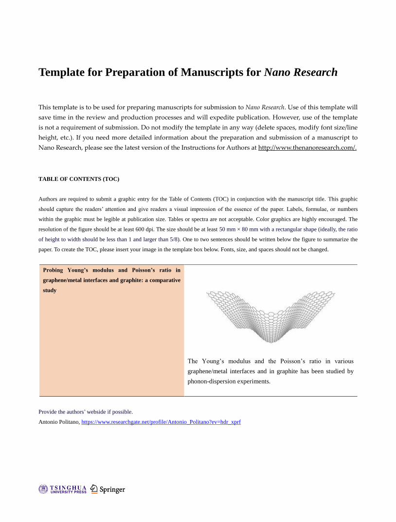
Probing Young’s modulus and Poisson’s ratio in
graphene/metal interfaces and graphite: a comparative
study
The Young’s modulus and the Poisson’s ratio in various
graphene/metal interfaces and in graphite has been studied by
phonon-dispersion experiments.
Provide the authors’ webside if possible.
Antonio Politano, https://www.researchgate.net/profile/Antonio_Politano?ev=hdr_xprf
Probing Young’s modulus and Poisson’s ratio in
graphene/metal interfaces and graphite: a comparative
study
Antonio Politano1 () and Gennaro Chiarello1,2
Received: day month year
Revised: day month year
Accepted: day month year
(automatically inserted by
the publisher)
© Tsinghua University Press
and Springer-Verlag Berlin
Heidelberg 2014
KEYWORDS
Young’s modulus,
Poisson’s ration, elastic
properties, graphene
ABSTRACT
By analyzing phonon dispersion, we have evaluated the average Young’s
modulus and Poisson’s ratio in graphene grown on Ru(0001), Pt(111), Ir(111),
Ni(111), BC3/NbB2(0001) and, moreover, in graphite. In both flat and
corrugated graphene sheets and graphite, we find a Poisson’s ratio of 0.19
and a Young’s modulus of 342 N/m. The unique exception is graphene/Ni(111),
for which we find different values (0.36 and 310 N/m, respectively) due to the
stretching of C-C bonds occurring in this commensurate overstructure. Such
findings are in excellent agreement with calculations performed for a
free-standing graphene membrane. The high crystalline quality of graphene
grown on metal substrates leads to macroscopic samples of high tensile
strength and bending flexibility to be used for technological applications such
as electromechanical devices and carbon-fiber reinforcements.
1 Introduction
The elastic moduli of single-layer graphene sheets
have attracted considerable interest in recent years.
In fact, the extraordinary intrinsic strength of
graphene[1] makes graphene a suitable material for
applications such as actuators[2] and
nano-electromechanical devices[3, 4] and, moreover,
as carbon-fiber reinforcement in polymeric
nanocomposites[5].
Graphene can be formed by graphite exfoliation [6],
thermal decomposition of SiC [7] and by epitaxial
growth on metal surfaces[8]. The preparation of
highly ordered monolayer graphene could be
extended up to the millimeter scale when graphene is
Nano Research
DOI (automatically inserted by the publisher)
Address correspondence to Antonio Politano, [email protected]
Research Article Please choose one
| www.editorialmanager.com/nare/default.asp
2 Nano Res.
epitaxially grown on transition-metal substrates. [9]
Furthermore, the possibility of transfer of the
graphene sheet onto insulating substrates may be a
promising route toward large scale production of
graphene devices [10]. Thus, it is important to know
the interaction strength between the graphene layer
and the metallic substrate in order to discern
between physisorption and chemisorption of
graphene and, moreover, to appraise the quality of
the contacts between metallic electrodes and
graphene devices[11-13].
Graphene has been grown on different transition
metal substrates: Pt(111)[14], Ni(111)[15],
Ru(0001)[16], Ir(111)[17], Rh(111)[18], Pd(111)[19],
Re(0001)[20], Cu(111)[21], and Co(0001)[22]. Among
the above-mentioned graphene systems, three
general classes may be distinguished.
Firstly, for the class of Ni and Co substrates, the
mismatch in the lattice parameter is negligible, thus
the graphene unit cell may be directly matched with
the substrate unit cell by slightly quenching or
stretching the bonds between carbon atoms of the
graphene lattice. In this case, a strong hybridization
between the substrate d bands and the π bands of
graphene occurs[11]. Thus, graphene is chemisorbed
onto these substrates with a small graphene-substrate
distance (2.1 Å for Ni [23] and 1.5-2.2 Å for Co [22]).
Whenever the mismatch in the lattice parameter
approaches 5-10%, a Moiré pattern appears. In this
case, the graphene sheet may be weakly (Pt, Ir) or
strongly bonded (Ru, Re, Pd, Rh) to the substrate.
The strong interaction occurring for graphene on
Ru(0001) [24] and Re(0001) [20] causes a strong
corrugation of the graphene sheet.
To date, a comparative investigation of elastic moduli
of graphene/metal interfaces is hitherto missing. Such
a comparative analysis could clarify whether the
elastic properties of periodically rippled graphene on
Ru(0001)[8, 25, 26] are different with respect to
systems in which the graphene overstructure is
nearly flat. Moreover, it would be interesting to
understand the influence of the stretching of C-C
bonds occurring in graphene/Ni(111) on the elastic
properties of the graphene sheet.
Herein, we estimate the average elastic properties
(Young’s modulus and the Poisson’s ratio) in
graphene epitaxially grown on Ru(0001), Pt(111),
Ir(111), Ni(111), BC3/NbB2(0001) and graphite, based
on the investigation of the phonon dispersion.
In most cases, we estimate the same values of the
Poisson’s ratio (0.19) and the Young’s modulus
(342 N/m) of the graphene sheet. The unique
exception is represented by graphene/Ni(111), for
which theirs values are 0.36 and 310 N/m,
respectively. Despite the macroscopic size of our
graphene sample which usually reduces the
tensile strength for the presence of defects and
grain boundaries, the above parameters well agree
with results reported for suspended graphene
membranes[27] with diameter of 1.0-1.5 μm.
Hence, our results demonstrate that high-quality
and macroscopic samples of epitaxial graphene on
metal substrates exhibit the tensile strength
predicted by theory. Moreover, we have
demonstrated that surface corrugation and the
graphene-substrate interaction do not play any
peculiar role on the elastic moduli of the graphene
sheet.
2 Experimental
Experiments were carried out in an ultra-high
vacuum (UHV) chamber operating at a base
pressure of 5∙10-9 Pa. The samples were single
crystals delivered from MaTecK GmbH.
Substrates have been cleaned by repeated cycles
of ion sputtering and annealing at 1300 K.
Surface cleanliness and order were checked using
Auger electron spectroscopy (AES) and
low-energy electron diffraction (LEED)
measurements, respectively. Graphene was obtained by dosing ethylene onto the
clean substrate held at 1150 K, with the exception of
graphene on Ni(111), for which a lower sample
temperature was used (800 K) [28]. The presence of a
single sheet of graphene in the whole sample has
been confirmed by ex-situ Raman spectroscopy[29].
Similar conclusions have been reported in other
works on the same systems in the same experimental
conditions [14, 30].
The inspection of the LEED pattern clearly shows the
presence of well-resolved spots which are
fingerprint of the order of the graphene
overstructure.
Graphene grows on Ru(0001) with a single

www.theNanoResearch.com∣www.Springer.com/journal/12274 | Nano Research
3 Nano Res.
)sinE
E1(sin
mE2q S
p
lossi
p
macroscopic domain which extend up to millimeter
scale [9]. Similar results have been obtained for
graphene on Ni(111) [31]. By contrast, micrometric
graphene domains grow on Ir(111) [32] and Pt(111)
[33, 34] with two and three rotational orientations,
respectively.
High-resolution electron energy loss spectroscopy
(HREELS) experiments were performed by using an
electron energy loss spectrometer (Delta 0.5, SPECS).
The energy resolution of the spectrometer was
degraded to 4 meV so as to increase the
signal-to-noise ratio of loss peaks. Dispersion of the
loss peaks, i.e., Eloss(q||), was measured by moving
the analyzer while keeping the sample and the
monochromator in a fixed position. To measure the
dispersion relation, values for the parameters Ep,
impinging energy and θi , the incident angle, were
chosen so as to obtain the highest signal-to-noise
ratio. The primary beam energy used for the
dispersion, Ep=20 eV, provided, in fact, the best
compromise among surface sensitivity, the highest
cross-section for phonon excitation and q||
resolution.
As
the parallel momentum transfer, q|| depends on Ep,
Eloss, θi and θs according to:
where Eloss is the energy loss and θs is the electron
scattering angle [35].
Accordingly, the integration window in reciprocal
space [36] is
where α is the angular acceptance of the apparatus
(±0.5° in our case). For the investigated range of q||,
the indeterminacy has been found to range from
0.005 (near ) to 0.022 Å -1 (for higher momenta).
The phonon dispersion for all systems has been
measured with the sample aligned along the
M . To obtain the energies of loss peaks, a
polynomial background was subtracted from each
spectrum. The resulting spectra were fitted by a
Gaussian line shape (not shown herein).
All measurements were made at room temperature.
3 Results and discussion
HREEL spectra, recorded as a function of the
scattering angle, show several dispersing features,
all assigned to phonon excitations. As a selected
case, we show in Figure 1 measurements of
graphene/Ru(0001) recorded at Ep=20 eV as a
function of the parallel momentum transfer q||.
Phonon modes are excited in electron scattering by
the impact mechanism[37]. Thus, the intensity of
phonon modes notably increases with q|| (Figure 1),
even if they are noticeable also at small momenta
just by increasing the acquisition time for improving
the signal-to-noise ratio (not shown).
In graphene, two kinds of phonons exist: lattice vibrations
in the plane of the sheet giving rise to transverse and
longitudinal acoustic (TA and LA) and optical (TO and
LO) branches, and lattice vibrations out of the plane
of the layer which give rise to the so-called flexural
phonons (ZA and ZO). Modes classified with “T” are
shear in-plane phonon excitations; “L” modes are
longitudinal in-plane vibrations; while “Z” indicates
out-of-plane polarization. In graphite and graphene, the
ZO mode is significantly softened with respect to the other
two optical modes, i.e. TO and LO. This is due to the
higher freedom for atom motion perpendicular to the plane
with respect to the in-plane motion. Figure 2 shows a
selected HREELS spectrum showing the above-mentioned
six phonon modes of the graphene lattice. The sharpness
of phonon modes observed in Figure 2 indicates an
excellent crystalline order in the graphene sample.
In principle, EELS planar scattering from an isolated
graphene sheet does not allow the observation of the
TA branch for selection rules inhibiting the
observation of odd phonons under reflection
symmetries[37]. However, the presence of an
underlying substrate acts as a symmetry breaking
and this allows to record a weak signal also from the
TA mode. Such a reduced intensity implies that long
acquisition time is required for detecting TA with a
sufficient signal-to-noise ratio.
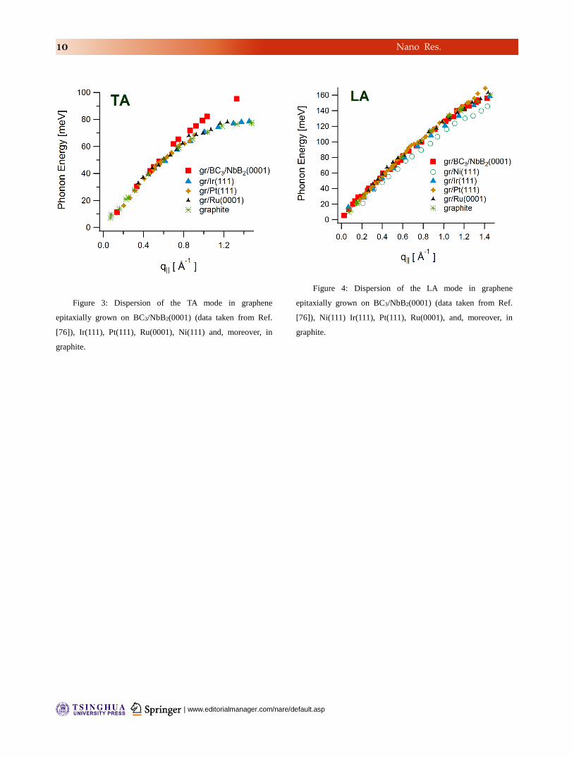
Figures 3 and 4 report the dispersion of the TA and
LA phonons, respectively, for different systems, that
is graphene epitaxially grown on Ni(111), Pt(111),
Ir(111), Ru(0001), BC3/NbB2(0001) and, moreover,
graphite.

| www.editorialmanager.com/nare/default.asp
4 Nano Res.
Sound velocities have been extracted from the
experimental slope of the acoustic branches in the
low-q|| limit, for which TA and LA phonons along
the K and M directions coincide. In
particular, we define vL, longitudinal sound velocity,
and vT, transverse sound velocity, as
and where and are the
frequencies of longitudinal and transverse acoustic
phonons, respectively.
We obtain 14.0 and 22.0 km/s for the TA and the LA
branches, respectively. The only exception is the
transverse sound velocity for graphene/Ni(111),
which is found to be lower by 11% (12.4 km/s).
The difference for graphene/Ni(111) arises from the
fact that the C−C bonds of the graphene layer are
stretched by 1.48% to form a 1 × 1 structure. The
energetically most favorable configuration is that
with one carbon atom is on top of a Ni atom and the
other carbon atom on a hollow site[38].
Data on the dispersion of acoustic phonons of a
graphene can provide information on its elastic
properties. According to the procedure illustrated in
Ref. [39], the sound velocities of the TA and LA
branches could be used for calculating the in-plane
stiffness (the 2D analogous of the bulk modulus)
and the shear modulus of the graphene sheet,
respectively:
D
T
D
L
v
v
2
2
Thus, we estimate and to be 211 and 144 N/m for
all systems with the exception of graphene/Ni(111),
for which their values are 244 and 114 N/m,
respectively. It is worth noticing that graphene is a
true 2D material, therefore its elastic behavior is
properly described by 2D properties with units of
force/length.
On the other hand, the 2D shear and bulk moduli are
also defined as a function of the Poisson’s ratio and
the Young’s modulus for 2D samples E2D:
)1(2
)1(22
2
D
D
E
E
Hence, from and it is possible to estimate the
Poisson’s ratio, i.e. the ratio of transverse contraction
strain to longitudinal extension strain in the direction
of the stretching force:
19.0
1
1
The obtained value for most systems, i.e. 0.19, agrees
well with results for graphite in the basal plane (0.165)
[40, 41] while it is 0.28 in carbon nanotubes [42]. It
represents an intermediate value with respect to
those reported by calculations for graphene, as
shown in Table I. Instead, for graphene/Ni(111) its
value is 0.36.
It is worth noticing that, according to molecular
dynamics calculations[43], the Poisson’s ratio
increases with the size of the graphene sample up to
reach a saturation value and it also depends on
temperature. This opens the possibility to tailor the
mechanical properties of graphene for engineering
applications.
The Poisson’s ratio could be used as a powerful test
among the various existing calculations on phonon
dispersion in graphene. As an example, the
calculated LA and TA modes in Ref. [44] would lead
to a clearly underestimated value of the Poisson’s
ratio (≈0.05).
It is also possible to estimate the Young’s modulus
E2D, which is a measure of the stiffness of
an isotropic elastic material. It is defined as the ratio
of the uniaxial stress over the uniaxial strain.
Table I. Poisson’s ratio ν, as reported in different
experimental and theoretical works. Poisson’s
ratio ν
Experimental (HREELS), graphene on Pt(111),
Ru(0001), Ir(111), BC3/NbB2(0001), graphite
0.19
Experimental, basal plane of graphite, Refs. [40, 41] 0.165
Experimental (HREELS), graphene/Ni(111) 0.36
Atomistic Monte Carlo, Ref. [45] 0.12
Tersoff-Brenner potential, Ref. [46] 0.149
Continuum plate theory, Ref. [47] 0.16

www.theNanoResearch.com∣www.Springer.com/journal/12274 | Nano Research
5 Nano Res.
Density functional theory, Ref. [48] 0.162
First-principles total-energy calculations, combined
to continuum elasticity, Ref. [49]
0.169
Ab initio, Ref. [50] 0.173
Ab initio, Ref. [51] 0.178
DFT, Ref. [52] 0.18
Ab initio, Ref. [53] 0.186
Ab initio, Ref. [54] 0.19
Valence force model, Ref. [55] 0.20
LDA, Ref. [56] 0.20
Cellular material mechanics theory, Ref. [57] 0.21
Molecular dynamics, Ref. [43] 0.22
Molecular dynamics, Ref. [58] 0.22
Empirical force-constant calculations, Ref. [59] 0.227
Brenner’s potential, Ref. [52] 0.27
continuum elasticity theory and tight-binding
atomistic simulations, Ref. [60]
0.31
Ab initio, Ref. [61] 0.32
Molecular dynamics, Ref. [62] 0.32
Brenner’s potential, Ref. [63] 0.397
Multiple component correlation model, Ref. [64] 0.4
Molecular dynamics, Ref. [65] 0.45
As reported in Table II, many theoretical works
found Young’s moduli ranging from 307 to 356 N/m.
The obtained value of E2D for most graphene/metal
interfaces, i.e. 342 N/m, agrees well with most
theoretical results (Table II), a part from calculations
in Ref. [66] (underestimated E2D). In particular, a
good agreement exists between present results and
first-principles total-energy calculations, combined to
continuum elasticity, reported in Ref. [49].
It will be helpful to compare present results with the
case of three-dimensional (3D) materials and, in
particular, with bulk graphite. To obtain the
corresponding 3D parameter for the selected case of
graphene/Pt(111), the value of E2D should be divided
by the distance between the graphene and the
underlying Pt(111) substrate (3.31 Å )[67, 68]. Thus,
E2D as obtained by vibrational measurements
corresponds to a 3D Young’s modulus E=1.03 TPa.
This is in fair agreement with experiments on bulk
graphite yielding 1.02 TPa for the in-plane Young’s
modulus[40]. For the sake of completeness, the
Young’s modulus obtained for single-walled carbon
nanotubes ranges from 0.45 and 1.47 TPa [69], while
for multi-walled carbon nanotubes it was found to
range from 0.27 to 0.95 TPa [70]. In graphene/Ni(111)
E2D is instead 310 N/m.
Table II. 2D Young’s modulus E2D, expressed in N/m, as
reported in different experimental and theoretical works. Young's modulus E2D (N/m)
Experimental (HREELS),
graphene on Pt(111), Ru(0001),
Ir(111), BC3/NbB2(0001),
graphite
342
Experimental (HREELS),
graphene/Ni(111)
310
Experimental (AFM) on
graphene/copper foils, Ref. [71]
339±17
Experimental (AFM) on graphene
membranes, Ref. [27]
340±50
Experimental (AFM) on graphene
membranes, Ref. [72]
350±50
Tersoff-Brenner potential, Ref.
[66]
235
Energetic model, Ref. [73] 307
continuum elasticity theory and
tight-binding atomistic
simulations, Ref. [60]
312
DFT, Ref. [52] 330
Brenner’s potential, Ref. [63] 336
First-principles total-energy
calculations, combined to
continuum elasticity, Ref. [49]
344
Tersoff-Brenner potential, Ref.
[46]
345
Ab initio, Ref. [53] 350
Atomistic Monte Carlo, Ref. [45] 353
Density functional theory, Ref.
[48]
356
Empirical force-constant
calculations, Ref. [59]
384
Experimental (AFM) on
graphene/copper foils, Ref. [74]
55
Recently, nanoindentation AFM measurements [74]
have been performed on graphene grown by CVD on
copper foils and successively transferred onto silicon
nitride grids with arrays of pre-patterned holes.
These experiments have revealed a notably reduced
E2D (55 N/m) with respect to the present finding (342
N/m). Such extremely low value of E2D could be a
consequence of the modification of the membrane
structure induced by the transfer process (see Ref. [74]
for more details).
In addition, in the linear elastic regime, it is possible
to estimate the elastic constants C11 and C12 , from E2D
and ν:
11
12
11
2
12
2
112
C
C
C
CCE D
This, C11=422 N/m and C12=80 N/m, which are in
good agreement with values reported by Cadelano et
| www.editorialmanager.com/nare/default.asp
6 Nano Res.
al.[49] (354 and 60 N/m).
Their corresponding 3D values are 1.27 and 0.24,
respectively, which agree well with experimental
findings for graphite reported in Ref. [75] (1.11 and
0.18 TPa).
Conclusions
We have demonstrated that the elastic properties in
graphene/metal interfaces are the same recorded in
graphite and free-standing graphene, with the
exception of graphene/Ni(111), where C-C bonds are
stretched by 1.48%. This implies a variation of the 2D
Young’s modulus by 9% (310 N/m versus 342 N/m in
the other systems).
The excellent crystalline quality of graphene grown
on metal substrates (with a reduced number of
defects and grain boundaries) leads to macroscopic
samples of high bending flexibility and tensile
strength, which could be used for applications in
advanced nanocomposites. Due to its thermal
stability up to 1200 K, chemical stability and
robustness, epitaxial graphene represents a
promising candidate for application in
nano-electromechanical devices.
Acknowledgements
We thank Davide Campi and Fernando de Juan for
helpful discussions. References
[1] Zhang, Y. Y.;Wang, C. M.;Cheng, Y.; Xiang, Y.
Mechanical properties of bilayer graphene sheets coupled by sp3
bonding. Carbon 2011, 49, 4511-4517.
[2] Park, S.;An, J.;Suk, J. W.; Ruoff, R. S. Graphene-Based
Actuators. Small 2010, 6, 210-212.
[3] Wang, Y.;Yang, R.;Shi, Z.;Zhang, L.;Shi, D.;Wang, E.;
Zhang, G. Super-Elastic Graphene Ripples for Flexible Strain
Sensors. ACS Nano 2011, 5, 3645-3650.
[4] Scharfenberg, S.;Rocklin, D. Z.;Chialvo, C.;Weaver, R.
L.;Goldbart, P. M.; Mason, N. Probing the mechanical properties
of graphene using a corrugated elastic substrate. Appl. Phys.
Lett. 2011, 98, 091908.
[5] Alzari, V.;Sanna, V.;Biccai, S.;Caruso, T.;Politano,
A.;Scaramuzza, N.;Sechi, M.;Nuvoli, D.;Sanna, R.; Mariani, A.
Tailoring the physical properties of nanocomposite films by the
insertion of graphene and other nanoparticles. Composites Part
B: Engineering 2014, 60, 29-35.
[6] Li, D.;Windl, W.; Padture, N. P. Toward Site-Specific
Stamping of Graphene. Adv. Mater. 2009, 21, 1243-1246.
[7] Emtsev, K. V.;Bostwick, A.;Horn, K.;Jobst, J.;Kellogg,
G. L.;Ley, L.;McChesney, J. L.;Ohta, T.;Reshanov, S. A.;Rohrl,
J.;Rotenberg, E.;Schmid, A. K.;Waldmann, D.;Weber, H. B.;
Seyller, T. Towards wafer-size graphene layers by atmospheric
pressure graphitization of silicon carbide. Nat. Mater. 2009, 8,
203-207.
[8] Borca, B.;Barja, S.;Garnica, M.;Minniti, M.;Politano,
A.;Rodriguez-García, J. M.;Hinarejos, J. J.;Farías, D.;Vázquez
de Parga, A. L.; Miranda, R. Electronic and geometric
corrugation of periodically rippled, self-nanostructured
graphene epitaxially grown on Ru(0001). New J. Phys. 2010, 12,
093018.
[9] Pan, Y.;Zhang, H.;Shi, D.;Sun, J.;Du, S.;Liu, F.; Gao,
H.-j. Highly Ordered, Millimeter-Scale, Continuous,
Single-Crystalline Graphene Monolayer Formed on Ru (0001).
Adv. Mater. 2009, 21, 2777-2780.
[10] Sutter, P. W.;Albrecht, P. M.; Sutter, E. A. Graphene
growth on epitaxial Ru thin films on sapphire. Appl. Phys. Lett.
2010, 97, 213101.
[11] Giovannetti, G.;Khomyakov, P. A.;Brocks, G.;Karpan, V.
M.;van den Brink, J.; Kelly, P. J. Doping Graphene with Metal
Contacts. Phys. Rev. Lett. 2008, 101, 026803.
[12] Politano, A.; Chiarello, G. Quenching of plasmons
modes in air-exposed graphene-Ru contacts for plasmonic
devices. Appl. Phys. Lett. 2013, 102, 201608-201604.
[13] Politano, A.; Chiarello, G. Unravelling suitable
graphene-metal contacts for graphene-based plasmonic devices.
Nanoscale 2013, 5, 8215-8220.
[14] Politano, A.;Marino, A. R.;Formoso, V.;Farías,
D.;Miranda, R.; Chiarello, G. Evidence for acoustic-like
plasmons on epitaxial graphene on Pt(111). Phys. Rev. B 2011,
84, 033401.
[15] Dedkov, Y. S.;Fonin, M.; Laubschat, C. A possible
source of spin-polarized electrons: The inert graphene/Ni(111)
system. Appl. Phys. Lett. 2008, 92, 052506.
[16] Politano, A.;Borca, B.;Minniti, M.;Hinarejos, J.
J.;Vázquez de Parga, A. L.;Farías, D.; Miranda, R. Helium
reflectivity and Debye temperature of graphene grown
www.theNanoResearch.com∣www.Springer.com/journal/12274 | Nano Research
7 Nano Res.
epitaxially on Ru(0001). Phys. Rev. B 2011, 84, 035450.
[17] Pletikosić, I.;Kralj, M.;Pervan, P.;Brako, R.;Coraux,
J.;N'Diaye, A. T.;Busse, C.; Michely, T. Dirac cones and
minigaps for graphene on Ir(111). Phys. Rev. Lett. 2009, 102,
056808.
[18] Sicot, M.;Bouvron, S.;Zander, O.;Rudiger, U.;Dedkov,
Y. S.; Fonin, M. Nucleation and growth of nickel nanoclusters on
graphene Moiré on Rh(111). Appl. Phys. Lett. 2010, 96,
093115-093113.
[19] Kwon, S.-Y.;Ciobanu, C. V.;Petrova, V.;Shenoy, V.
B.;Bareno, J.;Gambin, V.;Petrov, I.; Kodambaka, S. Growth of
Semiconducting Graphene on Palladium. Nano Lett. 2009, 9,
3985-3990.
[20] Miniussi, E.;Pozzo, M.;Baraldi, A.;Vesselli, E.;Zhan, R.
R.;Comelli, G.;Menteş, T. O.;Nino, M. A.;Locatelli, A.;Lizzit,
S.; Alfè, D. Thermal Stability of Corrugated Epitaxial Graphene
Grown on Re(0001). Phys. Rev. Lett. 2011, 106, 216101.
[21] Reddy, K. M.;Gledhill, A. D.;Chen, C.-H.;Drexler, J. M.;
Padture, N. P. High quality, transferrable graphene grown on
single crystal Cu(111) thin films on basal-plane sapphire. Appl.
Phys. Lett. 2011, 98, 113117.
[22] Eom, D.;Prezzi, D.;Rim, K. T.;Zhou, H.;Lefenfeld,
M.;Xiao, S.;Nuckolls, C.;Hybertsen, M. S.;Heinz, T. F.; Flynn,
G. W. Structure and Electronic Properties of Graphene
Nanoislands on Co(0001). Nano Lett. 2009, 9, 2844-2848.
[23] Gamo, Y.;Nagashima, A.;Wakabayashi, M.;Terai, M.;
Oshima, C. Atomic structure of monolayer graphite formed on
Ni(111). Surf. Sci. 1997, 374, 61-64.
[24] Wang, B.;Günther, S.;Wintterlin, J.; Bocquet, M.-L.
Periodicity, work function and reactivity of graphene on
Ru(0001) from first principles. New J. Phys. 2010, 12, 043041.
[25] Politano, A.;Campi, D.;Formoso, V.; Chiarello, G.
Evidence of confinement of the π plasmon in periodically
rippled graphene on Ru(0001). Phys. Chem. Chem. Phys. 2013,
15, 11356-11361.
[26] Politano, A.; Chiarello, G. Periodically rippled graphene
on Ru(0001): A template for site-selective adsorption of
hydrogen dimers via water splitting and hydrogen-spillover at
room temperature. Carbon 2013, 61, 412-417.
[27] Lee, C.;Wei, X.;Kysar, J. W.; Hone, J. Measurement of
the Elastic Properties and Intrinsic Strength of Monolayer
Graphene. Science 2008, 321, 385-388.
[28] Cupolillo, A.;Ligato, N.; Caputi, L. S. Two-dimensional
character of the interface-π plasmon in epitaxial graphene on
Ni(111). Carbon 2012, 50, 2588-2591.
[29] Cazzanelli, E.;Caruso, T.;Castriota, M.;Marino, A.
R.;Politano, A.;Chiarello, G.;Giarola, M.; Mariotto, G.
Spectroscopic characterization of graphene films grown on
Pt(111) surface by chemical vapor deposition of ethylene. J.
Raman Spectrosc. 2013, 44, 1393-1397.
[30] Gao, M.;Pan, Y.;Huang, L.;Hu, H.;Zhang, L. Z.;Guo, H.
M.;Du, S. X.; Gao, H. J. Epitaxial growth and structural property
of graphene on Pt(111). Appl. Phys. Lett. 2011, 98, 033101.
[31] Odahara, G.;Otani, S.;Oshima, C.;Suzuki, M.;Yasue, T.;
Koshikawa, T. Macroscopic single-domain graphene sheet on
Ni(111). Surf. Interface Anal. 2011, 43, 1491-1493.
[32] Starodub, E.;Bostwick, A.;Moreschini, L.;Nie,
S.;Gabaly, F. E.;McCarty, K. F.; Rotenberg, E. In-plane
orientation effects on the electronic structure, stability, and
Raman scattering of monolayer graphene on Ir(111). Phys. Rev.
B 2011, 83, 125428.
[33] Liang, Z.;Khosravian, H.;Uhl, A.;Meyer, R. J.; Trenary,
M. Graphene domain boundaries on Pt(111) as nucleation sites
for Pt nanocluster formation. Surf. Sci. 2012, 606, 1643–1648.
[34] Politano, A.;Marino, A. R.;Formoso, V.;Farías,
D.;Miranda, R.; Chiarello, G. Quadratic dispersion and damping
processes of π plasmon in monolayer graphene on Pt(111).
Plasmonics 2012, 7, 369-376.
[35] Rocca, M. Low-Energy EELS Investigation of Surface
Electronic Excitations on Metals. Surf. Sci. Rep. 1995, 22, 1-71.
[36] Politano, A.;Formoso, V.;Colavita, E.; Chiarello, G.
Probing collective electronic excitations in as-deposited and
modified Ag thin films grown on Cu(111). Phys. Rev. B 2009,
79, 045426.
[37] Ibach, H.; Mills, D. L. Electron Energy Loss
Spectroscopy and Surface Vibrations; Academic Press: San
Francisco, 1982.
[38] Allard, A.; Wirtz, L. Graphene on metallic substrates:
suppression of the Kohn anomalies in the phonon dispersion.
Nano Lett. 2010, 10, 4335-4340.
[39] Sánchez-Portal, D.;Artacho, E.;Soler, J. M.;Rubio, A.;
Ordejón , P. Ab initio structural, elastic, and vibrational
properties of carbon nanotubes. Phys. Rev. B 1999, 59, 12678.
[40] Blakslee, O. L.;Proctor, D. G.;Seldin, E. J.;Spence, G. B.;
| www.editorialmanager.com/nare/default.asp
8 Nano Res.
Weng, T. Elastic Constants of Compression-Annealed Pyrolytic
Graphite. J. Appl. Phys. 1970, 41, 3373-3382.
[41] Seldin, E. J.; Nezbeda, C. W. Elastic Constants and
Electron-Microscope Observations of Neutron-Irradiated
Compression-Annealed Pyrolytic and Single-Crystal Graphite.
J. Appl. Phys. 1970, 41, 3389-3400.
[42] Lu, J. P. Elastic Properties of Carbon Nanotubes and
Nanoropes. Phys. Rev. Lett. 1997, 79, 1297.
[43] Jiang, J.-W.;Wang, J.-S.; Li, B. Young's modulus of
graphene: A molecular dynamics study. Phys. Rev. B 2009, 80,
113405.
[44] Adamyan, V.; Zavalniuk, V. Phonons in graphene with
point defects. J. Phys.: Condens. Matt. 2011, 23, 015402.
[45] Zakharchenko, K. V.;Katsnelson, M. I.; Fasolino, A.
Finite Temperature Lattice Properties of Graphene beyond the
Quasiharmonic Approximation. Phys. Rev. Lett. 2009, 102,
046808.
[46] Kudin, K. N.;Scuseria, G. E.; Yakobson, B. I. C2F, BN,
and C nanoshell elasticity from ab initio computations. Phys.
Rev. B 2001, 64, 235406.
[47] Arghavan, S.; Singh, A. V. Free Vibration of Single
Layer Graphene Sheets: Lattice Structure Versus Continuum
Plate Theories. J. Nanotechnol. Eng. Med. 2011, 2,
031005-031006.
[48] Bera, S.;Arnold, A.;Evers, F.;Narayanan, R.; Wölfle, P.
Elastic properties of graphene flakes: Boundary effects and
lattice vibrations. Phys. Rev. B 2010, 82, 195445.
[49] Cadelano, E.;Palla, P. L.;Giordano, S.; Colombo, L.
Elastic properties of hydrogenated graphene. Phys. Rev. B 2010,
82, 235414.
[50] Gui, G.;Li, J.; Zhong, J. Band structure engineering of
graphene by strain: First-principles calculations. Phys. Rev. B
2008, 78, 075435.
[51] Peng, Q.;Liang, C.;Ji, W.; De, S. A theoretical analysis
of the effect of the hydrogenation of graphene to graphane on its
mechanical properties. Phys. Chem. Chem. Phys. 2013, 15,
2003-2011.
[52] Fair, K. M.;Arnold, M. D.; Ford, M. J. Determination of
the elastic properties of graphene by indentation and the validity
of classical models of indentation. J. Phys.: Condens. Matter
2014, 26, 015307.
[53] Liu, F.;Ming, P.; Li, J. Ab initio calculation of ideal
strength and phonon instability of graphene under tension. Phys.
Rev. B 2007, 76, 064120.
[54] Wang, R.;Wang, S.;Wu, X.; Liang, X. First-principles
calculations on third-order elastic constants and internal
relaxation for monolayer graphene. Physica B 2010, 405,
3501-3506.
[55] Perebeinos, V.; Tersoff, J. Valence force model for
phonons in graphene and carbon nanotubes. Phys. Rev. B 2009,
79, 241409.
[56] Wagner, P.;Ivanovskaya, V. V.;Rayson, M. J.;Briddon, P.
R.; Ewels, C. P. Mechanical properties of nanosheets and
nanotubes investigated using a new geometry independent
volume definition. J. Phys.: Condens. Matter 2013, 25, 155302.
[57] Scarpa, F.;Adhikari, S.; Srikantha Phani, A. Effective
elastic mechanical properties of single layer graphene sheets.
Nanotechnology 2009, 20, 065709.
[58] Kalosakas, G.;Lathiotakis, N. N.;Galiotis, C.; Papagelis,
K. In-plane force fields and elastic properties of graphene. J.
Appl. Phys. 2013, 113, 134307.
[59] Michel, K. H.; Verberck, B. Theory of the elastic
constants of graphite and graphene. Phys. Status Solidi B 2008,
245, 2177-2180.
[60] Cadelano, E.;Palla, P. L.;Giordano, S.; Colombo, L.
Nonlinear Elasticity of Monolayer Graphene. Phys. Rev. Lett.
2009, 102, 235502.
[61] Zhou, G.;Duan, W.; Gu, B. First-principles study on
morphology and mechanical properties of single-walled carbon
nanotube. Chem. Phys. Lett. 2001, 333, 344-349.
[62] Kam, K.;Scarpa, F.;Adhikari, S.; Chowdhury, R.
Graphene nanofilm as pressure and force sensor: A mechanical
analysis. Phys. Status Solidi B 2013, 250, 2085-2089.
[63] Arroyo, M.; Belytschko, T. Finite crystal elasticity of
carbon nanotubes based on the exponential Cauchy-Born rule.
Phys. Rev. B 2004, 69, 115415.
[64] Wang, C. G.;Lan, L.;Liu, Y. P.; Tan, H. F. Multiple
component correlation model for elastic modulus of single layer
graphene sheets. Physica E 2014, 56, 372-376.
[65] Zhou, L.;Wang, Y.; Cao, G. Elastic properties of
monolayer graphene with different chiralities. J. Phys.:
Condens. Matter 2013, 25, 125302.
[66] Zhou, J.; Huang, R. Internal lattice relaxation of
single-layer graphene under in-plane deformation. J. Mech.

www.theNanoResearch.com∣www.Springer.com/journal/12274 | Nano Research
9 Nano Res.
Phys. Solids 2008, 56, 1609-1623.
[67] Otero, G.;Gonzalez, C.;Pinardi, A. L.;Merino,
P.;Gardonio, S.;Lizzit, S.;Blanco-Rey, M.;Van de Ruit,
K.;Flipse, C. F. J.;Méndez, J.;de Andrés, P. L.; Martín-Gago, J.
A. Ordered Vacancy Network Induced by the Growth of
Epitaxial Graphene on Pt(111). Phys. Rev. Lett. 2010, 105,
216102.
[68] Gao, M.;Pan, Y.;Zhang, C.;Hu, H.;Yang, R.;Lu, H.;Cai,
J.;Du, S.;Liu, F.; Gao, H. J. Tunable interfacial properties of
epitaxial graphene on metal substrates. Appl. Phys. Lett. 2010,
96, 053109.
[69] Yu, M.-F.;Files, B. S.;Arepalli, S.; Ruoff, R. S. Tensile
Loading of Ropes of Single Wall Carbon Nanotubes and their
Mechanical Properties. Phys. Rev. Lett. 2000, 84, 5552.
[70] Yu, M.-F.;Lourie, O.;Dyer, M. J.;Moloni, K.;Kelly, T. F.;
Ruoff, R. S. Strength and Breaking Mechanism of Multiwalled
Carbon Nanotubes Under Tensile Load. Science 2000, 287,
637-640.
[71] Lee, G.-H.;Cooper, R. C.;An, S. J.;Lee, S.;van der
Zande, A.;Petrone, N.;Hammerberg, A. G.;Lee, C.;Crawford,
B.;Oliver, W.;Kysar, J. W.; Hone, J. High-Strength
Chemical-Vapor–Deposited Graphene and Grain Boundaries.
Science 2013, 340, 1073-1076.
[72] Clark, N.;Oikonomou, A.; Vijayaraghavan, A. Ultrafast
quantitative nanomechanical mapping of suspended graphene.
Phys. Status Solidi B 2013, 250, 2672-2677.
[73] Reddy, C. D.;Ramasubramaniam, A.;Shenoy, V. B.;
Zhang, Y.-W. Edge elastic properties of defect-free single-layer
graphene sheets. Appl. Phys. Lett. 2009, 94, 101904.
[74] Ruiz-Vargas, C. S.;Zhuang, H. L.;Huang, P. Y.;van der
Zande, A. M.;Garg, S.;McEuen, P. L.;Muller, D. A.;Hennig, R.
G.; Park, J. Softened Elastic Response and Unzipping in
Chemical Vapor Deposition Graphene Membranes. Nano Lett.
2011, 11, 2259-2263.
[75] Klintenberg, M.;Lebègue, S.;Ortiz, C.;Sanyal,
B.;Fransson, J.; Eriksson, O. Evolving properties of
two-dimensional materials: from graphene to graphite. J. Phys.:
Condens. Matter 2009, 21, 335502.
[76] Yanagisawa, H.;Tanaka, T.;Ishida, Y.;Matsue,
M.;Rokuta, E.;Otani, S.; Oshima, C. Analysis of phonons in
graphene sheets by means of HREELS measurement and ab
initio calculation. Surf. Interface Anal. 2005, 37, 133-136.
Figure 1: HREELS spectra recorded as a function of the
parallel momentum transfer q|| for monolayer graphene on
Ru(0001). The green arrow indicates the weak TA mode.
Figure 2: HREELS spectrum reporting the six phonon
modes of graphene on Ru(0001) for a selected value of the
parallel momentum transfer q|| (~0.95 Å-1). Measurements have
been carried out with a primary electron beam energy of 20 eV,
with a fixed incidence angle of 80° with respect to the sample
normal (grazing incidence). The loss energy is measured in
meV in the bottom panel, while in the top panel the
corresponding value in cm-1 is reported.
| www.editorialmanager.com/nare/default.asp
10 Nano Res.
Figure 3: Dispersion of the TA mode in graphene
epitaxially grown on BC3/NbB2(0001) (data taken from Ref.
[76]), Ir(111), Pt(111), Ru(0001), Ni(111) and, moreover, in
graphite.
Figure 4: Dispersion of the LA mode in graphene
epitaxially grown on BC3/NbB2(0001) (data taken from Ref.
[76]), Ni(111) Ir(111), Pt(111), Ru(0001), and, moreover, in
graphite.
www.theNanoResearch.com∣www.Springer.com/journal/12274 | Nano Research
Nano Res.
Electronic Supplementary Material
Click and type your title here. The font is Helvetica bold
18. Capitalize the initial letter of the first word only. The
title must be brief and grammatically correct
Type author names here. The font is "Helvetica 10". Please spell out first names and surnames. Do not
include professional or official titles or academic degrees. Place an “()” by the corresponding author(s).
For example, First A. Firstauthor1,2 (), Second B. Secondauthor2,†, and Third C. Thirdauthor1 ()
Supporting information to DOI 10.1007/s12274-****-****-* (automatically inserted by the publisher)
INFORMATION ABOUT ELECTRONIC SUPPLEMENTARY MATERIAL. The font is Palatino Linotype 10.
Electronic supplementary material (ESM) may include large artwork, lengthy descriptions, and extensive data.
Each supporting information artwork, description, and data file should be referred to in the manuscript at least
once. The format is “Fig. S1 in the Electronic Supplementary Material (ESM)”, or “Figure S2 in the ESM” at the
beginning of a sentence. A brief description of the ESM should be included in the main text, before the
references section, in a paragraph entitled “Electronic Supplementary Material:”. ESM should be submitted
with the manuscript and will be sent to referees during peer review. Authors should ensure that it is clearly
presented.
Address correspondence to First A. Firstauthor, email1; Third C. Thirdauthor, email2