probing the conformational states of the sh1−sh2 helix in myosin: a cross-linking approach ...
TRANSCRIPT

Probing the Conformational States of the SH1-SH2 Helix in Myosin: ACross-Linking Approach†
Lisa K. Nitao and Emil Reisler*
Department of Chemistry and Biochemistry and Molecular Biology Institute, UniVersity of California,Los Angeles, California 90095
ReceiVed July 17, 1998; ReVised Manuscript ReceiVed September 24, 1998
ABSTRACT: Previous biochemical studies have shown that the SH1 (Cys707) and SH2 (Cys697) groupson rabbit skeletal myosin subfragment 1 (S1) can be cross-linked by using reagents of different cross-linking lengths. In the presence of nucleotide, this cross-linking is accelerated. In the crystal structureof S1, the SH1 and SH2 residues are located on anR-helix, 19 Å apart. Thus, the cross-linking resultscould be indicative of helix melting or increased flexibility in the presence of nucleotides. Nucleotide-induced changes in this region were examined in this study by monitoring the cross-linking of SH1 andSH2 on S1 with dimaleimide reagents of spans ranging from 5 to 15 Å. A method was devised to directlymeasure the kinetic effects of nucleotides on the rates of cross-linking reactions. The slow and reagent-insensitive rates of the SH1-SH2 cross-linking in the absence of nucleotides reveal that the equipartitioningof the SH1-SH2 helix among states with different SH1-SH2 separations occurs infrequently. In thepresence of MgADP, MgATP, and MgATPγS, the rates of SH1 and SH2 cross-linking were increased∼2-7-fold for the shortest reagent (5-8 Å). Rate accelerations were much greater for the longer reagents(9-15 Å): 40-50-fold for MgADP, 25-40-fold for MgATP, and 80-270-fold for MgATPγS. To accountfor any nucleotide-dependent differences in the reactivities of the reagents toward SH2, the rates ofmonofunctional SH2 modification on SH1-labeled S1 were also measured for each reagent. Theseexperiments showed that the nucleotide-induced increases in the rates of SH2 modification were similarfor all of the reagents. Thus, the changes observed in the cross-linking rates are due not only to the typeof nucleotide bound in the active site but also to the span of the cross-linking reagent. These findings areinterpreted in terms of nucleotide-induced shifts in the equilibria among conformational states of theSH1-SH2 helix.
Myosin is a molecular motor that converts the chemicalenergy of ATP to mechanical work. For years, considerableeffort has focused on how myosin functions. It is believedthat ATP hydrolysis induces structural changes in the myosinhead, which result in the directed movement of the actinfilament. As ATP is hydrolyzed, different transitional statesare created in the myosin head (1). These transitional statesare associated with conformational changes that are necessaryto transduce the chemical energy for force production.Recently, X-ray crystallographic structures of the myosinmotor domain complexed with nucleotide and phosphateanalogues were determined (2-4). When bound to myosin,these analogues represent both the prehydrolysis state(MgADP‚BeFx, MgATPγS, MgAMPPNP) and the post-hydrolysis state (MgADP‚AlF4
-, MgADP‚VO4). When thestructures of these different states are compared, severalregions of the motor domain appear to shift. A proposedmechanism for hydrolysis suggests that changes in theseregions are transmitted to and then amplified by the lightchain binding domain (5). Therefore, studies of these regionsmay reveal what structural changes are occurring in the
myosin head as it converts the energy of ATP to motorfunction.
One site on myosin subfragment 1 (S1)1 that has been ofparticular interest is the helix containing the reactive sul-fhydryls, Cys707 (SH1) and Cys697 (SH2). The SH1-SH2helix has been shown previously to undergo nucleotide-induced structural changes (6). A variety of reagentsspecifically modify and cross-link these two groups. In thepresence of nucleotide, SH1 and SH2 can be cross-linkedwith reagents of spans ranging from 3 to 14 Å (7, 8). Undercertain conditions, a disulfide bond may even be formedbetween the two groups (9). From the atomic structure ofthe S1, these cysteine residues are located on opposite sidesof a bent helix, and the distance between the sulfur atoms isapproximately 19 Å (10). Therefore, in the presence ofnucleotide, the helix must undergo some conformationalchanges in order for SH1 and SH2 to come close to eachother. To account for these changes, two explanations can
† This work was supported by grants from USPHS National ResearchService Award GM07185 (to L.K.N.) and USPHS AR22031 and NSFMCB-9630997.
1 Abbreviations: ATPγS, adenosine 5′-(3-thiotriphosphate); BM,1,1′-(methylenedi-4,1-phenylene)bismaleimide; mPDM,N,N′-1,3-phe-nylenedimaleimide; DTT, dithiothreitol; NDM, naphthalene-1,5-di-maleimide; NEM,N-ethylmaleimide; oPDM,N,N′-1,2-phenylenedi-maleimide; pPDM,N,N′-1,4-phenylenedimaleimide; S1, myosin sub-fragment 1; SH1, Cys707 in the rabbit (or chicken) skeletal S1 sequence;SH2, Cys697 on S1.
16704 Biochemistry1998,37, 16704-16710
10.1021/bi9817212 CCC: $15.00 © 1998 American Chemical SocietyPublished on Web 11/01/1998

be suggested. Upon nucleotide binding, the helix may becollapsing or melting. Alternatively, the helix may bebending or have increased flexibility, which would then allowSH1 and SH2 to approach each other.
In previous cross-linking studies, the effect of nucleotideson the SH1-SH2 helix was estimated from results whichhad not separated SH1 modification and subsequent cross-linking activities (11, 12). In this study, a scheme wasdevised to isolate the cross-linking reaction. With thisscheme, the actual changes induced by nucleotide can bedetermined. Cross-linking reagents of different spans wereutilized to probe the distance separating SH1 and SH2. Thespans of the reagents range from approximately 5 Å for theshortest reagent (oPDM) to 15 Å for the longest reagent(BM). The effect of three nucleotide states, MgADP,MgATPγS, and MgATP, on the SH1-SH2 cross-linking wasexamined in this study. Our results show that the effect ofnucleotide on the cross-linking rates depends on both thenucleotide state and the span of the cross-linking reagent.These results are discussed in terms of the nucleotide-inducedflexibility of the SH1-SH2 helix.
MATERIALS AND METHODS
Reagents. N-Ethylmaleimide (NEM), ADP, and SephadexG-50 were purchased from Sigma (St. Louis, MO).N,N′-1,2-Phenylenedimaleimide (oPDM) andN,N′-1,4-phenylene-dimaleimide (pPDM) were from Research Organics (Cleve-land, OH). N,N′-1,3-Phenylenedimaleimide (mPDM), naph-thalene-1,5-dimaleimide (NDM), and 1,1′-(methylenedi-4,1-phenylene)bismaleimide (BM) were from Aldrich (Milwau-kee, WI). Adenosine 5′-(3-thiotriphosphate) (ATPγS) wasobtained from Boehringer Mannheim (Germany).
Determination of Cross-Linking Spans. Each cross-linkingreagent was drawn in the ChemDraw program and transferredto the MacroModel program which used energy minimal-ization in determining the cross-linking distances for differentconformers. The reagents and their cross-linking spans areshown in Figure 1.
Proteins. Myosin from rabbit psoas muscle was preparedaccording to Godfrey and Harrington (13). Subfragment 1(S1) was prepared by chymotryptic digestion of myosinfilaments as described by Weeds and Pope (14). Theconcentration of S1 was determined spectrophotometricallyby using an extinction coefficient ofE1%280) 7.5 cm-1.
ATPase ActiVities. Ca2+- and EDTA- (K+-)ATPase activi-ties of S1 were determined at 37°C according to Fiske andSubbarow (15). The Ca2+-ATPase assay solution contained600 mM KCl, 50 mM Tris-HCl (pH 7.6), 5.0 mM CaCl2,and 2.0 mM ATP. The EDTA-ATPase assay solutioncontained 444 mM KCl, 50 mM histidine, 50 mM Tris-HCl(pH 7.6), 5.0 mM EDTA, and 2.0 mM ATP.
Cross-Linking Experiments. All cross-linking reactionswere carried out in three steps. In the first step, bifunctionalreagents were attached to SH1 on S1. This involved reactingthe cross-linking reagent (18µM) with S1 (12 µM) in 10mM KCl, 10 mM imidazole, pH 7.0 at 4°C. Because theoPDM reaction was much slower than the other reactions,1.0 mM MgADP was included in the modification of SH1by this reagent only. For all of the reactions, the conditionswere chosen to optimize SH1 modification without signifi-cant SH1-SH2 cross-linking. The modification reactions
were monitored by measuring the Ca2+- and EDTA-ATPaseactivities. In the second step, after SH1 was modified, theexcess reagent was removed from the mixtures. The reactionmixtures were applied to Sephadex G-50 spin columns,which were equilibrated with 10 mM KCl, 10 mM imidazole,pH 7.0. Excess reagent was removed by centrifugation, andthe samples were equilibrated to 25°C. In the third step,the cross-linking reaction was monitored. After splitting thereaction mixture into two equal samples, nucleotide (1.0 mMMgADP, 1.0 mM MgATPγS, or 3.0 mM MgATP) wasadded to one of the samples while the other sample was usedto monitor the cross-linking reaction without nucleotide. Thereaction was quenched with 1.0 mM DTT in aliquots takenat various time points, and Ca2+- and EDTA-ATPaseactivities of each time point were measured. Because ofprotein dilutions involved in these assays, the final concen-trations of nucleotides carried over from the cross-linkingreactions to the activity assays were 10µM MgADP andMgATPγS and 30µM MgATP. Although these levels ofMg2+ inhibited somewhat the Ca2+- and EDTA-ATPaseactivities (data not shown), they did not complicate thedetermination of these activities. The ATPase activities wereanalyzed to calculate the cross-linking rates for all thereagents.
Rates of SH2 Modification. SH1 was modified by additionof 5-fold excess of NEM over S1 (20-25 µM). After 90
FIGURE 1: Chemical structures and cross-linking spans of thereagents utilized in this study. The structures were drawn inChemDraw and transferred to the MacroModel program. The rangesof distances given for each reagent reflect the possible attachmentsof the sulfhydryls to the maleimide group in the different conformersof the reagents.
SH1-SH2 Cross-Linking in Myosin Biochemistry, Vol. 37, No. 47, 199816705

min, the reaction was terminated with the addition of 1.0mM DTT. All SH1 modification reactions were carried outin 10 mM KCl, 10 mM imidazole, pH 7.0 at 4°C. Ca2+-and EDTA-ATPase activities were measured to determinethe extent of SH1 modification. Under these conditions, 95-98% of the SH1 groups were modified (data not shown).SH1-NEM S1 was then dialyzed overnight in 10 mM KCl,10 mM imidazole, pH 7.0, to remove excess reagent andDTT. SH2 was modified by the addition of 4-fold excessof cross-linking reagent (oPDM, mPDM, pPDM, NDM, BMin dimethylformamide) to S1 (10µM). The modificationreactions were performed at 25°C in the absence or presenceof 1.0 mM MgADP or 1.0 mM MgATPγS. To preventaggregation, 0.2 M sucrose was added to the solutions. Atvarious time points, the reaction was quenched in aliquotswith 1.0 mM DTT, and the Ca2+-ATPase activity of eachsample was measured to monitor the extent of SH2 modi-fication. As SH2 was modified, the Ca2+-ATPase of S1decreased. The rates of SH2 modification were determinedfrom semilogarithmic plots of Ca2+-ATPase activities of themodified samples versus reaction time.
RESULTS
Cross-Linking Rate Determination. To ascertain thekinetic effect of nucleotides on the SH1-SH2 cross-linkingof S1, a method was devised to directly measure the cross-linking rates. Results of the oPDM reaction with S1 arepresented in Figures 2 and 3 as representative of reactionswith all the cross-linking reagents. Initially, an SH1-modification reaction profile was determined for each cross-linking reagent (Figure 2). Conditions (pH 7.0, 4°C, 10min for oPDM) were optimized to promote SH1 modificationwithout significant SH2 cross-linking or monofunctionallabeling of SH2. According to established procedures (16),the modification reaction was monitored by measuring theCa2+- and EDTA-ATPase activities of S1. As SH1 ismodified, the Ca2+-ATPase activity increases, and the EDTA-ATPase activity decreases (Figure 2). When the rate of SH1modification slows down, the Ca2+-ATPase begins to plateau
and then decreases (due to SH2 cross-linking). The EDTA-ATPase activity also begins to level off. At this time, atthe plateau of the Ca2+-ATPase, the separation between thelevels of Ca2+-ATPase and EDTA-ATPase activities islargest and provides the greatest range for monitoring thecross-linking reaction. Once the time of maximal separationwas established for each reagent, the reaction was carriedout for that time and then excess reagent was removedquickly by centrifugation of the reaction mixtures on spincolumns. Subsequently, Ca2+- and EDTA-ATPase activitieswere measured to monitor the cross-linking reactions.
To determine the cross-linking rates, the ATPase activitieswere analyzed in detail as shown for the oPDM reaction withS1. The EDTA-ATPase assay measures only the activityof unlabeled S1 in either the unmodified or the modified S1samples. The plot of the EDTA-ATPase activity versus timeafter removal of excess reagent shows no change in activity,indicating that there is no new modification occurring ateither SH1 or SH2 on unlabeled S1 (Figure 3A). Therefore,the removal of excess reagent was complete. From theEDTA-ATPase, which is a linear function of unlabeled S1,the fraction of unlabeled S1 in the modified sample can bedetermined. By comparing the EDTA-ATPase activity of
FIGURE 2: oPDM modification of S1. Ca2+- (b) and EDTA- (1)ATPase activities of samples taken at various time points weremeasured and plotted as a function of time. For oPDM modificationsonly, 1.0 mM MgADP was added to increase the rate of SH1modification. As seen from this plot, the time chosen for maximalseparation between Ca2+- and EDTA-ATPases in this experimentwas 10 min.
FIGURE 3: ATPase activity of S1 after the removal of excess oPDM.(A) EDTA-ATPase (0, 9); (B) Ca2+-ATPase (O, b, 2); activitiesof S1 cross-linked in the absence (0, O) and presence of 1.0 mMMgADP (9, b) and 1.0 mM MgATPγS (2). After the removal ofexcess reagent, samples were taken at various time points, andATPase assays were performed to determine the cross-linking rate.EDTA-ATPase remains unchanged (A), indicating that the removalof excess oPDM was complete. The EDTA-ATPase for the samplewith MgADP was displaced for graphical purposes. Semilogarithmicplots of Ca2+-ATPase (B) show the decrease in this activity as aresult of cross-linking. Addition of MgADP increased the rate ofcross-linking by 2-fold. For MgATPγS, the increase in cross-linkingwas 7-fold.
16706 Biochemistry, Vol. 37, No. 47, 1998 Nitao and Reisler
unmodified S1 to that of the modified S1, the percentage ofunlabeled S1 in the modified samples is calculated. In theexample shown in Figure 3A, the EDTA-ATPase activityof the modified S1 was 54% that of the unmodified S1; i.e.,the modified sample contained 54% unlabeled S1. Thisvalue is needed in the subsequent analysis of the Ca2+-ATPase activity results.
The Ca2+-ATPase activity assay measures the activity ofboth labeled and unlabeled S1. The percentage of unlabeledS1 in each sample has been calculated via the EDTA-ATPaseassay. Thus, the Ca2+-ATPase activity contributed byunlabeled S1 can be subtracted from the total Ca2+-ATPaseof the modified S1 sample. The remaining Ca2+-ATPase isthat of SH1-labeled S1. This activity decreases as a resultof SH1-SH2 cross-linking. The first time point at whichdata for the cross-linking reaction were acquired was setarbitrarily as the zero time point. All subsequent data forthis reaction were normalized to the zero time point. InFigure 3B, the corrected Ca2+-ATPase activity is plotted ina semilogarithmic form as a function of time. As SH2 ismodified due to cross-linking, the Ca2+-ATPase activitydecreases. Since no new modification is occurring, thechanges observed in the Ca2+-ATPase activity are due tothe cross-linking reaction alone. The reaction follows a first-order process and yields the cross-linking rates for oPDM(Figure 3B). For this reagent, the cross-linking rate (k2) inthe absence of nucleotide is 0.010 min-1, and the cross-linking rates (k2N) in the presence of nucleotides are 0.02and 0.07 min-1 for MgADP and MgATPγS, respectively.
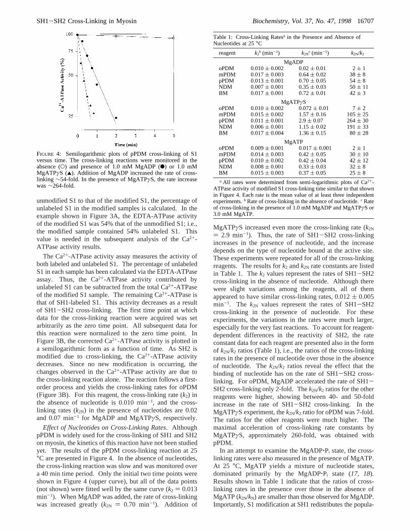
Effect of Nucleotides on Cross-Linking Rates. AlthoughpPDM is widely used for the cross-linking of SH1 and SH2on myosin, the kinetics of this reaction have not been studiedyet. The results of the pPDM cross-linking reaction at 25°C are presented in Figure 4. In the absence of nucleotides,the cross-linking reaction was slow and was monitored overa 40 min time period. Only the initial two time points wereshown in Figure 4 (upper curve), but all of the data points(not shown) were fitted well by the same curve (k2 ) 0.013min-1). When MgADP was added, the rate of cross-linkingwas increased greatly (k2N ) 0.70 min-1). Addition of
MgATPγS increased even more the cross-linking rate (k2N
) 2.9 min-1). Thus, the rate of SH1-SH2 cross-linkingincreases in the presence of nucleotide, and the increasedepends on the type of nucleotide bound at the active site.These experiments were repeated for all of the cross-linkingreagents. The results fork2 andk2N rate constants are listedin Table 1. Thek2 values represent the rates of SH1-SH2cross-linking in the absence of nucleotide. Although therewere slight variations among the reagents, all of themappeared to have similar cross-linking rates, 0.012( 0.005min-1. The k2N values represent the rates of SH1-SH2cross-linking in the presence of nucleotide. For theseexperiments, the variations in the rates were much larger,especially for the very fast reactions. To account for reagent-dependent differences in the reactivity of SH2, the rateconstant data for each reagent are presented also in the formof k2N/k2 ratios (Table 1), i.e., the ratios of the cross-linkingrates in the presence of nucleotide over those in the absenceof nucleotide. Thek2N/k2 ratios reveal the effect that thebinding of nucleotide has on the rate of SH1-SH2 cross-linking. For oPDM, MgADP accelerated the rate of SH1-SH2 cross-linking only 2-fold. Thek2N/k2 ratios for the otherreagents were higher, showing between 40- and 50-foldincrease in the rate of SH1-SH2 cross-linking. In theMgATPγS experiment, thek2N/k2 ratio for oPDM was 7-fold.The ratios for the other reagents were much higher. Themaximal acceleration of cross-linking rate constants byMgATPγS, approximately 260-fold, was obtained withpPDM.
In an attempt to examine the MgADP‚Pi state, the cross-linking rates were also measured in the presence of MgATP.At 25 °C, MgATP yields a mixture of nucleotide states,dominated primarily by the MgADP‚Pi state (17, 18).Results shown in Table 1 indicate that the ratios of cross-linking rates in the presence over those in the absence ofMgATP (k2N/kN) are smaller than those observed for MgADP.Importantly, S1 modification at SH1 redistributes the popula-
FIGURE 4: Semilogarithmic plots of pPDM cross-linking of S1versus time. The cross-linking reactions were monitored in theabsence (O) and presence of 1.0 mM MgADP (b) or 1.0 mMMgATPγS (2). Addition of MgADP increased the rate of cross-linking ∼54-fold. In the presence of MgATPγS, the rate increasewas∼264-fold.
Table 1: Cross-Linking Ratesa in the Presence and Absence ofNucleotides at 25°C
reagent k2b (min-1) k2N
c (min-1) k2N/k2
MgADPoPDM 0.010( 0.002 0.02( 0.01 2( 1mPDM 0.017( 0.003 0.64( 0.02 38( 8pPDM 0.013( 0.001 0.70( 0.05 54( 8NDM 0.007( 0.001 0.35( 0.03 50( 11BM 0.017( 0.001 0.72( 0.01 42( 3
MgATPγSoPDM 0.010( 0.002 0.072( 0.01 7( 2mPDM 0.015( 0.002 1.57( 0.16 105( 25pPDM 0.011( 0.001 2.9( 0.07 264( 30NDM 0.006( 0.001 1.15( 0.02 191( 33BM 0.017( 0.004 1.36( 0.15 80( 28
MgATPoPDM 0.009( 0.001 0.017( 0.001 2( 1mPDM 0.014( 0.003 0.42( 0.05 30( 10pPDM 0.010( 0.002 0.42( 0.04 42( 12NDM 0.008( 0.001 0.33( 0.03 32( 8BM 0.015( 0.003 0.37( 0.05 25( 8a All rates were determined from semi-logarithmic plots of Ca2+-
ATPase activity of modified S1 cross-linking time similar to that shownin Figure 4. Each rate is the mean value of at least three independentexperiments.b Rate of cross-linking in the absence of nucleotide.c Rateof cross-linking in the presence of 1.0 mM MgADP and MgATPγS or3.0 mM MgATP.
SH1-SH2 Cross-Linking in Myosin Biochemistry, Vol. 37, No. 47, 199816707

tion of transition states toward the MgATP state (19).Clearly, without such information for the reagents used inthis study, it is difficult to determine how much each of thesestates contributes to the overall effect on the cross-linking.However, in view of such redistribution of states, for theratios obtained in the MgATP experiments to be smaller thanthose seen in both the MgATPγS and MgADP states, theMgADP‚Pi state must either inhibit or have no effect on theSH1-SH2 cross-linking.
SH2 Modification Reaction. To test for any potentialdifferences in the reactivity of the reagents toward SH2 inthe presence of nucleotides (MgADP or MgATPγS), mono-functional labeling of SH2 was examined as well. The rateof SH2 modification was measured by preblocking SH1 withNEM and then following the modification of SH2 for eachreagent. The results of these experiments are presented inTable 2. The rate of SH2 modification (k2) for oPDM was0.007 min-1. For the other reagents, the rates (k2) wereapproximately 0.03 min-1, and there was little variationamong the individual reagents. In the presence of nucleotide,the rate of SH2 modification (k2N) by oPDM increased to0.025 min-1 for MgADP and 0.038 min-1 for MgATPγS.For the other reagents, the rates (k2N) were also increased inthe presence of nucleotide, to 0.133 min-1 for MgADP and0.230 min-1 for MgATPγS. Although the individual ratesfor oPDM and all the other reagents were different, the ratiosof rates (k2N/k2) were similar for all of the reagents (3-4-fold for MgADP and 5-7-fold for MgATPγS.
DISCUSSION
It has been shown in previous studies on SH1 and SH2that the binding of nucleotide to S1 accelerates both themodification and cross-linking of these two cysteines (6-9,11, 12, 20). Reagents with spans of up to 14 Å and downto 3 Å were used to cross-link SH1 to SH2. It is evenpossible for SH1 and SH2 to come close enough to oneanother to form a disulfide bond (9). However, for thisdisulfide bond formation to occur, nucleotides must bepresent and the reaction requires a time span of 24 h or more.Cross-linking with shorter reagents such as F2DNB (1,5-difluoro-2,4-dinitrobenzene) also requires the presence ofnucleotides and long reaction times (7, 8). Because nosystematic method of evaluating the rates of cross-linking
reactions has been used so far, these reactions may besampling infrequent, extreme, and low probability conforma-tions of the helix.
From the crystal structure of S1, SH1 and SH2 arecalculated to be approximately 19 Å apart. To accommodatefor the above cross-linking reactions, the SH1-SH2 helixmust be undergoing drastic changes. At least two speculativeexplanations can be suggested to describe what might beoccurring in the helix. The helix or a part of it may bemelting upon nucleotide binding, allowing SH1 and SH2 toapproach each other. Alternatively, the helix may haveincreased flexibility in the presence of nucleotides. Thisincreased flexibility could possibly be caused by rotationsabout glycine residues (699, 703, and 710) present in thesequence of the SH1-SH2 helix. Kinose et al. (21) andPatterson et al. (22) have recently shown that a mutation ofthe Gly699 residue (Gly680 inDictyostelium discoideummyosin) severely hinders the motor function of myosin. Thisconserved residue is therefore necessary and may be requiredto maintain the rotational flexibility of the helix. The goalof this work was to explore the events occurring in the SH1-SH2 helix by kinetic analysis of the cross-linking of thesecysteine groups.
Methodological Considerations. In this study, a newapproach was taken to examine the cross-linking of SH1 andSH2. By isolating the cross-linking event, it was possibleto determine what kinetic effect the nucleotides had on SH1-SH2 cross-linking. Several maleimide-based cross-linkingreagents of different spans (5-15 Å) were used, allowingus to probe the distances separating SH1 and SH2 and todetermine how the distances changed upon nucleotidebinding.
One concern in the experiments was to minimize thepossibility of monofunctional SH2 labeling. Several mea-sures were taken to eliminate such a possibility. Theseinclude low temperature (4°C), lower pH (pH 7.0), and lowmolar ratio of reagent to S1 (1.5:1.0). Until SH1 is labeled,the SH2 group under such conditions is not accessible tomost reagents, including phenylmaleimide, a monfunctionalequivalent of the dimaleimide cross-linking reagents (12, 23,24). Any monofunctional SH2 labeling on S1 carrying across-linking reagent attached to SH1 was unlikely becauseof the rapid removal of excess reagent and the relatively slowrates of such labeling (Table 2). However, even if it occursto some extent, such labeling, as well as the SH1-SH2 cross-linking completed prior to the first assay point of the cross-linking reaction, would be inconsequential to the subsequentkinetic analysis of the cross-linking. S1 labeled at both SH1and SH2 and the cross-linked S1 have no ATPase activity.Thus, after correction of the activity for the contribution dueto the unlabeled S1, the remaining Ca2+-ATPase representedonly SH1-labeled S1, and changes in the Ca2+-ATPase werea result of the SH1-SH2 cross-linking. From experimentto experiment, the population of SH1-labeled S1 availablefor monitoring the SH1-SH2 cross-linking varied from 30to 50%. However, the cross-linking rates determined fromsuch different experiments were reproducible irrespective ofthe fraction of SH1-labeled S1. The data acquired from thisstudy can be used to describe the changes in the SH1-SH2helix due to nucleotide binding.
Significance of SH1-SH2 Cross-Linking Rates. As ex-pected, our results show that the addition of nucleotide to
Table 2: SH2 Modification Ratesa in the Presence and Absence ofNucleotides at 25°C
reagent k2b (min-1) k2N
c (min-1) k2N/k2
MgADPoPDM 0.007( 0.0005 0.025( 0.002 3.6( 0.5mPDM, pPDM,
NDM, BM0.031( 0.014 0.133( 0.030 4.3( 1.9
MgATPγSoPDM 0.007( 0.0005 0.038( 0.005 5.4( 1.1mPDM, pPDM,
NDM, BM0.031( 0.014 0.230( 0.070 7.4( 3.3
a All rates were obtained from semi-logarithmic plots of the Ca2+-ATPase activity of modified S1 versus time of reaction. The rates arethe mean values of at least two independent experiments for eachreagent. For mPDM, pPDM, NDM, and BM, the rates were similarand are, therefore, the mean values for the four reagents.b Rate of SH2modification in the absence of nucleotide.c Rate of SH2 modificationin the presence of 1.0 mM nucleotide.
16708 Biochemistry, Vol. 37, No. 47, 1998 Nitao and Reisler
the SH1-SH2 cross-linking reactions increases the rate ofsuch reactions. The increases, however, depended on thereagent used. This can be attributed to either the differentspans of the reagents or the differing reactivities of thereagents toward SH2 in the presence of nucleotide. Toensure that the effects of nucleotide were due to the reagentspan and not the reactivity, the rate of SH2 modification wasdetermined for each reagent. The individual rates in thepresence and absence of nucleotides (MgADP or MgATPγS)differed for oPDM and the other reagents, but the effect ofnucleotide on the rates on SH2 labeling was similar for allof the reagents. This important conclusion is documentedby thek2N/k2 ratios shown in Table 2, which reveal very littleif any reagent-dependent variation for the SH2 labeling. Incontrast to that, thek2N/k2 ratios determined for the SH1-SH2 cross-linking depend strongly on the reagent length(Table 1). The ratio representation of cross-linking ratesfactors out any differences in SH2 reactivities towardreagents and corrects for experimental variation in the three-step cross-linking reaction.
The overall picture emerging fromk2 data in Table 1 isthat of S1 equilibrium between multiple states of the SH1-SH2 helix, with a relatively low probability for the statescorresponding to any of the distances probed by thebifunctional reagents (5-15 Å). As judged fromk2N data,nucleotides cause a dramatic shift in these equilibria,increasing drastically the population of helix states coveringSH1-SH2 distances between 9 and 15 Å. Thus, resultspresented in the form ofk2N/k2 ratios are indicative of changesin the population of helix states (i.e., distances between SH1and SH2) as a result of nucleotide binding.
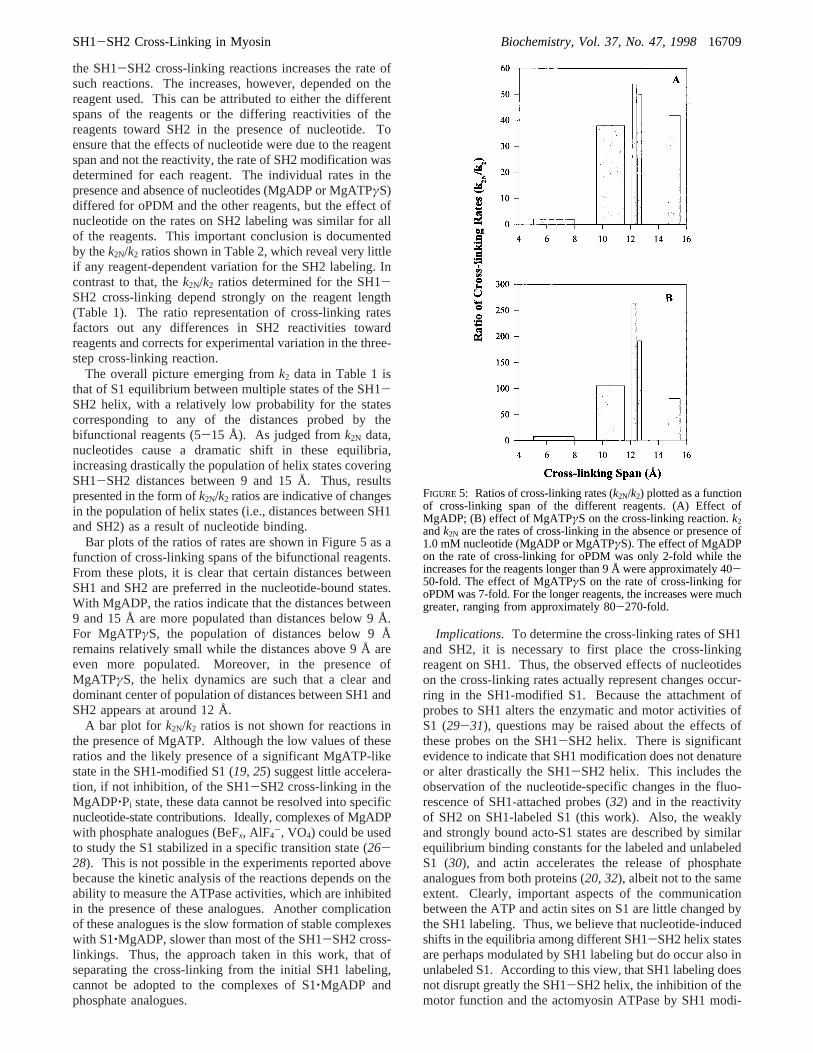
Bar plots of the ratios of rates are shown in Figure 5 as afunction of cross-linking spans of the bifunctional reagents.From these plots, it is clear that certain distances betweenSH1 and SH2 are preferred in the nucleotide-bound states.With MgADP, the ratios indicate that the distances between9 and 15 Å are more populated than distances below 9 Å.For MgATPγS, the population of distances below 9 Åremains relatively small while the distances above 9 Å areeven more populated. Moreover, in the presence ofMgATPγS, the helix dynamics are such that a clear anddominant center of population of distances between SH1 andSH2 appears at around 12 Å.
A bar plot for k2N/k2 ratios is not shown for reactions inthe presence of MgATP. Although the low values of theseratios and the likely presence of a significant MgATP-likestate in the SH1-modified S1 (19, 25) suggest little accelera-tion, if not inhibition, of the SH1-SH2 cross-linking in theMgADP‚Pi state, these data cannot be resolved into specificnucleotide-state contributions. Ideally, complexes of MgADPwith phosphate analogues (BeFx, AlF4
-, VO4) could be usedto study the S1 stabilized in a specific transition state (26-28). This is not possible in the experiments reported abovebecause the kinetic analysis of the reactions depends on theability to measure the ATPase activities, which are inhibitedin the presence of these analogues. Another complicationof these analogues is the slow formation of stable complexeswith S1‚MgADP, slower than most of the SH1-SH2 cross-linkings. Thus, the approach taken in this work, that ofseparating the cross-linking from the initial SH1 labeling,cannot be adopted to the complexes of S1‚MgADP andphosphate analogues.
Implications. To determine the cross-linking rates of SH1and SH2, it is necessary to first place the cross-linkingreagent on SH1. Thus, the observed effects of nucleotideson the cross-linking rates actually represent changes occur-ring in the SH1-modified S1. Because the attachment ofprobes to SH1 alters the enzymatic and motor activities ofS1 (29-31), questions may be raised about the effects ofthese probes on the SH1-SH2 helix. There is significantevidence to indicate that SH1 modification does not denatureor alter drastically the SH1-SH2 helix. This includes theobservation of the nucleotide-specific changes in the fluo-rescence of SH1-attached probes (32) and in the reactivityof SH2 on SH1-labeled S1 (this work). Also, the weaklyand strongly bound acto-S1 states are described by similarequilibrium binding constants for the labeled and unlabeledS1 (30), and actin accelerates the release of phosphateanalogues from both proteins (20, 32), albeit not to the sameextent. Clearly, important aspects of the communicationbetween the ATP and actin sites on S1 are little changed bythe SH1 labeling. Thus, we believe that nucleotide-inducedshifts in the equilibria among different SH1-SH2 helix statesare perhaps modulated by SH1 labeling but do occur also inunlabeled S1. According to this view, that SH1 labeling doesnot disrupt greatly the SH1-SH2 helix, the inhibition of themotor function and the actomyosin ATPase by SH1 modi-
FIGURE 5: Ratios of cross-linking rates (k2N/k2) plotted as a functionof cross-linking span of the different reagents. (A) Effect ofMgADP; (B) effect of MgATPγS on the cross-linking reaction.k2andk2N are the rates of cross-linking in the absence or presence of1.0 mM nucleotide (MgADP or MgATPγS). The effect of MgADPon the rate of cross-linking for oPDM was only 2-fold while theincreases for the reagents longer than 9 Å were approximately 40-50-fold. The effect of MgATPγS on the rate of cross-linking foroPDM was 7-fold. For the longer reagents, the increases were muchgreater, ranging from approximately 80-270-fold.
SH1-SH2 Cross-Linking in Myosin Biochemistry, Vol. 37, No. 47, 199816709
fications would arise from the uncoupling of the helix fromstructural elements of S1 involved in these functions.
There may be several reasons why different states of thehelix are not visualized in the X-ray structures of S1dC (2,3, 10). Among them could be different flexibilities of thishelix (because of different interactions with other structuralelements in rabbit andDictyosteliumS1), the effect of thelever arm truncation in S1dC on the cysteine helix, and thetrapping of a particular helix state by the crystallizationprocess. Assuming that the lever arm of S1 can be viewedas a rigid body (obviously a simplification), the multiplestates of the helix, which require its pivoting around glycineresidues, would result in variable orientations of the leverarm with respect to the catalytic domain (33). The disorderedstate of S1 weakly bound to actin, frequently observed instructural and spectroscopic studies, may be a simplereflection of that effect. A discussion of possible implica-tions of such transitions in the SH1-SH2 helix and the leverarm to the cross-bridge cycle must await a better analysis ofthe helix in the S1‚ADP‚Pi state, as well as information onits dynamic properties in the ternary complex of S1 withactin and MgADP. Such experiments and simulations oflever arm states as a function of SH1-SH2 helix transitionsare part of the subsequent studies.
ACKNOWLEDGMENT
We thank Dr. Miguel Garcia-Garibay for his help withthe cross-linking span determinations and Dr. AndreyBobkov for his many helpful discussions.
REFERENCES
1. Bagshaw, C. R., and Trentham, D. R. (1975)J. Supramol.Struct. 3, 315-322.
2. Fisher, A., Smith, C., Thoden, J., Smith, R., Sutoh, K., Holden,H., and Rayment, I. (1995)Biochemistry 34, 8960-8972.
3. Gulick, A. M., Bauer, C. B., Thoden, J. B., and Rayment, I.(1997)Biochemistry 36, 11619-11628.
4. Smith, C. A., and Rayment, I. (1996)Biochemistry 35, 5404-5417.
5. Rayment, I. (1996)J. Biol. Chem. 271, 15850-15853.6. Reisler, E., Burke, M., Himmelfarb, S., and Harrington, W.
F. (1974)Biochemistry 13, 3837-3840.7. Burke, M., and Reisler, E. (1977)Biochemistry 16, 5559-
5563.
8. Wells, J. A., Knoeber, C., Sheldon, M. C., Werber, M. M.,and Yount, R. G. (1980)J. Biol. Chem. 255, 11135-11140.
9. Wells, J. A., and Yount, R. G. (1980)Biochemistry 19, 1711-1717.
10. Rayment, I., Rypniewski, W., Schmidt-Base, K., Smith, R.,Tomchick, D., Benning, M., Winkelmann, D., Wesenberg, G.,and Holden, H. (1993)Science 261, 50-58.
11. Miller, L., Coppedge, J., and Reisler, E. (1982)Biochem.Biophys. Res. Commun. 106, 117-122.
12. Polosukhina, K., and Highsmith, S. (1997)Biochemistry 36,11952-11958.
13. Godfrey, J. E., and Harrington, W. F. (1970)Biochemistry 9,886-895.
14. Weeds, A., and Pope, B. (1977)J. Mol. Biol. 111, 129-157.15. Fiske, C. H., and Subbarow, Y. (1925)J. Biol. Chem. 60, 375-
400.16. Sekine, T., Barnett, L. M., and Kielley, W. W. (1962)J. Biol.
Chem. 237, 2769-2772.17. Trentham, D. R., Eccleston, J. F., and Bagshaw, C. R. (1976)
Q. ReV. Biophys. 9, 217-281.18. Cooke, R. (1997)Physiol. ReV. 77, 671-697.19. Ostap, E. M., White, H. D., and Thomas, D. D. (1993)
Biochemistry 32, 6712-6720.20. Phan, B. C., Peyser, Y. M., Reisler, E., and Muhlrad, A. (1997)
Eur. J. Biochem. 243, 636-642.21. Kinose, F., Wang, S. X., Kidambi, U. S., Moncman, C. L.,
and Winkelmann, D. A. (1996)J. Cell Biol. 134, 895-909.22. Patterson, B., Ruppel, K. M., Wu, Y., and Spudich, J. A. (1997)
J. Biol. Chem. 272, 27612-27617.23. Reisler, E. (1982)Methods Enzymol. 85 Pt. B, 84-93.24. Xie, L., Li, W. X., Barnett, V. A., and Schoenberg, M. (1997)
Biophys. J. 72, 858-865.25. Sleep, J. A., Trybus, K. M., Johnson, K. A., and Taylor, E.
W. (1981)J. Muscle Res. Cell Motil. 2, 373-399.26. Goodno, C. C., and Taylor, E. W. (1982)Proc. Natl. Acad.
Sci. U.S.A. 79, 21-25.27. Phan, B. C., Faller, L. D., and Reisler, E. (1993)Biochemistry
32, 7712-7719.28. Maruta, S., Henry, G. D., Sykes, B. D., and Ikebe, M. (1993)
J. Biol. Chem. 268, 7093-7100.29. Marriott, G., and Heidecker, M. (1996)Biochemistry 35,
3170-3174.30. Bobkov, A. A., Bobkova, E. A., Homsher, E., and Reisler, E.
(1997)Biochemistry 36, 7733-7738.31. Root, D. D., and Reisler, E. (1992)Biophys J. 63, 730-740.32. Phan, B. C., Cheung, P., Stafford, W. F., and Reisler, E. (1996)
Biophys. Chem. 59, 341-349.33. Burghardt, T. P., Garamszegi, S. P., Park, S., and Ajtai, K.
(1998)Biochemistry 37, 8035-8047.
BI9817212
16710 Biochemistry, Vol. 37, No. 47, 1998 Nitao and Reisler