priyatharisni kaniappan universiti sains malaysia...
TRANSCRIPT

DEVELOPMENT OF A NUCLEIC ACID AMPLIFICATION TEST FOR THE DETECTION OF
MYCOBACTERIUM TUBERCULOSIS
PRIYATHARISNI KANIAPPAN
UNIVERSITI SAINS MALAYSIA
2015

DEVELOPMENT OF A NUCLEIC ACID AMPLIFICATION
TEST FOR THE DETECTION OF MYCOBACTERIUM TUBERCULOSIS
By
PRIYATHARISNI KANIAPPAN
Thesis submitted in fulfillment of the requirements for the degree of Master of Science
UNIVERSITI SAINS MALAYSIA
OCTOBER 2015

ii
ACKNOWLEDGEMENTS
First and foremost, I would like to express my gratitude to my supervisor,
Professor Dr. Tang Thean Hock, for providing me the opportunity to pursue MSc.
Degree under his supervision. His guidance, patience and constructive criticism have
always been supportive at all times during my work. I am very deeply indebted to my
field supervisor, Datin Dr Ganeswrie Rajasekaram, Hospital Sultanah Aminah Johor
Bahru for her advice and continuous support and all insights.
I thank the Infectomic Cluster, Advanced Diagnostic Laboratory (AMDI) and
Microbiology Unit of Hospital Sultanah Aminah for providing the bacterial cultures,
which has contributed significantly to this work. I am thankful to the Ministry of Health
(MOH) for awading me scholarship ‘Hadiah Latihan Persekutuan’. I am grateful to the
management staffs of AMDI, USM who rendered me with valuable experiences outside
of the laboratory. I would also like to thank the family of Infectomic Cluster, AMDI, in
particular Mdm Siti Aminah, Dr. Hoe Chee Hock, Dr. Citartan Marimuthu, Ms. Lee Li
Pin, Ms. Thiviyaa Othaya Kumar, Ms. Nitya Ravichandran and Mr. Emmanuel Jayaraj
Moses for their assistance and support.
A huge thank you to my other half, Mr. Nageswaran Muniandy for
understanding my ambition and my inner need to pursue higher education. Last but not
least, I am grateful to the Almighty God for all His guidance, blessings, lessons and
‘companionship’ when I was away from my family to pursue my MSc. Words alone
can’t explain the degree of gratitude towards my family for showering me with love,
care and motivations for me to strive and complete this work successfully. This thesis
would not have been possible without their supports. My prayers for my late friend and
housemate Ms.Shailaja Balasubramaniam Menon whom is with angels right now.
Lastly, I offer my sincerest thanks to my friends and to those who supported me in any
respect during the completion of the project.

iii
TABLE OF CONTENTS Page
ACKNOWLEDGEMENTS ............................................................................................ ii
TABLE OF CONTENTS ............................................................................................... iii
LIST OF TABLES ........................................................................................................ vii
LIST OF FIGURES ..................................................................................................... viii
LIST OF ABBREVIATIONS AND SYMBOLS ........................................................... x
PEMBANGUNAN UJIAN AMPLIFIKASI ASID NUKLEIK BAGI TUJUAN
PENGESANAN KUMAN Mycobacterium tuberculosis ............................................. xiv
ABSTRAK ..................................................................................................................... xiv
ABSTRACT ................................................................................................................... xvi
CHAPTER 1: .................................................................................................................... 1
INTRODUCTION ............................................................................................................ 1
1.1. Mycobacteria ........................................................................................................... 1
1.2 Non Tuberculous Mycobacteria (NTM) .................................................................. 1
1.3 M. leprae .................................................................................................................. 2
1.4 M. tuberculosis complex (MTBC) ............................................................................ 2
1.5. Mycobacterium tuberculosis ................................................................................... 5
1.5.1. Cell wall of M. tuberculosis confers pathogenesis and acid fast property ....... 5
1.5.2 Transmission and immune response upon TB infection .................................. 7

iv
1.5.3 Epidemiology of Tuberculosis .......................................................................... 8
1.6 Diagnostic of Tuberculosis .................................................................................... 11
1.6.1 Smear Microscopy .......................................................................................... 11
1.6.2 Culture method ................................................................................................ 14
1.6.3 Nucleic acid-based detection of M. tuberculosis ............................................ 16
1.7 Polymerase chain reaction (PCR) .......................................................................... 16
1.7.1 Non-protein coding RNAs (npcRNA) -Potential Diagnostic marker of PCR-
based detection of M. tuberculosis ........................................................................... 17
1.7.2 IS6110, the widely employed gene target for PCR-based detection of
M. tuberculosis ......................................................................................................... 18
1.8. Research objectives ............................................................................................... 21
CHAPTER 2 ................................................................................................................... 23
MATERIALS AND METHODS .................................................................................. 23
2.1 Materials ................................................................................................................. 23
2.1.1 Chemicals ........................................................................................................ 23
2.1.2 Culture and bacterial strains ......................................................................... 24
2.1.3 Plasmid ............................................................................................................ 26
2.2 Methods .................................................................................................................. 27
2.2.1 Cultivation of Mycobacteria ........................................................................... 27
2.2.2 Bacterial culture .............................................................................................. 27

v
2.2.3 Mycobacterial Genomic DNA extraction ....................................................... 28
2.2.4 Bacterial DNA Extraction ............................................................................... 29
2.2.5 Bioinformatic-based search of npcRNA genes specific for M. tuberculosis .. 30
2.2.6 Primer Design .................................................................................................. 30
2.3 PCR assay ............................................................................................................... 30
2.3.1 Gel electrophoresis .......................................................................................... 31
2.4 Specificity of the mPCR Assay .............................................................................. 31
2.5 Sensitivity of the mPCR Assay .............................................................................. 32
2.6 Validation of the mPCR ......................................................................................... 32
CHAPTER 3 ................................................................................................................... 33
RESULTS AND DISCUSSION .................................................................................... 33
3.1. npcTB 6715 specific for MTBC is chosen as the diagnostic marker for mPCR
assay ............................................................................................................................. 34
3.2. IS6110 , en masse with npcTB 6715, as the diagnostic markers for the mPCR
assay ............................................................................................................................. 40
3.2. IS6110 , en masse with npcTB 6715, as the diagnostic markers for the mPCR
assay ............................................................................................................................. 40
3.3. Development of mPCR assay for the detection of M. tuberculosis using IS6110
and npcTB 6715 as the target ....................................................................................... 43
3.3.1. Optimization of the mPCR ............................................................................. 43
3.3.1.1. Optimization of annealing temperature by Gradient PCR ...................... 44

vi
3.3.1.2 Primer Concentration Optimization ......................................................... 46
3.3.1.3 Incorporation of Amplification Internal Control (AIC) ........................... 48
3.3.1.4. Optimization of deoxyribonucleotide triphosphate (dNTPs) .................. 50
3.3.1.5. Optimization of MgCl2 ............................................................................ 52
3.3.2. Final optimized PCR parameters ................................................................... 54
3.4. Determination of the specificity of the mPCR ...................................................... 57
3.5. Determination of the sensitivity of the mPCR ...................................................... 60
3.6. Validation of the developed mPCR with culture positive samples ....................... 63
CHAPTER 4: CONCLUSION ...................................................................................... 69
REFERENCE ................................................................................................................. 71
APPENDICES A: Preparation of Buffers and Reagents ............................................. 88

vii
LIST OF TABLES
Page
Table 3.1 : Primers suggested for npcTB 6715 39
Table 3.2 : Primers suggested for IS6110 42
Table 3.3 : Final optimized PCR parameters 55
Table 3.4 : Possible outcome of mPCR and the interpretations 64
Table 3.5 : Performance of npcTB 6715 and IS6110 in 500
Mycobacteria culture
64
Table 3.6 : Performance of Genotype Mycobacterium CM/AS in 500
Mycobacteria culture positive sample
65
Table 3.7 : Performance of mPCR (npcTB6715 and IS6110) compared
to LPA assay in 500 Mycobacteria culture
65

viii
LIST OF FIGURES
Figure 1.1 : Phylogenetic linage of non-tubercoulosis mycobacteria,
M. leprae and M. tuberculosis complex
4
Figure 1.2 : Mycobacterial cell wall 6
Figure 1.3 (A) : Estimated new TB rates for year 2012 10
Figure 1.3 (B) : TB Mortality Rate in Malaysia, 1990-2012 10
Figure 1.4 (A) : Ziehl-Neelsen Stain 13
Figure 1.4 (B) : Auramine-Rhodamine Stain 13
Figure 1.5 (A) : M. tuberculosis colony appearance 15
Figure 1.5 (B) : M. tuberculosis colony on Lowenstein-Jensen media 15
Figure 1.6 : Diagram of IS6110 20
Figure 3.1 : Percentage of potential non-coding RNA contig sequence
subjected to BlastN
36
Figure 3.2 : Sequence of npcTB 6715 for M. tuberculosis 37
Figure 3.3: : The genomic location and orientation of non-coding
RNA gene npcTB 6715 in M. tuberculosis H37Rv
38
Figure 3.4: : A graphical representation showing different target
regions used for PCR detection of M. tuberculosis.
41
ix
Figure 3.5 (A) : Gradient PCR optimization IS6110 45
Figure 3.5 (B) : Gradient PCR optimization npcTB 6715 45
Figure 3.5 (C) : Gradient PCR optimization npcTB 6715 Internal
Amplification Control (AIC).
45
Figure 3.6 : Optimization of primer amounts of IS6110 and npcTB
6715 in multiplex PCR
47
Figure 3.7 : Optimization of the amount of AIC in the mPCR assay 49
Figure 3.8 : Agarose gel electrophoresis of multiplex PCR using
different concentration of dNTPs
51
Figure 3.9 : Optimization of the amount of MgCl2 53
Figure 3.10 : Agarose gel electrophoresis of the multiplex PCR using
the final optimized multiplex reaction
56
Figure 3.11 (A) : Specificity determination of the mPCR assay 58
Figure 3.11 (B) : Specificity determination of the mPCR assay 58
Figure 3.11 (C) : Specificity determination of the mPCR assay 58
Figure 3.12 : Sensitivity determination of the mPCR assay 62

x
LIST OF ABBREVIATIONS AND SYMBOLS
A Adenine
bp Base pair(s)
BLAST
Megablast
Basic Local Alignment Search Tool.
Mega Basic Local Alignment Search Tool
C Cytosine
°C Degrees Celsius
CFU Colony Forming Unit
dATP Deoxyadenosine triphosphate
dCTP Deoxycytidine triphosphate
ddH2O Double-distilled water
dGTP Deoxyguanosine triphosphate
DNA Deoxyribonucleic acid
dNTP Deoxyribonucleotide triphosphate
dsDNA Double-stranded DNA
dTTP Deoxythymidine triphosphate
EDTA Ethylenediaminetetraacetic Acid
ELISA Enzyme-Linked Immunosorbent Assay
et al. and others
EtBr 3, 8-diamino-5-Ethyl-6-phenyl phenanthridinium Bromide
g Gram
G Guanine

xi
GAPDH Glyceraldehyde-3-phospate-dehydrogenate
HCl Hydrochloric acid
H Hour (s)
IAC
KH2PO4
Internal Amplification Control
Potassium phosphate
KCl Potassium chloride
kDa Kilodalton
L Litre
LB Luria Bertani
M Mol/Liter, molar
mg Milligram
MgCl2 Magnesuim Chloride
min Minute (s)
mL Milliliter
mM
mRNA
mPCR
Millimolar
Messenger RNA
Multiplex Polymerase Chain Reaction
NaCl Sodium chloride
NaOAc. 3H2O Sodium acetate trihydrate
NaOH Sodium hydroxide
ng Nanogram
NH2HPO4 Sodium hydrogen phosphate
nt Nucleotide (s)
xii
OD Optical Density
npcRNA Non-protein-coding gene
PBS Phosphate buffered saline
PCR
pg
Polymerase Chain Reaction
Picogram
RBS Ribosomal Binding Site
RNA Ribonucleic acid
RNase Ribonuclease
RPM
rRNA
Revolutions per minute
Ribosomal RNA
RT Room temperature
sec Second (s)
SDS Sodium dodecyl sulfate
siRNA
sRNA
Small interfering RNA
Small RNA
ssDNA Single-stranded DNA
T Thymine
TAE Tris–Acetic Acid–EDTA
Tris
Tris-HCl
Tris-(Hydroxymethyl)-Aminomethane
Tris-(Hydroxymethyl)-Aminomethane Hydrocloride
tRNA Transfer RNA
UV Ultraviolet
V Volt (s)

xiii
v/v Volume per volume
XLD Xylose Lysine Deoxycholate
µL Micro liter
µM Micro molar
%
β
Percentage
Beta
xiv
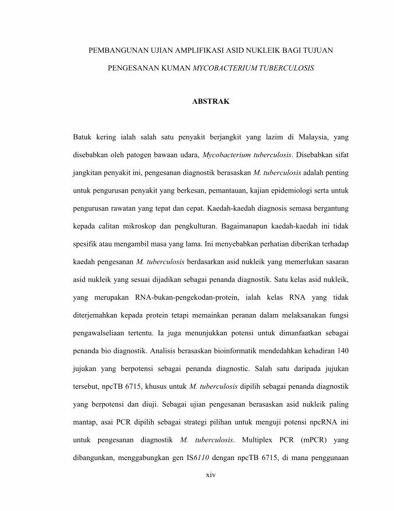
PEMBANGUNAN UJIAN AMPLIFIKASI ASID NUKLEIK BAGI TUJUAN
PENGESANAN KUMAN MYCOBACTERIUM TUBERCULOSIS
ABSTRAK
Batuk kering ialah salah satu penyakit berjangkit yang lazim di Malaysia, yang
disebabkan oleh patogen bawaan udara, Mycobacterium tuberculosis. Disebabkan sifat
jangkitan penyakit ini, pengesanan diagnostik berasaskan M. tuberculosis adalah penting
untuk pengurusan penyakit yang berkesan, pemantauan, kajian epidemiologi serta untuk
pengurusan rawatan yang tepat dan cepat. Kaedah-kaedah diagnosis semasa bergantung
kepada calitan mikroskop dan pengkulturan. Bagaimanapun kaedah-kaedah ini tidak
spesifik atau mengambil masa yang lama. Ini menyebabkan perhatian diberikan terhadap
kaedah pengesanan M. tuberculosis berdasarkan asid nukleik yang memerlukan sasaran
asid nukleik yang sesuai dijadikan sebagai penanda diagnostik. Satu kelas asid nukleik,
yang merupakan RNA-bukan-pengekodan-protein, ialah kelas RNA yang tidak
diterjemahkan kepada protein tetapi memainkan peranan dalam melaksanakan fungsi
pengawalseliaan tertentu. Ia juga menunjukkan potensi untuk dimanfaatkan sebagai
penanda bio diagnostik. Analisis berasaskan bioinformatik mendedahkan kehadiran 140
jujukan yang berpotensi sebagai penanda diagnostic. Salah satu daripada jujukan
tersebut, npcTB 6715, khusus untuk M. tuberculosis dipilih sebagai penanda diagnostik
yang berpotensi dan diuji. Sebagai ujian pengesanan berasaskan asid nukleik paling
mantap, asai PCR dipilih sebagai strategi pilihan untuk menguji potensi npcRNA ini
untuk pengesanan diagnostik M. tuberculosis. Multiplex PCR (mPCR) yang
dibangunkan, menggabungkan gen IS6110 dengan npcTB 6715, di mana penggunaan

xv
npcRNA ini mampu mengesan M. tuberculosis yang tidak mempunyai gen IS6110,
sekaligus mampu mengelakkan keputusan negatif palsu. mPCR ini menunjukkan had
pengesanan sebanyak 10 pg. Evaluasi mPCR dengan 500 kultur Mycobacterium
menunjukkan nilai-nilai 98.4%, 96.1%, 98.6% dan 95.4% masing-masing untuk
kepekaan, kekhususan, Nilai Ramalan Positif dan Nilai Ramalan Negatif. Nilai-nilai
yang diperolehi mencadangkan potensi diagnostik mPCR, sebagai ujian diagnostik
untuk mengesan M. tuberculosis dan menyokong potensi diagnostik npcTB 6715.

xvi
DEVELOPMENT OF A NUCLEIC ACID AMPLIFICATION TEST FOR THE
DETECTION OF MYCOBACTERIUM TUBERCULOSIS
ABSTRACT
Tuberculosis is one of the common infectious diseases in Malaysia, caused by
the airborne pathogen, Mycobacterium tuberculosis. Due to the infectious nature of the
disease, diagnostic-based detection of Mycobacterium tuberculosis is vital for effective
disease management, surveillance, epidemiological purpose and for the administration of
a prompt treatment. Current diagnostic methods rely on smear microscopy and culture
method; however these strategies suffer from several pitfalls. This shifts the attention
towards the nucleic-acid based detection of M. tuberculosis, which requires the
identification of suitable nucleic acid target as the diagnostic marker. One novel class of
nucleic acid, which is the non-protein-coding RNAs, is a class of RNA that is not
translated into protein but perform certain regulatory functions, suggesting the
potentiality to be harnessed as a diagnostic biomarker. Bioinformatic-based analysis
revealed the presence of 140 npcRNA genes, one of it, npcTB 6715, which is specific
for M. tuberculosis is chosen as the potential diagnostic marker. As the most established
nucleic acid-based detection platform, PCR assay is chosen as the platform of choice to
test the potentiality of this npcRNA for the diagnostic-based detection of
M. tuberculosis. The PCR assay developed also incorporates IS6110 as the counterpart
of npcTB 6715, whereby the usage of the npcRNA is able to detect the IS6110-deficient
strains of M. tuberculosis, averting false-negative results. The multiplex PCR assay
developed showed a detection limit of 10 pg. Evaluation with 500 Mycobacterium
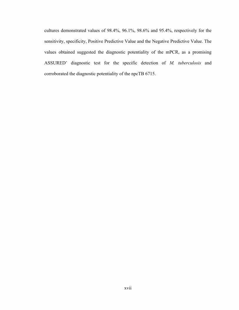
xvii
cultures demonstrated values of 98.4%, 96.1%, 98.6% and 95.4%, respectively for the
sensitivity, specificity, Positive Predictive Value and the Negative Predictive Value. The
values obtained suggested the diagnostic potentiality of the mPCR, as a promising
ASSURED’ diagnostic test for the specific detection of M. tuberculosis and
corroborated the diagnostic potentiality of the npcTB 6715.

1
CHAPTER 1:
INTRODUCTION
1.1. Mycobacteria
Mycobacteria are aerobic, non-motile, non-sporulation rod shaped
microorganisms, GC rich nucleotide containing microorganism with size range of 2-
4µm (length) and 0.5-0.2µm (wide). Classified under actinobacteria phylum,
mycobacteria are further categorized into the family of Mycobacteriaceae. There are
over a hundred species of Mycobacterium, which are genetically closely related as
reported by16S rRNA sequencing studies (Tortoli, 2006). Generally, mycobacteria can
be divided into three major categories (Figure 1.1): Mycobacterium tuberculosis
complex (MTBC), non-tuberculosis mycobacteria (NTM) and Mycobacterium leprae
(M. Leprae) (Jagielski et al., 2014).
1.2 Non Tuberculous Mycobacteria (NTM)
NTM are ubiquitous in the environment. Most of the NTM are non-pathogenic
organisms which are known to colonize water sources, soil, dust particles, and food
supplies. NTM can be cultivated on common liquid and solid culture media. Depending
on the growth period, they are divided into rapidly growing mycobacteria such as

2
Mycobacteria fortuitum, Mycobacteria chelonae, and Mycobacteria abscessus as well
as slow growing species such as Mycobacteria avium, Mycobacteria kansasii, and
Mycobacteria marinum. About 160 different species of NTM have been identified.
1.3 M. leprae
One of the earliest identified slow growing NTM is the M. leprae. M. leprae
causes leprosy (Hansen's disease) in human (Cole et al., 2001). M. leprae is an
intracellular, pleomorphic and acid-fast bacterium. It is an aerobic bacillus (rod-shaped)
surrounded by the characteristic waxy coating unique to mycobacteria. M. leprae is
different from other mycobacteria in terms of the arrangement in smear, as it is
arranged in parallel chains, just like cigarettes in a pack. Leprosy infections are mainly
focused on the skin and peripheral nerves. Therefore, their diagnoses are largely
established based on skin and neurologic examination of the patient.
1.4 M. tuberculosis complex (MTBC)
MTBC is a group of closely related mycobacterial species consisting of
M. tuberculosis, M. bovis, M. africanum, M. microti, M. canetti, M. caprae, M.
pinnipedii and M. mungi and M. suricattae.

3
M. bovis, the main cause of TB in bovine animal is known to cause cross species
infection from bovine to humans through consumption of contaminated milk (Cosivi et
al., 1998). Originating from the African sub-continent, M. africanum represents another
major cause of TB especially in Sub-Saharan Africa. However, reports on the presence
of M. africanum have emerged in countries other than Sub-Saharan Africa such as
England, Spain, and the United States (Desmond et al., 2004; Grange and Yates, 1989;
Remacha Esteras et al., 2003). On the other hand, M. microtii is a rare pathogen that
usually infects rodents and shrews (Cavanagh et al., 2002). M. canetti causes infection in
humans although the prevalence of this subspecies in not common. Out of the many
mycobacterial species of the MTBC, M. tuberculosis is the most common causative
agent of TB among humans.

4
Figure 1.1 Phylogenetic lineage of Non-Tubercoulosis Mycobacteria, M. leprae and M.
tuberculosis complex [Adapted from (Haning et al., 2014)].

5
1.5. Mycobacterium tuberculosis
M. tuberculosis is a non-motile, slender, straight, or slightly curved bacillus. It
is an obligate aerobic organism; hence it can only survive in an oxygen-containing
environment. M. tuberculosis bacilli are 2-4 micrometers in length and 0.2-0.5 µm in
width. The generation time of M. tuberculosis, in synthetic medium or infected animals
is approximately 24 hours (Ginsberg and Spigelman, 2007). The slow growing feature
of M. tuberculosis contributes to the chronic nature of the disease, which requires a
prolonged treatment. In addition, the compositional complexity of its cell wall could
also account for the nature of the disease, which represents a formidable impediment
for researchers and health care system.
1.5.1. Cell wall of M. tuberculosis confers pathogenesis and acid fast property
The cell wall of M. tuberculosis, is composed of peptidoglycan, arabinogalactan
and mycolic acids covalently linked to each other (Figure 1.2) (Chatterjee, 1997). Due
to the presence of mycolic acids, the primary stain used for gram staining does not
penetrate well into the cell wall, making it unsuitable for gram staining (Washington,
1996). As an alternative, acid-fast staining can be used to stain M. tuberculosis. The
stain forms a complex with the mycolic acids, thereby resist decolorization by acid
containing decolorization agent (Cheesbrough, 2006). These mycolic acids are also
responsible for the pathogenesis of TB (Forrellad et al., 2013).

6
Figure 1.2: Mycobacterial cell wall [Reproduced from (Medjahed et al., 2010)].

7
1.5.2 Transmission and immune response upon TB infection
TB infection spreads via droplets in the air expelled by an infected person during
common daily acts such as sneezing, coughing, laughing, and even talking. The amount
of bacilli present in the droplet, UV light exposure, ventilation factor and aerosolization
occurrence plays important role in transmission (American Thoracic Society and CDC,
2000). The spread of TB is enhanced by factors such as rapid increase in global travel,
malnutrition, overcrowding, and co-infection of HIV and TB.
TB infection is a tug-of-war between the host immune system and the
pathogen’s virulence. The first mechanism triggered upon the infection is the physical
defense mechanism, which sets to expel any foreign molecules from invading the
human body. Likewise, when a person inhales droplets containing M. tuberculosis
bacilli, they are trapped in the upper respiratory. Following this, the mucus secreting
Goblet cells on the respiratory tract would secrets mucus, trapping the pathogen and
pushing it upwards to be expelled from the system. However, in some cases, the M.
tuberculosis bacilli that managed to elude this physical defense system travels to the
alveoli in the lungs and is predisposed to another mechanism known as the innate
immune system. In this defense system, alveolar macrophage surrounds and phagocytes
the M. tuberculosis, attempting to destroy the bacilli.
At this stage, destruction of the bacilli is reliant on the interplay between
immunity of the host and the virulence of the bacilli itself (van Crevel et al., 2002). As
8
such, in immuno-compromised individual, viable M. tuberculosis that infects lungs
cause pulmonary tuberculosis. The classic clinical symptoms of TB is productive cough
that produces thick cloudy sputum for more than 2 weeks, chest pain, shortness of
breath, hemoptysis, fever, chills, night sweats, loss of appetite and sudden weight loss.
Apart from residing in the alveolus, viable M. tuberculosis can also travel to other
tissue via the lymphatic system causing extrapulmonary TB (Dale et al., 2003). In
addition, the infected individual also has the potential to incur infection on other
individuals. In contrast, in the case of Latent TB, the infected person is not responsible
for any transmission of TB. Only 5-10% of latent TB infection would develop into
active TB.
1.5.3 Epidemiology of Tuberculosis
World Health Organization (WHO) estimated that 8.6 million people were newly
infected by TB and 1.3 million deaths occurred due to TB infection in 2012 (WHO,
2013). Out of the 8.6 million TB cases, 2.9 million cases were women and 0.5 million
cases occurred among children. Over 400000 deaths and 74000 deaths were women and
children, respectively. Nearly 1 million deaths and 0.3 million deaths occurred among
non-HIV infected individuals and HIV infected individuals, respectively. Global
surveillance data indicated that most of the cases in 2012 occurred in Asia (58%) and
the African region (27%) [Figure 1.3(A)]. Additionally, 22 countries around the world
have been identified as 'High Burden' for TB and among them are the Asian countries
such as India, China, Indonesia, Thailand, Bangladesh, and Vietnam.
9
In Malaysia, according to the Ministry of Health Malaysia (MOHM), TB is one
of the major 5 communicable diseases for the past 15 years (MOHM, 1997-2013). TB
cases in Malaysia have steadily increased since 2005 and tend to rise further. In 2012,
TB incidence rate including among HIV infected individuals was estimated at 24000
(MOH, 2012). Mortality rates have been between 4 and 6% during the years from 1990
to 2011 [Figure 1.3(B)]. Approximately 1414 deaths were reported and occurred among
non-HIV infected individuals in 2012, with a mortality rate of 4.9% (WHO, 2014). Due
to this alarming rate of TB infection, MOHM aims to strengthen the case detection and
provide prompt treatment to TB infected individuals in Malaysia. In line with this, The
National TB Control Program is currently implementing 'National Strategic Plan to
Control TB, 2011-2015' to enhance all activities to reduce the burden of TB in the
country (MOH, 2012). An effective diagnostic method for efficient management of the
disease is also part of the endeavor to alleviate the problem of TB in the country.

10
(A)
(B)
Figure 1.3 (A): Estimated new TB rates for year 2012 (Adapted from Global
tuberculosis Report 2013) (B): TB Mortality Rate in Malaysia, 1990-2012 (Adapted
from Ministry of Health Malaysia Annual Report 2012).
11
1.6 Diagnostic of Tuberculosis
The very first step towards controlling TB infection is the suitable diagnostic
method. This added with the infectious nature of TB, particularly which of the
pulmonary TB, stresses the need for a fast and accurate diagnostic strategy. To date, a
potpourri of diagnostic methods were devised, ranging from simple microscopy
examination to intricate molecular methods. These methods vary in terms of duration,
sensitivity, simplicity, or even specificity.
1.6.1 Smear Microscopy
Smear microscopy represents the most common method for TB diagnosis. The
microscopy method is easy, rapid, in-expensive and could identify individuals with TB
at the infectious stage (Aber et al., 1980, Huebner et al., 1993). Due to the low cost,
most low and middle-income countries utilize smear microscopy with Ziehl-Neelsen
(ZN) staining as the primary tool to diagnose TB (Eyangoh et al., 2008). Samples,
commonly sputum, were smeared directly on the slides without prior processing and are
subjected to ZN staining. The acid-fast bacteria mycobacteria appear bright red with
blue background following ZN staining [Figure1.4 (A)]. WHO has recommended 2 or 3
sputum samples collected for laboratory examination (WHO, 1998).
Investigation of Acid Fast Bacilli (AFB) yield in serial sputum examination
reported that 85.8% of TB cases were detected in the first sputum sample while an

12
increment of 11.9% and 3.1% were reported consecutively for the second and third
samples (WHO, 2009). However, the shortcoming of smear microscopy is that it has
low sensitivity and specificity; whereby the detection is limited only to 10,000 bacilli in
1 mL of sample. This hampers diagnosis, especially for HIV-associated TB cases and
among children who are unable to provide quality sputum samples. Furthermore, the
presence of M. tuberculosis cannot be confirmed with smear microscopy alone.
The disadvantages associated with conventional compound microscope can be
possibly addressed by the usage of Flourescence Microscopy (WHO, 2011). This
microscope method relies on acid-fast fluorochrome stain such as auromine O or
auramine-rhodamine instead of normal acid fast stain. Image visualization is actualized
via powerful light source such as halogen, high presure mercury vapour lamp or Light-
emitting Diod (LED). Mycobacterium fluoresces against dark back ground as shown in
Figure1.4 (B). FM increases sputum positive detection by 10 to 37% (Annam et al.,
2009, Hooja et al., 2011, Laifangbam et al., 2009). The application of FM consumes
lesser time than conventional microscopy, as the former uses lower power objective
lense (usually 25x) than that of the latter (usually 100x) (Kivihya-Ndugga et al., 2003;
Steingart et al., 2006). Furthermore, florochrome staining method is relatively easier
than Ziehl-Neelsen method (Holst et al., 1959). Although FM clearly has advantages
over conventional microscopy, its use is limited by the high cost and it requires skillful
personnel. Moreover, cases such as incorporation of florochrome dye into the inorganic
objects and the ability of the dead cells to be stained with florochrome stain result in
false positivity (Steingart et al., 2006; Washington, 2006).

13
(A)
(B)
Figure 1.4: (A) Ziehl-Neelsen Stain (B) Auramine-rhodamine stain. The acid-fast
bacteria mycobacteria appear bright red following Ziehl-Neelsen staining whereas in
Auramine-Rhodamine staining the bacillus appears in orange colors (extracted on 15
December 2014 from www below).
http://www.digitalpathology.uct.ac.za/topics/classiccasesoftb/post_pulmonarytb.html).

14
1.6.2 Culture method
Culture method is considered as the ‘gold-standard’ for TB diagnostic. Firstly,
sputum samples, are decontaminated and concentrated to maximize the recovery of M.
tuberculosis. The digestion and decontamination method recommended by WHO is the
Modified Petroff method (WHO, 1998). Finally, the decontaminated sample will be
concentrated by centrifugation prior to the inoculation of the sediment into the culturing
media. Cultivation of the M. tuberculosis requires egg-enriched media containing
glycerol, asparagine and agar or liquid medium supplemented with serum or bovine
albumin to grow (WHO, 1998). Most commonly used solid culture media to cultivate
M. tuberculosis is Lowenstein-Jensen (Rageade et al., 2014). However, due to the slow-
growing nature of M. tuberculosis; the conventional culture method takes a period of 6-
8 weeks prior to the reporting of the result. Figure 1.5 (A) and (B) shows M.
tuberculosis colonies grown on Lowenstein-Jensen media.
The slow-growing property that results in the delay of reporting M. tuberculosis
cases can be possibly addressed with liquid culture using radiometric medium. The
commercial liquid culture (Mycobacterium Growth Indicator Tube (MGIT), developed
by Benton Dickinson, USA, was endorsed by WHO in 2007 (Boehme et al., 2013).
Studies have reported that MGIT culture extensively reduces the time of detection to as
low as 5 to 12 days and increases recovery chances of mycobacteria from clinical
sample from 10 to 15%. The liquid culture system reportedly reduces number of false
negativityby 50% (Chan et al., 2008; Hillemann et al., 2006; Satti et al., 2010).

15
However, contamination, cost and complexity of liquid culture system hinders its usage
especially in low income countries (Boehme et al., 2013).
Figure 1.5: (A) M. tuberculosis colony is buff to yellow in colour with rough and
wrinkle appearance, (B) M. tuberculosis colony on Lowenstein-Jensen media, which
takes 2-6 weeks to get visual colony growth (extracted on 15 Dec 2014 from www
below).
http://www.microbeworld.org/component/jlibrary/?view=article&id=6641
(A) (B)
16
1.6.3 Nucleic acid-based detection of M. tuberculosis
The caveats associated with microscopy and culture method, which is time-
consuming and non-specific draws the attention towards the direct detection of M.
tuberculosis sample. In tandem with this, Nucleic Acid Amplification Tests (NAAT)
are harnessed as the diagnostic tool for the direct detection of M. tuberculosis. NAAT
provides faster, more accurate results compared to the conventional biochemical based
tests, allowing timely identification of active TB. Generally, NAAT is carried out in
several steps. Firstly, the sample containing M. tuberculosis cells are lysed and the
lysed samples are further processed to remove proteins or other unwanted compounds.
Subsequently, specific nucleic acid sequences of M. tuberculosis are amplified to
expedite detection by fluorescence or lateral flow assay (Boehme et al., 2013). The
most common nucleic acid amplification method is the Polymerase Chain Reaction
(PCR).
1.7 Polymerase chain reaction (PCR)
Polymerase chain reaction (PCR) is a method first conceived in the 1980’s by
Kary Mullis. PCR is used extensively in the field of molecular biology due to its
specificity. The aim of PCR is to exponentially synthesize a predeterminated DNA
sequence in vitro by using short oligonucleotides (termed primers). The first step in
PCR is the denaturation process, whereby the reaction mixture containing DNA
templates are heated at high temperature (90-95oC) for 30 sec to 5 min. This is followed

17
by the annealing step, the process at which both the forward and reverse primers anneal
to their complementary sequences on the template DNA under the temperature between
40-60oC. Next, is the elongation step, by which the temperature is raised to 72oC to
allow the enzyme Taq Polymerase to carry out synthesis of new copies of sequences in
the presence of magnesium ion, and excess amounts of dNTPs. This amplification,
which occurs exponentially, allows easy detection of the amplicon, for example by
visualization after running PCR product on the gel electrophoresis. The most important
criterion prior to the development of a PCR assay is the identification of the suitable
target for amplification.
1.7.1 Non-protein coding RNAs (npcRNA) -Potential Diagnostic marker of PCR-
based detection of M. tuberculosis
Non-protein coding RNAs (npcRNA) are RNA transcripts capable of
performing specific functions but are not translated into protein. One cut off size-based
subgroup of npcRNAs, small RNAs, having the size range between 20-300 nts,
participate in various cellular processes; housekeeping, virulence, pathogenesis,
regulation of DNA replication, transcription, and mRNA stability (Bartel, 2004;
Huttenhofer et al., 2005). To date, a large number of sRNA were identified and
characterized (Gopinath et al., 2010; Storz, 2002, Washietl et al., 2005; Yong et al.,
2013; Zhou and Xie, 2011). Moreover, some of these molecules have been suggested as
molecular markers of genetic diseases and cancer (Calin et al., 2004; Costa, 2005;
Esteller, 2011; Hall and Russell, 2005; Meng et al., 2013; Ren et al., 2013;

18
Rozhdestvensky et al., 2007, Tang et al., 2005). One example is the MALAT1 sRNA
that is reported as markers to determine the overall survival of patients (Zheng et al.,
2014).
On the other hand, infectious agents such as Helicobacter pylori (Sharma et al.,
2010) and Vibrio cholera (Livny and Waldor, 2010; Raabe et al., 2011) have been the
subjects of immense interest towards the discovery of sRNAs. Previous studies carried
out by our group have led to the discovery of 97 sRNAs from the human pathogen
Salmonella Typhi, a causative agent of salmonellosis (Chinni et al., 2010). Recent study
by Nithya et al., (2015) also have demonstrated the potentiality of the sRNA genes as
the diagnostic marker for the differentiation of Salmonella species. Despite the growing
evidence that sRNAs can serve as biological markers for human diseases, investigation
into the use of small sRNAs as targets for the diagnosis of infectious agents has not
been explored exhaustively.
1.7.2 IS6110, the widely employed gene target for PCR-based detection of
M. tuberculosis
Amongst the available gene targets, IS6110, remains the most popular target for
laboratory-developed tests against M. tuberculosis (Balamurugan et al., 2006; Chaidir
et al., 2012; Deshpande et al., 2007; Maurya et al., 2011; Mokrousov et al., 2006;
Mokrousov et al., 2003; Nandagopal et al., 2010). IS elements are segments of DNA
that can insert at multiple sites in a target molecule (Mahillon and Chandler, 1998). In
19
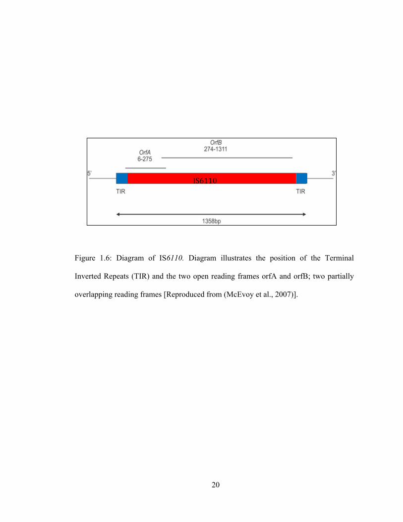
M. tuberculosis, one particular IS element, IS6110 with the length of 1.36 kbp has 28-
bp terminal inverted repeats (TIR) at its ends (Figure 1.6). IS6110 is associated with the
adaptation to the host, activation of genes during infection, evolution and is involved in
activation of downstream genes with an orientation-dependent activity promoter
(Alonso et al., 2013). IS6110 is present in multiple copies usually up to 6-25 times
copies in the M. tuberculosis genome incontras with M. bovis, which contains only one
copy (Philipp, 1996).
Its high specificity to MTBC, the presence in multiple copies and stability in the
genome of M. tuberculosis renders IS6110 an ideal biomarker. Moreover location of an
IS6110 copy in a specific strain can be used for rapid identification and the
differentiation of that particular strain amidst the other strains (Millan-Lou et al., 2012).
IS6110 has been extensively used in diagnostic and epidemiology study of M.
tuberculosis. de Lassence and colleagues stipulated that the utilization of IS6110 in
PCR is more sensitive than other identification protocol (de Lassence et al., 1992).
Other studies, utilizes IS6110 gene detecting in M. tuberculosis with various success
rates (Barani et al., 2011, Chaidir et al., 2012; Kulkarni et al., 2012). However, absence
or low copy of IS6110 in certain strains of M. tuberculosis suggests that an additional
target(s) could be amalgamated with IS6110 to elevate the efficiency and accuracy of
the PCR-based diagnostic assay.
20
Figure 1.6: Diagram of IS6110. Diagram illustrates the position of the Terminal
Inverted Repeats (TIR) and the two open reading frames orfA and orfB; two partially
overlapping reading frames [Reproduced from (McEvoy et al., 2007)].
IS6110

21
1.8. Research objectives
In concert with the effort to test the potentiality of the npcRNA genes as the
potential diagnostic markers for M. tuberculosis detection, the incorporation of IS6110
as the counterpart in PCR amplification is viewed to be able to ward off false negative
results derived from IS6110-deficient M. tuberculosis strains.
Thus, the initial aim of the project was to identify npcRNA genes that could act
as the potential diagnostic markers for M. tuberculosis detection. The identified target is
then coupled with IS6110 towards the development and optimization of multiplex PCR.
Collectively, the objectives are as follows;
1. Identification of npcRNA genes specific for the detection of M. tuberculosis via
BLASTn analysis Software.
2. Development and optimization of the multiplex PCR using the identified
npcRNA genes and IS6110 as the targets. The parameters are:-
i) Optimization of annealing temperature
ii) Optimization of primer concentration
iii) Incorporation of Internal Amplification Control
iv) Optimization of deoxyribonucleotide triphosphate
v) Optimization of MgCl2
vi) Determination of the specificity of mPCR
vii) Determination of the sensitivity of the mPCR

22
3. Evaluation of the mPCR assay
i) Validation of the developed mPCR with culture positive samples

23
CHAPTER 2
MATERIALS AND METHODS
2.1 Materials
2.1.1 Chemicals
Chemicals and reagents used in this study were of analytical grade and are listed below:
Manufacturer Chemicals
Amresco (USA)
Bio basic inc (Canada)
Bio-Rad (Hercules,USA)
Biotools (Canada)
Laborotarious Conda (Madrid,Spain)
Merck (Darmstadt,Germany)
Invitrogen ® (Carlsbad,CA)
Promega (Madison,USA)
Sodium acetate; Sodium hydroxide;
Potassium chloride anhydrous.
EDTA; PCR primers.
Ethidium bromide (10mg/mL);
Sodium deodecyl sulfate (SDS)
10X PCR buffer; Magnesium
chloride (MgCl2) ; dNTP mix; DNA
polymerase.
LB agar (lennox); LB broth (lennox),
Middlebrooke 7H9, Middlebrooke
7H10
Ethanol; Acetic acid (glacial) 100%;
Sodium hydroxide; Glycerol
Ultra-pure Tris
100bp DNA ladder; Agarose powder

24
2.1.2 Culture and bacterial strains
Bacterial strains used in this study are from the stock collection, obtained from
the Department of Microbiology & Parasitology, School of Medical Sciences, USM
and Infectomic Cluster, Advance Medical and Dental Institute (AMDI), USM.
Bacterial Strains Description Source
Mycobacterium
tuberculosis H37Rv
Virulent laboratory strain AMDI
Mycobacterium
tuberculosis H37Ra
Attenuated tubercle bacillus closely
related M. tuberculosis H37Rv
AMDI
Mycobacterium avium Non tuberculosis Mycobacterium AMDI
Mycobacterium
abscessus
Non tuberculosis Mycobacterium AMDI
Mycobacterium
fortuitum
Non tuberculosis Mycobacterium AMDI
Mycobacterium
fortuitum
Non tuberculosis Mycobacterium AMDI
Mycobacterium
gordonae
Non tuberculosis Mycobacterium AMDI
Mycobacterium
scroferaceum
Non tuberculosis Mycobacterium AMDI
Mycobacterium
intracellulare
Non tuberculosis Mycobacterium AMDI
Mycobacterium
kansasii
Non tuberculosis Mycobacterium AMDI