principles of human anatomy and physiology, 11e1 chapter 6 the skeletal system: bone tissue lecture...
TRANSCRIPT
Principles of Human Anatomy and Physiology, 11e 1
Chapter 6
The Skeletal System: Bone Tissue
Lecture Outline
Principles of Human Anatomy and Physiology, 11e 2
INTRODUCTION
• Bone is made up of several different tissues working together.
• Each individual bone is an organ; the bones, along with their cartilages, make up the skeletal system.
• The study of bone structure and the treatment of bone disorders is called osteology.
Principles of Human Anatomy and Physiology, 11e 3
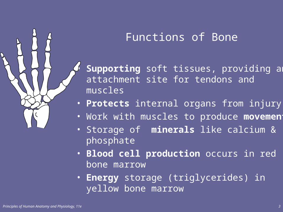
Functions of Bone
• Supporting soft tissues, providing an attachment site for tendons and muscles
• Protects internal organs from injury• Work with muscles to produce movement• Storage of minerals like calcium & phosphate• Blood cell production occurs in red bone
marrow • Energy storage (triglycerides) in yellow bone
marrow
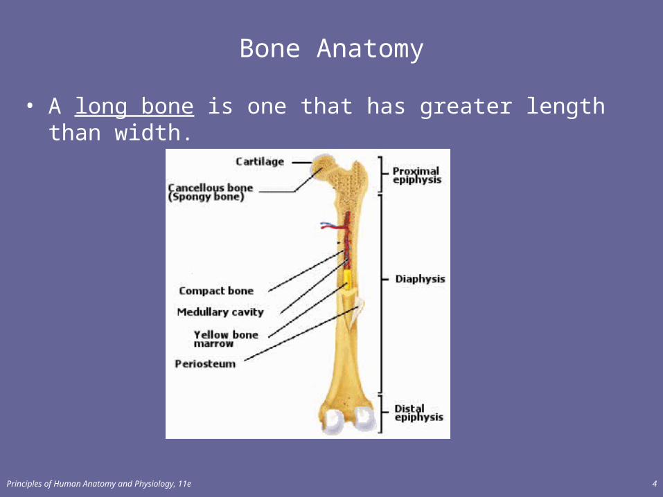
Bone Anatomy
• A long bone is one that has greater length than width.
Principles of Human Anatomy and Physiology, 11e 4
Principles of Human Anatomy and Physiology, 11e 5
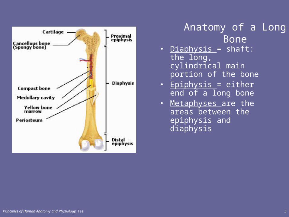
Anatomy of a Long Bone
• Diaphysis = shaft: the long, cylindrical main portion of the bone
• Epiphysis = either end of a long bone
• Metaphyses are the areas between the epiphysis and diaphysis
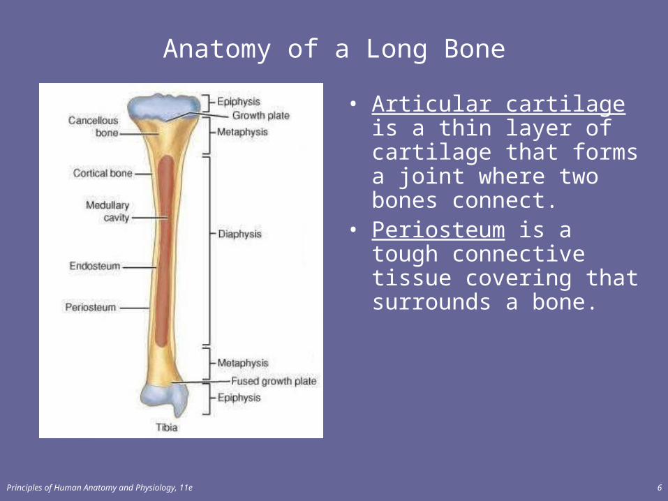
Anatomy of a Long Bone
• Articular cartilage is a thin layer of cartilage that forms a joint where two bones connect.
• Periosteum is a tough connective tissue covering that surrounds a bone.
Principles of Human Anatomy and Physiology, 11e 6

Principles of Human Anatomy and Physiology, 11e 7
Anatomy of a Long Bone
• The medullary cavity or marrow cavity is the space in the diaphysis that stores yellow bone marrow.
• The endosteum is a thin membrane that lines the medullary cavity.
Principles of Human Anatomy and Physiology, 11e 8
HISTOLOGY OF BONE TISSUE
• Bone (osseous) tissue consists of widely separated cells surrounded by large amounts of matrix.
• The matrix of bone is made up of water, collagen, and salts.• These salts are used to form new bone tissue, which is
called calcification.
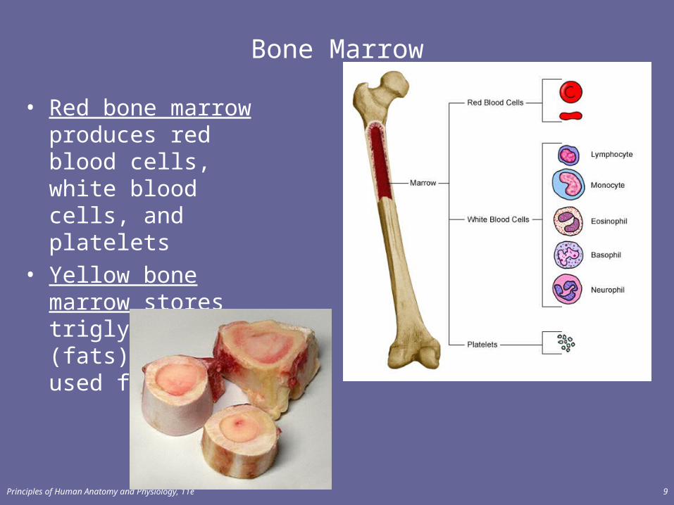
Bone Marrow
• Red bone marrow produces red blood cells, white blood cells, and platelets
• Yellow bone marrow stores triglycerides (fats) which are used for energy
Principles of Human Anatomy and Physiology, 11e 9
Principles of Human Anatomy and Physiology, 11e 10
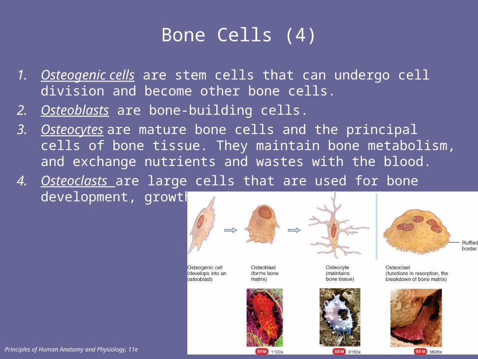
Bone Cells (4)
1. Osteogenic cells are stem cells that can undergo cell division and become other bone cells.
2. Osteoblasts are bone-building cells.
3. Osteocytes are mature bone cells and the principal cells of bone tissue. They maintain bone metabolism, and exchange nutrients and wastes with the blood.
4. Osteoclasts are large cells that are used for bone development, growth, maintenance, and repair
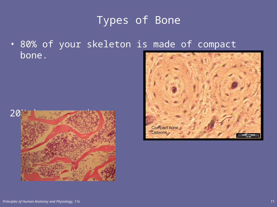
Types of Bone
• 80% of your skeleton is made of compact bone.
20% is spongy bone.
Principles of Human Anatomy and Physiology, 11e 11
Principles of Human Anatomy and Physiology, 11e 12
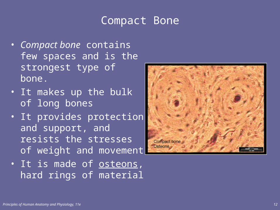
Compact Bone
• Compact bone contains few spaces and is the strongest type of bone.
• It makes up the bulk of long bones
• It provides protection and support, and resists the stresses of weight and movement
• It is made of osteons, hard rings of material
Principles of Human Anatomy and Physiology, 11e 13
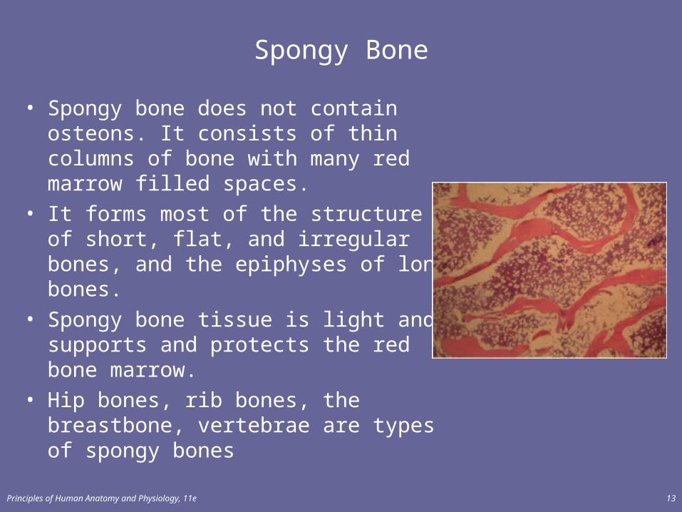
Spongy Bone
• Spongy bone does not contain osteons. It consists of thin columns of bone with many red marrow filled spaces.
• It forms most of the structure of short, flat, and irregular bones, and the epiphyses of long bones.
• Spongy bone tissue is light and supports and protects the red bone marrow.
• Hip bones, rib bones, the breastbone, vertebrae are types of spongy bones
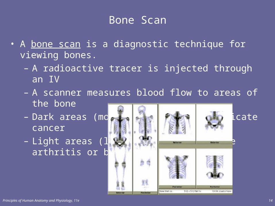
Bone Scan
• A bone scan is a diagnostic technique for viewing bones.– A radioactive tracer is injected through an IV– A scanner measures blood flow to areas of the bone– Dark areas (more blood flow) may indicate cancer– Light areas (less blood) may indicate arthritis or bone
loss.
Principles of Human Anatomy and Physiology, 11e 14
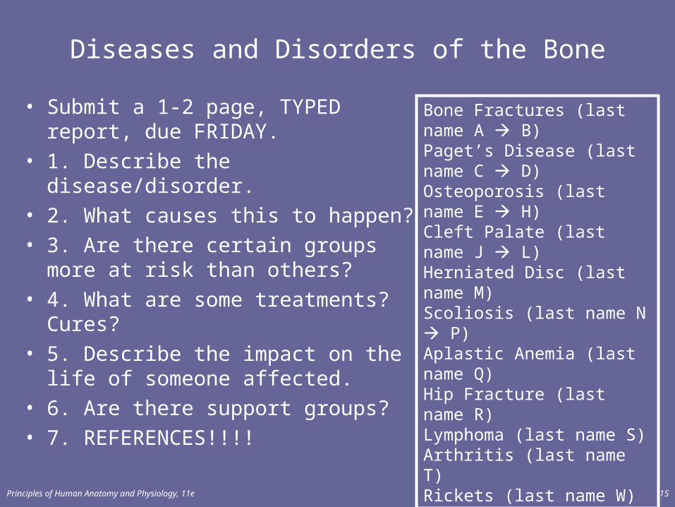
Diseases and Disorders of the Bone
• Submit a 1-2 page, TYPED report, due FRIDAY.
• 1. Describe the disease/disorder.• 2. What causes this to happen?• 3. Are there certain groups more at
risk than others? • 4. What are some treatments?
Cures? • 5. Describe the impact on the life of
someone affected.• 6. Are there support groups?• 7. REFERENCES!!!!
Principles of Human Anatomy and Physiology, 11e 15
Bone Fractures (last name A B)Paget’s Disease (last name C D) Osteoporosis (last name E H) Cleft Palate (last name J L)Herniated Disc (last name M) Scoliosis (last name N P)Aplastic Anemia (last name Q) Hip Fracture (last name R)Lymphoma (last name S) Arthritis (last name T)Rickets (last name W)
Principles of Human Anatomy and Physiology, 11e 16
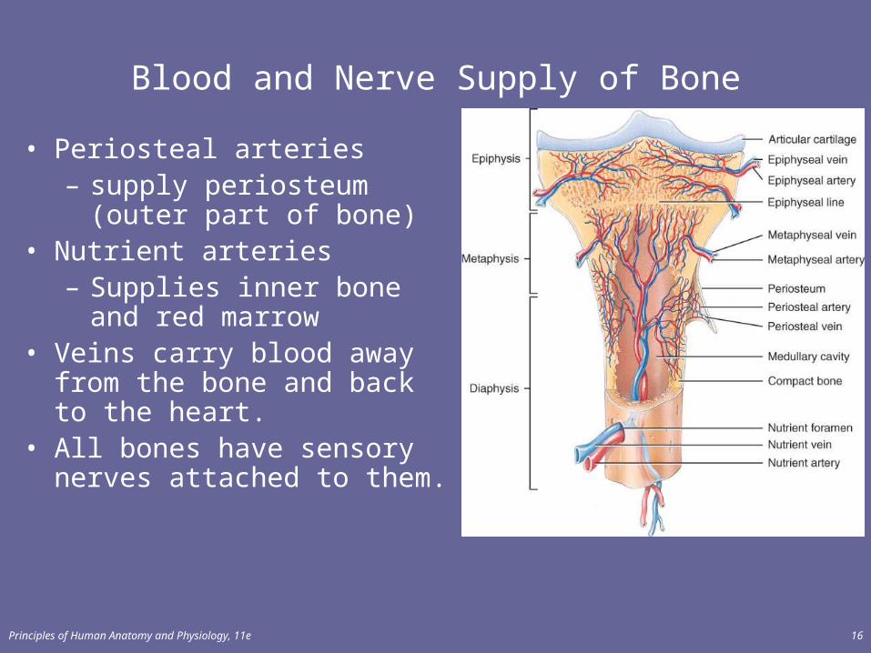
Blood and Nerve Supply of Bone
• Periosteal arteries– supply periosteum (outer
part of bone)• Nutrient arteries
– Supplies inner bone and red marrow
• Veins carry blood away from the bone and back to the heart.
• All bones have sensory nerves attached to them.
Principles of Human Anatomy and Physiology, 11e 17
BONE FORMATION
• Bone formation is termed osteogenesis or ossification• Two types of ossification occur.
– Intramembranous ossification– Endochondrial ossification
Intramembranous Ossification
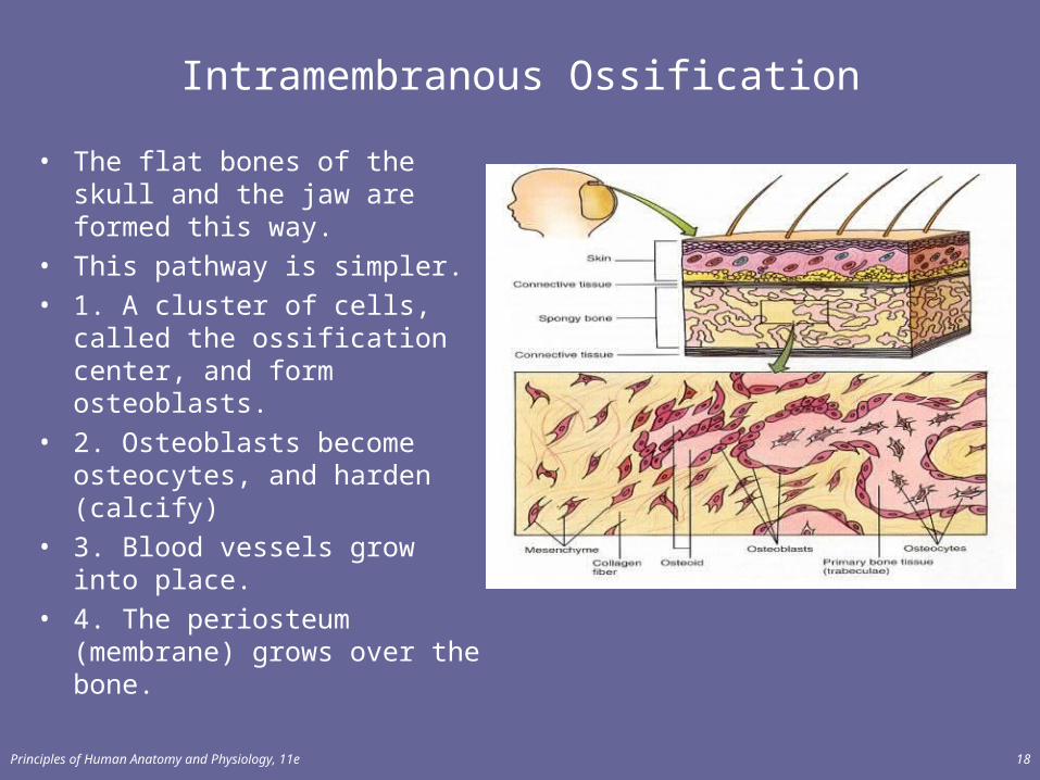
• The flat bones of the skull and the jaw are formed this way.
• This pathway is simpler.• 1. A cluster of cells, called the
ossification center, and form osteoblasts.
• 2. Osteoblasts become osteocytes, and harden (calcify)
• 3. Blood vessels grow into place.
• 4. The periosteum (membrane) grows over the bone.
Principles of Human Anatomy and Physiology, 11e 18
Endochondral Ossification
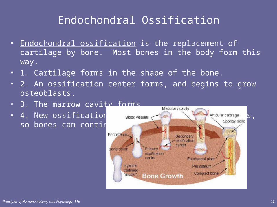
• Endochondral ossification is the replacement of cartilage by bone. Most bones in the body form this way.
• 1. Cartilage forms in the shape of the bone.• 2. An ossification center forms, and begins to grow osteoblasts.• 3. The marrow cavity forms.• 4. New ossification centers form at each epiphysis, so bones can
continue to elongate.
Principles of Human Anatomy and Physiology, 11e 19
Principles of Human Anatomy and Physiology, 11e 20
Growth in Length
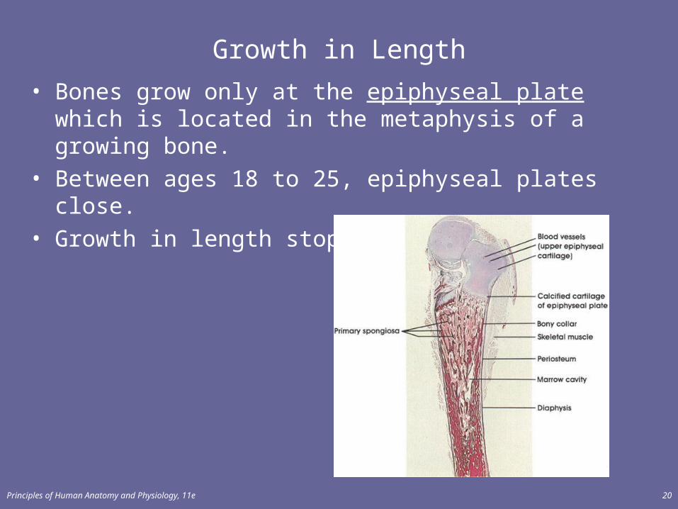
• Bones grow only at the epiphyseal plate which is located in the metaphysis of a growing bone.
• Between ages 18 to 25, epiphyseal plates close.• Growth in length stops at age 25
Growth in Thickness
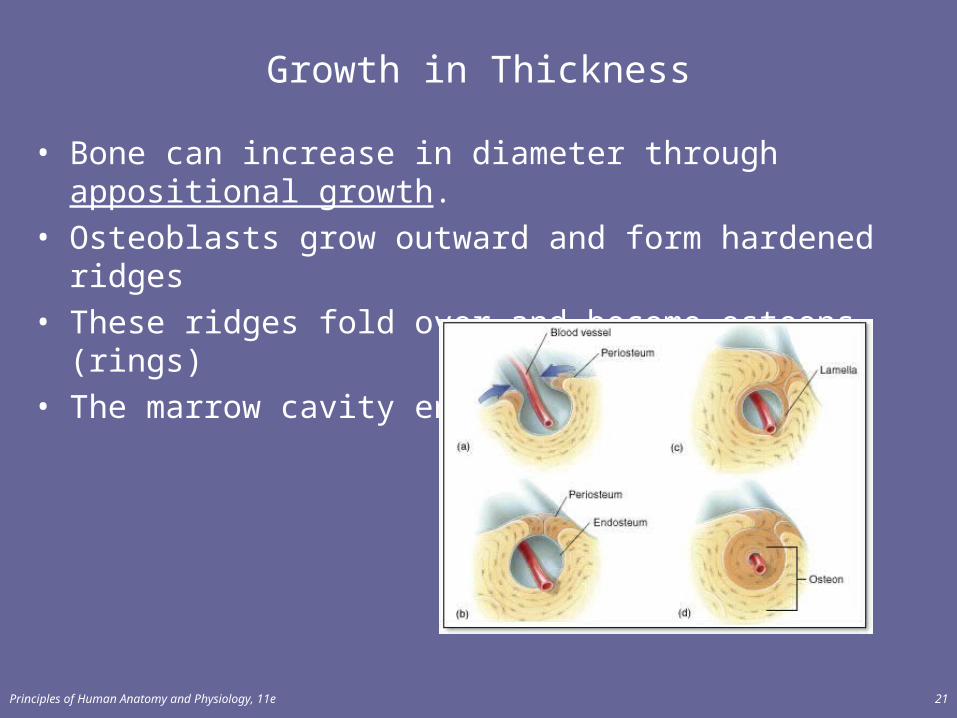
• Bone can increase in diameter through appositional growth.• Osteoblasts grow outward and form hardened ridges• These ridges fold over and become osteons (rings)• The marrow cavity enlarges.
Principles of Human Anatomy and Physiology, 11e 21

Principles of Human Anatomy and Physiology, 11e 22
Bone Remodeling
• Remodeling is the ongoing replacement of old bone tissue by new bone tissue.– Old bone is constantly destroyed by osteoclasts (bone
resorption), whereas new bone is constructed by osteoblasts (bone deposition).
– 20% of spongy bone and 4% of compact bone is remodeled each year!
– This can be triggered by exercise, diet, or lack of exercise.
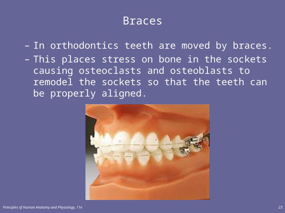
Braces
– In orthodontics teeth are moved by braces.– This places stress on bone in the sockets causing
osteoclasts and osteoblasts to remodel the sockets so that the teeth can be properly aligned.
Principles of Human Anatomy and Physiology, 11e 23
Principles of Human Anatomy and Physiology, 11e 24
Factors Affecting Bone Growth
• Nutrition– adequate levels of minerals and vitamins
• calcium and phosphorus for bone growth• vitamin C for collagen formation, Vitamin A for
osteoblasts• vitamins K and B12 for protein synthesis
Factors affecting Bone Growth
• Sufficient levels of specific hormones– during childhood need insulinlike growth factor (IGF)– Human Growth Hormone (too much=gigantism, not
enough=pituitary dwarfism)– sex steroids at puberty– At puberty the sex hormones, estrogen and testosterone,
stimulate sudden growth and modifications of the skeleton to create the male and female forms (wider pelvis).
Principles of Human Anatomy and Physiology, 11e 25
Principles of Human Anatomy and Physiology, 11e 26
Fracture and Repair of Bone
A fracture is any break in a bone.• Fractures are named according to their severity, the shape
of the fracture, or the physician who first described fractures.
Common Fractures
• Open Fracture: Broken bones protrude through skin• Closed Fracture: Broken bones do NOT show through• Comminuted fracture: Bone splinters at site of impact• Greenstick Fracture: One side of the bone breaks, the other
bends (only occurs in children)• Impacted fracture: One bone is driven into another• Pott’s fracture: fracture of the fibula (leg bone)• Colles’ fracture: fracture of the forearm bone• Stress fracture: Microscopic fissures but no broken bones
– Results from repeated, strenuous activity like running, jumping, or dancing
Principles of Human Anatomy and Physiology, 11e 27
Principles of Human Anatomy and Physiology, 11e 28
Fracture & Repair of Bone• Healing is faster in bone than in cartilage since bones have
better blood supplies• Healing of bone is still slow process due to vessel damage• Clinical treatment:
– closed reduction = restore pieces to normal position by manipulation
– open reduction = realignment during surgery• Both are followed by immobilization in a cast, sling, or
bandage
Principles of Human Anatomy and Physiology, 11e 29
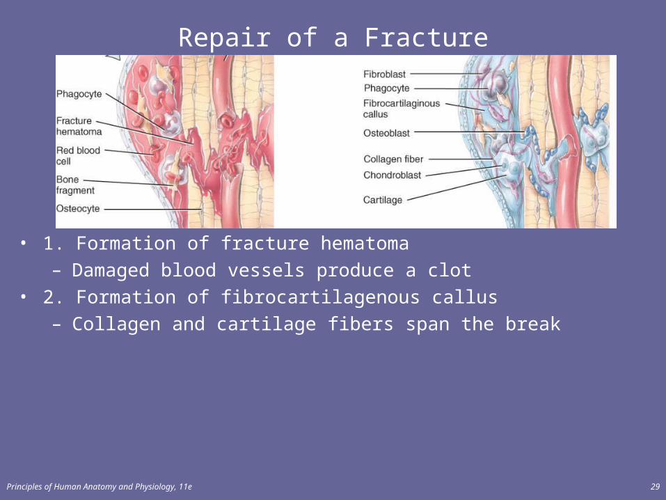
Repair of a Fracture
• 1. Formation of fracture hematoma– Damaged blood vessels produce a clot
• 2. Formation of fibrocartilagenous callus– Collagen and cartilage fibers span the break
Principles of Human Anatomy and Physiology, 11e 30
Repair of a Fracture
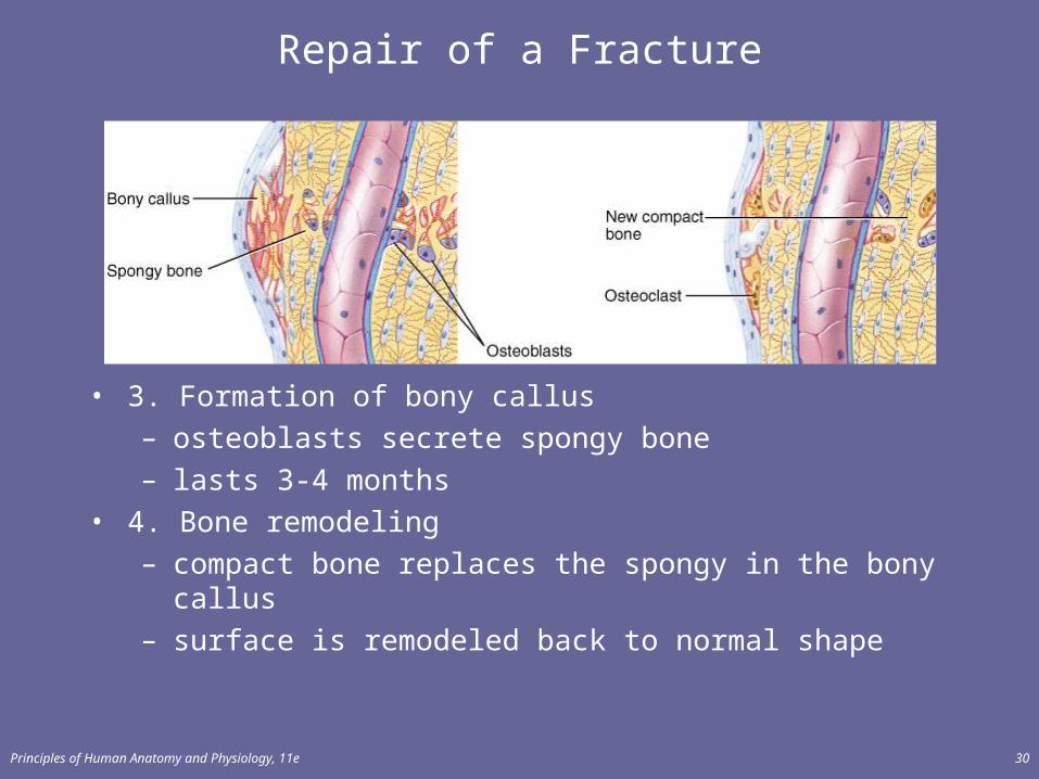
• 3. Formation of bony callus– osteoblasts secrete spongy bone– lasts 3-4 months
• 4. Bone remodeling– compact bone replaces the spongy in the bony callus– surface is remodeled back to normal shape
Principles of Human Anatomy and Physiology, 11e 31
Calcium Homeostasis & Bone Tissue
• Skeleton is a reservoir of Calcium & Phosphate• Calcium ions involved with many body systems
– nerve & muscle cell function– blood clotting– enzyme function in many biochemical reactions
• Small changes in blood levels of Ca+2 can be deadly– cardiac arrest if too high– respiratory arrest if too low
Principles of Human Anatomy and Physiology, 11e 32
EXERCISE AND BONE TISSUE
• Within limits, bone has the ability to alter its strength in response to mechanical stress by increasing deposition of mineral salts and production of collagen fibers.– Removal of mechanical stress leads to weakening of
bone through demineralization (loss of bone minerals) and collagen reduction.
• reduced activity while in a cast• astronauts in weightless environment• bedridden person
– Weight-bearing activities, such as walking or moderate weightlifting, help build and retain bone mass.
Principles of Human Anatomy and Physiology, 11e 33
AGING AND BONE TISSUE
• Loss of calcium which may result in osteoporosis.– very rapid in women 40-45 as estrogens levels decrease – in males, begins after age 60
• Decreased rate of protein synthesis– decrease in collagen production which gives bone its
tensile strength– decrease in growth hormone– bone becomes brittle & susceptible to fracture

Principles of Human Anatomy and Physiology, 11e 34
Osteoporosis
• Decreased bone mass resulting in porous bones • Those at risk
– white, thin menopausal, smoking, drinking female with family history
– athletes who are not menstruating due to decreased body fat & decreased estrogen levels
– people allergic to milk or with eating disorders whose intake of calcium is too low
• Prevention or decrease in severity– adequate diet, weight-bearing exercise, & estrogen
replacement therapy (for menopausal women)– behavior when young may be most important factor
Principles of Human Anatomy and Physiology, 11e 35
Disorders of Bone Ossification
• Rickets• calcium salts are not deposited properly• bones of growing children are soft• bowed legs, skull, rib cage, and pelvic deformities
result• Osteomalacia
• new adult bone produced during remodeling fails to ossify
• hip fractures are common