primitive neuroectodermal tumor (pnet) of the kidney: a case report
TRANSCRIPT
BioMed CentralBMC Cancer
ss
Open AcceCase reportPrimitive Neuroectodermal Tumor (PNET) of the kidney: a case reportGiorgio Pomara*1, Francesco Cappello2, Maria G Cuttano1, Francesca Rappa2, Girolamo Morelli1, Pierantonio Mancini1 and Cesare Selli1Address: 1Department of Surgery – Urology Section – S. Chiara Hospital – University of Pisa, Pisa – Italy and 2Department of Experimental Medicine – Human Anatomy Section – University of Palermo, Palermo – Italy
Email: Giorgio Pomara* - [email protected]; Francesco Cappello - [email protected]; Maria G Cuttano - [email protected]; Francesca Rappa - [email protected]; Girolamo Morelli - [email protected]; Pierantonio Mancini - [email protected]; Cesare Selli - [email protected]
* Corresponding author
Renal PNETEwing's Sarcomarhabdomyosarcoma tumorstem cellscarcinogenesis
AbstractBackground: A case of Primitive Neuroectodermal Tumor (PNET) of the kidney in a 27-year-oldwoman is presented. Few cases are reported in the literature with a variable, nonspecificpresentation and an aggressive behaviour. In our case, a radical nephrectomy withlymphadenectomy was performed and there was no residual or recurrent tumour at 24-monthfollow-up.
Methods: The surgical specimens were formalin-fixed and paraffin embedded. The sections werestained with routinary H&E. Immunohistochemistry was performed.
Results: The immunohistochemical evaluation revealed a diffuse CD99 positivity in the cytoplasmof the neoplastic cells. Pankeratin, cytokeratin AE1/AE3, vimentin, desmin, S100, cromogranin werenegative. The clinical presentation and the macroscopic aspect, together with the histologicalpattern, the cytological characteristic and the cellular immunophenotype addressed the diagnosistowards primary PNET of kidney.
Conclusions: Since sometimes it is difficult to discriminate between PNET and Ewing's tumour,we reviewed the difficulties in differential diagnosis. These tumors have a common precursor butthe stage of differentiation in which it is blocked is probably different. This could also explain theirdifferent biological behaviour and prognosis.
BackgroundThe peripheral Primitive Neuroectodermal Tumor(PNET), firstly recognized by Arthur Purdy Stout in 1918,is a member of the family of "small round-cell tumors".Primitive renal localization is very rare. There are almost50 cases reported in the literature, although it is difficult
to estimate the exact number since often it has not beendifferentiated from Ewing's Sarcoma [1-13]. Renal PNETis more aggressive than in the other sites. It frequentlyarises during childhood or adolescence, having an aggres-sive clinical course towards metastatic disease and death.It often recurs locally and metastasises early to regional
Published: 26 January 2004
BMC Cancer 2004, 4:3
Received: 13 September 2003Accepted: 26 January 2004
This article is available from: http://www.biomedcentral.com/1471-2407/4/3
© 2004 Pomara et al; licensee BioMed Central Ltd. This is an Open Access article: verbatim copying and redistribution of this article are permitted in all media for any purpose, provided this notice is preserved along with the article's original URL.
Page 1 of 5(page number not for citation purposes)
BMC Cancer 2004, 4 http://www.biomedcentral.com/1471-2407/4/3
lymph nodes, lungs, liver, bone and bone marrow, result-ing in a poor prognosis. The 5-year disease-free survivalrate, for patients presenting well confined extra-skeletalPNET, is around 45–55% and cases with advanced diseaseat presentation have a median relapse-free survival of only2 years [1].
Case presentationA 27-year-old woman was referred because of a mild leftflank pain and haematuria. Ultrasonography identified aleft renal mass homogeneously hyperechogenic in com-parison with renal parenchyma. CT scan showed a 11 mm× 8 mm × 6 mm tumor replacing the upper half of the leftkidney with extension into the renal vein. Chest x-ray was
negative. Pathological stage after radical nephrectomy wasT3aN0Mx.
The surgical specimens were formalin-fixed and paraffinembedded. The sections were stained with routinary H&E.Immunohistochemistry was performed using avidinbiotin complex technique and diaminobenzidine as chro-mogen. The antibodies used included CD99 (Dako,M3601), pankeratin (Dako, M0821), cytokeratin AE1/AE3 (Dako, M3515), vimentin (Dako, M7010), desmin(Dako, M0760), S100 (Dako, Z0311), and chromograninA (Dako, M0869), at suggested dilution. We performedalso appropriate routinely positive and negative controls.
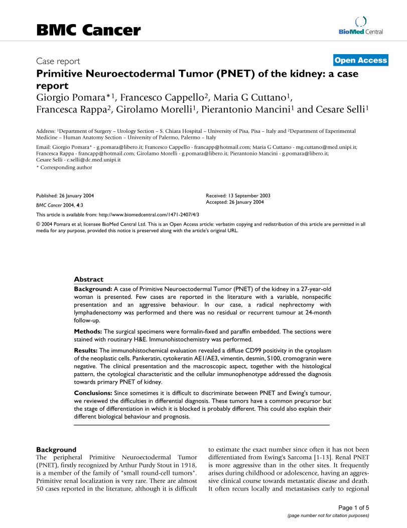
H&E features of the tumour (original magnification 10×; inset 40 ×): sheets of monotonous cells infiltrating vessels (small arrow) even near the capsule (long arrow)Figure 1H&E features of the tumour (original magnification 10×; inset 40 ×): sheets of monotonous cells infiltrating vessels (small arrow) even near the capsule (long arrow). Homer-Wright rosettes were common (inset).
Page 2 of 5(page number not for citation purposes)
BMC Cancer 2004, 4 http://www.biomedcentral.com/1471-2407/4/3
The tumor was multilobular, grey, glistening, focally hem-orrhagic, surrounded by a capsule and with a sharpdemarcation from the uninvolved kidney. Histologically,the tumor consisted of small round cells with roundnuclei and scant cytoplasm. It presented different pat-terns, with cohesive lobules or rosettes and perivascularpseudo-rosettes or, in some areas, spindle cellular ele-ments (fig. 1).
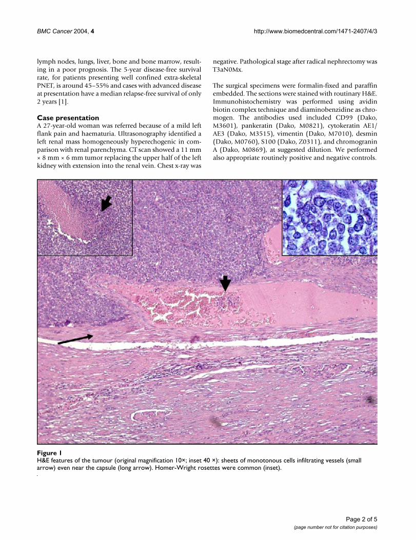
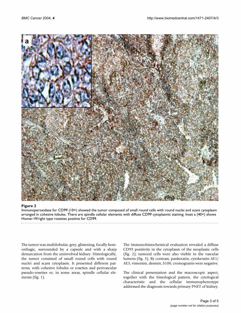
The immunohistochemical evaluation revealed a diffuseCD99 positivity in the cytoplasm of the neoplastic cells(fig. 2); tumoral cells were also visible in the vascularlumens (fig. 3). By contrast, pankeratin, cytokeratin AE1/AE3, vimentin, desmin, S100, cromogranin were negative.
The clinical presentation and the macroscopic aspect,together with the histological pattern, the cytologicalcharacteristic and the cellular immunophenotypeaddressed the diagnosis towards primary PNET of kidney.
Immunoperoxidase for CD99 (10×) showed the tumor composed of small round cells with round nuclei and scant cytoplasm arranged in cohesive lobulesFigure 2Immunoperoxidase for CD99 (10×) showed the tumor composed of small round cells with round nuclei and scant cytoplasm arranged in cohesive lobules. There are spindle cellular elements with diffuse CD99 cytoplasmic staining. Inset a (40×) shows Homer-Wright type rosettes positive for CD99.
Page 3 of 5(page number not for citation purposes)
BMC Cancer 2004, 4 http://www.biomedcentral.com/1471-2407/4/3
A bone scan did not reveal positive areas. Eight cycles ofchemotherapy with Vincristine, Ifosfamide and Adriamy-cin, four cycles of Ifosfamide and VP16 and eight sittingsof local radiotherapy were sequentially performed. Fol-low-up examinations with CT and bone scan failed toshow residual or recurrent tumor after 24 months.
ConclusionsPrimitive Neuroectodermal Tumor of the kidney is a rareentity. The few cases reported revealed a variable presenta-tion and an aggressive behaviour. The distinction fromother primary malignancies of the kidneys is crucial for
prognosis. The differential diagnosis includes extra-osseous Ewing's sarcoma, rhabdomyosarcoma, Wilm'stumor, carcinoid, neuroblastoma, clear cell sarcoma ofthe kidney, lymphoma, the small cell variant of osteosar-coma, desmoplastic small round cell tumor and nephrob-lastoma [5].
The Homer-Wright type rosettes, commonly scarce ofnumber or less defined in extra skeletal Ewing's sarcoma(ES), are a typical histological feature for PNET and canaddress the diagnosis although they can be found also inneuroblastoma [5]. To better address the diagnosis, an
Immunoperoxydase for CD99 (X25) revealed positive tumoral cells in the vascular lumens (arrow)Figure 3Immunoperoxydase for CD99 (X25) revealed positive tumoral cells in the vascular lumens (arrow).
Page 4 of 5(page number not for citation purposes)
BMC Cancer 2004, 4 http://www.biomedcentral.com/1471-2407/4/3
immunohistochemical analysis is necessary. In our casethe presence of MIC-2 gene products, known also asCD99, 12E7, E2, 013 and HBA71, suggested a PNET diag-nosis. Primitive neuroectodermal tumors only immunor-reactive to CD99, even if uncommon, are reported in theliterature [13]. The reactivity to vimentin, NSE and S-100may facilitate the diagnosis but is not patognomonic,while CD 99 positivity is nowadays a clue for the diagno-sis. Moreover cytogenetic studies (not performed in ourcase) demonstrated that PNET and Ewing's sarcoma canboth be associated to a translocation of the long arms ofchromosome 11 and 22, t(11;22)(q22;q12) [5]. Despitetheir genetic and antigenic similarity, many authors cur-rently recognize PNET and extra-skeletal Ewing's sarcomaof the kidney as separate entities. It is also important tokeep separate renal PNET and malignant rhabdomyosar-coma tumor (MRT). Weeks et al reported 8 cases sugges-tive for PNET but mimicking MRT [14]. Although renalPNET and MRT show similar clinico-pathological fea-tures, the latter usually occurs in very young children, hav-ing a more aggressive prognosis.
Rodriguez et al postulated that these two renal neoplasmsshare a common undifferentiated precursor to explaintheir similarity and we agree with these Authors [12].Indeed, the hypothesis that tumors arise from stem cells(SCs) as a consequence of a maturative arrest is now grow-ing [15]. SCs are present in almost all tissues and mayoriginate different cellular lineages by the multi-step proc-ess named "differentiation". The role of SCs in tumorigen-esis was clearly demonstrated in a number of carcinogenicmodels showing that solid and haematopoietic cancerscould arise from tissue-specific SCs [16-19]. In agreementwith Sell and Pierce, we retain that the degree of malig-nancy of a carcinoma depends by the stage in which SCsdifferentiation stopped during carcinogenesis [19]. In par-ticular, since PNET, Ewing's tumour and MRT have a sim-ilar morphology, our hypothesis is that the mesenchimalstem precursor of these tumors is the same, but the stageof differentiation in which it is blocked is different. Thiscould explain why sometimes it is difficult to discriminatebetween these tumors, notwithstanding they present a dif-ferent biological behaviour.
Competing interestsNone declared.
Authors' contributionAll authors contributed.
AcknowledgementsWritten consent was obtained from the patient for publication of the patient's details
References1. Casella R, Moch H, Rochlitz C et al.: Metastatic primitive neur-
oectodermal tumor of the kidney in adults. Eur Urol 2001,39:613-617.
2. Cuesta Alcala JA, Solchaga Martinez A, Caballero Martinez MC et al.:Primary neuroectodermal tumor (PNET)of the kidney:26cases. Current status of its diagnosis and treatment. Arch EspUrol 2001, 54:1081-1093.
3. Dogra PN, Goel A, Kumar R et al.: Extra-osseous Ewing's Sar-coma of the kidney. Urol Int 2002, 69:150-152.
4. Friedrichs N, Vorreuther R, Poremba C et al.: Primitive neuroec-todermal tumor in the differential diagnosis of malignantkidney tumors. Pathol Res Pract 2002, 198:563-569.
5. Gonsulen G, Ergin M, Paydas S et al.: Primitive neuroectodermaltumor of the kidney: a rare entity. Int Urol Nephrol 2001,33:449-451.
6. Jimenez RE, Folpe AL, Lapham RL et al.: Primary Ewing's Sar-coma/Primitive neuroectodermal tumor of the kidney. Aclinicopathologic and immunohistochemical analysis of 11cases. Am J Surg Pathol 2002, 26:320-327.
7. Karnes RJ, Gettman MT, Anderson PM et al.: Primitive neuroec-todermal tumor (extraskeletal Ewing's Sarcoma) of the kid-ney with vena caval tumor thrombus. J Urol 2000, 164:772.
8. Kuroda M, Urano M, Abe M et al.: Primary primitive neuroecto-dermal tumor of the kidney. Pathol Int 2000, 50:967-972.
9. Lam JS, Hensle TW et al.: Organ-confined Primitive Neuroecto-dermal tumor arising from the kidney. J Pediatr Surger 2003,38:619-621.
10. Premalata CS, Gayathri DV et al.: Primitive Neuroectodermaltumor of the kidney. A report of two cases diagnosed by fineneedle aspiration cytology. Acta Cytol 2003, 47:475-479.
11. Ranadive NU, Urmi C, Kumar M: Primary primitive neuroecto-dermal tumor (PNET) of the kidney: a case report. Arch EspUrol 1999, 52:190-192.
12. Rodriguez-Galindo C, Marina NM, Fletcher BD et al.: Is primitiveneuroectodermal tumor of kidney a distinct entity? Cancer1997, 79:2243-2250.
13. Thomas JC, Sebek BA, Krishnamurthi V: Primitive Neuroectoder-mal tumor of the kidney with inferior cava and atrial tumorthrombus. J Urol 2002, 168:1486-1487.
14. Weeks DA, Beckwith JB, Mierau GW, Zuppan CW: Renal neo-plasms mimicking rhabdoid tumor of kidney. A report fromthe National Wilms' Tumor Study Pathology Center. Am JSurg Pathol 1991, 15:1042-54.
15. Reya T, Morrison JS, Clarke MF et al.: Stem cells, cancer, and can-cer stem cells. Nature 2001, 414:105-111.
16. Andrews PW: From teratocarcinomas to embryonic stemcells. Philos Trans R Soc Lond B Biol Sci 1977, 357:405-417.
17. Novikoff PM, Yam A: Stem cells and rat liver carcinogenesis:contributions of confocal and electron microscopy. J Histo-chem Cytochem 1998, 46:613-626.
18. Pathak S: Organ- and tissue-specific stem cells andcarcinogenesis. Anticancer Res 2002, 22:1353-1356.
19. Sell S, Pierce GB: Maturation arrest of stem cell differentiationin a common pathway for the cellular origin of teratocarci-nomas and epithelial cancers. Lab Invest 1994, 70:6-22.
Pre-publication historyThe pre-publication history for this paper can be accessedhere:
http://www.biomedcentral.com/1471-2407/4/3/prepub
Page 5 of 5(page number not for citation purposes)