prevalence and genetic diversity of endosymbiotic bacteria infecting
TRANSCRIPT
Ghosh et al. BMC Microbiology (2015) 15:93 DOI 10.1186/s12866-015-0425-5
RESEARCH ARTICLE Open Access
Prevalence and genetic diversity ofendosymbiotic bacteria infecting cassavawhiteflies in AfricaSaptarshi Ghosh, Sophie Bouvaine and MN Maruthi*
Abstract
Background: Cassava provides over half of the dietary requirement for more than 200 million poor in Africa. Inrecent years, cassava has been affected by an epidemic of a virus disease called cassava brown streak disease(CBSD) that is spreading in much of eastern and central Africa, affecting food security and the economicdevelopment of the poor. The viruses that cause CBSD are transmitted by the insect vector whitefly (Bemisia tabaci),which have increased to very high numbers in some African countries. Strains of endosymbiotic bacteria infectingwhiteflies have been reported to interact specifically with different whitefly populations with varied effects on itshost biology and efficiency of virus transmission. The main aim of this study was therefore to investigate theprevalence and diversity of the secondary endosymbiotic bacteria infecting cassava whiteflies with a view to betterunderstand their role on insect population dynamics and virus disease epidemics.
Results: The genetic diversity of field-collected whitefly from Tanzania, Malawi, Uganda and Nigeria was determinedby mitochondrial DNA based phylogeny and restriction fragment length polymorphism. Cassava in these countrieswas infected with five whitefly populations, and each one was infected with different endosymbiotic bacteria.Incidences of Arsenophonus, Rickettsia, Wolbachia and Cardinium varied amongst the populations. Wolbachia wasthe most predominant symbiont with infection levels varying from 21 to 97%. Infection levels of Arsenophonusvaried from 17 to 64% and that of Rickettsia was 0 to 53%. Hamiltonella and Fritschea were absent in all the samples.Multiple locus sequence typing identified four different strains of Wolbachia infecting cassava whiteflies. A commonstrain of Wolbachia infected the whitefly population Sub-Saharan Africa 1-subgroup 1 (SSA1-SG1) and SSA1-SG2,while others were infected with different strains. Phylogeny based on 16S rDNA of Rickettsia and 23S rDNA ofArsenophonus also identified distinct strains.
Conclusions: Genetically diverse bacteria infect cassava whiteflies in Africa with varied prevalence across differenthost populations, which may affect their whitefly biology. Further studies are required to investigate the role ofendosymbionts to better understand the whitefly population dynamics.
Keywords: Cassava, Whitefly, mtCOI, Wolbachia, Rickettsia, Arsenophonus
BackgroundThe whitefly, Bemisia tabaci (Hemiptera: Aleyrodidae)has gained importance as one of the most important agri-cultural pests owing to its wide geographic spread, largehost range of over 500 hosts, and most significantly as avector of over 100 different plant viruses in the tropicaland subtropical regions of the world [1,2]. B. tabaci is a
* Correspondence: [email protected] Resources Institute, University of Greenwich, Chatham ME4 4 TB,Kent, UK
© 2015 Ghosh et al.; licensee BioMed Central.Commons Attribution License (http://creativecreproduction in any medium, provided the orDedication waiver (http://creativecommons.orunless otherwise stated.
cryptic species complex comprising at least 24 morpho-logically indistinguishable species [3] with a proposedorigin in sub-Saharan Africa (SSA) and with high variabil-ity in mitochondrial cytochrome oxidase I (mtCOI) nu-cleotide sequences amongst major geographical clades[2,4]. Cassava, a key food security crop throughout SSA,suffers devastating yield losses due to B. tabaci-bornecassava mosaic begomoviruses (CMBs) and cassavabrown streak viruses (CBSVs). These cause cassavamosaic disease (CMD) and cassava brown streak disease(CBSD), respectively [5,6]. Five genetically distinct groups
This is an Open Access article distributed under the terms of the Creativeommons.org/licenses/by/4.0), which permits unrestricted use, distribution, andiginal work is properly credited. The Creative Commons Public Domaing/publicdomain/zero/1.0/) applies to the data made available in this article,
Ghosh et al. BMC Microbiology (2015) 15:93 Page 2 of 17
of B. tabaci, named Sub-Saharan Africa 1 to 5 (SSA 1–5)colonise cassava in SSA. These have been generally referredto as cassava whiteflies in this and other studies. SSA1 oc-curs throughout the SSA, SSA2 in East and West Africa,SSA3 in Cameroon and Togo, SSA4 only in Cameroon andSSA5 in South Africa [6]. Based on mtCOI sequence diver-gences, SSA1 was further divided into four subgroups;SSA1- subgroup 1 (SSA1-SG1), SSA1-SG2, SSA1-SG3 andSSA1-SG4 [6].Superabundant B. tabaci populations, commonly num-
bering more than 1000 adults per top five leaves, of cas-sava plants, have been associated with the rapid spread ofCMD pandemic in East and Central Africa since the late1990s. SSA2, the then super abundant population whichwas also described as the ‘invader or UG2’, was associatedwith the spread of the CMD pandemic [7,8]. In recentyears, a shift in the B. tabaci population has occurred withthe relative frequency of SSA1-SG1 increasing from 24.6%to 89.2%, while the frequencies of SSA2 and SSA1-SG2decreasing significantly from 63.9% to 1.4%, and 11.5% to1.4%, respectively, between 1997 and 2010 [6]. The rea-sons for this natural shift in cassava population remainunknown. Similar shift in genetic diversity of populationsare reported for B. tabaci species, mainly with respect tothe population replacement by the invasive MiddleEast-Asia Minor 1 (MEAM1, previously B-biotype) andMediterranean (MED, previously Q-biotype) populations[9-11].In addition to the primary endosymbiont Portiera
aleyrodidarum, the species B. tabaci has been reportedto harbour six vertically transmitted secondary endosym-bionts, Arsenophonus, Wolbachia, Hamiltonella, Cardi-nium, Fritschea and Rickettsia [12-14]. Recently, a newbacterium named Candidatus hemipteriphilus asiaticuswas also found to infect B. tabaci from China [15]. Severalof these endosymbionts can affect the biology and behav-iour of B. tabaci. Wolbachia and Cardinium in particularare known to induce cytoplasmic incompatibility (CI), aprocess in which the host reproduction is manipulatedto allow rapid spread of bacteria through insect popula-tions [16]. Whether such phenotypes are induced bythese bacteria in B. tabaci remains unknown. Rickettsia,when infecting MEAM1, provided fitness benefits by in-creased fecundity and survival [17], increased heat stresstolerance [18], defence against pathogens [19] but occa-sionally also increased the susceptibility to insecticides[20]. Fritschea has reported negative impact with reducedfecundity and narrowing the host range of infected NewWorld species of whiteflies [21]. Endosymbionts can alsoalter the vector ability of B. tabaci. Hamiltonella inMEAM1 and Arsenophonus in Asia II populations facili-tated virus transmission by releasing a bacterial chaper-onin GroEL that binds and protects virus particles duringtheir transit through the insect body [22,23]. Hamiltonella
in the MED and Rickettsia in MEAM1 populations arealso reported to increase acquisition, retention and trans-mission of Tomato yellow leaf curl virus [24,25]. The studyof intracellular bacterial communities in these whitefliesand their impact on the host was essential for understand-ing the dynamics of insect populations and their vectorabilities. In this study, we identified the endosymbionts in-fecting cassava whiteflies, determined their infection fre-quencies in different populations and characterised thediverse bacterial species by sequencing. We have also de-veloped a cost effective and reliable restriction fragmentlength polymorphism (RFLP) diagnostic method for themolecular typing of the cassava whitefly populations.
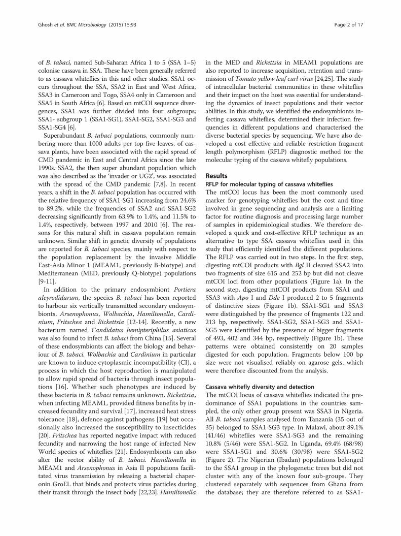
ResultsRFLP for molecular typing of cassava whitefliesThe mtCOI locus has been the most commonly usedmarker for genotyping whiteflies but the cost and timeinvolved in gene sequencing and analysis are a limitingfactor for routine diagnosis and processing large numberof samples in epidemiological studies. We therefore de-veloped a quick and cost-effective RFLP technique as analternative to type SSA cassava whiteflies used in thisstudy that efficiently identified the different populations.The RFLP was carried out in two steps. In the first step,digesting mtCOI products with Bgl II cleaved SSA2 intotwo fragments of size 615 and 252 bp but did not cleavemtCOI loci from other populations (Figure 1a). In thesecond step, digesting mtCOI products from SSA1 andSSA3 with Apo I and Dde I produced 2 to 5 fragmentsof distinctive sizes (Figure 1b). SSA1-SG1 and SSA3were distinguished by the presence of fragments 122 and213 bp, respectively. SSA1-SG2, SSA1-SG3 and SSA1-SG5 were identified by the presence of bigger fragmentsof 493, 402 and 344 bp, respectively (Figure 1b). Thesepatterns were obtained consistently on 20 samplesdigested for each population. Fragments below 100 bpsize were not visualised reliably on agarose gels, whichwere therefore discounted from the analysis.
Cassava whitefly diversity and detectionThe mtCOI locus of cassava whiteflies indicated the pre-dominance of SSA1 populations in the countries sam-pled, the only other group present was SSA3 in Nigeria.All B. tabaci samples analysed from Tanzania (35 out of35) belonged to SSA1-SG3 type. In Malawi, about 89.1%(41/46) whiteflies were SSA1-SG3 and the remaining10.8% (5/46) were SSA1-SG2. In Uganda, 69.4% (68/98)were SSA1-SG1 and 30.6% (30/98) were SSA1-SG2(Figure 2). The Nigerian (Ibadan) populations belongedto the SSA1 group in the phylogenetic trees but did notcluster with any of the known four sub-groups. Theyclustered separately with sequences from Ghana fromthe database; they are therefore referred to as SSA1-
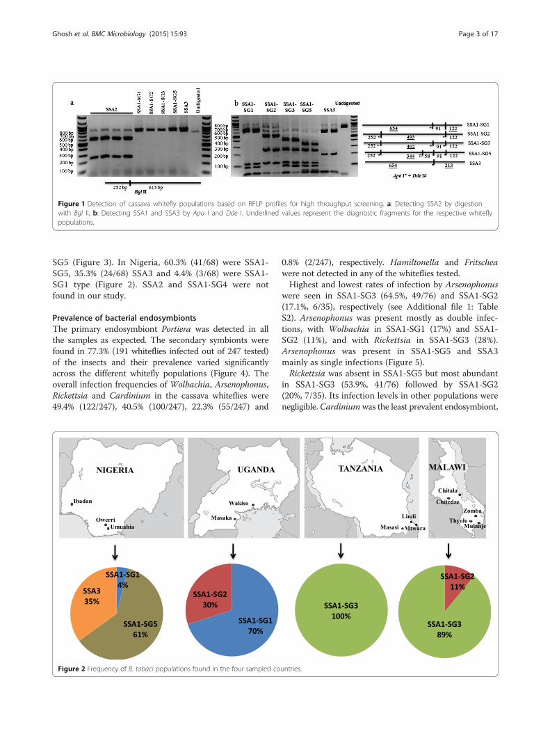
Figure 1 Detection of cassava whitefly populations based on RFLP profiles for high throughput screening. a: Detecting SSA2 by digestionwith Bgl II, b: Detecting SSA1 and SSA3 by Apo I and Dde I. Underlined values represent the diagnostic fragments for the respective whiteflypopulations.
Ghosh et al. BMC Microbiology (2015) 15:93 Page 3 of 17
SG5 (Figure 3). In Nigeria, 60.3% (41/68) were SSA1-SG5, 35.3% (24/68) SSA3 and 4.4% (3/68) were SSA1-SG1 type (Figure 2). SSA2 and SSA1-SG4 were notfound in our study.
Prevalence of bacterial endosymbiontsThe primary endosymbiont Portiera was detected in allthe samples as expected. The secondary symbionts werefound in 77.3% (191 whiteflies infected out of 247 tested)of the insects and their prevalence varied significantlyacross the different whitefly populations (Figure 4). Theoverall infection frequencies of Wolbachia, Arsenophonus,Rickettsia and Cardinium in the cassava whiteflies were49.4% (122/247), 40.5% (100/247), 22.3% (55/247) and
Figure 2 Frequency of B. tabaci populations found in the four sampled co
0.8% (2/247), respectively. Hamiltonella and Fritscheawere not detected in any of the whiteflies tested.Highest and lowest rates of infection by Arsenophonus
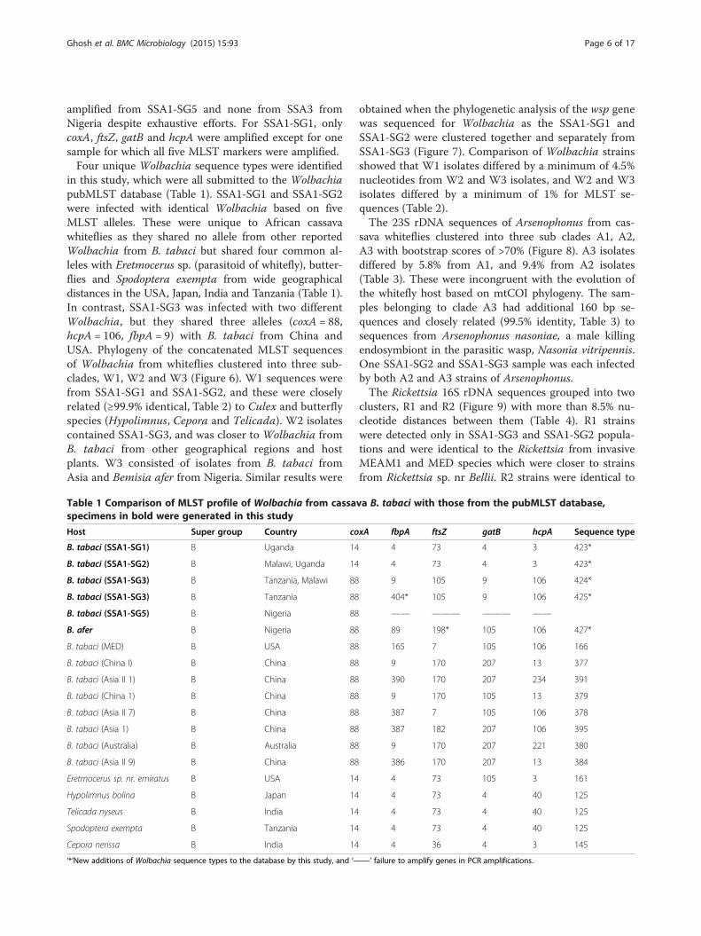
were seen in SSA1-SG3 (64.5%, 49/76) and SSA1-SG2(17.1%, 6/35), respectively (see Additional file 1: TableS2). Arsenophonus was present mostly as double infec-tions, with Wolbachia in SSA1-SG1 (17%) and SSA1-SG2 (11%), and with Rickettsia in SSA1-SG3 (28%).Arsenophonus was present in SSA1-SG5 and SSA3mainly as single infections (Figure 5).Rickettsia was absent in SSA1-SG5 but most abundant
in SSA1-SG3 (53.9%, 41/76) followed by SSA1-SG2(20%, 7/35). Its infection levels in other populations werenegligible. Cardiniumwas the least prevalent endosymbiont,
untries.
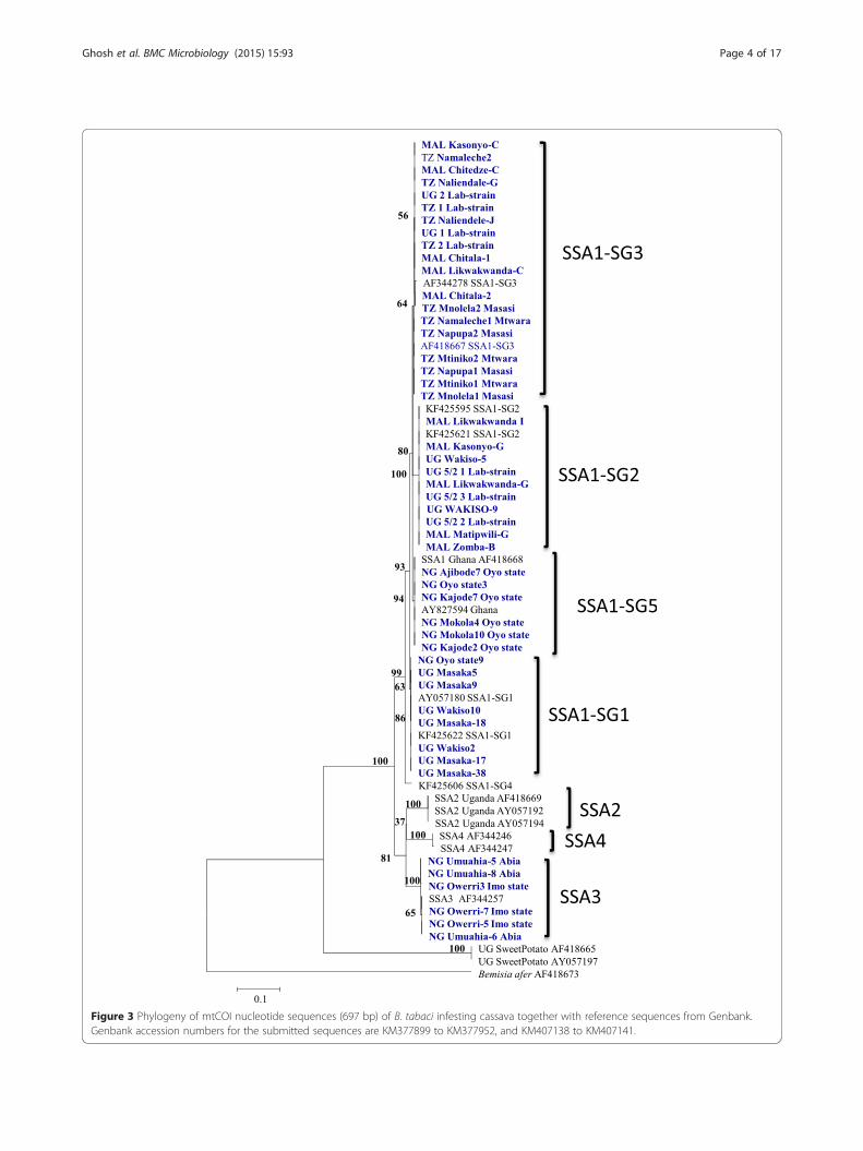
Figure 3 Phylogeny of mtCOI nucleotide sequences (697 bp) of B. tabaci infesting cassava together with reference sequences from Genbank.Genbank accession numbers for the submitted sequences are KM377899 to KM377952, and KM407138 to KM407141.
Ghosh et al. BMC Microbiology (2015) 15:93 Page 4 of 17
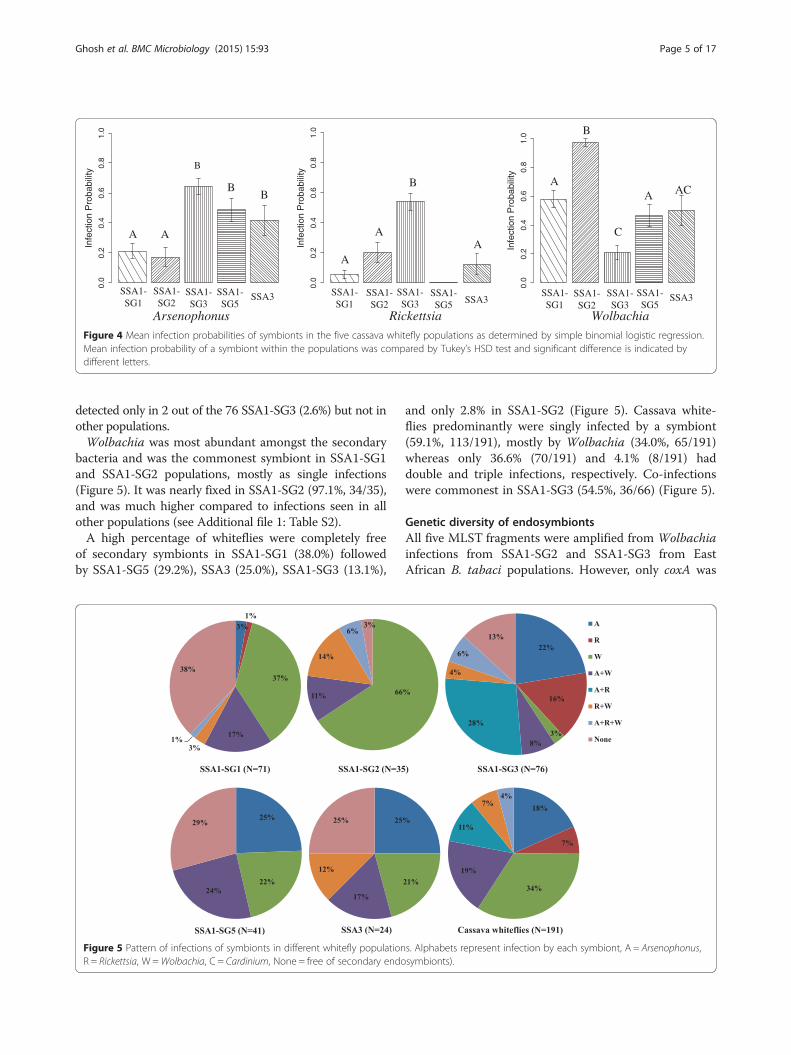
Figure 4 Mean infection probabilities of symbionts in the five cassava whitefly populations as determined by simple binomial logistic regression.Mean infection probability of a symbiont within the populations was compared by Tukey’s HSD test and significant difference is indicated bydifferent letters.
Ghosh et al. BMC Microbiology (2015) 15:93 Page 5 of 17
detected only in 2 out of the 76 SSA1-SG3 (2.6%) but not inother populations.Wolbachia was most abundant amongst the secondary
bacteria and was the commonest symbiont in SSA1-SG1and SSA1-SG2 populations, mostly as single infections(Figure 5). It was nearly fixed in SSA1-SG2 (97.1%, 34/35),and was much higher compared to infections seen in allother populations (see Additional file 1: Table S2).A high percentage of whiteflies were completely free
of secondary symbionts in SSA1-SG1 (38.0%) followedby SSA1-SG5 (29.2%), SSA3 (25.0%), SSA1-SG3 (13.1%),
Figure 5 Pattern of infections of symbionts in different whitefly populationR = Rickettsia, W =Wolbachia, C = Cardinium, None = free of secondary endo
and only 2.8% in SSA1-SG2 (Figure 5). Cassava white-flies predominantly were singly infected by a symbiont(59.1%, 113/191), mostly by Wolbachia (34.0%, 65/191)whereas only 36.6% (70/191) and 4.1% (8/191) haddouble and triple infections, respectively. Co-infectionswere commonest in SSA1-SG3 (54.5%, 36/66) (Figure 5).
Genetic diversity of endosymbiontsAll five MLST fragments were amplified from Wolbachiainfections from SSA1-SG2 and SSA1-SG3 from EastAfrican B. tabaci populations. However, only coxA was
s. Alphabets represent infection by each symbiont, A = Arsenophonus,symbionts).
Ghosh et al. BMC Microbiology (2015) 15:93 Page 6 of 17
amplified from SSA1-SG5 and none from SSA3 fromNigeria despite exhaustive efforts. For SSA1-SG1, onlycoxA, ftsZ, gatB and hcpA were amplified except for onesample for which all five MLST markers were amplified.Four unique Wolbachia sequence types were identified
in this study, which were all submitted to the WolbachiapubMLST database (Table 1). SSA1-SG1 and SSA1-SG2were infected with identical Wolbachia based on fiveMLST alleles. These were unique to African cassavawhiteflies as they shared no allele from other reportedWolbachia from B. tabaci but shared four common al-leles with Eretmocerus sp. (parasitoid of whitefly), butter-flies and Spodoptera exempta from wide geographicaldistances in the USA, Japan, India and Tanzania (Table 1).In contrast, SSA1-SG3 was infected with two differentWolbachia, but they shared three alleles (coxA = 88,hcpA = 106, fbpA = 9) with B. tabaci from China andUSA. Phylogeny of the concatenated MLST sequencesof Wolbachia from whiteflies clustered into three sub-clades, W1, W2 and W3 (Figure 6). W1 sequences werefrom SSA1-SG1 and SSA1-SG2, and these were closelyrelated (≥99.9% identical, Table 2) to Culex and butterflyspecies (Hypolimnus, Cepora and Telicada). W2 isolatescontained SSA1-SG3, and was closer to Wolbachia fromB. tabaci from other geographical regions and hostplants. W3 consisted of isolates from B. tabaci fromAsia and Bemisia afer from Nigeria. Similar results were
Table 1 Comparison of MLST profile of Wolbachia from cassaspecimens in bold were generated in this study
Host Super group Country co
B. tabaci (SSA1-SG1) B Uganda 14
B. tabaci (SSA1-SG2) B Malawi, Uganda 14
B. tabaci (SSA1-SG3) B Tanzania, Malawi 88
B. tabaci (SSA1-SG3) B Tanzania 88
B. tabaci (SSA1-SG5) B Nigeria 88
B. afer B Nigeria 88
B. tabaci (MED) B USA 88
B. tabaci (China I) B China 88
B. tabaci (Asia II 1) B China 88
B. tabaci (China 1) B China 88
B. tabaci (Asia II 7) B China 88
B. tabaci (Asia 1) B China 88
B. tabaci (Australia) B Australia 88
B. tabaci (Asia II 9) B China 88
Eretmocerus sp. nr. emiratus B USA 14
Hypolimnus bolina B Japan 14
Telicada nyseus B India 14
Spodoptera exempta B Tanzania 14
Cepora nerissa B India 14
‘*’New additions of Wolbachia sequence types to the database by this study, and ‘—
obtained when the phylogenetic analysis of the wsp genewas sequenced for Wolbachia as the SSA1-SG1 andSSA1-SG2 were clustered together and separately fromSSA1-SG3 (Figure 7). Comparison of Wolbachia strainsshowed that W1 isolates differed by a minimum of 4.5%nucleotides from W2 and W3 isolates, and W2 and W3isolates differed by a minimum of 1% for MLST se-quences (Table 2).The 23S rDNA sequences of Arsenophonus from cas-
sava whiteflies clustered into three sub clades A1, A2,A3 with bootstrap scores of >70% (Figure 8). A3 isolatesdiffered by 5.8% from A1, and 9.4% from A2 isolates(Table 3). These were incongruent with the evolution ofthe whitefly host based on mtCOI phylogeny. The sam-ples belonging to clade A3 had additional 160 bp se-quences and closely related (99.5% identity, Table 3) tosequences from Arsenophonus nasoniae, a male killingendosymbiont in the parasitic wasp, Nasonia vitripennis.One SSA1-SG2 and SSA1-SG3 sample was each infectedby both A2 and A3 strains of Arsenophonus.The Rickettsia 16S rDNA sequences grouped into two
clusters, R1 and R2 (Figure 9) with more than 8.5% nu-cleotide distances between them (Table 4). R1 strainswere detected only in SSA1-SG3 and SSA1-SG2 popula-tions and were identical to the Rickettsia from invasiveMEAM1 and MED species which were closer to strainsfrom Rickettsia sp. nr Bellii. R2 strains were identical to
va B. tabaci with those from the pubMLST database,
xA fbpA ftsZ gatB hcpA Sequence type
4 73 4 3 423*
4 73 4 3 423*
9 105 9 106 424*
404* 105 9 106 425*
—— ——— ——— ——
89 198* 105 106 427*
165 7 105 106 166
9 170 207 13 377
390 170 207 234 391
9 170 105 13 379
387 7 105 106 378
387 182 207 106 395
9 170 207 221 380
386 170 207 13 384
4 73 105 3 161
4 73 4 40 125
4 73 4 40 125
4 73 4 40 125
4 36 4 3 145
—’ failure to amplify genes in PCR amplifications.
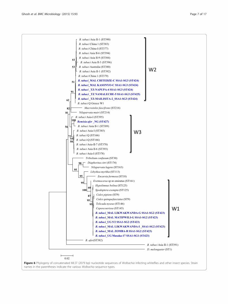
Figure 6 Phylogeny of concatenated MLST (2079 bp) nucleotide sequences of Wolbachia infecting whiteflies and other insect species. Strainnames in the parentheses indicate the various Wolbachia sequence types.
Ghosh et al. BMC Microbiology (2015) 15:93 Page 7 of 17
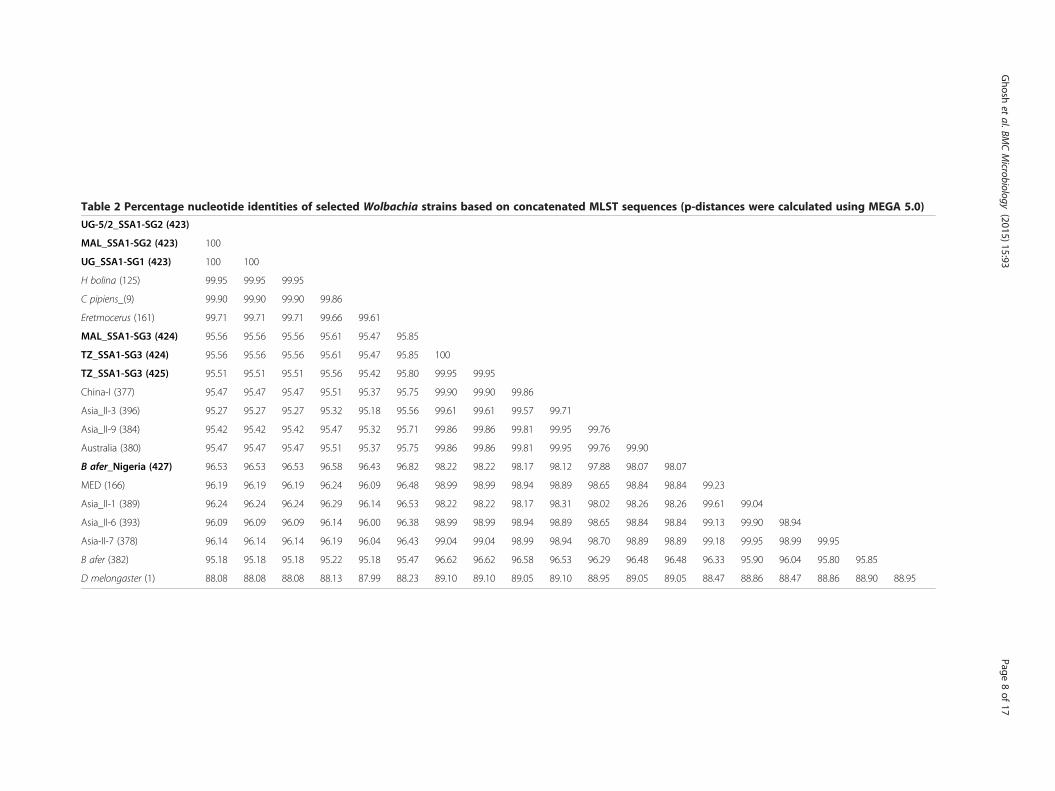
Table 2 Percentage nucleotide identities of selected Wolbachia strains based on concatenated MLST sequences (p-distances were calculated using MEGA 5.0)
UG-5/2_SSA1-SG2 (423)
MAL_SSA1-SG2 (423) 100
UG_SSA1-SG1 (423) 100 100
H bolina (125) 99.95 99.95 99.95
C pipiens_(9) 99.90 99.90 99.90 99.86
Eretmocerus (161) 99.71 99.71 99.71 99.66 99.61
MAL_SSA1-SG3 (424) 95.56 95.56 95.56 95.61 95.47 95.85
TZ_SSA1-SG3 (424) 95.56 95.56 95.56 95.61 95.47 95.85 100
TZ_SSA1-SG3 (425) 95.51 95.51 95.51 95.56 95.42 95.80 99.95 99.95
China-I (377) 95.47 95.47 95.47 95.51 95.37 95.75 99.90 99.90 99.86
Asia_II-3 (396) 95.27 95.27 95.27 95.32 95.18 95.56 99.61 99.61 99.57 99.71
Asia_II-9 (384) 95.42 95.42 95.42 95.47 95.32 95.71 99.86 99.86 99.81 99.95 99.76
Australia (380) 95.47 95.47 95.47 95.51 95.37 95.75 99.86 99.86 99.81 99.95 99.76 99.90
B afer_Nigeria (427) 96.53 96.53 96.53 96.58 96.43 96.82 98.22 98.22 98.17 98.12 97.88 98.07 98.07
MED (166) 96.19 96.19 96.19 96.24 96.09 96.48 98.99 98.99 98.94 98.89 98.65 98.84 98.84 99.23
Asia_II-1 (389) 96.24 96.24 96.24 96.29 96.14 96.53 98.22 98.22 98.17 98.31 98.02 98.26 98.26 99.61 99.04
Asia_II-6 (393) 96.09 96.09 96.09 96.14 96.00 96.38 98.99 98.99 98.94 98.89 98.65 98.84 98.84 99.13 99.90 98.94
Asia-II-7 (378) 96.14 96.14 96.14 96.19 96.04 96.43 99.04 99.04 98.99 98.94 98.70 98.89 98.89 99.18 99.95 98.99 99.95
B afer (382) 95.18 95.18 95.18 95.22 95.18 95.47 96.62 96.62 96.58 96.53 96.29 96.48 96.48 96.33 95.90 96.04 95.80 95.85
D melongaster (1) 88.08 88.08 88.08 88.13 87.99 88.23 89.10 89.10 89.05 89.10 88.95 89.05 89.05 88.47 88.86 88.47 88.86 88.90 88.95
Ghosh
etal.BM
CMicrobiology
(2015) 15:93 Page
8of
17
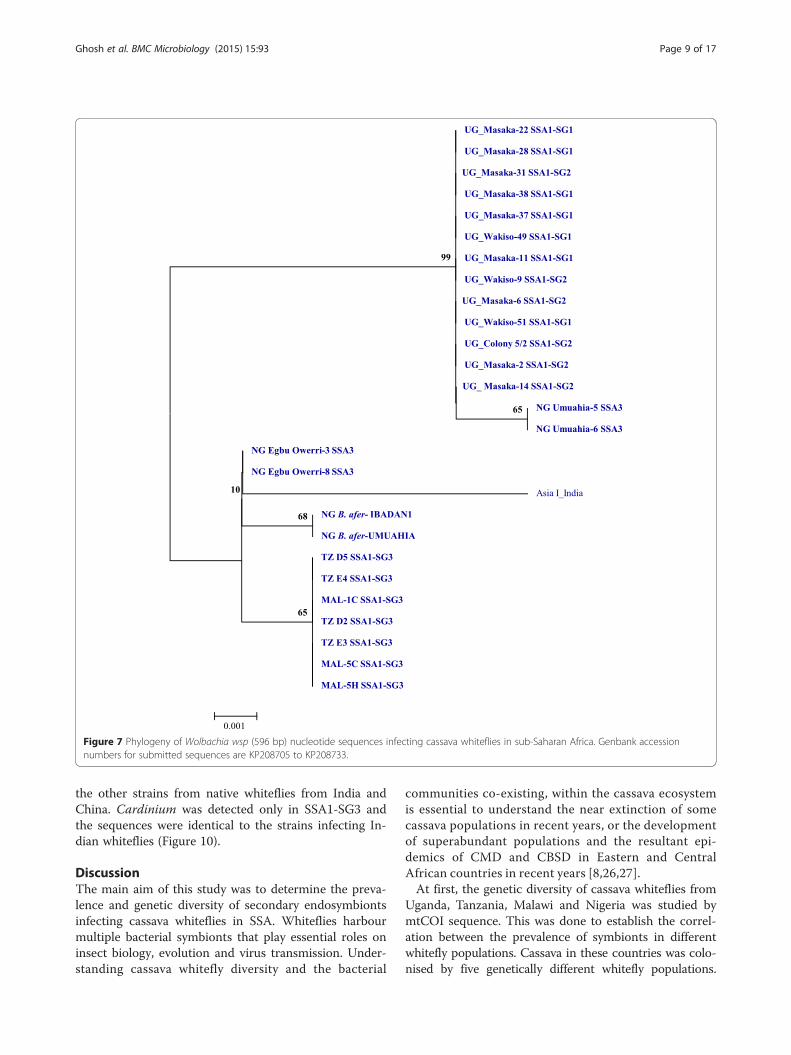
Figure 7 Phylogeny of Wolbachia wsp (596 bp) nucleotide sequences infecting cassava whiteflies in sub-Saharan Africa. Genbank accessionnumbers for submitted sequences are KP208705 to KP208733.
Ghosh et al. BMC Microbiology (2015) 15:93 Page 9 of 17
the other strains from native whiteflies from India andChina. Cardinium was detected only in SSA1-SG3 andthe sequences were identical to the strains infecting In-dian whiteflies (Figure 10).
DiscussionThe main aim of this study was to determine the preva-lence and genetic diversity of secondary endosymbiontsinfecting cassava whiteflies in SSA. Whiteflies harbourmultiple bacterial symbionts that play essential roles oninsect biology, evolution and virus transmission. Under-standing cassava whitefly diversity and the bacterial
communities co-existing, within the cassava ecosystemis essential to understand the near extinction of somecassava populations in recent years, or the developmentof superabundant populations and the resultant epi-demics of CMD and CBSD in Eastern and CentralAfrican countries in recent years [8,26,27].At first, the genetic diversity of cassava whiteflies from
Uganda, Tanzania, Malawi and Nigeria was studied bymtCOI sequence. This was done to establish the correl-ation between the prevalence of symbionts in differentwhitefly populations. Cassava in these countries was colo-nised by five genetically different whitefly populations.
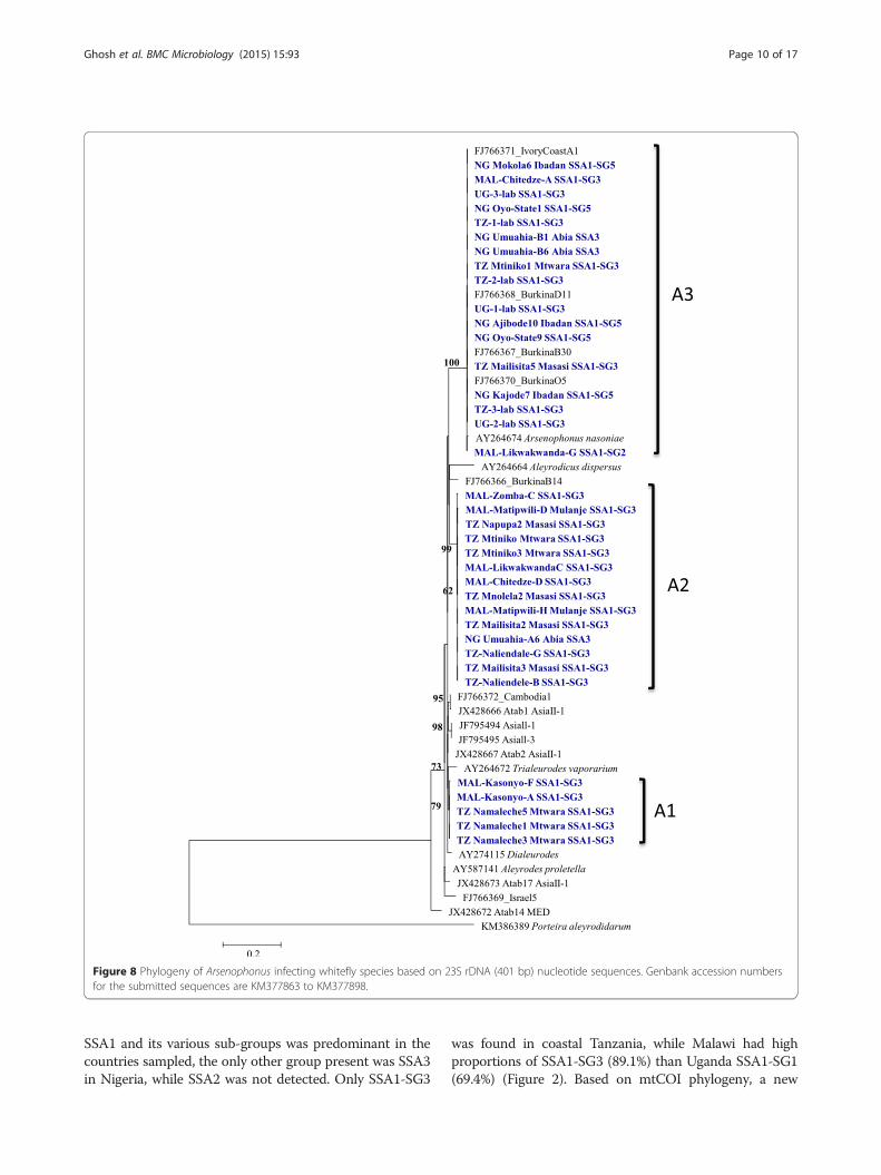
Figure 8 Phylogeny of Arsenophonus infecting whitefly species based on 23S rDNA (401 bp) nucleotide sequences. Genbank accession numbersfor the submitted sequences are KM377863 to KM377898.
Ghosh et al. BMC Microbiology (2015) 15:93 Page 10 of 17
SSA1 and its various sub-groups was predominant in thecountries sampled, the only other group present was SSA3in Nigeria, while SSA2 was not detected. Only SSA1-SG3
was found in coastal Tanzania, while Malawi had highproportions of SSA1-SG3 (89.1%) than Uganda SSA1-SG1(69.4%) (Figure 2). Based on mtCOI phylogeny, a new
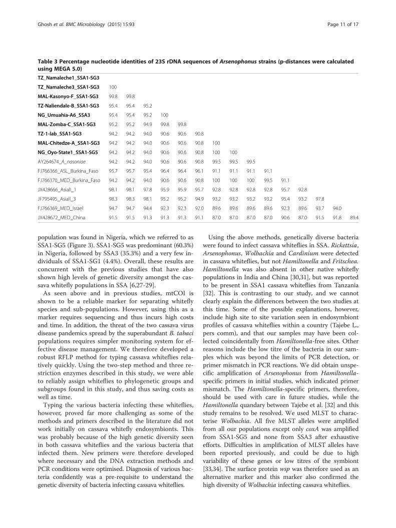
Table 3 Percentage nucleotide identities of 23S rDNA sequences of Arsenophonus strains (p-distances were calculatedusing MEGA 5.0)
TZ_Namaleche1_SSA1-SG3
TZ_Namaleche3_SSA1-SG3 100
MAL-Kasonyo-F_SSA1-SG3 99.8 99.8
TZ-Naliendale-B_SSA1-SG3 95.4 95.4 95.2
NG_Umuahia-A6_SSA3 95.4 95.4 95.2 100
MAL-Zomba-C_SSA1-SG3 95.2 95.2 94.9 99.8 99.8
TZ-1-lab_SSA1-SG3 94.2 94.2 94.0 90.6 90.6 90.8
MAL-Chitedze-A_SSA1-SG3 94.2 94.2 94.0 90.6 90.6 90.8 100
NG_Oyo-State1_SSA1-SG5 94.2 94.2 94.0 90.6 90.6 90.8 100 100
AY264674_A_nasoniae 94.2 94.2 94.0 90.6 90.6 90.8 99.5 99.5 99.5
FJ766366_ASL_Burkina_Faso 95.7 95.7 95.4 96.4 96.4 96.1 91.1 91.1 91.1 91.1
FJ766370_MED_Burkina_Faso 94.2 94.2 94.0 90.6 90.6 90.8 100 100 100 99.5 91.1
JX428666_AsiaII_1 98.1 98.1 97.8 95.9 95.9 95.7 92.8 92.8 92.8 92.8 95.7 92.8
JF795495_Asiall_3 98.3 98.3 98.1 95.2 95.2 94.9 93.2 93.2 93.2 93.2 95.4 93.2 97.8
FJ766369_MED_Israel 94.7 94.7 94.4 92.3 92.3 92.0 89.6 89.6 89.6 89.6 92.3 89.6 93.7 94.0
JX428672_MED_China 91.5 91.5 91.3 91.3 91.3 91.1 87.0 87.0 87.0 87.0 90.6 87.0 91.5 91.8 89.4
Ghosh et al. BMC Microbiology (2015) 15:93 Page 11 of 17
population was found in Nigeria, which we referred to asSSA1-SG5 (Figure 3). SSA1-SG5 was predominant (60.3%)in Nigeria, followed by SSA3 (35.3%) and a very few in-dividuals of SSA1-SG1 (4.4%). Overall, these results areconcurrent with the previous studies that have alsoshown high levels of genetic diversity amongst the cas-sava whitefly populations in SSA [6,27-29].As seen above and in previous studies, mtCOI is
shown to be a reliable marker for separating whiteflyspecies and sub-populations. However, using this as amarker requires sequencing and thus incurs high costsand time. In addition, the threat of the two cassava virusdisease pandemics spread by the superabundant B. tabacipopulations requires simpler monitoring system for ef-fective disease management. We therefore developed arobust RFLP method for typing cassava whiteflies rela-tively quickly. Using the two-step method and three re-striction enzymes described in this study, we were ableto reliably assign whiteflies to phylogenetic groups andsubgroups found in this study, and thus saving costs aswell as time.Typing the various bacteria infecting these whiteflies,
however, proved far more challenging as some of themethods and primers described in the literature did notwork initially on cassava whitefly endosymbionts. Thiswas probably because of the high genetic diversity seenin both cassava whiteflies and the various bacteria thatinfected them. New primers were therefore developedwhere necessary and the DNA extraction methods andPCR conditions were optimised. Diagnosis of various bac-teria confidently was a pre-requisite to understand thegenetic diversity of bacteria infecting cassava whiteflies.
Using the above methods, genetically diverse bacteriawere found to infect cassava whiteflies in SSA. Rickettsia,Arsenophonus, Wolbachia and Cardinium were detectedin cassava whiteflies, but not Hamiltonella and Fritschea.Hamiltonella was also absent in other native whiteflypopulations in India and China [30,31], but was reportedto be present in SSA1 cassava whiteflies from Tanzania[32]. This is contrasting to our study, and we cannotclearly explain the differences between the two studies atthis time. Some of the possible explanations, however,include high site to site variation seen in endosymbiontprofiles of cassava whiteflies within a country (Tajebe L.,pers comm), and that our samples may have been col-lected coincidentally from Hamiltonella-free sites. Otherreasons include the low titre of the bacteria in our sam-ples which was beyond the limits of PCR detection, orprimer mismatch in PCR reactions. We did obtain unspe-cific amplification of Arsenophonus from Hamiltonella-specific primers in initial studies, which indicated primermismatch. The Hamiltonella-specific primers, therefore,should be used with care in future studies, while theHamiltonella quandary between Tajebe et al. [32] and thisstudy remains to be resolved. We used MLST to charac-terise Wolbachia. All five MLST alleles were amplifiedfrom all our populations except only coxA was amplifiedfrom SSA1-SG5 and none from SSA3 after exhaustiveefforts. Difficulties in amplification of MLST alleles havebeen reported previously, and could be due to highvariability of these genes or low titres of the symbiont[33,34]. The surface protein wsp was therefore used as analternative marker and this marker also confirmed thehigh diversity of Wolbachia infecting cassava whiteflies.
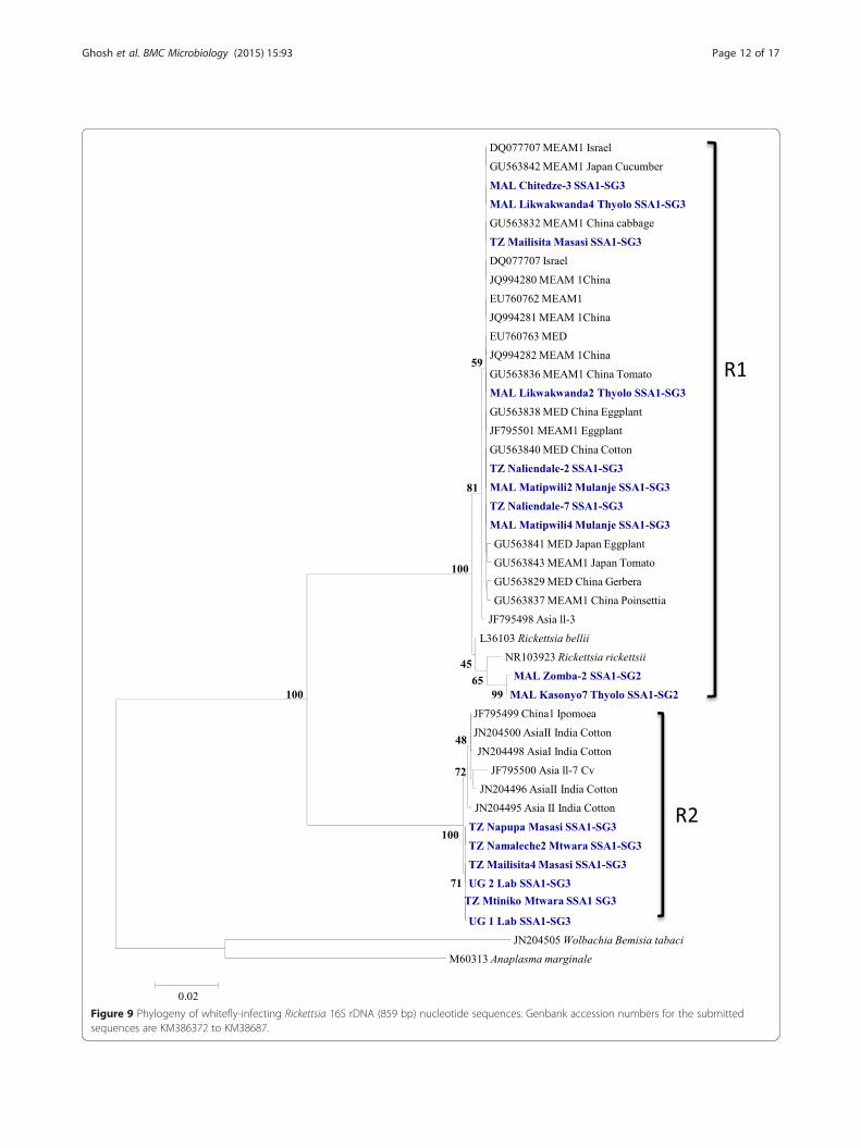
Figure 9 Phylogeny of whitefly-infecting Rickettsia 16S rDNA (859 bp) nucleotide sequences. Genbank accession numbers for the submittedsequences are KM386372 to KM38687.
Ghosh et al. BMC Microbiology (2015) 15:93 Page 12 of 17
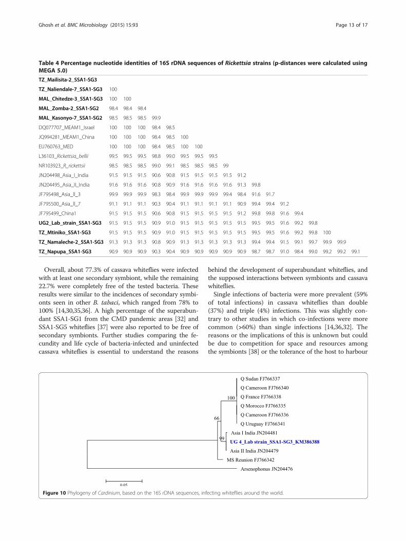
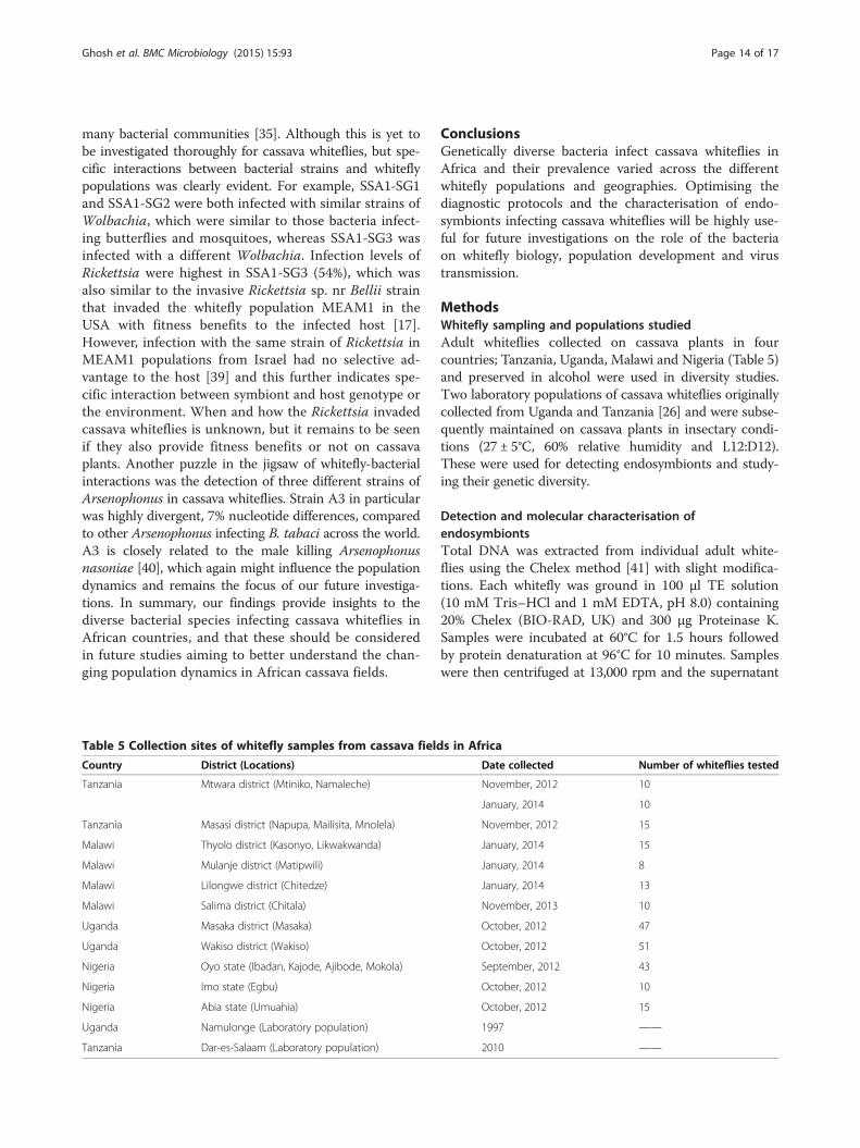
Table 4 Percentage nucleotide identities of 16S rDNA sequences of Rickettsia strains (p-distances were calculated usingMEGA 5.0)
TZ_Mailisita-2_SSA1-SG3
TZ_Naliendale-7_SSA1-SG3 100
MAL_Chitedze-3_SSA1-SG3 100 100
MAL_Zomba-2_SSA1-SG2 98.4 98.4 98.4
MAL_Kasonyo-7_SSA1-SG2 98.5 98.5 98.5 99.9
DQ077707_MEAM1_Israel 100 100 100 98.4 98.5
JQ994281_MEAM1_China 100 100 100 98.4 98.5 100
EU760763_MED 100 100 100 98.4 98.5 100 100
L36103_Rickettsia_bellii 99.5 99.5 99.5 98.8 99.0 99.5 99.5 99.5
NR103923_R_rickettsii 98.5 98.5 98.5 99.0 99.1 98.5 98.5 98.5 99
JN204498_Asia_I_India 91.5 91.5 91.5 90.6 90.8 91.5 91.5 91.5 91.5 91.2
JN204495_Asia_II_India 91.6 91.6 91.6 90.8 90.9 91.6 91.6 91.6 91.6 91.3 99.8
JF795498_Asia_ll_3 99.9 99.9 99.9 98.3 98.4 99.9 99.9 99.9 99.4 98.4 91.6 91.7
JF795500_Asia_ll_7 91.1 91.1 91.1 90.3 90.4 91.1 91.1 91.1 91.1 90.9 99.4 99.4 91.2
JF795499_China1 91.5 91.5 91.5 90.6 90.8 91.5 91.5 91.5 91.5 91.2 99.8 99.8 91.6 99.4
UG2_Lab_strain_SSA1-SG3 91.5 91.5 91.5 90.9 91.0 91.5 91.5 91.5 91.5 91.5 99.5 99.5 91.6 99.2 99.8
TZ_Mtiniko_SSA1-SG3 91.5 91.5 91.5 90.9 91.0 91.5 91.5 91.5 91.5 91.5 99.5 99.5 91.6 99.2 99.8 100
TZ_Namaleche-2_SSA1-SG3 91.3 91.3 91.3 90.8 90.9 91.3 91.3 91.3 91.3 91.3 99.4 99.4 91.5 99.1 99.7 99.9 99.9
TZ_Napupa_SSA1-SG3 90.9 90.9 90.9 90.3 90.4 90.9 90.9 90.9 90.9 90.9 98.7 98.7 91.0 98.4 99.0 99.2 99.2 99.1
Ghosh et al. BMC Microbiology (2015) 15:93 Page 13 of 17
Overall, about 77.3% of cassava whiteflies were infectedwith at least one secondary symbiont, while the remaining22.7% were completely free of the tested bacteria. Theseresults were similar to the incidences of secondary symbi-onts seen in other B. tabaci, which ranged from 78% to100% [14,30,35,36]. A high percentage of the superabun-dant SSA1-SG1 from the CMD pandemic areas [32] andSSA1-SG5 whiteflies [37] were also reported to be free ofsecondary symbionts. Further studies comparing the fe-cundity and life cycle of bacteria-infected and uninfectedcassava whiteflies is essential to understand the reasons
Figure 10 Phylogeny of Cardinium, based on the 16S rDNA sequences, inf
behind the development of superabundant whiteflies, andthe supposed interactions between symbionts and cassavawhiteflies.Single infections of bacteria were more prevalent (59%
of total infections) in cassava whiteflies than double(37%) and triple (4%) infections. This was slightly con-trary to other studies in which co-infections were morecommon (>60%) than single infections [14,36,32]. Thereasons or the implications of this is unknown but couldbe due to competition for space and resources amongthe symbionts [38] or the tolerance of the host to harbour
ecting whiteflies around the world.
Ghosh et al. BMC Microbiology (2015) 15:93 Page 14 of 17
many bacterial communities [35]. Although this is yet tobe investigated thoroughly for cassava whiteflies, but spe-cific interactions between bacterial strains and whiteflypopulations was clearly evident. For example, SSA1-SG1and SSA1-SG2 were both infected with similar strains ofWolbachia, which were similar to those bacteria infect-ing butterflies and mosquitoes, whereas SSA1-SG3 wasinfected with a different Wolbachia. Infection levels ofRickettsia were highest in SSA1-SG3 (54%), which wasalso similar to the invasive Rickettsia sp. nr Bellii strainthat invaded the whitefly population MEAM1 in theUSA with fitness benefits to the infected host [17].However, infection with the same strain of Rickettsia inMEAM1 populations from Israel had no selective ad-vantage to the host [39] and this further indicates spe-cific interaction between symbiont and host genotype orthe environment. When and how the Rickettsia invadedcassava whiteflies is unknown, but it remains to be seenif they also provide fitness benefits or not on cassavaplants. Another puzzle in the jigsaw of whitefly-bacterialinteractions was the detection of three different strains ofArsenophonus in cassava whiteflies. Strain A3 in particularwas highly divergent, 7% nucleotide differences, comparedto other Arsenophonus infecting B. tabaci across the world.A3 is closely related to the male killing Arsenophonusnasoniae [40], which again might influence the populationdynamics and remains the focus of our future investiga-tions. In summary, our findings provide insights to thediverse bacterial species infecting cassava whiteflies inAfrican countries, and that these should be consideredin future studies aiming to better understand the chan-ging population dynamics in African cassava fields.
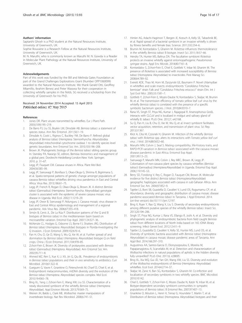
Table 5 Collection sites of whitefly samples from cassava field
Country District (Locations)
Tanzania Mtwara district (Mtiniko, Namaleche)
Tanzania Masasi district (Napupa, Mailisita, Mnolela)
Malawi Thyolo district (Kasonyo, Likwakwanda)
Malawi Mulanje district (Matipwili)
Malawi Lilongwe district (Chitedze)
Malawi Salima district (Chitala)
Uganda Masaka district (Masaka)
Uganda Wakiso district (Wakiso)
Nigeria Oyo state (Ibadan, Kajode, Ajibode, Mokola)
Nigeria Imo state (Egbu)
Nigeria Abia state (Umuahia)
Uganda Namulonge (Laboratory population)
Tanzania Dar-es-Salaam (Laboratory population)
ConclusionsGenetically diverse bacteria infect cassava whiteflies inAfrica and their prevalence varied across the differentwhitefly populations and geographies. Optimising thediagnostic protocols and the characterisation of endo-symbionts infecting cassava whiteflies will be highly use-ful for future investigations on the role of the bacteriaon whitefly biology, population development and virustransmission.
MethodsWhitefly sampling and populations studiedAdult whiteflies collected on cassava plants in fourcountries; Tanzania, Uganda, Malawi and Nigeria (Table 5)and preserved in alcohol were used in diversity studies.Two laboratory populations of cassava whiteflies originallycollected from Uganda and Tanzania [26] and were subse-quently maintained on cassava plants in insectary condi-tions (27 ± 5°C, 60% relative humidity and L12:D12).These were used for detecting endosymbionts and study-ing their genetic diversity.
Detection and molecular characterisation ofendosymbiontsTotal DNA was extracted from individual adult white-flies using the Chelex method [41] with slight modifica-tions. Each whitefly was ground in 100 μl TE solution(10 mM Tris–HCl and 1 mM EDTA, pH 8.0) containing20% Chelex (BIO-RAD, UK) and 300 μg Proteinase K.Samples were incubated at 60°C for 1.5 hours followedby protein denaturation at 96°C for 10 minutes. Sampleswere then centrifuged at 13,000 rpm and the supernatant
s in Africa
Date collected Number of whiteflies tested
November, 2012 10
January, 2014 10
November, 2012 15
January, 2014 15
January, 2014 8
January, 2014 13
November, 2013 10
October, 2012 47
October, 2012 51
September, 2012 43
October, 2012 10
October, 2012 15
1997 ——
2010 ——

Ghosh et al. BMC Microbiology (2015) 15:93 Page 15 of 17
was collected and stored at −20°C. Whitefly mtCOI genesand the endosymbiont 16S or 23S rDNA were amplifiedby polymerase chain reactions (PCR) using genus specificprimers (see Additional file 1). New primers were de-signed for Cardinium and Wolbachia to increase effi-ciency and specificity of detection. Multilocus sequencetyping (MLST) based on the diversity of five conservedhousekeeping genes; coxA, fbpA, ftsZ, gatB and hcpA havebeen used as a standard tool for strain typing and evolu-tionary studies of Wolbachia. The MLST approach wasused to characterize the Wolbachia infecting cassavawhiteflies using standard primers and protocols [42]. TheWolbachia surface protein (wsp) gene was also used as anadditional marker for characterisation. Amplification ofthese genes was carried out in 25 μl volumes using 2 μlDNA lysate as template, 0.4 μM of each primer, 0.15 mMof dNTPs, 1 × DreamTaq Green buffer and 0.5 unitDreamTaq Green DNA polymerase (Thermo ScientificLtd., UK). Amplifications consisted of 94°C for 3 minutesfollowed by 38 cycles of 94°C for 30 seconds, annealingfor 45 seconds (Additional file 1: Table S1), 72°C for1.5 minutes and final extension for 7 minutes at 72°C.PCR products were visualised on 1% agarose gels contain-ing RedSafe nucleic acid staining solution (Intron Bio-technology, Korea). PCR products were purified andsubmitted for Sanger sequencing (Source Bioscience, UK)in both directions per whitefly sample, and five sampleswere sequenced for each location. Endosymbionts werealso detected and sequences from two laboratory whiteflystrains (Table 5). Sequences were compared to known se-quences in databases using the BLAST algorithm inNCBI.
Developing a diagnostic tool for cassava whitefliesThe mtCOI fragments from five whitefly samples per lo-cation were sequenced, followed by phylogenetic analysiswith reference sequences of haplotypes [6] for the identi-fication of consensus haplotype groupings. The whiteflymtCOI sequences generated were analysed to identifyunique restriction endonuclease sites using the softwarepackage NEBcutter (http://tools.neb.com/NEBcutter2).Three enzymes Bgl II (A/GATCT), Apo I (R/AATTY)and Dde I (C/TNAG) were found to produce uniquepatterns across SSA populations. The mtCOI fragmentswere re-amplified from at least 20 adults for each cas-sava whitefly population using 3 μl of DNA templateand 1 unit of DreamTaq DNA polymerase in 30 μl vol-ume reactions (40 cycles) for higher yields. Previouslyextracted DNA from four SSA2 whitefly samples wereused in this assay as reference samples [26]. The RFLPwas carried out in a two-step procedure. First, 15 μl ofPCR products were digested with 5 units of Bgl II.Second, the remaining 15 μl of PCR products weredigested with 5 units each of Apo I and Dde I at 37°C
for 1.5 hours. Digested products were electrophoresedseparately on 2% agarose gels.
Phylogenetic and statistical analysisThe mtCOI sequences from the whitefly, the 16S or 23SrDNA sequences from the endosymbionts and the MLSTsequences from Wolbachia were aligned separately usingClustalW of MEGA 5.2 [43]. Phylogenetic trees wereconstructed by the maximum-likelihood method usingMEGA 5.2. Different nucleotide substitution modelswere used based on the lowest Bayesian information cri-terion scores obtained. Phylogenetic trees for mtCOIand Wolbachia were generated using the T93 + G + Isubstitution model, the HKY + G substitution modelfor Arsenophonus, the K2 + G substitution model forRickettsia and the K2 substitution model for Cardinium[44]. The robustness of the clades was assessed by 1000bootstrap replicates.The probabilities of bacterial infections in cassava
whitefly populations were predicted using simple bino-mial logistic regression. Each bacterium was used as thedependent variable and the whitefly populations as inde-pendent variables. Differences in infection patternsamong groups were evaluated by Tukey’s HSD test usingthe glht function from multcomp package of R [45].
Availability of supporting dataThe data sets supporting the results of this article areavailable in the MLST and EMBL database with uniquesequence and accession numbers. These are currentlypublicly available.Genbank accession numbers generated in this study
are as below; mtCOI sequences KM377899 to KM377952,and KM407138 to KM407141; Wolbachia wsp KP208705to KP208733; Arsenophonus 23S rDNA KM377863 toKM377898, Rickettsia 16S rDNA.KM386372 to KM38687; and Cardinium KM386388.
The accession number for the MLST sequence types onthe pubMLST database for the Wolbachia infecting cas-sava whitefly are 423–425 and 427.
Additional file
Additional file 1: Table S1. Primer sequences and annealingtemperatures used for PCR amplification. Table S2. Multiple comparisonsof mean infection incidence of symbionts: Tukey contrasts.
Competing interestsThe authors declare that they have no competing interests.
Authors’ contributionsMNM conceived the work, designed research, collected samples andcorrected the paper extensively. SB helped with analysis and corrected thepaper. SG designed and performed research, carried out most of the analysisand made initial draft of the paper. All authors read and approved the finalmanuscript.
Ghosh et al. BMC Microbiology (2015) 15:93 Page 16 of 17
Authors’ informationSaptarshi Ghosh is a PhD student at the Natural Resources Institute,University of Greenwich, UK.Sophie Bouvaine is a Research Fellow at the Natural Resources Institute,University of Greenwich, UK.M. N. Maruthi, who is commonly known as Maruthi M. N. Gowda is a Readerin Molecular Plant Pathology at the Natural Resources Institute, University ofGreenwich, UK.
AcknowledgementsPart of this work was funded by the Bill and Melinda Gates Foundation aspart of the Grand Challenges Explorations Grant (Number OPP1060099)awarded to the Natural Resources Institute. We thank Gerald Otti, GeoffreyMkamillo, Ibrahim Benesi and Peter Wasswa for their cooperation incollecting whitefly samples in the fields. SG received a scholarship from theUniversity of Greenwich for his PhD.
Received: 24 November 2014 Accepted: 15 April 2015
References1. Jones DR. Plant viruses transmitted by whiteflies. Eur J Plant Path.
2003;(109):195–219.2. De Barro PJ, Liu SS, Boykin LM, Dinsdale AB. Bemisia tabaci: a statement of
species status. Ann Rev Entomol. 2011;56:1–19.3. Dinsdale A, Cook L, Riginos C, Buckley YM, De Barro P. Refined global
analysis of Bemisia tabaci (Hemiptera: Sternorrhyncha: Aleyrodoidea:Aleyrodidae) mitochondrial cytochrome oxidase 1 to identify species levelgenetic boundaries. Ann Entomol Soc Am. 2010;103:196–208.
4. Brown JK. Phylogenetic biology of the Bemisia tabaci sibling species group.In: Stansley PA, Naranjo SE, editors. Bemisia: bionomics and management ofa global pest. Dordrecht-Heidelberg-London-New York: Springer;2010. p. 31–67.
5. Legg JP, Fauquet CM. Cassava viruses in Africa. Plant Mol Biol.2004;56:585–99.
6. Legg JP, Sseruwagi P, Boniface S, Okao-Okuja G, Shirima R, Bigirimana S,et al. Spatio-temporal patterns of genetic change amongst populations ofcassava Bemisia tabaci whiteflies driving virus pandemics in East and CentralAfrica. Virus Res. 2013;186:61–75.
7. Legg JP, French R, Rogan D, Okao-Okuja G, Brown JK. A distinct Bemisiatabaci (Gennadius) (Hemiptera: Sternorrhyncha: Aleyrodidae) genotypecluster is associated with the epidemic of severe cassava mosaic virusdisease in Uganda. Mol Ecol. 2002;11:1219–29.
8. Legg JP, Owor B, Sseruwagi P, Ndunguru J. Cassava mosaic virus disease inEast and Central Africa: epidemiology and management of a regionalpandemic. Adv Virus Res. 2006;67:355–418.
9. Simón B, Cenis JL, De La Rúa P. Distribution patterns of the Q and Bbiotypes of Bemisia tabaci in the mediterranean basin based onmicrosatellite variation. Entomol Exp Appl. 2007;124:327–36.
10. McKenzie CL, Hodges G, Osborne LS, Byrne FJ, Shatters JRG. Distribution ofBemisia tabaci (Hemiptera: Aleyrodidae) biotypes in Florida-investigating theQ invasion. J Econ Entomol. 2009;102:670–6.
11. Pan H, Chu D, Ge D, Wang S, Wu Q, Xie W, et al. Further spread of anddomination by Bemisia tabaci (Hemiptera: Aleyrodidae) biotype Q on fieldcrops. China J Econ Entomol. 2011;104:978–85.
12. Zchori-Fein E, Brown JK. Diversity of prokaryotes associated with Bemisiatabaci (Gennadius) (Hemiptera: Aleyrodidae). Ann Entomol Soc Am.2002;95:711–8.
13. Ahmed MZ, Ren S, Xue X, Li XX, Jin G, Qiu BL. Prevalence of endosymbiontsin Bemisia tabaci populations and their in vivo sensitivity to antibiotics. CurrMicrobiol. 2010;61:322–8.
14. Gueguen G, Vavre F, Gnankine O, Peterschmitt M, Charif D, Chiel E, et al.Endosymbiont metacommunities, mtDNA diversity and the evolution of theBemisia tabaci (Hemiptera: Aleyrodidae) species complex. Mol Ecol.2010;19:4365–78.
15. Bing XL, Yang J, Zchori-Fein E, Wang XW, Liu SS. Characterization of anewly discovered symbiont of the whitefly Bemisia tabaci (Hemiptera:Aleyrodidae). Appl Environ Microb. 2013;79:569–75.
16. Werren JH, Baldo L, Clark ME. Wolbachia: master manipulators ofinvertebrate biology. Nat Rev Microbiol. 2008;6:741–51.
17. Himler AG, Adachi-Hagimori T, Bergen JE, Kozuch A, Kelly SE, Tabashnik BE,et al. Rapid spread of a bacterial symbiont in an invasive whitefly is drivenby fitness benefits and female bias. Science. 2011;332:254–6.
18. Brumin M, Kontsedalov S, Ghanim M. Rickettsia influences thermotolerancein the whitefly Bemisia tabaci B biotype. Insect Sci. 2011;18:57–66.
19. Hendry TA, Hunter MS, Baltrus DA. The facultative symbiont Rickettsiaprotects an invasive whitefly against entomopathogenic Pseudomonassyringae strains. Appl Env Microb. 2014;80:7161–8.
20. Kontsedalov S, Zchori‐Fein E, Chiel E, Gottlieb Y, Inbar M, Ghanim M. Thepresence of Rickettsia is associated with increased susceptibility of Bemisiatabaci (Homoptera: Aleyrodidae) to insecticides. Pest Manag Sci.2008;64:789–92.
21. Everett KDE, Thao M, Horn M, Dyszynski GE, Baumann P. Novel chlamydiaein whiteflies and scale insects: endosymbionts “Candidatus Fritscheabemisiae” strain Falk and “Candidatus Fritschea eriococci” strain Elm. Int JSyst Evol Micr. 2005;55:1581–7.
22. Gottlieb Y, Zchori-Fein E, Mozes-Daube N, Kontsedalov S, Skaljac M, BruminM, et al. The transmission efficiency of tomato yellow leaf curl virus by thewhitefly Bemisia tabaci is correlated with the presence of a specificsymbiotic bacterium species. J Virol. 2010;84:9310–7.
23. Rana VS, Singh ST, Priya NG, Kumar J, Rajagopal R. Arsenophonus GroELinteracts with CLCuV and is localized in midgut and salivary gland ofwhitefly B. tabaci. PLoS One. 2012;7, e42168.
24. Su Q, Pan H, Liu B, Chu D, Xie W, Wu Q, et al. Insect symbiont facilitatesvector acquisition, retention, and transmission of plant virus. Sci Rep.2013;3:1367.
25. Kliot A, Cilia M, Czosnek H, Ghanim M. Infection of the whitefly Bemisiatabaci with Rickettsia spp. alters its interactions with Tomato yellow leaf curlvirus. J Virol. 2014;88:5652–60.
26. Maruthi MN, Colvin J, Seal S. Mating compatibility, life‐history traits, andRAPD‐PCR variation in Bemisia tabaci associated with the cassava mosaicdisease pandemic in East Africa. Entomol Exp Appl.2001;99:13–23.
27. Sseruwagi P, Maruthi MN, Colvin J, Rey MEC, Brown JK, Legg JP.Colonisation of non-cassava plant species by cassava whiteflies (Bemisiatabaci) (Gennadius) (Hemiptera:Aleyrodidae) in Uganda. Entomol Exp Appl.2006;119:145–53.
28. Berry SD, Fondong V, Rey C, Rogan D, Fauquet CM, Brown JK. Molecularevidence for five distinct Bemisia tabaci (Homoptera:Aleyrodidae)geographic haplotypes associated with cassava in sub-Saharan Africa. AnnEntomol Soc Am. 2004;97:852–9.
29. Tajebe LS, Boni SB, Guastella D, Cavalieri V, Lund OS, Rugumamu CP, et al.Abundance, diversity and geographic distribution of cassava mosaic diseasepandemic‐associated Bemisia tabaci in Tanzania. J Appl Entomol. 2014.(on-line version) doi:10.1111/jen.12197.
30. Bing X, Ruan Y, Rao Q, Wang X, Liu S. Diversity of secondary endosymbiontsamong different putative species of the whitefly Bemisia tabaci. Insect Sci.2013;20:194–206.
31. Singh ST, Priya NG, Kumar J, Rana VS, Ellango R, Joshi A, et al. Diversity andphylogenetic analysis of endosymbiotic bacteria from field caught Bemisiatabaci from different locations of North India based on 16S rDNA libraryscreening. Infect Genet Evol. 2012;12:411–9.
32. Tajebe LS, Guastella D, Cavalieri V, Kelly SE, Hunter MS, Lund OS, et al.Diversity of symbiotic bacteria associated with Bemisia tabaci (Homoptera:Aleyrodidae) in cassava mosaic disease pandemic areas of Tanzania. AnnAppl Biol. 2014;166:297–310.
33. Augustinos AA, Santos-Garcia D, Dionyssopoulou E, Moreira M,Papapanagiotou A, Scarvelakis M, et al. Detection and characterization ofWolbachia infections in natural populations of aphids: is the hidden diversityfully unravelled? PLoS One. 2011;6, e28695.
34. Bing XL, Xia WQ, Gui JD, Yan GH, Wang XW, Liu SS. Diversity and evolutionof the Wolbachia endosymbionts of Bemisia (Hemiptera: Aleyrodidae)whiteflies. Ecol Evol. 2014;4:2714–37.
35. Skaljac M, Zanic K, Ban SG, Kontsedalov S, Ghanim M. Co-infection andlocalization of secondary symbionts in two whitefly species. BMC Microbiol.2010;10:142.
36. Chiel E, Gottlieb Y, Zchori-Fein E, Mozes Daube N, Katzir N, Inbar M, et al.Biotype-dependent secondary symbiont communities in sympatricpopulations of Bemisia tabaci. B Entomol Res. 2007;97:407–13.
37. Gnankine O, Mouton L, Henri H, Terraz G, Houndete T, Martin T, et al.Distribution of Bemisia tabaci (Homoptera: Aleyrodidae) biotypes and their
Ghosh et al. BMC Microbiology (2015) 15:93 Page 17 of 17
associated symbiotic bacteria on host plants in West Africa. Insect ConservDivers. 2013;2013(6):411–21.
38. Vautrin E, Vavre F. Interactions between vertically transmitted symbionts:coperation or conflict? Trends Microbiol. 2009;17:95–9.
39. Chiel E, Inbar M, Mozes-Daube N, White JA, Hunter MS, Zchori-Fein E.Assessments of fitness effects by the facultative symbiont Rickettsia in thesweetpotato whitefly (Hemiptera: Aleyrodidae). Ann Entomol Soc Am.2009;102:413–8.
40. Gherna RL, Werren JH, Weisburg W, Cote R, Woese CR, Mandelco L, et al.NOTES: Arsenophonus nasoniae gen. nov., sp. nov., the causative agent ofthe son-killer trait in the parasitic wasp Nasonia vitripennis. Int J SystBacteriol. 1991;41:563–5.
41. Walsh PS, Metzger DA, Higuchi R. Chelex 100 as a medium for simpleextraction of DNA for PCR-based typing from forensic material.Biotechniques. 1991;10:506–13.
42. Baldo L, Dunning Hotopp JC, Jolley KA, Bordenstein SR, Biber SA,Choudhury RR, et al. Multilocus sequence typing system for theendosymbiont Wolbachia pipientis. Appl Env Microb. 2006;72:7098–110.
43. Tamura K, Peterson D, Peterson N, Stecher G, Nei M, Kumar S. MEGA5:molecular evolutionary genetics analysis using maximum likelihood,evolutionary distance, and maximum parsimony methods. Mol Biol Evol.2011;28:2731–9.
44. Posada D. Selecting models of evolution. In: The phylogenetic handbook. Apractical approach to DNA and protein phylogeny. Cambridge: CambridgeUniversity Press; 2003. p. 256–82.
45. R Development Core Team. R:a language and environment for statisticalcomputing. Vienna, Austria: R Foundation for Statistical Computing; 2011.http://www.R-project.org.
Submit your next manuscript to BioMed Centraland take full advantage of:
• Convenient online submission
• Thorough peer review
• No space constraints or color figure charges
• Immediate publication on acceptance
• Inclusion in PubMed, CAS, Scopus and Google Scholar
• Research which is freely available for redistribution
Submit your manuscript at www.biomedcentral.com/submit