prenatal testing (nipt) the evolution of prenatal ... · pdf filethe evolution of prenatal...
TRANSCRIPT
The Evolution of Prenatal Diagnosis from Invasive Procedures to Non-invasivePrenatal Testing (NIPT)Yuval Yaron1,2,*, and Rachel Michaelson-Cohen3,4
1Prenatal Genetic Diagnosis Unit, Genetic Institute, Tel Aviv Sourasky Medical Center, Tel Aviv, Israel2Sackler Faculty of Medicine, Tel Aviv University, Israel3Department of Obstetrics & Gynecology, Shaare Zedek Medical Center, Hebrew University of Jerusalem, Israel4Medical Genetics Institute, Shaare Zedek Medical Center, Hebrew University of Jerusalem, Israel*Corresponding author: Yuval Yaron, Prenatal Genetic Diagnosis Unit, Genetic Institute, Tel Aviv Sourasky Medical Center, 6 Weizmann Street, Tel Aviv, 64239,Israel, Tel: +972-3-6974704; Fax: +972-3-6974555; E-mail: [email protected]
Received date: Feb 26, 2014; Accepted date: April 25, 2014; Published date: April 28, 2014
Copyright: © 2013 Yaron Y, et al. This is an open-access article distributed under the terms of the Creative Commons Attribution License, which permits unrestricteduse, distribution, and reproduction in any medium, provided the original author and source are credited.Introduction
Prenatal diagnosis of fetal aneuploidy, such as Down syndrome(DS), is usually based on a two-stage model: screening tests directed atthe entire population of pregnant women and invasive diagnostic testsfor the high risk population as determined by the aforesaid screeningtests. Screening tests include different combinations of maternal serumbiochemical markers and various ultrasound markers. Currently, themost prevalent screening test is the combined first trimester screeningtest, which includes ultrasound measurement of nuchal translucencyin combination with maternal serum levels of PAPP-A and free beta-hCG [1,2]. This approach is gradually replacing second trimesterbiochemical screening which is based on maternal serum levels ofAFP, hCG, uE3 (with or without inhibin A). Additional approachesinclude various combinations of first- and second-trimester screeningmarkers, such as the integrated test [3] and contingent screening [4].Results of these screening tests are reported as risk figures: a patient isdetermined to be at high risk for Down syndrome if the calculated riskexceeds a predetermined cutoff (such as 1: 270 at the time ofscreening). Different screening tests have Down syndrome detectionrates ranging from 65% (2nd trimester triple test) to as high as 94%(fully integrated test). However, this relatively high rate is achieved at acost of a 5% False Positive Rate (FPR) and at a relatively low PositivePredictive Value (PPV) of 2% - 4%. Thus most of the screen positivepatients who ultimately undergo invasive testing are subsequentlyfound to carry chromosomally normal fetuses.
Diagnostic tests are offered to patients determined to be at highrisk, as determined by such screening tests; family or personal historyof chromosomal anomalies or abnormal ultrasound findings.Diagnostic tests rely on chromosomal analysis (karyotype) of fetal cellswhich must be obtained by an invasive procedure such as ChorionicVillus Sampling (CVS) or amniocentesis. While diagnostic testsprovide definitive results, they are associated with a slight buttroubling risk of pregnancy loss [5].
Over the last two decades numerous attempts have been made tofind noninvasive techniques for diagnosing fetal aneuploidy. Initialresearch focused on the isolation of fetal cells from the maternal blood[6]. However the isolation of such cells proved to be technicallydifficult and inconsistent. In recent years the focus has shifted to cellfree fetal DNA (cfDNA) in maternal circulation.
What is cfDNA? As a result of cell turnover, short (~200 bp)fragments of cfDNA are released into the bloodstream. Duringpregnancy, about 10% of cfDNA is of fetal origin (cffDNA) [7]. Cellfree DNA was first used for non-invasive prenatal diagnosis of fetal sex
using PCR amplification of Y-chromosome specific fragments [8].Subsequently, cfDNA was used to determine fetal Rhesus (Rh) statusin Rh-negative women and to exclude paternally-derived mutations[9-11].
Recently cfDNA has been used to detect fetal chromosomalanomalies, by a technique generally referred to as Non-InvasivePrenatal Testing (NIPT). Most of these tests are based on sequencingand quantification of cfDNA in the maternal plasma. Non-invasiveprenatal testing is based on the assumption that when the fetus has anormal constitution of 46 chromosomes, there is a constant ratiobetween the number of fragments derived from each chromosome. Incontrast, when the fetus is affected by a chromosomal numericaberration, there is a deviation from the expected ratio. For example, ifthe fetus has trisomy 21, more fetal cfDNA fragments fromchromosome 21 are released into the maternal circulation. While theabsolute increase in chromosome 21-derived fragments is quite low,sequencing and counting of numerous fragments provides statisticalsignificant results [12,13]. This approach only became feasible with theintroduction of shot-gun or Massive Parallel Sequencing (MPS), whichallows the simultaneous sequencing and counting of millions ofcfDNA fragments [14,15].
NIPT was first offered for trisomy 21 [16], but soon expanded toinclude trisomy 18 and 13 and sex chromosome anomalies such asTurner syndrome (45,X), Klinefelter syndrome (47,XXY), and others[17-19]. Recently, NIPT has been reported as efficient in the detectionof sub-chromosomal anomalies that are usually detected bychromosomal microarrays such as Velo-cardio-facial syndrome (VCF)caused by a microdeletion in chromosome 22q [20]. An alternative toMPS employs targeted sequencing of only a few chromosomes ofclinical interest. An example of this approach based on quantificationof pre-selected non-polymorphic loci by digital analysis of selectedregions (DANSRTM) [21-25]. Another targeted approach is based onsequencing of polymorphic loci on chromosomes of interest, which isthen compared with the expected allele distribution based onmaternal, and occasionally parental, genotypes [26,27]. Since suchtargeted approaches sequence only specific fragments of interest, thecost is expected to decrease.
Initial studies evaluated the clinical utility of NIPT in high-riskpatients. Reported sensitivities for detection of trisomy 21 ranged from98.6% to 100% and specificities from 99.7% to 100% (Table 1). Giventhe high sensitivity and the low false positive rates (<0.1% for trisomy21), these tests are expected to reduce the number of unnecessaryinvasive procedures, while maintaining high Positive Predictive Values
Yaron and Michaelson-Cohen, J Med Diagn Meth 2014, 3:2DOI: 10.4172/2168-9784.1000156
Short Communication Open Access
J Med Diagn MethISSN:2168-9784 Medical Diagnostics, an Open Access Journal
Volume 3 • Issue 2 • 1000156
Jour
nal o
f M
edical Diagnostic
Methods
ISSN: 2168-9784
Journal of Medical Diagnostic Methods
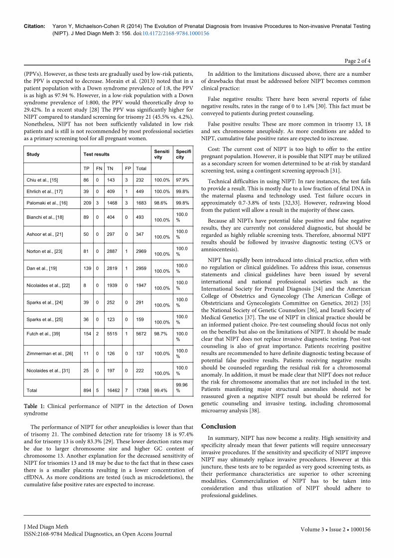
(PPVs). However, as these tests are gradually used by low-risk patients,the PPV is expected to decrease. Morain et al. (2013) noted that in apatient population with a Down syndrome prevalence of 1:8, the PPVis as high as 97.94 %. However, in a low-risk population with a Downsyndrome prevalence of 1:800, the PPV would theoretically drop to29.42%. In a recent study [28] The PPV was significantly higher forNIPT compared to standard screening for trisomy 21 (45.5% vs. 4.2%).Nonetheless, NIPT has not been sufficiently validated in low riskpatients and is still is not recommended by most professional societiesas a primary screening tool for all pregnant women.
Study Test results Sensitivity
Specificity
TP FN TN FP Total
Chiu et al., [15] 86 0 143 3 232 100.0% 97.9%
Ehrlich et al., [17] 39 0 409 1 449 100.0% 99.8%
Palomaki et al., [16] 209 3 1468 3 1683 98.6% 99.8%
Bianchi et al., [18] 89 0 404 0 493 100.0%100.0%
Ashoor et al., [21] 50 0 297 0 347 100.0%100.0%
Norton et al., [23] 81 0 2887 1 2969 100.0%100.0%
Dan et al., [19] 139 0 2819 1 2959 100.0%100.0%
Nicolaides et al., [22] 8 0 1939 0 1947 100.0%100.0%
Sparks et al., [24] 39 0 252 0 291 100.0%100.0%
Sparks et al., [25] 36 0 123 0 159 100.0%100.0%
Futch et al., [39] 154 2 5515 1 5672 98.7% 100.0%
Zimmerman et al., [26] 11 0 126 0 137 100.0% 100.0%
Nicolaides et al., [31] 25 0 197 0 222 100.0%100.0%
Total 894 5 16462 7 17368 99.4%99.96%
Table 1: Clinical performance of NIPT in the detection of Downsyndrome
The performance of NIPT for other aneuploidies is lower than thatof trisomy 21. The combined detection rate for trisomy 18 is 97.4%and for trisomy 13 is only 83.3% [29]. These lower detection rates maybe due to larger chromosome size and higher GC content ofchromosome 13. Another explanation for the decreased sensitivity ofNIPT for trisomies 13 and 18 may be due to the fact that in these casesthere is a smaller placenta resulting in a lower concentration ofcffDNA. As more conditions are tested (such as microdeletions), thecumulative false positive rates are expected to increase.
In addition to the limitations discussed above, there are a numberof drawbacks that must be addressed before NIPT becomes commonclinical practice:
False negative results: There have been several reports of falsenegative results, rates in the range of 0 to 1.4% [30]. This fact must beconveyed to patients during pretest counseling.
False positive results: These are more common in trisomy 13, 18and sex chromosome aneuploidy. As more conditions are added toNIPT, cumulative false positive rates are expected to increase.
Cost: The current cost of NIPT is too high to offer to the entirepregnant population. However, it is possible that NIPT may be utilizedas a secondary screen for women determined to be at-risk by standardscreening test, using a contingent screening approach [31].
Technical difficulties in using NIPT: In rare instances, the test failsto provide a result. This is mostly due to a low fraction of fetal DNA inthe maternal plasma and technology used. Test failure occurs inapproximately 0.7-3.8% of tests [32,33]. However, redrawing bloodfrom the patient will allow a result in the majority of these cases.
Because all NIPTs have potential false positive and false negativeresults, they are currently not considered diagnostic, but should beregarded as highly reliable screening tests. Therefore, abnormal NIPTresults should be followed by invasive diagnostic testing (CVS oramniocentesis).
NIPT has rapidly been introduced into clinical practice, often withno regulation or clinical guidelines. To address this issue, consensusstatements and clinical guidelines have been issued by severalinternational and national professional societies such as theInternational Society for Prenatal Diagnosis [34] and the AmericanCollege of Obstetrics and Gynecology (The American College ofObstetricians and Gynecologists Committee on Genetics, 2012) [35]the National Society of Genetic Counselors [36], and Israeli Society ofMedical Genetics [37]. The use of NIPT in clinical practice should bean informed patient choice. Pre-test counseling should focus not onlyon the benefits but also on the limitations of NIPT. It should be madeclear that NIPT does not replace invasive diagnostic testing. Post-testcounseling is also of great importance. Patients receiving positiveresults are recommended to have definite diagnostic testing because ofpotential false positive results. Patients receiving negative resultsshould be counseled regarding the residual risk for a chromosomalanomaly. In addition, it must be made clear that NIPT does not reducethe risk for chromosome anomalies that are not included in the test.Patients manifesting major structural anomalies should not bereassured given a negative NIPT result but should be referred forgenetic counseling and invasive testing, including chromosomalmicroarray analysis [38].
ConclusionIn summary, NIPT has now become a reality. High sensitivity and
specificity already mean that fewer patients will require unnecessaryinvasive procedures. If the sensitivity and specificity of NIPT improveNIPT may ultimately replace invasive procedures. However at thisjuncture, these tests are to be regarded as very good screening tests, astheir performance characteristics are superior to other screeningmodalities. Commercialization of NIPT has to be taken intoconsideration and thus utilization of NIPT should adhere toprofessional guidelines.
Citation: Yaron Y, Michaelson-Cohen R (2014) The Evolution of Prenatal Diagnosis from Invasive Procedures to Non-invasive Prenatal Testing(NIPT). J Med Diagn Meth 3: 156. doi:10.4172/2168-9784.1000156
Page 2 of 4
J Med Diagn MethISSN:2168-9784 Medical Diagnostics, an Open Access Journal
Volume 3 • Issue 2 • 1000156

References1. Malone FD, Canick JA, Ball RH, Nyberg DA, Comstock CH, et al. (2005)
First- and Second-Trimester Evaluation of Risk (FASTER) ResearchConsortium. First-trimester or second-trimester screening, or both, forDown's syndrome. N Engl J Med 353: 2001-2011.
2. Driscoll DA, Gross S (2009) Clinical practice. Prenatal screening foraneuploidy. N Engl J Med 360: 2556-2562.
3. Wald NJ, Watt HC, Hackshaw AK (1999) Integrated screening forDown's syndrome on the basis of tests performed during the first andsecond trimesters. N Engl J Med 341: 461-467.
4. Wright D, Bradbury I, Benn P, Cuckle H, Ritchie K (2004) Contingentscreening for Down syndrome is an efficient alternative to non-disclosure sequential screening. Prenat Diagn 24: 762-766.
5. Tabor A, Alfirevic Z (2010) Update on procedure-related risks forprenatal diagnosis techniques. Fetal Diagn Ther 27: 1-7.
6. Bianchi DW, Simpson JL, Jackson LG, Evans MI, Elias S, et al. (1999)Fetal cells in maternal blood: NIFTY clinical trial interim analysis. DM-STAT. NICHD fetal cell study (NIFTY) group. Prenat Diagn 19: 994-995.
7. Lo YM (2000) Fetal DNA in maternal plasma: biology and diagnosticapplications. Clin Chem 46: 1903-1906.
8. Lo YM, Patel P, Wainscoat JS, Sampietro M, Gillmer MD, et al. (1989)Prenatal sex determination by DNA amplification from maternalperipheral blood. Lancet 2: 1363-1365.
9. Lo YM, Hjelm NM, Fidler C, Sargent IL, Murphy MF, et al. (1998)Prenatal diagnosis of fetal RhD status by molecular analysis of maternalplasma. N Engl J Med 339: 1734-1738.
10. Finning KM, Martin PG, Soothill PW, Avent ND (2002) Prediction offetal D status from maternal plasma: introduction of a new noninvasivefetal RHD genotyping service. Transfusion 42: 1079-1085.
11. Clausen FB, Christiansen M, Steffensen R, Jørgensen S, Nielsen C, et al.(2012) Report of the first nationally implemented clinical routinescreening for fetal RHD in D-pregnant women to ascertain therequirement for antenatal RhD prophylaxis. Transfusion 52: 752-758.
12. Lo YM, Lun FM, Chan KC, Tsui NB, Chong KC, et al. (2007) Digital PCRfor the molecular detection of fetal chromosomal aneuploidy. Proc NatlAcad Sci U S A 104: 13116-13121.
13. Fan HC, Quake SR (2007) Detection of aneuploidy with digitalpolymerase chain reaction. Anal Chem 79: 7576-7579.
14. Fan HC, Blumenfeld YJ, Chitkara U, Hudgins L, Quake SR (2008)Noninvasive diagnosis of fetal aneuploidy by shotgun sequencing DNAfrom maternal blood. Proc Natl Acad Sci U S A 105: 16266-16271.
15. Chiu RW, Chan KC, Gao Y, Lau VY, Zheng W, et al. (2008) Noninvasiveprenatal diagnosis of fetal chromosomal aneuploidy by massively parallelgenomic sequencing of DNA in maternal plasma. Proc Natl Acad Sci U SA 105: 20458-20463.
16. Palomaki GE, Kloza EM, Lambert-Messerlian GM, Haddow JE, NeveuxLM, et al. (2011) DNA sequencing of maternal plasma to detect Downsyndrome: an international clinical validation study. Genet Med 13:913-920.
17. Ehrich M, Deciu C, Zwiefelhofer T, Tynan JA, Cagasan L, et al. (2011)Noninvasive detection of fetal trisomy 21 by sequencing of DNA inmaternal blood: a study in a clinical setting. Am J Obstet Gynecol 204:205.
18. Bianchi DW, Platt LD, Goldberg JD, Abuhamad AZ, Sehnert AJ, et al.(2012) Genome-wide fetal aneuploidy detection by maternal plasmaDNA sequencing. Obstet Gynecol 119: 890-901.
19. Dan S, Wang W, Ren J, Li Y, Hu H, et al. (2012) Clinical application ofmassively parallel sequencing-based prenatal noninvasive fetal trisomytest for trisomies 21 and 18 in 11,105 pregnancies with mixed risk factors.Prenat Diagn 32: 1225-1232.
20. Yu SC, Jiang P, Choy KW, Chan KC, Won HS, et al. (2013) Noninvasiveprenatal molecular karyotyping from maternal plasma. PLoS One 8:e60968.
21. Ashoor G, Syngelaki A, Wagner M, Birdir C, Nicolaides KH (2012)Chromosome-selective sequencing of maternal plasma cell-free DNA forfirst-trimester detection of trisomy 21 and trisomy 18. Am J ObstetGynecol 206: 322.
22. Nicolaides KH, Syngelaki A, Ashoor G, Birdir C, Touzet G (2012)Noninvasive prenatal testing for fetal trisomies in a routinely screenedfirst-trimester population. Am J Obstet Gynecol 207: 374.
23. Norton ME, Brar H, Weiss J, Karimi A, Laurent LC, et al. (2012) Non-Invasive Chromosomal Evaluation (NICE) Study: results of a multicenterprospective cohort study for detection of fetal trisomy 21 and trisomy 18.Am J Obstet Gynecol 207: 137e1-137e 8.
24. Sparks AB, Struble CA, Wang ET, Song K, Oliphant A (2012)Noninvasive prenatal detection and selective analysis of cell-free DNAobtained from maternal blood: evaluation for trisomy 21 and trisomy 18.Am J Obstet Gynecol 206: 319.
25. Sparks AB, Wang ET, Struble CA, Barrett W, Stokowski R, et al. (2012)Selective analysis of cell-free DNA in maternal blood for evaluation offetal trisomy. Prenat Diagn 32: 3-9.
26. Zimmermann B, Hill M, Gemelos G, Demko Z, Banjevic M, et al. (2012)Noninvasive prenatal aneuploidy testing of chromosomes 13, 18, 21, X,and Y, using targeted sequencing of polymorphic loci. Prenat Diagn 32:1233-1241.
27. Nicolaides KH, Wright D, Poon LC, Syngelaki A, Gil M (2013) First-trimester contingent screening for trisomy 21 by biomarkers andmaternal blood cell-free DNA testing. Ultrasound Obstet Gynecol 42:41-50.
28. Bianchi DW, Parker RL, Wentworth J, Madankumar R, Saffer C, et al.(2014) DNA sequencing versus standard prenatal aneuploidy screening.N Engl J Med 370: 799-808.
29. Twiss P, Hill M1, Daley R1, Chitty LS2 (2014) Non-invasive prenataltesting for Down syndrome. Semin Fetal Neonatal Med 19: 9-14.
30. Canick JA, Palomaki GE, Kloza EM, Lambert-Messerlian GM, HaddowJE (2013) The impact of maternal plasma DNA fetal fraction on nextgeneration sequencing tests for common fetal aneuploidies. Prenat Diagn33: 667-674.
31. Nicolaides KH, Syngelaki A, Gil M, Atanasova V, Markova D (2013)Validation of targeted sequencing of single-nucleotide polymorphismsfor non-invasive prenatal detection of aneuploidy of chromosomes 13,18, 21, X, and Y. Prenat Diagn 33: 575-579.
32. Gil MM, Quezada MS, Bregant B, Ferraro M, Nicolaides KH (2013)Implementation of maternal blood cell-free DNA testing in earlyscreening for aneuploidies. Ultrasound Obstet Gynecol 42: 34-40.
33. Fernando MR, Chen K, Norton S, Krzyzanowski G, Bourne D, et al.(2010) A new methodology to preserve the original proportion andintegrity of cell-free fetal DNA in maternal plasma during sampleprocessing and storage. Prenat Diagn 30: 418-424.
34. Benn P, Borrell A, Cuckle H, Dugoff L, Gross S, et al. (2012) Prenataldetection of Down Syndrome using massively parallel sequencing (MPS):a rapid response statement from a committee on behalf of the Board ofthe International Society for Prenatal Diagnosis, 24 October 2011. PrenatDiagn 32:1–2.
35. The American College of Obstetricians and Gynecologists (ACOG)(2012) Committee on Genetics. The Society for Maternal–Fetal medicinepublications committee. Noninvasive prenatal testing for fetalaneuploidy. Committee opinion 545, 2012 Obstet Gynecol 120:1532-1534.
36. Devers PL, Cronister A, Ormond KE, Facio F, Brasington CK, et al.(2013) Noninvasive prenatal testing/noninvasive prenatal diagnosis: theposition of the National Society of Genetic Counselors. J Genet Couns 22:291-295.
37. Michaelson-Cohen R, Gershoni-Baruch R, Sharoni R, Mordechai S,Yaron Y, et al. (2013) Noninvasive fetal testing in maternal plasma forfetal chromosomal imbalances. Fetal Diagn Ther (in press).
38. Wapner RJ, Martin CL, Levy B, Ballif BC, Eng CM, et al. (2012)Chromosomal microarray versus karyotyping for prenatal diagnosis. NEngl J Med 367: 2175-2184.
Citation: Yaron Y, Michaelson-Cohen R (2014) The Evolution of Prenatal Diagnosis from Invasive Procedures to Non-invasive Prenatal Testing(NIPT). J Med Diagn Meth 3: 156. doi:10.4172/2168-9784.1000156
Page 3 of 4
J Med Diagn MethISSN:2168-9784 Medical Diagnostics, an Open Access Journal
Volume 3 • Issue 2 • 1000156

39. Futch T, Spinosa J, Bhatt S, de Feo E, Rava RP, et al. (2013) Initial clinicallaboratory experience in noninvasive prenatal testing for fetal aneuploidyfrom maternal plasma DNA samples. Prenat Diagn 33: 569-574.
Citation: Yaron Y, Michaelson-Cohen R (2014) The Evolution of Prenatal Diagnosis from Invasive Procedures to Non-invasive Prenatal Testing(NIPT). J Med Diagn Meth 3: 156. doi:10.4172/2168-9784.1000156
Page 4 of 4
J Med Diagn MethISSN:2168-9784 Medical Diagnostics, an Open Access Journal
Volume 3 • Issue 2 • 1000156