prefrontal cortex and spatial sequencing in macaque monkey
TRANSCRIPT
Exp Brain Res (1989) 78:447~64 Exp.erimental Brain Research �9 Springer-Verlag 1989
Prefrontal cortex and spatial sequencing in macaque monkey
P. Barone and J.-P. Joseph Laboratoire de Neuropsychologie Exp6rimentale, INSERM Unit~ 94, 16, Avenue du Doyen L@ine, F 69500 Bron, France
Summary. 1. Single neuron activity was recorded from the prefrontal cortex of two macaque mon- keys during the performance of a task involving spatial sequencing. The monkeys faced a panel displaying a central fixation point and three fixed targets (two lateral and one above the point of fixation). In the first phase of each trial, the three targets were turned on in random order: in the second phase, the animal had to press each target, still lighted, in the order of their illumination. Thus, successful performance of the task depended strongly on temporal memory. The animals were fitted with D C - E O G electrodes. 2. Three hundred and two task-related neurons were recorded in the superior arcuate area and caudal part of sulcus principalis. Among the cells whose pattern of activ- ity appeared to be related to the sequencing task, five classes were distinguished: Visual tonic (VT), fixation, context, saccade related and visual phasic cells. In addition, a small number of cells appeared to be related to other aspects of the behavior, but not to the sequencing task. Our present analysis concentrates on two groups of sequencing task- related cells (VT and context cells). 3. The VT cells (35/302-11.5%) were recorded exclusively from the superior arcuate area. All VT cells increased their firing rate (sustained activation) during fixation of the central fixation point (FP) following onset of one of the three targets used, specific for a given cell (directional or spatial selectivity). In one group of VT cells, a shift in the eye position towards the specific peripheral target resulted in the return of the cells' firing rate to the pre-trial level. In the other group of VT cells, reset of the firing rate to pre-trial level was not related to the onset of fixation of the peripheral target. Sustained activa-
O~t)rint requests to: J.-P. Joseph (address see above)
tion of the VT cells depended also on the sequential order of illumination of the specific target (tempo- ral selectivity). In twenty-four cells (68.5% of VT cells) sustained activation was observed when the target came first in the sequence. Onset of the tar- get in the second or third rank elicited either no response or only a short lasting phasic activation. In the remaining eleven cells (31.5% of VT cells), sustained activation was only observed when the target came second in a given sequence. The firing of the VT cells was correlated with the animals' performance of the task. On trials where the ani- mals selected successive targets in an incorrect order, the temporal pattern of activation of VT cells was different from that in the correctly per- formed trials. Thus, the correct temporal encoding of a target appeared to be a prerequisite for the correct performance of a sequence. 4. The context cells (36.5%-116/302) were activated when the ani- mal fixated a particular target during execution of the sequence and, like VT cells, were encoun- tered exclusively in the penetrations through the superior arcuate area. Activation also depended on the state (illuminated vs extinguished or hit vs non-hit) of the non-fixated targets and/or on their time-relationships with respect to the fixated target - hence context cells. 5. Properties of VT and con- text cells revealed in the present experiment are consistent with the idea that the superior arcuate area is involved in temporally and spatially ex- tended structures of behavior. If so, the arcuate area would constitute a specialized part of prefron- tal cortex implicated in construction of oculomotor plans.
Key words: Prefrontal cortex - Visual tonic cells - Content cells - Monkey
448
Introduction
In current theories of frontal lobe function, the control of behavior in novel and non-routine situa- tions is usually ascribed to the prefrontal cortex. Basic to this control is the capacity for ordering or for sequencing, i.e. "the ability to maintain and organize bits of information in meaningful se- quences.., or to extract crucial elements from one series (of information) and integrate them with data from other series" (Stuss and Benson 1987).
In human patients with frontal lobe lesion, defi- cits in concept formation, memory and cognition have often been associated with a dysfunction of sequencing. For example, Luria (1966) concluded from his studies of calculation disorders that "it is the sequential ordering of intellectual activity that is apparently disrupted in patients with a le- sion of the frontal lobes, being replaced by frag- mentary operations". The memory deficits which follow lesions of dorsolateral prefrontal cortex have been associated with impairments in sequenc- ing and with inability to conduct an orderly search through memory (Petrides and Milner 1982; Mil- ner et al. 1985; Stuss and Benson 1986, 1987).
Motor sequencing has also been investigated in humans with anterior lesions presumably involving prefrontal cortex. It has been shown that these patients have substantial difficulties copying sequences of movements o f t h e arm (Kolb and Milner 1981; Kimura 1982) or of the face (Kimura 1982). Thus, it appears that in the situations in which the memory is taxed, the prefrontal cortex plays a specific role in motor sequencing (Kolb and Milner 1981 ; De Renzi et al. 1983). The results of a number of studies in macaque monkey with fron- tal lobe lesions, which included the periarcuate area but spared the supplementary motor area (SMA), have also indicated that prefrontal cortex plays a role in motor sequencing (Pinto-Hamuy and Linck 1965; Brody and Pribram 1978). Passingham (1985b) has found that after destruction of either the sulcus principalis or the superior prefrontal convexity, monkeys were able to learn a fixed sequence of three movements normally, but follow- ing removal of arcuate cortex (areas 6 and 8) they were slow to learn the task.
In the present experiment, we sought to deter- mine the neural processes in the prefrontal cortex which play a role in preparation and execution of spatial sequences. We trained two monkeys to per- form a task that taxed memory for temporal order of spatial items. The animals had to keep in mem- ory the order of illumination of three fixed spatial targets and had to press them later in the same
order. Order of illumination of the targets was ran- domized between trials.
We recorded unit activity from the superior periarcuate area and sulcus principalis, that is from two regions of prefrontal cortex which receive spa- tial information from area PG of parietal cortex (Barbas and Mesulam 1981, 1985). We chose to fit the animals with D C - E O G electrodes since a previous experiment (Joseph and Barone 1987) had shown that control of eye-movements and subse- quent fixations contributed to an understanding of the neural processes that accompany the perfor- mance of spatial delayed tasks.
Our data suggest that the neural apparatus of the superior arcuate area plays a major role in planning and execution of spatial sequences: prior to execution of the sequence, one set of neurons stores positions and time-relationships of the spa- tial targets, while during execution of the sequence, another set of neurons indicates, at each stage, the current state of the sequence, i.e. the distribution of illuminated and extinguished targets and their time-relationships with the current task. We hypothesize that activation of these neural sets is the substrate of the storage and excution of the oculomotor plan.
Methods
Animals
Two adult Rhesus monkeys (Macaca mulatta) served as sub- jects in this experiment.
Apparatus
During the training sessions and experiments, monkeys were seated in a primate chair with head restrained while facing a panel located at arm's reach (18 cm from the eyes). The panel was composed of a central fixation point (FP), three peripheral light-targets and two hold levers. The FP was a 5 x 5 mm light- square. The light-targets were 1.5 x 1.5 cm translucent squares, illuminated from the rear by a yellow light-emitting diode (LED). One target was located 6.5 cm above FP while the other two targets were located 3 cm below FP and respectively 11 cm to the right and to the left of FP. The hold levers were located 7 cm below the right and the left light target. They were 1 cm wide and protruded by 2 cm. The hold lever that could be used during a trial depended on which arm the monkey was permitted to extend from its chair.
Procedure
The monkeys were trained in a delayed spatial sequencing task. In each trial depressing the hold lever on the side of the arm used for that trial resulted in illuminating the FP for 4/6 sec- onds. While the monkey fixated the FP (step 1, Fig. IA, B), the three peripheral light-targets were illuminated one after an- other (steps 2, 3, 4, Fig. 1). For each trial the order of illumina-
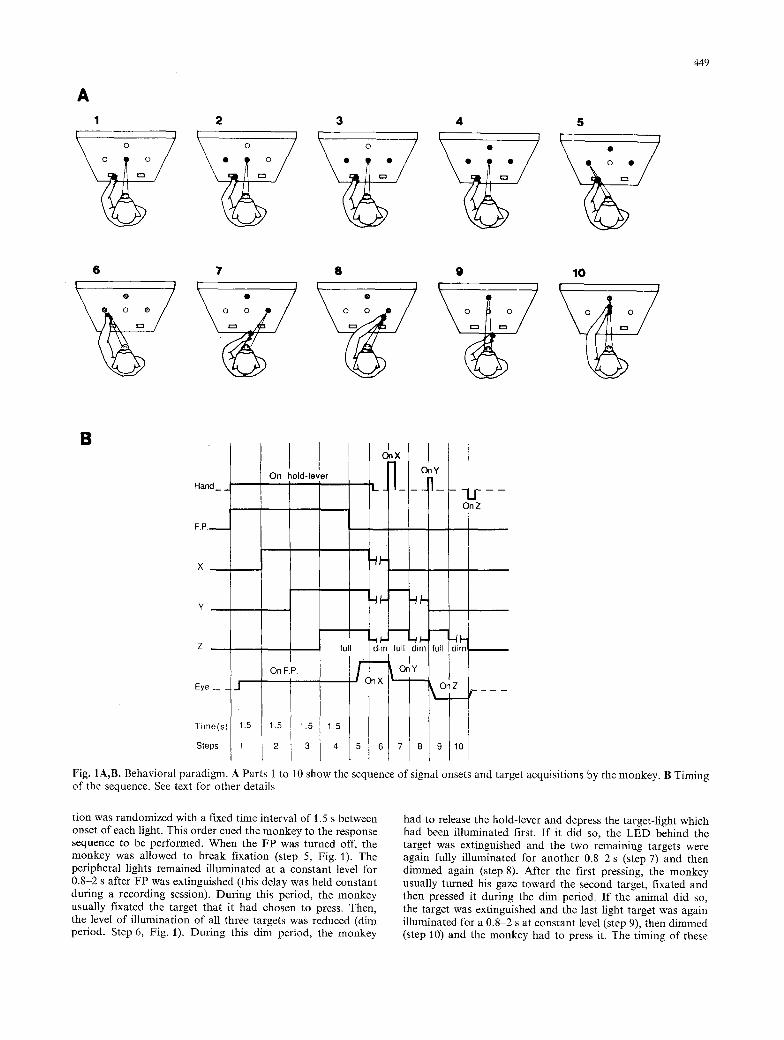
A 1
0 0
2 3 4 5
�9 O 5( ) 449
6 7 8 9 10 \ . / o o .
G 0 0
B
Hand
OnX I On hold-lever 1
I On Z
Z , , i I full I dim full diml full
Eye _ 1 J
Time(s) 1.5
Steps I 1
On F.P.
1.5 1 5
3 4
X
6 7 8
Fig. IA,B. Behavioral paradigm. A Parts 1 to 10 show the sequence of signal onsets and target acquisitions by the monkey. B Timing of the sequence. See text for other details
tion was randomized with a fixed time interval of 1.5 s between onset of each light. This order cued the monkey to the response sequence to be performed. When the FP was turned off, the monkey was allowed to break fixation (step 5, Fig. 1). The peripheral lights remained illuminated at a constant level for 0.8-2 s after FP was extinguished (this delay was held constant during a recording session). During this period, the monkey usually fixated the target that it had chosen to press. Then, the level of illumination of all three targets was reduced (dim period. Step 6, Fig. 1). During this dim period, the monkey
had to release the hold-lever and depress the target-light which had been illuminated first. If it did so, the LED behind the target was extinguished and the two remaining targets were again fully illuminated for another 0.8 2 s (step 7) and then dimmed again (step 8). After the first pressing, the monkey usually turned his gaze toward the second target, fixated and then pressed it during the dim period. If the animal did so, the target was extinguished and the last light target was again illuminated for a 0 . 8 ~ s at constant level (step 9), then dimmed (step 10) and the monkey had to press it. The timing of these
450
events is represented in Fig. lB. If the animal performed the sequential task correctly, it was rewarded with 0.2 ml of apple juice.
In this report, "1", "2" and "3" designate respectively the upper, right and left target. According to the order of onset of the three targets ("three-target" paradigm), six different se- quences could be performed: 123, 132, 213,231,312, 321. In the course of the recording sessions, the animals were also presented with sequences of two targets ("two-target" paradigm), by elimi- nating either one of the three targets.
The trial was aborted if the monkey made eye movements (measured by vertical and horizontal EOGs) outside a window centered on the FP before the FP was extinguished. The limits of the window corresponded to half of the eccentricity of the targets (32 ~ from FP for the right and left targets and 19 ~ for the upper target). The animals were neither rewarded nor punished, for releasing the hold-lever before the first dim peri- od, pressing the light-targets in a wrong order or before the dim period, or failing to perform the sequence within a 12 s time interval after the F P had been extinguished, but the trial was aborted in the all these cases. To prevent the animals from adopting oculo-motor stereotypes, a correction procedure (rep- etition of trial until it is successful) was applied throughout the training and recording sessions. The trials were performed under control of a microcomputer PDP 11-23. Successive trials were separated by an interval of I s during which the hold-lever was inactivated.
The animals daily, received forcements 200-300 reinforce until they reached a criterion of more than 80% correct re- sponses. They required 5-6 months to master the task. The animals received their entire daily fluid ration in the testing apparatus. Monkey-pellets were available ad libitum in the home cage.
Surgery and unit recording
During a pause in the training period, Ag/AgC1 electro- oculographic electrodes were implanted under sodium pen- tobarbital anaesthesia. When training was completed, the animals were re-operated. Stainless steel cylinders (18ram in diameter) were implanted in the skull. The center of the cylinders were aimed at A = 3 0 m m and L = 1 5 mm in the Horsley-Clarke coordinate system. During recording sessions the animal's head was held rigidly. Activity of single cells was recorded from both hemispheres through tungsten microelec- trodes (0.1-1 M) plated with platinum black and driven by a motorized microdrive (Trent Wells). Electrode penetrations were made on a millimeter grid of points usually separated by I mm but when a unit showed activity related to the task, subsequent electrode penetrations were aimed at the immediate vicinity.
to one or more changes of the task events were selected for graphic representation (raster display). Only those cells that could be studied during execution of the six sequences of the "three-target" paradigm are considered in the present study.
Histology
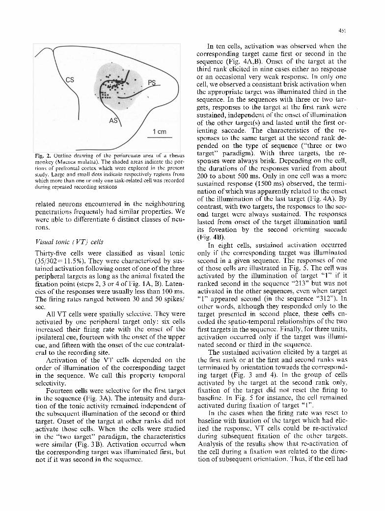
At the end of the experiments, several lesions were made at penetrations located at the crucial points within the explored area of the cortex by passing anodal current (20 I.tA for l rain) through a microelectrode. Within a day after the lesions, the animals were sacrificed with an overdose of Pentobarbital and perfused transcardially with isotonic saline followed by 4% for- maldehyde saline. Pins were inserted at known electrode coordi- nates. The brains were removed from the skulls, photographed and postfixed for 1 week. They were then sectioned on a freez- ing microtome at 40 l.tm and stained with cresyl violet. That sort of approach did not allow a reconstruction of each elec- trode penetration. However it allowed an identification of the general area from which we recorded the task-related neurons (Fig. 2).
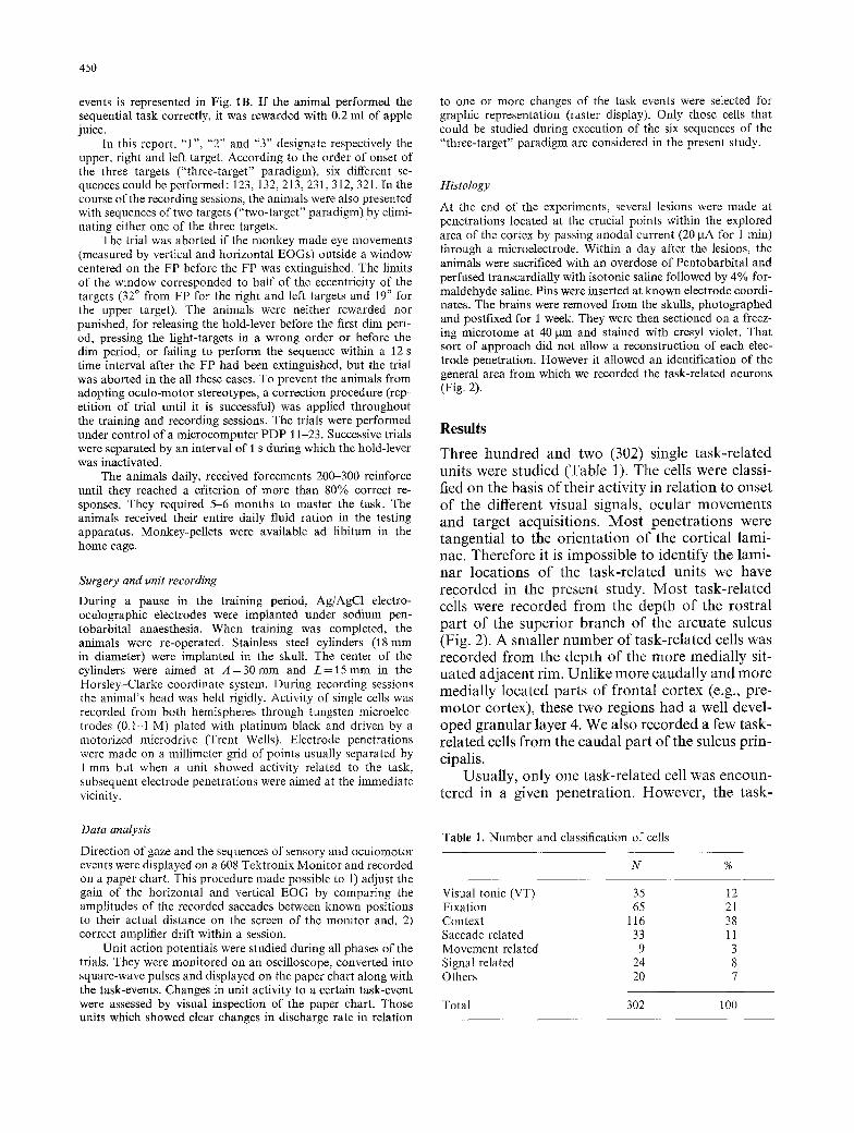
Results
Three hundred and two (302) single task-related units were studied (Table 1). The cells were classi- fied on the basis of their activity in relation to onset of the different visual signals, ocular movements and target acquisitions. Most penetrations were tangential to the orientation of the cortical lami- nae. Therefore it is impossible to identify the lami- nar locations of the task-related units we have recorded in the present study. Most task-related cells were recorded from the depth of the rostral part of the superior branch of the arcuate sulcus (Fig. 2). A smaller number of task-related cells was recorded from the depth of the more medially sit- uated adjacent rim. Unlike more caudally and more medially located parts of frontal cortex (e.g., pre- motor cortex), these two regions had a well devel- oped granular layer 4. We also recorded a few task- related cells from the caudal part of the sulcus prin- cipalis.
Usually, only one task-related cell was encoun- tered in a given penetration. However, the task-
Data analysis
Direction of gaze and the sequences of sensory and oculomotor events were displayed on a 608 Tektronix Moni tor and recorded on a paper chart. This procedure made possible to l) adjust the gain of the horizontal and vertical EOG by comparing the amplitudes of the recorded saccades between known positions to their actual distance on the screen of the monitor and, 2) correct amplifier drift within a session.
Unit action potentials were studied during all phases of the trials. They were monitored on an oscilloscope, converted into square-wave pulses and displayed on the paper chart along with the task-events. Changes in unit activity to a certain task-event were assessed by visual inspection of the paper chart. Those units which showed clear changes in discharge rate in relation
Table 1. Number and classification of cells
N %
Visual tonic (VT) 35 12 Fixation 65 21 Context 116 38 Saccade related 33 11 Movement related 9 3 Signal related 24 8 Others 20 7
Total 302 100
451
Fig. 2. Outline drawing of the periarcuate area of a rhesus monkey (Macaca mulatta). The shaded areas indicate the por- tions of prefrontal cortex which were explored in the present study. Large and small dots indicate respectively regions from which more than one or only one task-related cell was recorded during repeated recording sessions
related neurons encountered in the neighbouring penetrations frequently had similar properties. We were able to differentiate 6 distinct classes of neu- rons.
Visual tonic (VT) cells
Thirty-five cells were classified as visual tonic (35/302= 11.5%). They were characterized by sus- tained activation following onset of one of the three peripheral targets as long as the animal fixated the fixation point (steps 2, 3 or 4 of Fig. 1A, B). Laten- cies of the responses were usually less than 100 ms. The firing rates ranged between 30 and 50 spikes/ sec.
All VT cells were spatially selective. They were activated by one peripheral target only: six cells increased their firing rate with the onset of the ipsilateral cue, fourteen with the onset of the upper cue, and fifteen with the onset of the cue contralat- eral to the recording site.
Activation of the VT cells depended on the order of illumination of the corresponding target in the sequence. We call this property temporal selectivity.
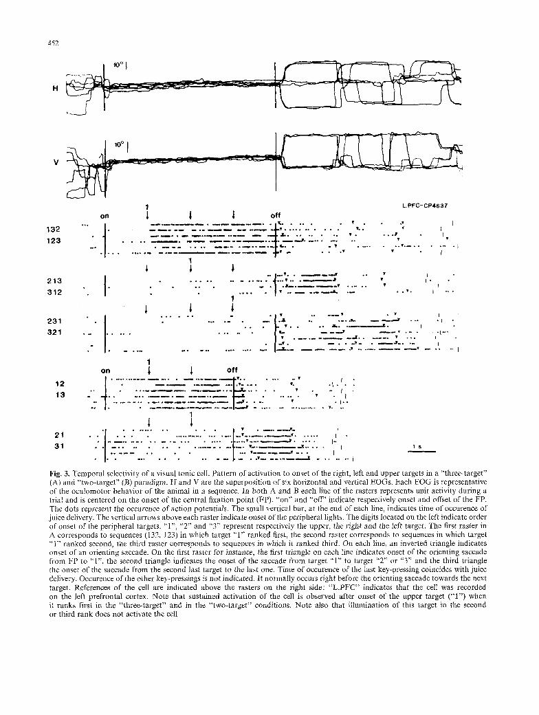
Fourteen cells were selective for the first target in the sequence (Fig. 3A). The intensity and dura- tion of the tonic activity remained independent of the subsequent illumination of the second or third target. Onset of the target at other ranks did not
�9 activate those cells. When the cells were studied in the "two target" paradigm, the characteristics were similar (Fig. 3 B). Activation occurred when the corresponding target was illuminated first, but not if it was second in the sequence.
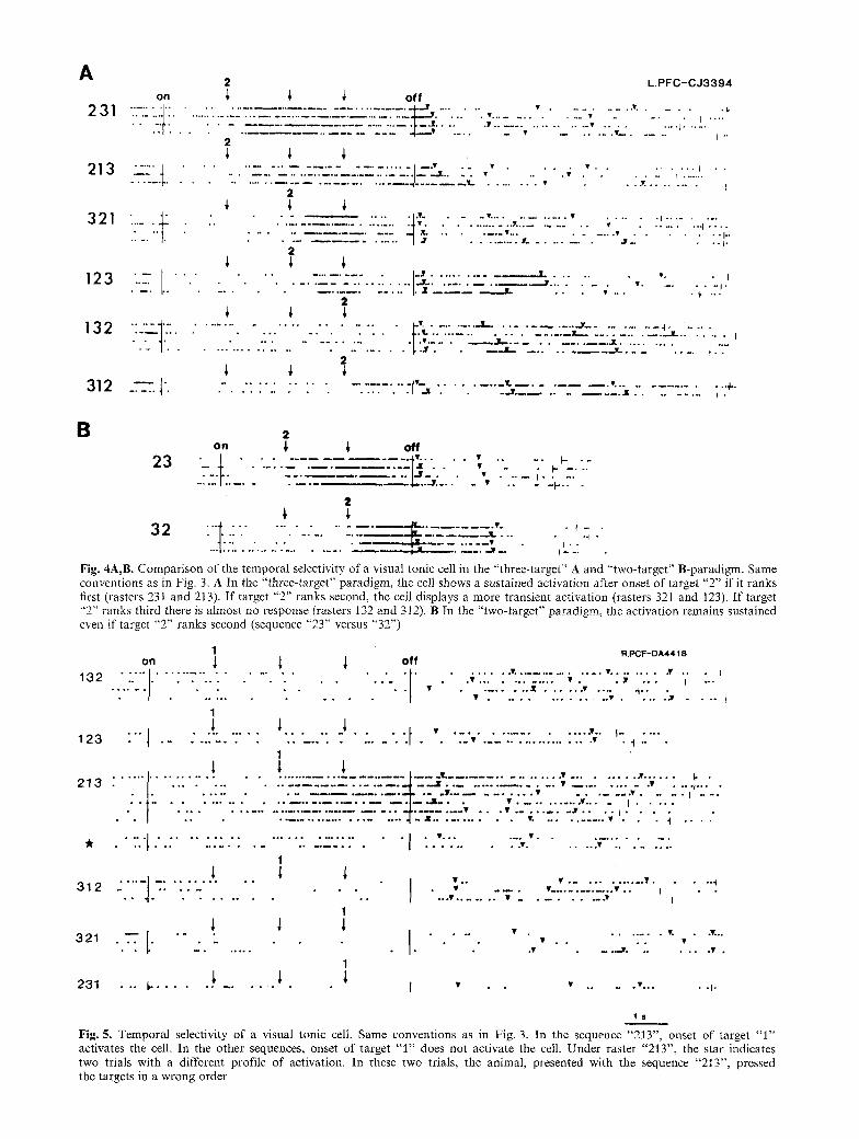
In ten cells, activation was observed when the corresponding target came first or second in the sequence (Fig. 4A,B). Onset of the target at the third rank elicited in nine cases either no response or an occasional very weak response. In only one cell, we observed a consistant brisk activation when the appropriate target was illuminated third in the sequence. In the sequences with three or two tar- gets, responses to the target at the first rank were sustained, independent of the onset of illumination of the other target(s) and lasted until the first or- ienting saccade. The characteristics of the re- sponses to the same target at the second rank de- pended on the type of sequence ("three or two target" paradigm). With three targets, the re- sponses were always brisk. Depending on the cell, the durations of the responses varied from about 200 to about 500 ms. Only in one cell was a more sustained response (1500 ms) observed, the termi- nation of which was apparently related to the onset of the illumination of the last target (Fig. 4A). By contrast, with two targets, the responses to the sec- ond target were always sustained. The responses lasted from onset of the target illumination until its foveation by the second orienting saccade (Fig. 4B).
In eight cells, sustained activation occurred only if the corresponding target was illuminated second in a given sequence. The responses of one of those cells are illustrated in Fig. 5. The cell was activated by the illumination of target "1" if it ranked second in the sequence "213" but was not activated in the other sequences, even when target "1" appeared second (in the sequence "312"). In other words, although they responded only to the target presented in second place, these cells en- coded the spatio-temporal relationships of the two first targets in the sequence. Finally, for three units, activation occurred only if the target was illumi- nated second or third in the sequence.
The sustained activation elicited by a target at the first rank or at the first and second ranks was terminated by orientation towards the correspond- ing target (Fig. 3 and 4). In the group of cells activated by the target at the second rank only, fixation of the target did not reset the firing to baseline. In Fig. 5 for instance, the cell remained activated during fixation of target "1".
In the cases when the firing rate was reset to baseline with fixation of the target which had elic- ited the response, VT cells could be re-activated during subsequent fixation of the other targets. Analysis of the results show that re-activation of the cell during a fixation was related to the direc- tion of subsequent orientation. Thus, if the cell had
H
I �9
. . . . . . I '~176 ~ '
o,, 1 I 1 o .
' " ' " '
1 2 3 �9 ' " �9 , , ~ �9 �9 �9 I
452
1 1 1 1
l I I
�9 ~ 1 4 9 " � 9 I "
�9 1 ~
�9 , � 9 o � 9 .
2 1 3 3 1 2
. . ~ , * . . �9 . . . u . . . ~ J
1 1
~
"I . . . . . . . . . . . . l ! . . . . . . " 2 1
�9 : . - - 2 ~ . . . " . . . . . - 7 : 7 . . . . . " 2 . . ' " ' . "2 ;L . -7 ." - ._ . . . . , " .'.'.'7 3 1 . . . . . . . . . . . . . . . . . . . . . . . �9 . . . . �9 . . . . . I" I 1 s
i . . . . . . . . . [ . . . . . . . . . ' ' ' " ' �9 �9 �9 . � 9 1 4 9 �9 �9 �9 � 9 1 4 9 . ~ ~ �9 � 9 1 6 5 m . ~ �9 . ~ ~ [
Fig. 3. Temporal selectivity of a visual tonic cell. Pattern of activation to onset of the right, left and upper targets in a "three-target" (A) and "two-target" (B) paradigm. H and V are the superposition of six horizontal and vertical EOGs. Each EOG is representative of the oculomotor behavior of the animal in a sequence. In both A and B each line of the rasters represents unit activity during a trial and is centered on the onset of the central fixation point (FP). "on" and "off" indicate respectively onset and offset of the FP. The dots represent the occurence of action potentiMs. The small vertical bar, at the end of each line, indicates time of occurence of juice delivery. The vertical arrows above each raster indicate onset of the peripheral lights. The digits located on the left indicate order of onset of the peripheral targets. "1", "2" and "3" represent respectively the upper, the right and the left target. The first raster in A corresponds to sequences (132, 123) in which target "1" ranked first, the second raster corresponds to sequences in which target "1" ranked second, the third raster corresponds to sequences in which it ranked third. On each line, an inverted triangle indicates onset of an orienting saccade. On the first raster for instance, the first triangle on each line indicates onset of the orienting saccade from FP to "1", the second triangle indicates the onset of the saccade from target "1" to target "2" or "3" and the third triangle the onset of the saccade from the second last target to the last one. Time of occurence of the last key-pressing coincides with juice delivery. Occurence of the other key-pressings is not indicated. It normally occurs right before the orienting saccade towards the next target. References of the cell are indicated above the rasters on the right side: "L.PI~C ' ' indicates that the cell was recorded on the left prefrontal cortex. Note that sustained activation of the cell is observed after onset of the upper target ("1") when it ranks first in the "three-target" and in the "two-target" conditions. Note also that illumination of this target in the second or third rank does not activate the cell
I
I I
1 1 . . . . . . . . / ' . . . �9 . . . . . . ' 2 3 1 I].... " . . . . . . . . . I . ~ f ~ . . . L . .~v" - - ~ , - - 'J . . . . . J I 3 2 1 . . . . . . . . . ' . . . . . . . . I-..
�9 �9 _ J . . v " I
i on 1 1 o f f
1 3 " " . ~ . . . . . . . . . . . . . " t :"
A
2 3 1
~ :-2 . . .
3 1 2 - - - l ' ,
2 L . P F C - C J 3 3 9 4
o n ~, ~, ~" o f f
7 2" " ' - - " _ - ' " ' - . 2 . - ' _ ' . - - . : _ ~ " : . - . L 2 _ - . _ . s . ' . . , . . . . . . . . . . . . . . . . . - . . . . . . . . . . . . . . . . . . . . . . . . . . . u �9
. . . . . . . . . ~ . . . . " . . . . . . . . . ' J ~Y . . �9 V . . . . �9 . . . . . . . . j . . - " - - - Y s . . . . . . . . . . - " ' . - - - " - ' " - " : - . ' 2 - ' . : . ' - . ' L " . _ " ' " ~ .. . . . . .,. . . . . . . . . ....... I
2
" ' ~ " �9 . . . . . . . . . . . . . Y" . . . . . . . . . . �9 . . . . . . . . . I
. . . . . . . . . �9 . . . . . . . . v j - . . . . . . I " �9 2 . . . . . . . . . . . . . . . I V . . . . . . . . . . . h
. . . . . . . . . . . . . . 3 . . . . . . . . . . . . . w W . v. I . ~ . . - - . ~ : . ' : : _ . , . . . . . . . . . ,.
. . . . . . . . . . . . . . . . . . ~ * * �9 . . . . ). . . -
2
. . . . . . . . . . . . . . . . . . . . . ~vo . . . . . . . . ~ . . . . . . . . . . . . . j . . . . . . . . . . . . ~ . . . . . .
" " ~ ' ~ . . . . . . . . . . . . . . . . . . ~ . . . . . . . . . . . - - ! - . . . . . . . I . . . . . . / . v . . . . . . L . . . . . . . . . . . .,.X . . . . . . . . . t " ' - -
. . . . . . . . . . . . . ~, . . I . . ~ ~r . . . . . . . . . �9 . . . . . . . . I . �9
2
. . . . . . . . . . . . . . . . . . . . . . . - - . ' - " . "" . ' 1 ' ~ . . . . . . . . . _ . ~ . _ _ _ " . . . . . . . . . . _ _ _ . _ . " " ; . . . . . . . . . ; " + ' .
B 2 o n ; ; o f f
2 3 . T-I . . . . . . - -- ,.._-.--'1~4..._,._' . . . . . ' . . . . " ~ :. . . " . . - ~. ~" . . . .
2
~ - T L - _-- ..'7"2-..-_; . . . . . . i - �9 , . . . . . . . J - " I - - -
F i g . 4A,B. Comparison of the temporal selectivity of a visual tonic cell in the "three-target" A and "two-target" B-paradigm. Same conventions as in Fig. 3. A In the "three-target" paradigm, the cell shows a sustained activation after onset of target "2" if it ranks first (rasters 231 and 213). If target "2" ranks second, the cell displays a more transient activation (rasters 321 and 123). If target "2"' ranks third there is almost no response (rasters 132 and 312). B In the "two-target" paradigm, the activation remains sustained even if target "2" ranks second (sequence "23" versus "32")
1 o n ~ ~ 1 o f f
1 1
1 2 3 " " ] . . . . . . . . .
R . P C F - D A 4 4 1 8
. . . . . . T , . . . . . . . . . . . . . . T ~ . . . . . . . . � 9 . . �9 I
�9 �9 . . . . . . . . . . . . �9 �9 �9 Y - - �9 I - ' " . . . . . . . �9 W . . . . . . �9 . . . . . i . .
�9 . . !
1 l ".t " . . . . " ' "
1
I . . . . . �9 " l -"
. . . . . . . . . . . . . . . . . . . . . . . . . . . . . . . . . . . . . . . , . ~ _ . . . , . . . . . . . . . . . . . . . . . . , . . . . . . . . . , . . . . . . . . . L . ~ . . . 2 1 3 n . . . . . . . . . . . . . . . . . . . . . . . . . . - ' ~ - " . . . . . . . . . . . . . . . �9 . . . . . . . . . . �9 -
~ J
4 . . . . . . . . . . . . . . . . . . . . . . . . . - . I - -, . . . �9 . . . . . . . . . . �9 . . . . , . . . . . I . . . . . . . . . . . . . . . . . . . . . . . . . . . . . . .,4. . . . . . . . . �9 . . . � 9 . . . . . . . . ,,v . . . . I . . . .
�9 I . . . . . . . . . . . . . . . . . . . . . . . . -I - , x . . . . . . . . . . . v , . . . . . . . . . . �9 �9 �9 �9 "1 . . . .
�9 ." . : . . . : . : . . . . . : . . . . . . . . . . . . . . ,, . , . . . . . . �9 . . . . . . . . . . .
1 1 ~ ~ � 9 , . . . . . . . . . . . . , .
3 1 2 5 . ' i ' ' t : : " : . : - " : . . . ' : I " �9 " - - " �9 . . . . . . . . . . . . v ' " ' . . . . . . �9 - - �9 - � 9 I
1
1 1 1 I . . . . �9 . . . . . . . ,. -,-.. 3 2 1
. . . . . . . . . I �9 _ , , . . J r . . . . . . . �9 .
1
231 . . . I- . . . . . . . . . . . . . I �9 �9 . . . . �9
- - - I
. .I-
1 s
Fig. 5. Temporal selectivity of a visual tonic cell. Same conventions as in Fig. 3. In the sequence "213", onset of target "1" activates the cell. In the other sequences, onset of target "1" does not activate the cell. Under raster "213", the star indicates two trials with a different profile of activation. In these two trials, the animal, presented with the sequence "213", pressed the targets in a wrong order
454
2 1 3 . . . .
2 3 1 . . . . . .
2 y
: : : ! ....... �9 �9 I Noolmaamoo 0 o �9 �9 �9
�9 �9 �9 �9 ~ �9 | e m n i a m o o o o e ~ ;
R.PCF-DR3805
�9 �9 I
- � 9 I
" � 9 ~ " t ~
�9 , .. f �9 v ,.. I
�9 �9 Y
123 *
3 2 1 �9 * " �9
. + . , _ . . . . . , . . , . . . .
I - - . . . . . . . V . . . . . I . . . . . . . . . V . "" I'" "
. . . . . . . . . v I o e n ~ e o o e e e e o e c u e � 9 1 4 9 1 4 9 e l e ~ �9 �9 �9 �9 I
w m � 9 1 4 9 1 4 9 1 4 9 1 4 9 oe e e �9 ~ e ~ eee io �9 i
�9 o � 9
e e �9
v . . . . . . _ Y . | - - - - . I . . . . 3 1 2 , . . . . - r . . . . . . . . . . . .
�9 �9 + N m �9 �9 e e o e �9 e ra �9 enmNoe I * �9
V �9 V . . . . . I"
1 s
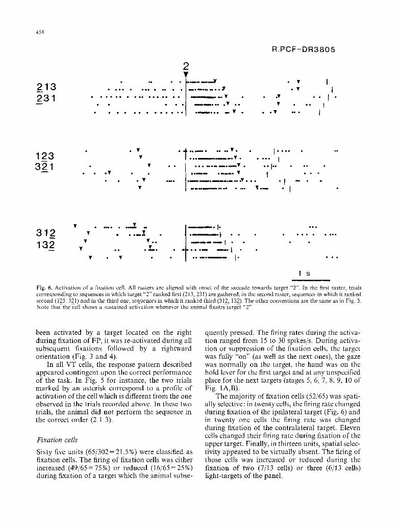
Fig. 6. Activation of a fixation cell. All rasters are aligned with onset of the saccade towards target "2". In the first raster, trials corresponding to sequences in which target "2" ranked first (213, 231) are gathered, in the second raster, sequences in which it ranked second (123,321) and in the third one, sequences in which it ranked third (312, 132). The other conventions are the same as in Fig. 3. Note that the cell shows a sustained activation whenever the animal fixates target "2"
been activated by a target located on the right during fixation of FP, it was re-activated during all subsequent fixations followed by a rightward orientation (Fig. 3 and 4).
In all VT cells, the response pattern described appeared contingent upon the correct performance of the task. In Fig. 5 for instance, the two trials marked by an asterisk correspond to a profile of activation of the cell which is different from the one observed in the trials recorded above. In these two trials, the animal did not perform the sequence in the correct order (2 1 3).
Fixation cells
Sixty five units (65/302= 21.5%) were classified as fixation cells. The firing of fixation cells was either increased (49/65=75%) or reduced (16/65=25%) during fixation of a target which the animal subs�9
quently pressed. The firing rates during the activa- tion ranged from 15 to 30 spikes/s. During activa- tion or suppression of the fixation cells, the target was fully "on" (as well as the next ones), the gaze was normally on the target, the hand was on the hold lever for the first target and at any unspecified place for the next targets (stages 5, 6, 7, 8, 9, 10 of Fig. 1A,B).
The majority of fixation cells (52/65) was spati- ally selective: in twenty cells, the firing rate changed during fixation of the ipsilateral target (Fig. 6) and in twenty one cells the firing rate was changed during fixation of the contralateral target. Eleven cells changed their firing rate during fixation of the upper target. Finally, in thirteen units, spatial selec- tivity appeared to be virtually absent. The firing of those cells was increased or reduced during the fixation of two (7/13 cells) or three (6/13 cells) light-targets of the panel.
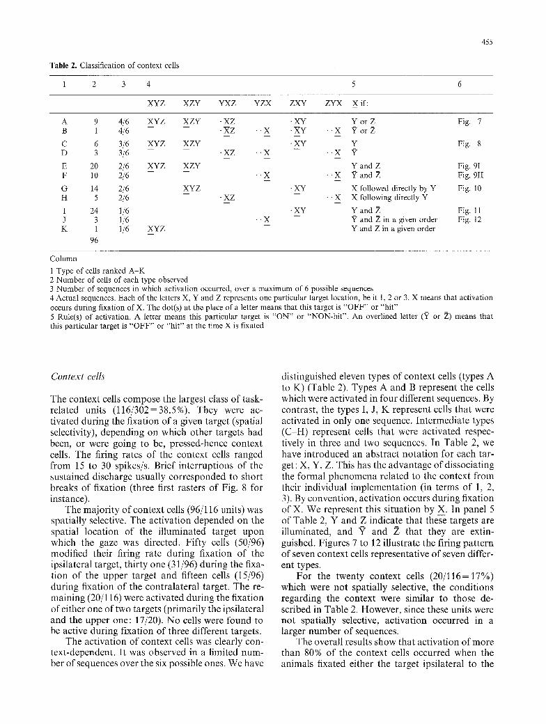
Table 2. Classification of context cells
455
1 2 3 4 5 6
XYZ XZY YXZ YZX ZXY ZYX X if:
A 9 4/6 XYZ B 1 4/6
C 6 3/6 XYZ D 3 3/6
E 20 2/6 XYZ F 10 2/6
O 14 2/6 H 5 2/6
I 24 1/6 J 3 1/6 K 1 1/6 XYZ
96
XZY . X Z - . X z . . x
XZY
XZY
XYZ
�9 XZ �9 "X
�9 XZ
" X
" X
�9 XY Y o r Z Fig. 7 �9 XY " X X~orZ
�9 XY Y Fig. 8 - - . . X ?
Y and Z Fig. 9I �9 �9 X x~ and Z Fig. 9II
XY X followed directly by Y Fig. 10 �9 - X X following directly Y
XY Y and Z Fig. 11 -- x7 and 2{ in a given order Fig. 12
Y and Z in a given order
Column
1 Type of cells ranked A - K 2 Number of cells of each type observed 3 Number of sequences in which activation occurred, over a maximum of 6 possible sequences 4 Actual sequences. Each of the letters X, Y and Z represents one particular target location, be it 1, 2 or 3. X means that activation occurs during fixation of X. The dot(s) at the place of a letter means that this target is " O F F " or "h i t" 5 Rule(s) of activation. A letter means this particular target is " O N " or "NON-hi t" . An overlined letter (Y or Z) means that this particular target is " O F F " or "h i t" at the time X is fixated
Context cells
The context cells compose the largest class of task- related units (116/302=38.5%). They were ac- tivated during the fixation of a given target (spatial selectivity), depending on which other targets had been, or were going to be, pressed-hence context cells. The firing rates of the context cells ranged from 15 to 30 spikes/s. Brief interruptions of the sustained discharge usually corresponded to short breaks of fixation (three first rasters of Fig. 8 for instance).
The majority of context cells (96/116 units) was spatially selective. The activation depended on the spatial location of the illuminated target upon which the gaze was directed. Fifty cells (50/96) modified their firing rate during fixation of the ipsilateral target, thirty one (31/96) during the fixa- tion of the upper target and fifteen cells (15/96) during fixation of the contralateral target�9 The re- maining (20/116) were activated during the fixation of either one of two targets (primarily the ipsilateral and the upper one: 17/20)�9 No cells were found to be active during fixation of three different targets.
The activation of context cells was clearly con- text-dependent. It was observed in a limited num- ber of sequences over the six possible ones. We have
distinguished eleven types of context cells (types A to K) (Table 2). Types A and B represent the cells which were activated in four different sequences. By contrast, the types I, J, K represent cells that were activated in only one sequence. Intermediate types (C-H) represent cells that were activated respec- tively in three and two sequences. In Table 2, we have introduced an abstract notation for each tar- get: X, Y, Z. This has the advantage of dissociating the formal phenomena related to the context from their individual implementation (in terms of 1, 2, 3). By convention, activation occurs during fixation of X. We represent this situation by X. In panel 5 of Table 2, Y and Z indicate that these targets are illuminated, and Y and Z that they are extin- guished. Figures 7 to 12 illustrate the firing pattern of seven context cells representative of seven differ- ent types.
For the twenty context cells (20/116=17%) which were not spatially selective, the conditions regarding the context were similar to those de- scribed in Table 2. However, since these units were not spatially selective, activation occurred in a larger number of sequences.
The overall results show that activation of more than 80% of the context cells occurred when the animals fixated either the target ipsilateral to the
456
213 231
O 0 O 0 �9 �9 �9 �9
�9 O 0 O0 (NIO
O 0 �9 �9 0 0 4 ) 4 0 Q O 0
O0 �9
R PFC-COZ'3230 2
7 , i |eaiDeoe~mmeo ,l~mo~ ~ e ~ Y
123 321
" W " . . . . . .
, �9 - e e m m o o m i e e e n o ~ m ~ m o ~ u Oo O 0 O e ~ e | ~ w i i
. . . �9 �9 �9 . . . . j . . . . . - . .
312 132
�9 . ~ . .v , ._. �9 . . . - , . , . 7
Y . Y .
Y . . Y.
. u Y
u . Y
o o o e �9 e o c o l e o e e e o e e e e $ o �9 �9 o o
e e �9 �9 o o o 4 ~ �9 �9 e e o �9 �9 �9
e o e o o o o e �9 c o o i o e o o e e e l e o �9 o e e e
�9 e o �9 �9 ee r i e e m o e o e o e e
�9 0 �9 �9 . . . J �9 �9 �9 e
m �9 . J �9 �9
�9 �9 �9 o e J �9 �9 �9 e e 4 o o
1 S i
Fig. 7. Activation of a context cell. All rasters are aligned with onset of the saccade towards target "2". The other conventions are the same as in Fig. 3. The cell shows a sustained activation when the animal fixates target "2" provided that targets "1" or "3" are still illuminated (upper raster, sequences "213" and "231" or middle raster, sequences "123 or "321"). Note that when targets "3" and ' '1' ' are both extinguished (last raster) the cell is poorly activated
recording site or the upper target, and depended on the state of the target(s) contralateral to the direc- tion of gaze.
We did not systematically study the context cells in the "two-target" paradigm. However, our data show that whenever the activation of a context cell depended on a particular target being already extinguished in the "three-target" paradigm, ac- tivation was also observed in the "two-target" par- adigm if the same target had not been introduced in the sequence from the beginning. In figure 11B, activation of the cell in the sequence "23" in which target "1" had not been introduced, is comparable to the activation observed in the sequence "123" in which target "1" has been extinguished by the ani- mal.
Saccade or movement related units
Activation of thirty three units (33/302 = 11%) was related to saccadic eye movements. The majority of these cells (28/33=85%) showed a short post-
saccadic activation and only five displayed some evidence of presaccadic activation.
Nine units showed a phasic increase of activity at the time of the arm movement upon the target light. In seven cells, this was observed during ac- quisition of whatever target-light and in two cells during acquisition of a particular target.
Signal related units
Eight units (8/302=2.5%) showed distinct phasic activity following presentation of a peripheral vi- sual cue. Three cells were activated by the contralat- eral signal, two by the ipsilateral signal and one by the upper one. One cell displayed an activation for the onset of the contralateral or upper cue and one by any one of the cues. Among these eight cells, five increased their discharge rate with the onset of a visual key only if it ranked first in the sequence.
Sixteen units were activated following the presentation of the central fixation light. Activation was transient in eight cells and sustained in the other eight.
10 ~ I
4 5 7
H o -
l o ~ I
V ~ ~
3 1 2
3 u
. . " . " . - - . . . . . . _ . . _ . , _
R.PCF-COZ3126
�9 - ~ I
�9 I . . v I
321 : - 1 - - . ,
�9 oo
~ ~ ~ I ~ �9
�9 . ~ I ~
�9 I
231 .v "" It- " - ' - ~ ' ' - ' ' v t,., �9 I �9 ~ V �9 . - - � 9 [
1 32 "* . . . . . ~ . . "11 . , . . . . - �9 I
�9 V �9 . " 5 "
�9 u , , - I �9 * [ , -
2 1 3 . . . . * " v I �9 " 1 " "
. V . . . . 17 I . . . . . 123 v . . . . v . . . . I" ,-- 1 s
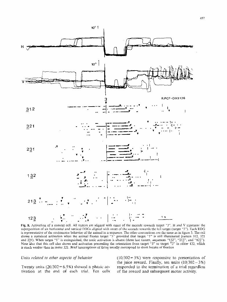
F i g . 8. Activation of a context cell. All rasters are aligned with onset of the saccade towards target "3". H and V represent the superposition of six horizontal and vertical EOGs aligned with onset of the saccade towards the left target (target "3"). Each EOG is representative of the oculomotor behavior of the animal in a sequence. The other conventions are the same as in figure 3. The cell shows a sustained activation when the animal fixates target "3" provided that target "1" is still illuminated (rasters 312, 321 and 231). When target "1" is extinguished, the tonic activation is absent (three last rasters, sequences "132", "213", and-"12_~'). Note also that this cell also shows and activation preceeding the orientation from target "3" to target "2" in raster 132, which is much weaker than in raster 321. Brief interruptions of firing usually correspond to short breaks of fixation
Units related to other aspects of behavior
Twenty units (20/302= 6.5%) showed a phasic ac- tivation at the end of each trial. Ten cells
(10/302=3%) were responsive to presentation of the juice reward. Finally, ten units (10/302= 3%) responded to the termination of a trial regardless of the reward and subsequent motor activity.
458
T �9 e � 9
1 2 3 �9 �9 i ' 3 2 . . . . . . . . . .
o o e �9 �9
�9 o e �9 �9 �9 e N �9 �9
�9 �9 �9 e e �9 e o e e
2 1 3 � 9
3 ] ' 2 " ' " �9 - - �9 n �9 e e �9 �9
2 3 1 " �9 . . . . � 9
3 2 T " ' � 9 � 9 . . . . . . . �9 - - ~ � 9 ~ ~ ~ �9
�9 , , � 9
1 Y
m o o e e e ~ ~ o V
L e ~ P e u m n m o i m e o ~ m m e n V �9 e o
o e l l l m ~ e o o ~ e e ~ e o o e o V e
�9 " - i - : - - - : ? . ;
R.PFC-C P3163
�9 Je eo
�9 I v .] v . [
�9 , . i �9
�9 ~ I
�9 �9 . . . . . . I . . . . . . .
�9 �9 �9 u = l
�9 e � 9 �9 �9 �9 I �9
�9 e e �9 �9 �9 I | �9 �9 e e e
�9 I
t n �9 �9 J o e I ~ ~ l
. I
. . . . . . . . . I1". �9 �9 I
E 2 3 1 2 1 3 " " "
m �9 e e
2 u
e e l * o , o l � 9
�9 o w e �9
=e �9
R PFC-DT4054
�9 I I
e e �9 �9 I
�9 I
. v l | . . . ..V. . . . . j v, . . . . . . . �9
1 2 3 . . . . , .... �9
- - , I , �9 �9 , �9 e . c o l �9
�9 �9 �9 , . a o . o , �9
I I I
e � 9
I J ~ 1 7 6
I - I "
0 0
D O
Q D
�9 D O
1 3 2 ' . . . . . . . . . . . . . d 1 3 1 2 ' ' . . . . . . I
�9 . . . . Ir . . . . . . . . . I
N
0 0
1 s
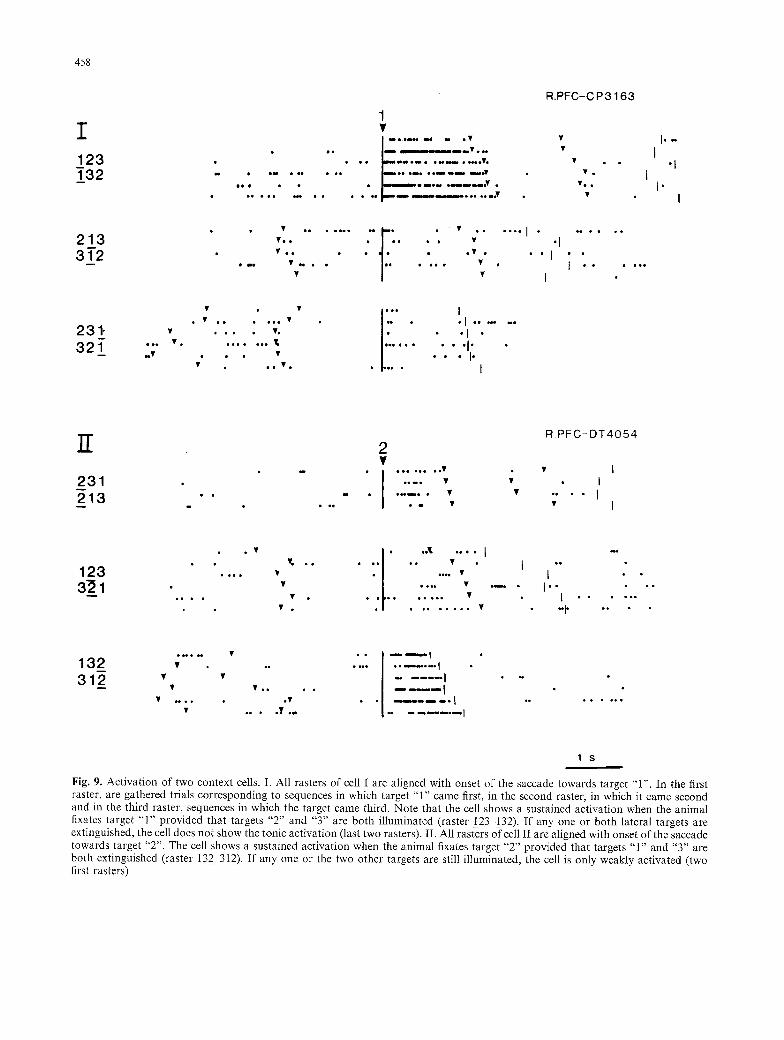
Fig. 9. Activation of two context cells. I. All rasters of cell I are aligned with onset of the saccade towards target "1". In the first raster, are gathered trials corresponding to sequences in which target "1" came first, in the second raster, in which it came second and in the third raster, sequences in which the target came third. Note that the cell shows a sustained activation when the animal fixates target "1" provided that targets "2" and "3" are both illuminated (raster 123-132). I f any one or both lateral targets are extinguished, the cell does not show the tonic activation (last two rasters). II. All rasters of cell II are aligned with onset of the saccade towards target "2". The cell shows a sustained activation when the animal fixates target "2" provided that targets "1" and "3" are both extinguished (raster 132 312). If any one or the two other targets are still illuminated, the cell is only weakly activated (two first rasters)
10~
V
,o~ i q-/I r -
4 5 9
132
1 R . P F C - D A 3 2 5 2
. . . . . . :: ' - = : ' - - : ! ; , : i " �9 " " �9 t �9
' . . . . . . . . I
123 ~
: : f ' " �9 ~ �9 . . . . . I
I .I
213 . . . . �9 . | . �9
" " � 9 . . . . . . . . . . . L~ . . . . . . . v. I - �9 - . �9 . . . . . . . . . . . . �9 - . I
-- . � 9 . . . . . . . . . �9 * * I . . . . �9 . . . . . . . . . . . . , . . . . . 1 ' * I *
312 . . - �9 ] . . . . . . . . . . . . . �9 �9 I
�9 � 9 I �9 I , �9 -" . ' . . ' - ' , "�9 �9 "t �9 " Y ' " * " I
231 �9 . . . . . . . . . . . . . . . . . � 9 1 4 9
. . . . . . I . . . .
m * , Y * * * * ~ �9 . T I . � 9 ~ .
321 .v. * . . . . . . I! . . . . . . . . �9 . . . . . �9 . . . . . . . . . . . . I " ~ ' � 9 1 4 9 " " 1 s
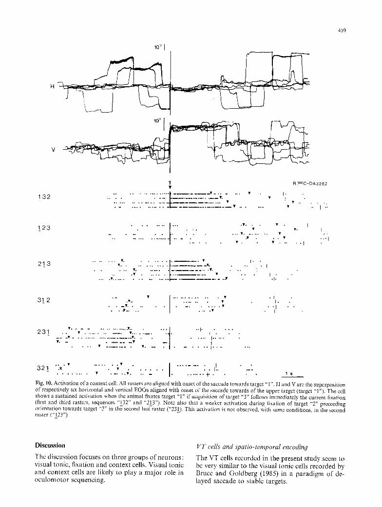
Fig. I 0. Activation of a context cell. All rasters are aligned with onset of the saccade towards target "1". H and V are the superposition of respectively six horizontal and vertical EOGs aligned with onset of the saccade towards of the upper target (target "1"). The cell shows a sustained activation when the animal fixates target "1" if acquisition of target "3" follows immediately the current fixation (first and third rasters, sequences "132" and "21_3") Note also that a weaker activation during fixation of target "2" preceeding orientation towards target "3" in the second last raster ("231). This activation is not observed, with same conditions, in the second raster ("123")
D i s c u s s i o n
T h e d i s c u s s i o n focuses o n t h r e e g r o u p s o f n e u r o n s : v i sua l ton ic , f i xa t i on a n d c o n t e x t cells. V i sua l t o n i c a n d c o n t e x t cells a re l ike ly to p l a y a m a j o r ro le in o c u l o m o t o r s e q u e n c i n g .
VT cells and spatio-temporal encoding
T h e V T cells r e c o r d e d in the p r e s e n t s t u d y seem to be ve ry s im i l a r to the v i sua l t o n i c cel ls r e c o r d e d b y Bruce a n d G o l d b e r g (1985) in a p a r a d i g m o f de- l ayed s a c c a d e to s t ab l e t a rge t s .
460
lo ~ I
l~176 I
R . P F C - C ~ 3 3 4 1
A 2 �9 " " " J ' . . . . . . . �9 " " ~ V I 2 1 3 . . . . �9 . . a I
1 . . . . " � 9 " " �9 I . . . . . �9 Y y � 9 �9 V I
�9 �9 � 9 1 4 9 �9 �9 , , , �9 J
231 0 I
1 0
!
�9 , . . � 9 3 . . ~ J r
�9 ~ � 9 1 4 9 1 4 9 �9
�9 . � 9 1 4 9 V �9
�9 �9 . , �9 . Y I I.
123 �9 . . t , !
I ,,-Z . " I r
J , .v. I
I 0
I �9
321 �9 | . . . . �9 I �9 , V . ] " " �9 - I
�9 " �9 �9 I �9 ~ , , , . V " ' l
�9 � 9 1 4 9 �9 .
B
132
312 �9
2 3
2 1
1 2
3 2
V ~ �9 �9 ~ ~ �9 . . �9 �9
�9 Y Y �9
l . �9 ..'I
Q Q
~ 0 I Q Q
O D QO Q ~ 0
O g
Y
. . . . ;
�9 ~176 ,1 , ~
I �9 |
�9 � 9 J
!
, J �9
~-:..-- --~..,.. j I , �9 �9
.Y v
J " ' ! 1 �9 �9 ~
i . . ,
�9 - " "1
�9 I
I
I
1 s
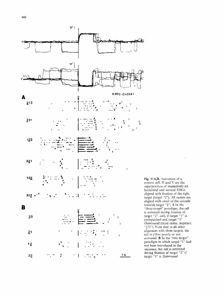
Fig. I1A,B. Activation of a context cell H and V are the superposition of respectively six horizontal and vertical EOGs aligned with fixation of the right target (target "2") All rasters are aligned with onset of the saccade towards target "2". A In the "'three-target" paradigm, the cell is activated during fixation of target "2", only if target "1" is extinguished and target "3" illuminated (third raster, sequence "I_2Y'). Note that in all other sequences with three targets, the cell is either poorly or not activated. B In the "two-target" paradigm in which target "1" had not been introduced in the sequence, the cell is activated during fixation of target "2" if target "3" is illuminated
4 6 1
132 123
1 u
�9 e o ~ e e l e e e ~ m m �9 �9 �9 ~ �9
L.PFC-C/] 3731
= i T ~ e � 9 �9 �9 �9 e o � 9 �9 �9
�9 . . . . �9 , . . �9 �9 ~ �9 �9
e � 9 �9 � 9 �9 l e e �9 �9 ' o � 9 �9 �9
�9 �9 = , i l l ~ 1 4 9 e o � 9 �9 e � 9 �9 o � 9 � 9 1 4 9 1 4 9 1 4 9 1 4 9
213 312 �9 ...ii.:"
�9 �9 �9 �9 * - o - ~ . I - * , ~ * o �9
�9 e �9 �9 �9 �9 e � 9 ~ 1 4 9 �9 �9 J e e
�9 �9 w , . J . . . . ,
. . . . �9 " I "
231 �9 . V , . . . V . " 1 ' ' " -
fe �9 �9 �9 �9 eve e e e �9 eOod, e ~ e
�9 �9 �9 �9 �9 �9 V �9 e e ~ �9 * .
�9 �9 �9 �9 o e ~ o e e e e * *
- I - . �9 �9 . " " - - "
- - "1 �9 " " " "
- . - ' 1 - - - " " " - - - ' " �9
�9 - . I . �9 �9 �9
321 �9 �9 T ~ e * e o �9 �9 *
t �9 Y . ~ N ~ �9 . .
V Y ~ �9 ~ �9
e ~ e o o �9 o e �9 6 V o e e o o ~ e e e e e �9 e o o e
�9 . . ~ o o ~ m o o e , J o ~ ~ �9 �9 �9
e ~ e u m ~ e : J �9 e e e �9 �9 e ~
e e ~ o ~ e �9 m e Q , t u e ~
oeeeee ~ e e o a m 4 8 e n m ~ eoemuo J u ~ o e ~ eeeeeQ e ~ e e ~ e o
�9 e e a m e e e e e m e e e e e m , m J e e e e
1 s
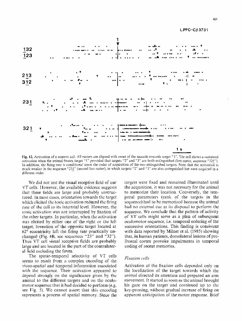
Fig. 12. Activation of a context cell. All rasters are aligned with onset of the saccade towards target "1". The cell shows a sustained activation when the animal fixates target "1" provided that targets "2" and "3" are both extinguished (last raster, sequence "321_"). In addition, the firing rate is conditional upon the order of acquisition of the two extinguished targets. Note that the activation is much weaker in the sequence "23_1" (second last raster), in which targets "2" and "3" are also extinguished but were acquired in a different order
We did not test the visual receptive field of our VT cells. However, the available evidence suggests that these fields are large and probably unstruc- tured. In most cases, orientation towards the target which elicited the tonic activation reduced the firing rate of the cell to its intertrial level. However, the tonic activation was not interrupted by fixation of the other targets. In particular, when the activation was elicited by either one of the right or the left target, foveation of the opposite target located at 62 ~ eccentricity left the firing rate practically un- changed (Fig. 4B, see sequences "23" and "32"). Thus VT cell visual receptive fields are probably large and are located in the part of the contralater- al field excluding the fovea.
The spatio-temporal selectivity of VT cells seems to result from a complex encoding of the visuo-spatial and temporal information associated with the sequence. Their activation appeared to depend strongly on the significance given by the animal to the different targets and on the oculo- motor sequence that it had decided to perform (e.g. see Fig. 5). We cannot assert that this encoding represents a process of spatial memory. Since the
targets were fixed and remained illuminated until the acquisition, it was not necessary for the animal to memorize their location. Conversely, the tem- poral parameters (rank of the targets in the sequence) had to be memorized because the animal had no external cue at its disposal to perform the sequence. We conclude that the pattern of activity of VT cells might serve as a plan of subsequent oculomotor sequence, i.e. temporal ordering of the successive orientations. This finding is consistent with data reported by Milner et al. (1985) showing that, in human patients, dorsolateral lesions of pre- frontal cortex provoke impairments in temporal coding of recent memories.
Fixation cells
Activation of the fixation cells depended only on the localization of the target towards which the animal directed its attention and prepared an arm movement. It started as soon as the animal brought his gaze on the target and continued up to the key-pressing, without gradual increase of firing on apparent anticipation of the motor response. Brief
462
interruptions of the sustained discharge usually corresponded to short breaks of fixation. Thus, activation seemed to be primarily related to visual attention.
Suzuki and Azuma (1977) recorded fixation like cells (their G cells) from a large portion of the prefrontal cortex including Sulcus Principalis and frontal eye field (FEF), in an experimental par- adigm which included a bar-pressing task in re- sponse to the dimming of a target. In the absence of visually-guided reaching, they observed no spatial selectivity. Bruce and Goldberg (1985) recorded from the FEF a small proportion of fixation-foveal neurons in a paradigm which included also a bar pressing task in response to the dimming of a peripheral light. They did not dissociate foveal and fixation neurons and did not mention spatial selec- tivity. The comparison of our data and of those obtained by these two groups seems to indicate that the activation of spatially selective fixation neurons only intervenes during preparation for visually guided reaching. It could be associated with some aspects of visual attention for goal-di- rected movements. One hypothesis is that the G neurons and our fixations neurons belong to the same population. Our experimental paradigm would have simply unveiled new properties of the G neurons.
Activation of the context cells
A major problem which has important implications concerns the origin of the activation of the context cells. Three main hypotheses - visual, motor and cognitive - are considered.
The first hypothesis would be that activation represents a visual response to illuminated tar- get(s). According to this hypothesis, the context cells would be VT cells with a receptive field (RF) different from those of the VT cells described above. For instance, the cell whose responses are illustrated in Fig. 7 (type A) would be a VT cell responding to stimuli located in the upper-left quad- rant. The cell whose responses are illustrated in Fig. 8 (type C), could be classified as a VT cell responding to stimuli located in the upper-right quadrant. For two other types of cells (types F and I), the visual hypothesis fits with the data although it requires a number of assumptions for which there is at present very little evidence. The cell whose responses are illustrated in Fig. 9II (type F) for instance would have an inhibitory field to the left and a foveal excitatory field modulated by the di- rection of gaze (to account for the spatial selectiv- ity). The activation of the cell in Fig. 11 (type I) could be related to a restricted RF to the left on the
horizontal meridian coupled with an inhibitory field up and to the left (thus the slight greater activity in sequences 213 and 231 than in sequences 132 and 312).
However, two lines of evidence argue against the hypothesis of visual responses of context cells. First, the context cells were never activated by the onset of peripheral targets during central fixation. If they were visual, this result would indicate that among the cells which are potentially visual (175), two thirds (the 116 context cells) have RFs which do not include regions located at 19 ~ on the superi- or vertical meridian and/or at 32 ~ on the horizontal meridian. This conclusion seems at variance with the data of Suzuki (1985) showing that the RFs in the superior arcuate area are eccentric but large. Second, our data show that activation of many context cells cannot be solely interpreted as a re- sponse to the current visual stimuli of the environ- ment, since it also depended on temporal con- straints. We found cells (23/96) that were activated if a particular target was pressed right after the current fixation (types K or G) or if a particular target (type H) or the other two (type J) had been already pressed at a given rank with respect to the current fixation.
The second hypothesis is that activation of con- text cells is related to the preparation of motor and oculomotor activity which follows the fixation period. This hypothesis accounts for the firing characteristics of 3 types of context cells. One type (type E) would be involved in preparation for the arm movement from hold-lever to target X (Fig. 9I). The firing pattern of cells of types A and G (23/96) fits with the hypothesis of a saccade preparation. Activation of the cell in Fig. 7 (type A) would be associated with saccade preparation to- wards targets " l " or "3", when the monkey fixated target "2". Activation of the cell in Fig. 10 (type G) could be associated with a saccade preparation towards "3" when the monkey fixated "1". How- ever, in the hypothesis o f a saccade preparation, the firing rate would be expected to show a gradual increase in apparent anticipation of the saccade. This was never observed. The activation was steady as soon as the animal's gaze was on target. For the other types of context cells, there is no evidence that activation is related to the preparation for a saccade. For instance in Fig. 8, the firing rate of the cell is very different in the second and in the fourth raster although in both cases the animal fixated target "3" and prepared for a saccade towards target "2". The firing rate of the cell in Fig. 11 is very different in the second and third rasters al- though the animal in both cases fixated target "2"
463
and prepared for a saccade towards target "3". In Fig. 9I, the cell is activated during fixation of target "1" if targets "2" and "3" are both illuminated. The firing rate is different in the other sequences al- though the goal of the saccades following fixation of target "1" was the same.
The third hypothesis concerning the mecha- nisms of activation of context cells considers that they are independent of the physical state - illumi- nated or extinguished - of the targets and of motor or oculomotor preparation. According to this hypothesis which we would call "cognitive hypoth- esis", the firing pattern of the cells could be the neural trace of a representation of the state of the sequence constructed by prefrontal cortex on the basis of cognitive information (memory of location and rank of the future targets provided by the VT cells on one hand, memory of the location and time of occurrence of the previous key pressings on the other). With respect to the direction of the current fixation, this representation would indicate regions where targets have been or are still to be pressed and their order of acquisition. The cognitive hypothesis is the only hypothesis which accounts for the firing pattern of all context cells including types B, H, J (Fig. 12) and K (10/96).
The cognitive hypothesis would explain the ani- mals' ability to perform the task when targets re- main illuminated after t h e press (unpublished re- sults). If the environment does not provide cues regarding the state of the sequence, the animal has to perform the task exclusively on the basis of cog- nitive (i.e. of memorized spatial and temporal) in- formation.
The cognitive hypothesis is consistent with the observations and interpretations of Fuster (1985) and Passingham (1985a) for whom the concept of working memory is critical to an understanding of the prefrontal cortex. In addition, it suggests that the working memory is continually updated with information acquired during performance of the tasks stored by the animal (in the VT cells).
Special role of superior arcuate area in oculomotor sequencing
Our data support the hypothesis that the superior arcuate area is concerned with construction of ocu- lomotor plans and "internally organized [...] eye scanning (Collin et al. 1982).
Bruce and Goldberg have suggested that the major role of FEF "is to construct the amplitude and direction of the saccade most adapted to corti- cal decisions" and to channel this information to
the subcortical oculomotor centers. In our experi- ment, the phasic activity of the saccade-related cells, the tonic activity of VT cells responding to the first target only and the tonic activity of some context cells (types A and G) may be directly impli- cated in the programming and execution of succes- sive saccades.
In the special case of oculomotor sequences, it is likely that the "cortical decisions" also involves selection of successive targets. In the framework of the cognitive hypothesis discussed above, some VT and context cells of the arcuate area could be re- sponsible for the selection seen as an integral part of the perception of motivationally important stimuli. At present there is no evidence that the arcuate area modulates the perception of the en- vironment but so far this scheme is consistent with the hypothesis that FEF, in relation with area PG of parietal cortex, is the neural basis of sequential distribution of attention within extrapersonal space (Mesulam 1981). It also agrees with clinical ob- servations and current theories of prefrontal func- tions which outline the role of prefrontal cortex in controlling the posterior areas and providing con- scious directions for the efficient processing of in- formation (Stuss and Benson 1987).
Organization of the prearcuate oculomotor area
Our recording sites include area 8 A and, apparent- ly, the caudal part of area 8 B. Since the discovery of an oculomotor region in the frontal lobe, rostral to the arcuate sulcus, much attention has been con- centrated on its extent (Schlag and Schlag-Rey 1987). Bruce and al. (1985) have delineated a low- threshold frontal-eye-field where saccades are eli- cited with stimulation current < 50 pA. This area corresponds to the restricted area lying along the posterior portion of the arcuate sulcus and con- fined to Walker's cytoarchitectonic areas 8 A (cau- dal part) and 45.
These data suggest that there may be a func- tional division of area 8 into a "supraoculomotor" area extending rostrally, involved in programming oculomotor strategies (see also Joseph and Barone 1987) and a more caudal area, dealing with execu- tion of saccades. However, this conclusion is lim- ited by the apparently homogeneous distribution of our cells over the two hypothetized divisions (Fig. 2) and the uncertainty of the spatial defini- tion of the core of the frontal eye field. Moreover, it is still unknown whether area 8B and rostral part of area 8 A send a modulating feedback signal to the core of the FEF.
464
Sulcus principalis and oculomotor sequencin9
Funahashi et al. (1989) have shown that neurons in different parts of sulcus principalis (SP) possess information concerning the location of visual cues during the delay period of an oculomotor delayed- response task (1.5 to 6 seconds). This result sug- gests that storage and/or execution of oculomotor programs based on memorized spatial items are also under control of SP.
In the present experiment, numerous penetra- tions were made in the area surrounding the caudal part of SP which receives a substantial input from parietal cortex (Barbas and Mesulam 1985). Surprisingly, we recorded neither visual tonic nor context cells in this region. This result seems to indicate that the caudal part of SP is not in- volved in programming oculomotor sequences. Clearly much more work has to be done to under- stand the nature of the relationships between the SP and the arcuate area.
Acknowledgements. The authors are grateful to M.E. Goldberg, J. and M. Schlag, and B. Dreher for reading an earlier draft and providing constructive comments. We also thank P. Giroud for surgical assistance and animal preparation, N. Boyer and S. Richard for histological assistance, and M. Soulier for secre- tarial services.
References
Barbas H, Mesulam MM (1981) Organization of afferent input to subdivisions of area 8 in the rhesus monkey. J Comp Neurol 200: 407-431
Barbas H, Mesulam MM (1985) Cortical afferent input to the principalis region of the rhesus monkey. Neuroscience 15 : 619-637
Brody BA, Pribram KH (1978) The role of frontal and parietal cortex in cognitive processing: test of spatial and sequence functions. Brain 101 : 607-633
Bruce CJ, Goldberg ME (1985) Primate FEF. I. Single neurons discharging before saccades. J Neurophysiol 53:603-635
Bruce CJ, Goldberg ME, Bushnell MC, Stanton GB (1985) Primate FEF. II. Physiological and anatomical correlates of electrically evoked eye movements. J Neurophysiol 53 : 714-734
Collin NG, Cowey A, Latto R, Marzi C (1982) The role of frontal eye field and superior colliculi in visual-search and non visual-search in rhesus monkey. Behav Brain Res 4:177-193
Crowne DP, Yeo CH, Russell IS (1981) The effect of unilateral frontal eye field lesions in the monkey: visual motor guid- ance and avoidance. Behav Brain Res 2:165-185
De Renzi E, Faglioni P, Lodesani M, Vecchi A (1983) Perfor- mance of left-brain damage patients on imitation of single movements and motor sequences: frontal and parietal in- jured patients compared. Cortex 19:333 343
Funahashi S, Bruce CJ, Goldman-Rakic PS (1989) Mnemonic coding of visual space in the monkey's dorsolateral prefron- tal cortex. J Neurophysiol 61:331-349
Fuster JM (1985) Temporal organization of behavior. Hum Neurobiol 4: 57-60
Goldman-Rakic PS (1987) Circuitry of primate prefrontal cor- tex and regulation of behavior by representational memory. In: Handbook of physiology. Chap. V. The nervous system 9 : 373-417
Joseph JP, Barone P (1987) Prefrontal unit activity during a delayed oculomotor task in the monkey. Exp Brain Res 67 : 460-468
Kimura D (1982) Left hemisphere control of oral and brachial- movements and their relation to communication. Philos Trans R Soc B298:135 149
Kolb B, Milner B (1981) Performance of complex arm and facial movement after focal brain lesions. Neuropsychologia 19 : 491-503
Luria AR (1966) Higher cortical functions in man. Tavistock, London
Mesulam MM (1981) A cortical network for directed attention and unilateral neglect. Ann Neurol 10:309 325
Milner B, Petrides M, Smitt ML (1985) Frontal lobes and the temporal organization of memory. Hum Neurobiol 4:137-142
Passingham RE (1985a) Memory of monkeys (Macaca mulatta) with lesions in prefrontal cortex. Behav Neurosci 99:3-21
Passingham RE (1985b) Prefrontal cortex and sequencing of movement in monkeys (Macaca mulatta). Neuropsycholo- gia 23 : 453-462
Petrides M, Milner B (1982) Deficit on subject-ordered task after frontal and temporal lobe lesion in man. Neuropsy- chologia 20 : 249-262
Petrides M (1985) Deficit in non-spatial conditionnal associative learning after periarcuate lesions in the monkey. Behav Brain Res 16:95-101
Pinto-Hamuy T, Link P (1965) Effects of frontal lesions on performance: sequential tasks by monkeys. Exp Neurol 12:96 107
Schlag J, Schlag-Rey M (1987) Evidence for a supplementary eye field. J Neurophysiol 57:179 200
Stuss DT, Benson DF (1986) The frontal lobe. Raven Press, New York
Stuss DT, Benson DF (1987) The frontal lobe and control of cognition and memory. In: The frontal lobe revisited. Perga- mon, pp 141-158
Suzuki H, Azuma H (1977) Prefrontal neuronal activity during gazing at a ligh spot in monkey. Brain Res 126:497-508
Suzuki H (1985) Distribution and organization of visual and auditory neurons in the monkey prefrontal cortex. Vision Res 25 : 465-469
Received March 22, 1989 / Accepted July 17, 1989