ppt on brain
TRANSCRIPT
By-Mr. ASHOK BISHNOI
Assist. Professor , JINR
The brain is the largest mass of nervous tissue in the body.
It lie in the cranial cavity
Weight of brain:-
In male is -1380 gm (48.6 oz)
In female is – 1250gm (44 oz)
Introduction:-

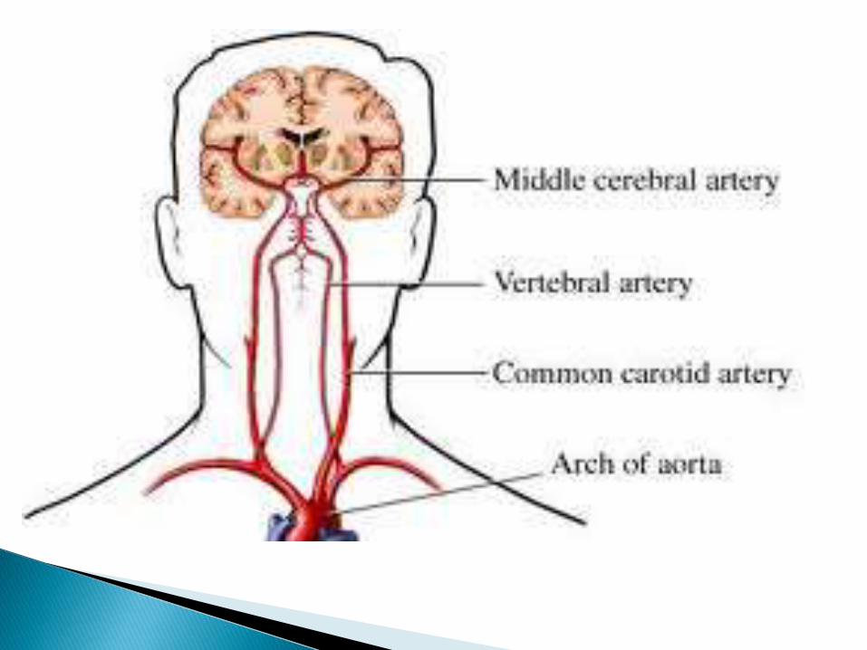
By carotid artery
Rt & lt anterior cerebral artery
Rt & lt posterior cerebral artery
Vertebral artery
Brain receive about 15% of the
cardiac output
Approximately 750 ml blood/mt
Blood supply to the brain;-

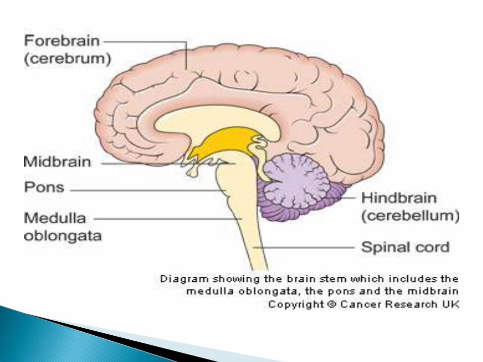
Cerebrum (Forebrain)
Mid brain
Pons
Medulla oblongata Brain steam
Cerebrum (Hindbrain)
Part of the brain:-
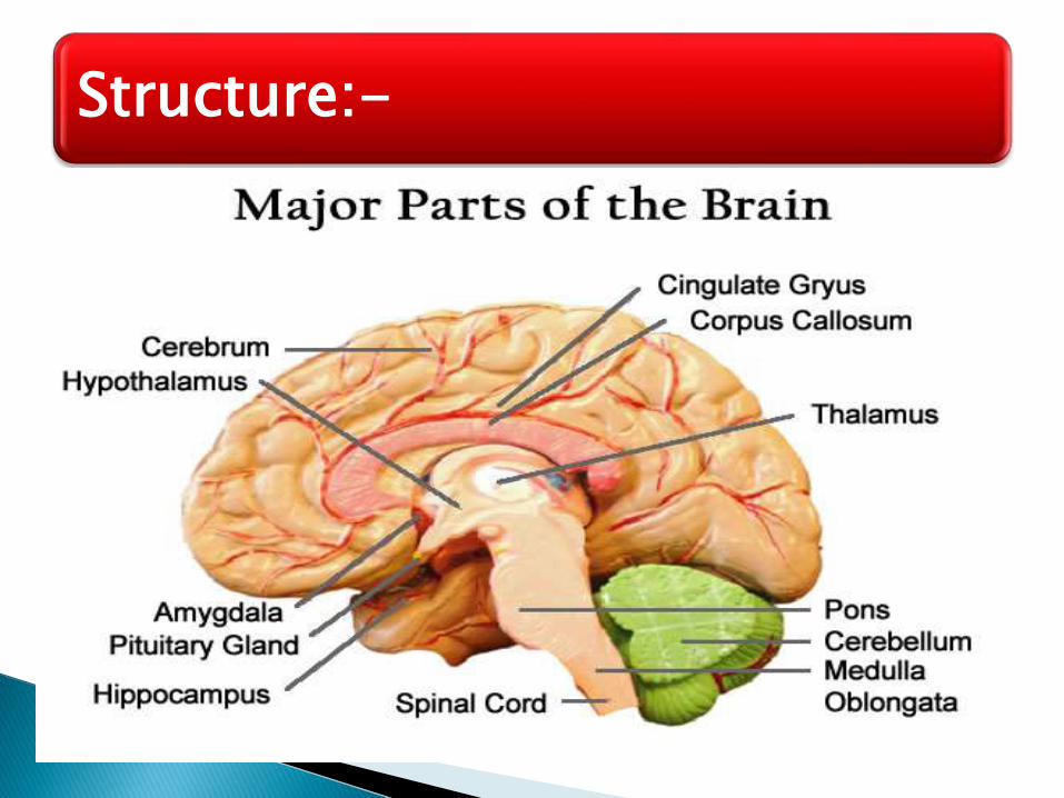
Structure:-
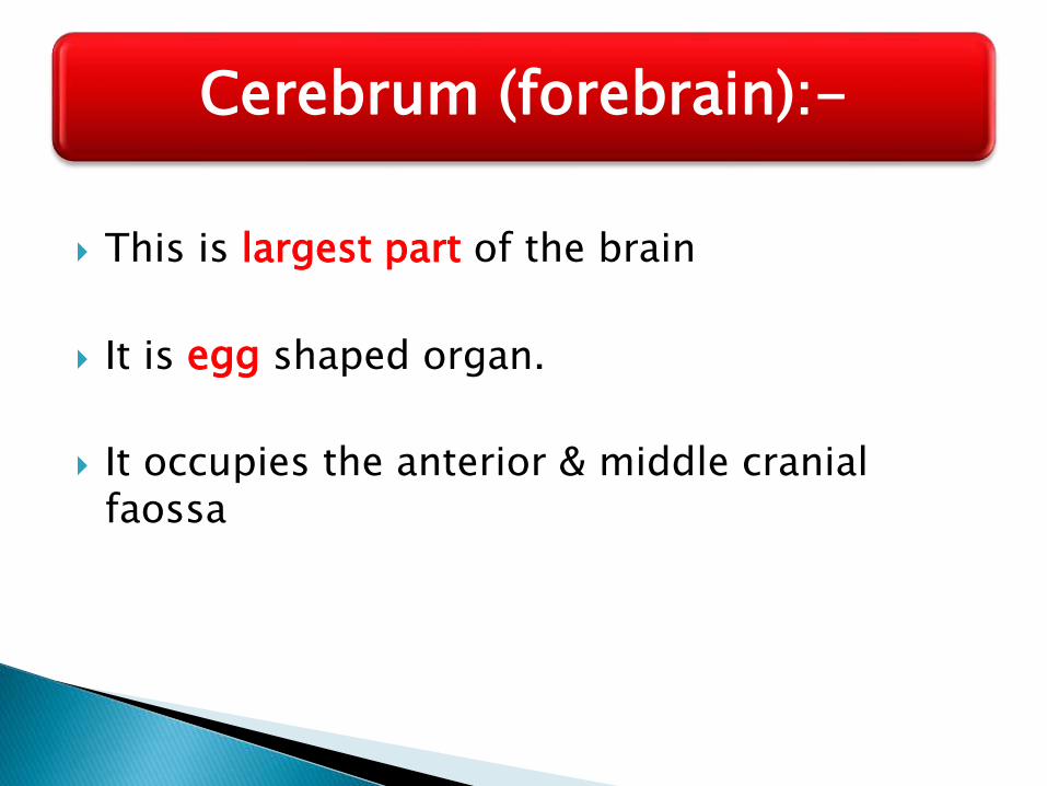
This is largest part of the brain
It is egg shaped organ.
It occupies the anterior & middle cranial faossa
Cerebrum (forebrain):-

Layers of the Cerebrum
Gray matter
Outer layer
Composed mostly of neuron cell bodies
White matter
Fiber tracts inside the gray matter
Example: corpus callosum connects hemispheres
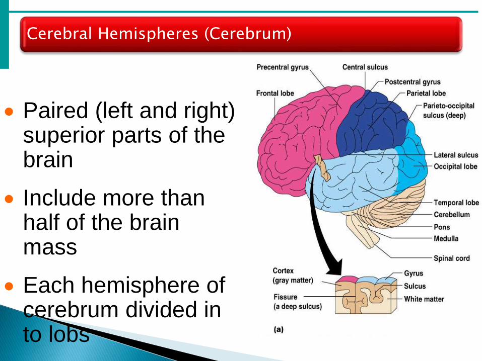
Cerebral Hemispheres (Cerebrum)
Paired (left and right) superior parts of the brain
Include more than half of the brain mass
Each hemisphere of cerebrum divided in to lobs
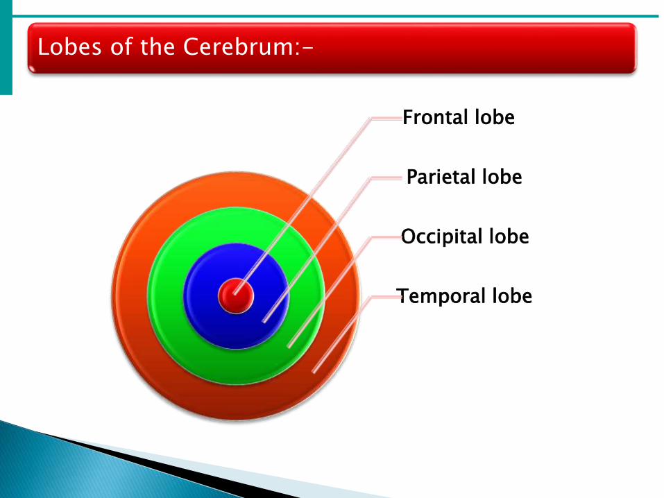
Lobes of the Cerebrum:-
Frontal lobe
Parietal lobe
Occipital lobe
Temporal lobe

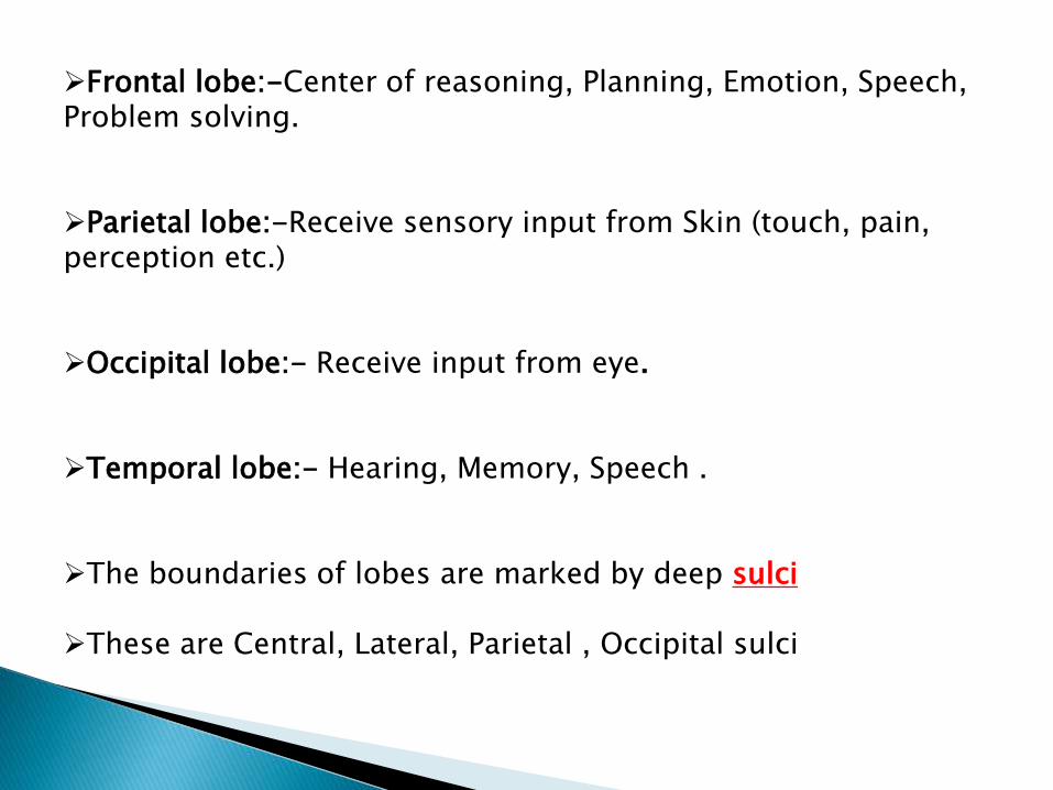
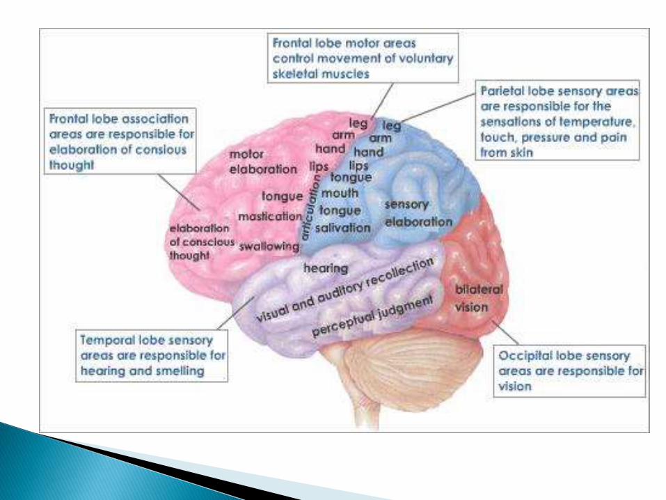
Frontal lobe:-Center of reasoning, Planning, Emotion, Speech, Problem solving.
Parietal lobe:-Receive sensory input from Skin (touch, pain, perception etc.)
Occipital lobe:- Receive input from eye.
Temporal lobe:- Hearing, Memory, Speech .
The boundaries of lobes are marked by deep sulci
These are Central, Lateral, Parietal , Occipital sulci

Functional Areas of the Cerebrum:-
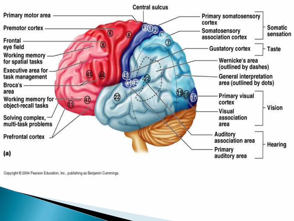
Motor area ( Lie in frontal lobe)– sends impulses to skeletal muscles.
Broca’s area (motor speech) – Situated in frontal lobe (Control muscle movement necessary for speech)
Sensory area – receives impulses from the body’s sensory receptors
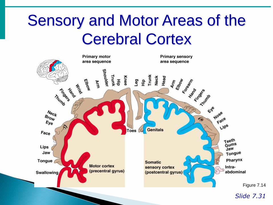
Sensory and Motor Areas of the
Cerebral Cortex
Slide 7.31
Figure 7.14

Sensory area:-
Slide 7.32a
Cerebral areas involved in special senses
Gustatory area (taste)
Visual area
Auditory area
Olfactory area
Gustatory area (taste):- Situated above the lateral sulcusimpulses from sensory receptor in taste buds are receive & perceive taste.
Visual area:- Situated in occipital lobe (optic nerve pass from the eye to this area.
Auditory area (Hearinf0:- Situated below the lateral sulcus in temporal bone. ( receive & interrupt impulse transmit from inner ear to auditory part)
Olfactory area (smell):-Situated in temporal lobe were impulse from nose transited through olfactory nerve are receive & interrupt.

Other areas:-
Thalamus:- Sensory input from skin or special sense.
Hypothalamus:- It is compose of number of nerve cell
It is situated front & below the thalamus.
Function:-
1. Appetite & satiety
2. Thirst & water balance
3. Regulate body temperature
4. Emotional reaction

Brain Stem
Attaches to the spinal cord
Parts of the brain stem
1. Midbrain
2. Pons
3. Medulla oblongata
4. Cerebrum (Hindbrain)
1. Mid brain:-
• It is the area of brain situated around the
cerebral aqueducts between cerebrum &
pons.
• It connect the cerebrum with lower part of brain & spinal cord
2. Pons:-
• I t is situated in front of the cerebellum.
• It consist of nerve fiber(white matter).
• Passing fiber from higher level of brain to spinal cord .

3. Medulla oblongata:-
• It extent from pons above &
• it continuous with spinal cord
below.
• About 2.5 cm long.
• Function:- ( the vital center)
• Cardiovascular center (HR control & BP regulation)
• Respiratory center
• Reflex center of Vomiting, Coughing, Sneezing, &
Swallowing

4. Cerebellum.:-
• It is situated behind the pons
• It occupying posterior cranial fossa
Function:-
• It help co-ordination of
voluntary muscular
movement, Posture
& balance.