potts spine new
TRANSCRIPT
1
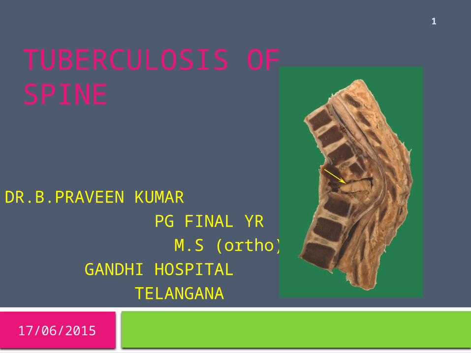
TUBERCULOSIS OF SPINE
DR.B.PRAVEEN KUMAR PG FINAL YR M.S (ortho) GANDHI HOSPITAL TELANGANA
17/06/2015

04/15/2023
2
Outline
1. Introduction 2. Clinical features3. Pathology , pathogenesis &
pathophysiology4. Diagnosis 5. Management
3
Introduction One fifth of TB population … in
India. Spinal tubercular account for 30-
60% of the Musculoskeletal TB infections
Always secondary Most common : 1st three decades SEX : M=F Most affected : Thoraco-lumbar
region
04/15/2023
4
REGIONAL DISTRIBUTION
CERVICAL 12% CERVICODORSAL 5% DORSAL 42%
(THORACIC) LUMBAR 26% DORSOLUMBAR 12% LUMBOSACRAL 3%
04/15/2023
5
Clinical features of spinal TB
Clinical kyphosis 95% Palpable cold abscess 20% Radiological paraverebral abscess 21% Neurological involvement 20% Tubercular sinuses (active/healed)
13% Associated extra spinal skeletal foci 12% Associated visceral foci 12% Skipped lesion in spine 7% Lateral shift(radiological ) 5%
6
A.Active stage1.Pain: Back pain (Commonest), Diffuse in early stages, but later become localised to the affected diseased segments.It may be a radicular pain. Depending upon the nerve root affected, it may present as: 1.Cervical root- Arm pain 2.Dorsal root- Girdle( pectoral ) pain 3.Dorso-lumbar root- Abdomen pain4.Lumbar root- Groin pain , or 5.Lumbo-Sacral root- Sciatic pain
CLINICAL FEATURES
04/15/20237
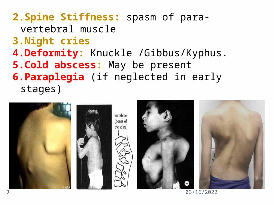
2.Spine Stiffness: spasm of para-vertebral muscle
3.Night cries4.Deformity: Knuckle /Gibbus/Kyphus.5.Cold abscess: May be present6.Paraplegia (if neglected in early stages)
04/15/20238
7.Constitutional Symptoms (Only in 20% cases): Malaise, weight loss, loss of appetite, night sweats, evening rise of temperature.
B. Healed stageNo systemic features but deformity persists.Radiological evidence of bone healing
But several of these signs and symptoms may be absent.
Important: c/f presentation depends on
1.Stage
2 Site
3.Presence of complications :neurologic
deficits, abscesses, or sinus tracts
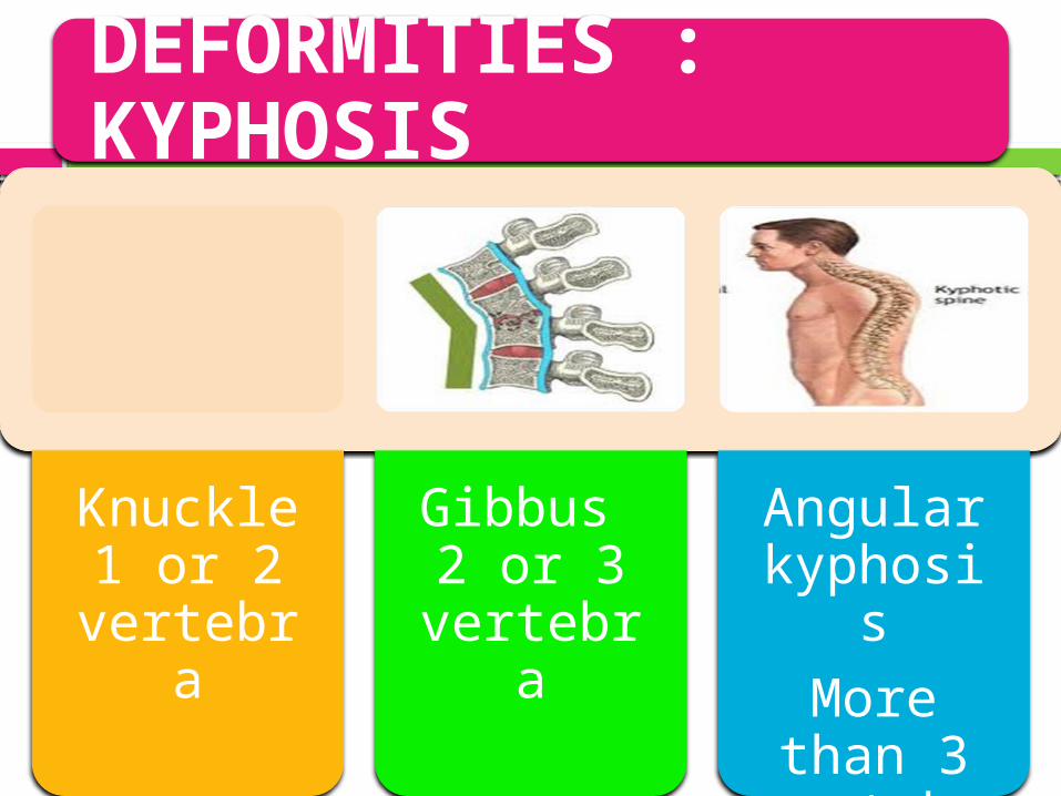
DEFORMITIES : KYPHOSIS
Knuckle 1 or 2
vertebra
Gibbus 2 or 3
vertebra
Angular kyphosis
More than 3
vertebra
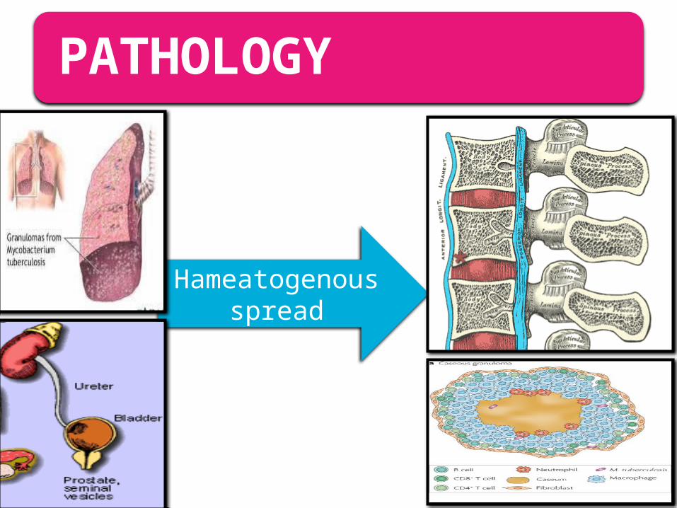
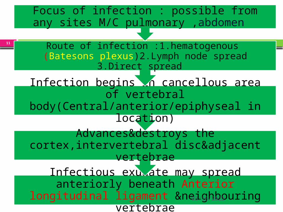
PATHOLOGY
Hameatogenous spread
04/15/2023
11
Infectious exudate may spread anteriorly beneath Anterior longitudinal ligament
&neighbouring vertebrae
Advances&destroys the cortex,intervertebral disc&adjacent vertebrae
Infection begins in cancellous area of vertebral body(Central/anterior/epiphyseal in
location)
Route of infection :1.hematogenous (Batesons plexus)2.Lymph node spread 3.Direct spread
Focus of infection : possible from any sites M/C pulmonary ,abdomen
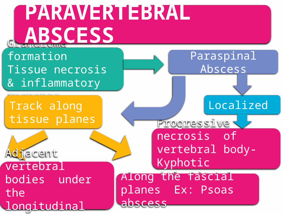
Granuloma formationTissue necrosis & inflammatory response
Paraspinal Abscess
Localized Track along tissue planes
Progressive necrosis of vertebral body-Kyphotic deformityAdjacent vertebral
bodies under the longitudinal ligaments
Along the fascial planes Ex: Psoas abscess
PARAVERTEBRAL ABSCESS

PARAVERTEBRAL ABSCESS
Cervical region• Between vertebral bodies, pharynx
and trachea
Upper thoracic• ‘V’ shaped shadow, stripping lung
apices laterally and downwards
Below T4 – Fusiform shape (Bird’s nest)• Below Diaphragm – unilateral &
blilateral psoas shadow.
04/15/2023
14
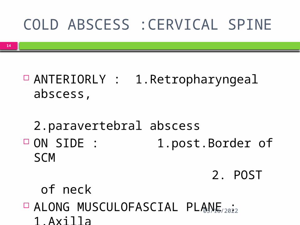
COLD ABSCESS :CERVICAL SPINE
ANTERIORLY : 1.Retropharyngeal abscess,
2.paravertebral abscess ON SIDE : 1.post.Border of SCM 2. POST of neck ALONG MUSCULOFASCIAL PLANE :
1.Axilla 2.Arm
04/15/2023
15
COLD ABSCESS :THORACIC SPINE ANTERIORLY 1.mediastinal abscess 2. paravertebral abscess ON SIDE : 1.psoas abscess 2. lumbar abscess ALONG MUSCULO-FASCIAL PLANE: 1.Ant. Chest wall 2.Mid-axillary line 3.posterior chest wall

04/15/2023
16
COLD ABSCESS :LUMBAR SPINE ANTERIORLY :prevertebral abscess : paravertebaral abscess ON THE SIDE : lumbar abscess : psoas abscess
ALONG MUSCULOFASCIAL PLANE : groin ,leg along sciatic nerve to pelvis, gluteal
region, posterior aspect of thigh and popliteal Region(KNEE)
Pathophysiology
Potts disease is usually secondary The basic lesion is a combination of
osteomyelitis and arthritis. The area usually affected is the anterior
aspect of the vertebral body Tuberculosis spread from that area to
adjacent intervertebral disks. disk is secondary to the spread of
infection from the vertebral body.
Progressive bone destruction leads to vertebral collapse, kyphosis & neurological involvement
Kyphotic deformity occurs in collapse of anterior spine.
Kyphotic def:; DORSAL SPINE THAN LUMBAR
The collapse is minimal in cervical spine because most of the body weight is borne through the articular processes.
Healing takes place by gradual fibrosis and calcification of the granulmatous tuberculous tissue:::FIROUS ANKYLOSIS
19
04/15/2023
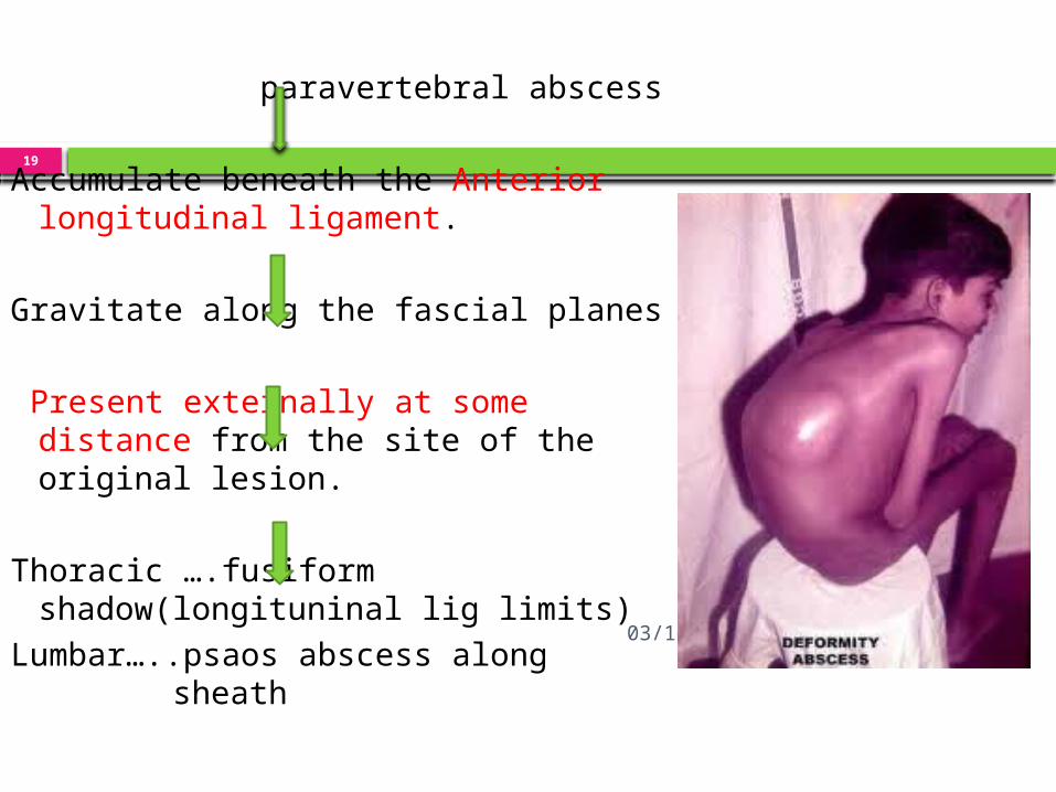
paravertebral abscess Accumulate beneath the Anterior
longitudinal ligament. Gravitate along the fascial planes Present externally at some
distance from the site of the original lesion.
Thoracic ….fusiform shadow(longituninal lig limits)
Lumbar…..psaos abscess along sheath
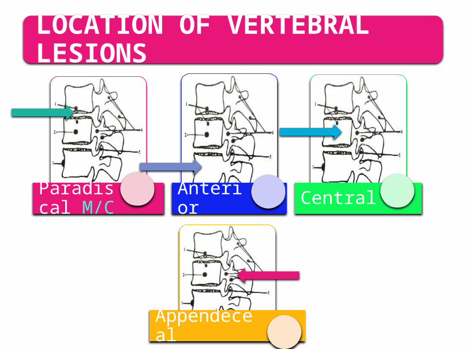
LOCATION OF VERTEBRAL LESIONS
Paradiscal M/C Anterior Central
Appendeceal
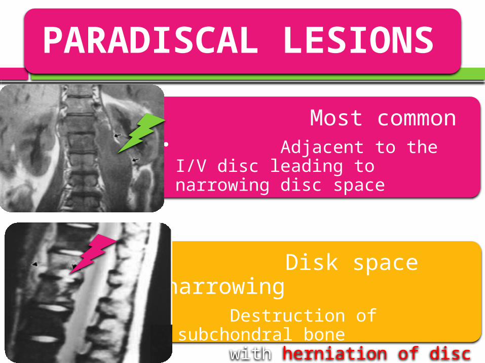
PARADISCAL LESIONS
Most common• Adjacent to the I/V disc
leading to narrowing disc space
Disk space narrowing • Destruction of subchondral
bone with herniation of disc into the body.
• Direct involvement of the disc.
04/15/2023
22
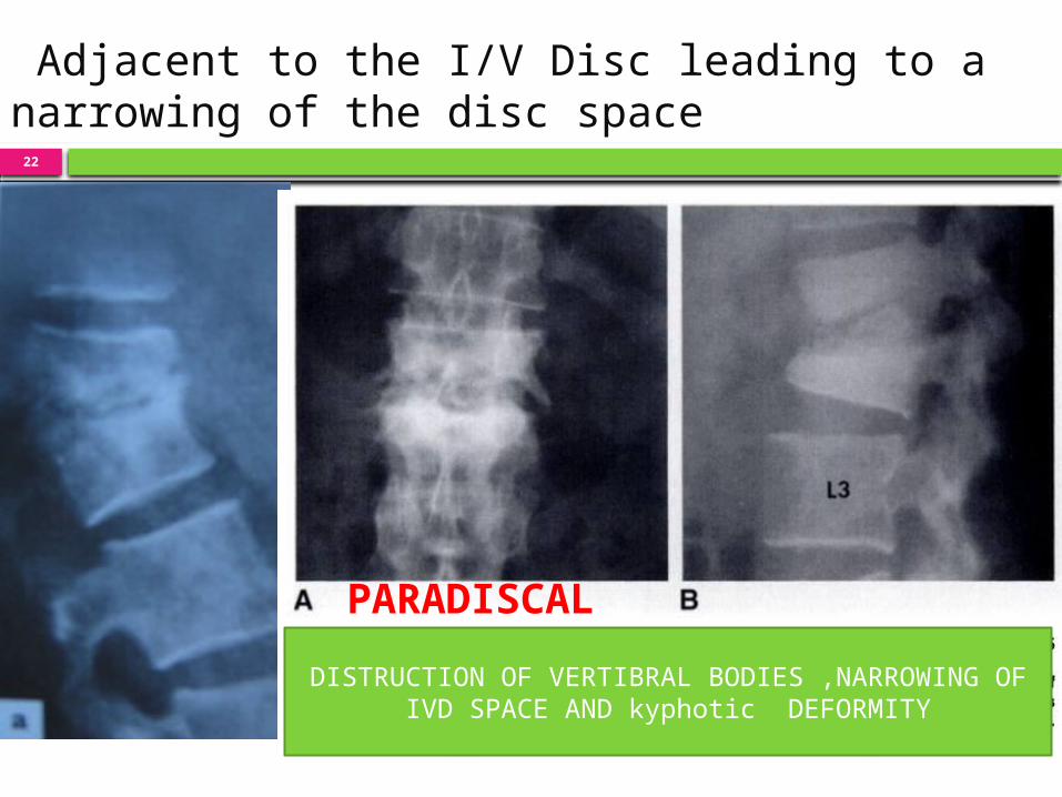
Adjacent to the I/V Disc leading to a narrowing of the disc space
PARADISCAL
DISTRUCTION OF VERTIBRAL BODIES ,NARROWING OF IVD SPACE AND kyphotic DEFORMITY
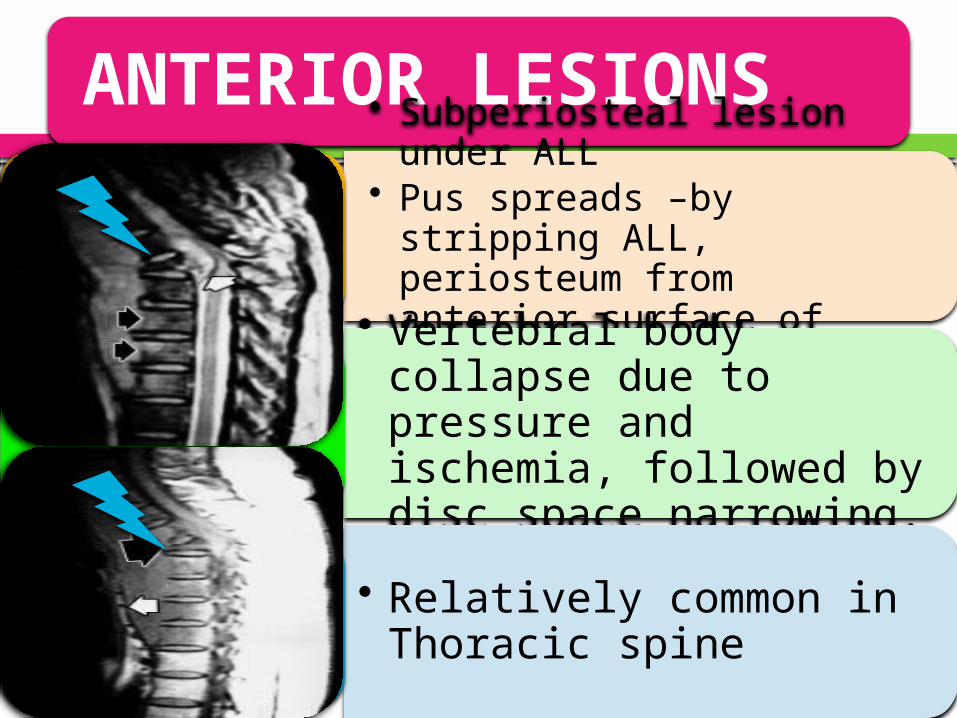
ANTERIOR LESIONS• Subperiosteal lesion under
ALL• Pus spreads –by stripping
ALL, periosteum from anterior surface of vertebral body• Vertebral body collapse due to pressure and ischemia, followed by disc space narrowing.
• Relatively common in Thoracic spine
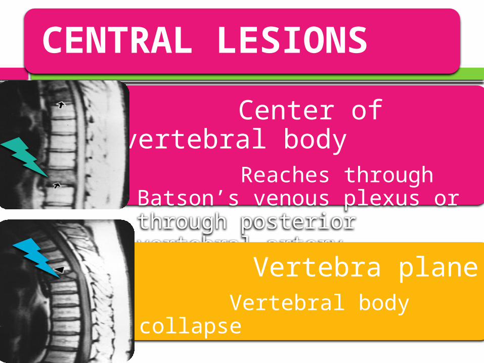
CENTRAL LESIONS
Center of vertebral body• Reaches through Batson’s
venous plexus or through posterior vertebral artery
Vertebra plane• Vertebral body collapse•
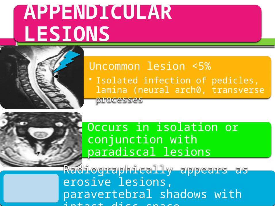
APPENDICULAR LESIONS
Uncommon lesion <5%• Isolated infection of pedicles, lamina
(neural arch0, transverse processes
Occurs in isolation or conjunction with paradiscal lesions
Radiographically appears as erosive lesions, paravertebral shadows with intact disc space.
04/15/2023
26
Management plan
DIAGNOSIS CLINICO RADIOLOGICAL & LAB STUDIES Microbiological studies Histopathological study CT SCAN MRI SCAN USG RADIONUCLIDE SCAN MYELOGRAPHY
DIAGNOSISComplete blood picture• ESR Increased / Increased Lymphocyte
countELISA• For antibody to mycobacterial antigen • Sensitivity 60-80%
PCR • Sensitivity of 40%
Chest radiograph
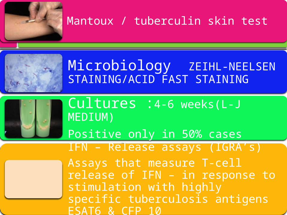
Mantoux / tuberculin skin test
Microbiology ZEIHL-NEELSEN STAINING/ACID FAST STAINING
Cultures :4-6 weeks(L-J MEDIUM)
Positive only in 50% cases
IFN – Release assays (IGRA’s)Assays that measure T-cell release of IFN – in response to stimulation with highly specific tuberculosis antigens ESAT6 & CFP 10
04/15/2023
29
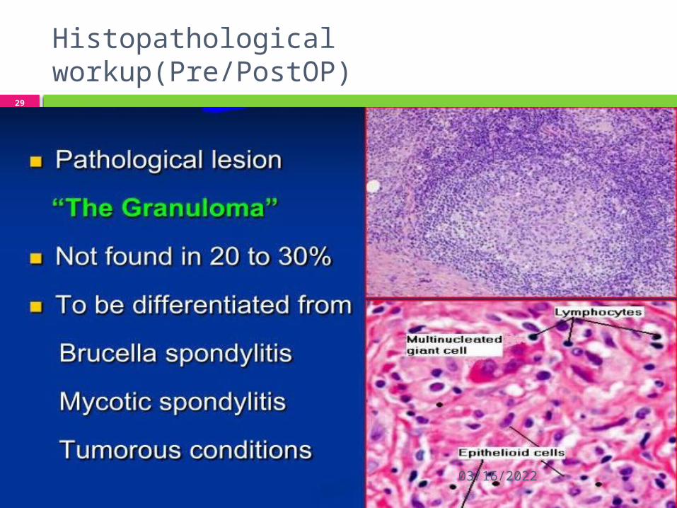
Histopathological workup(Pre/PostOP)
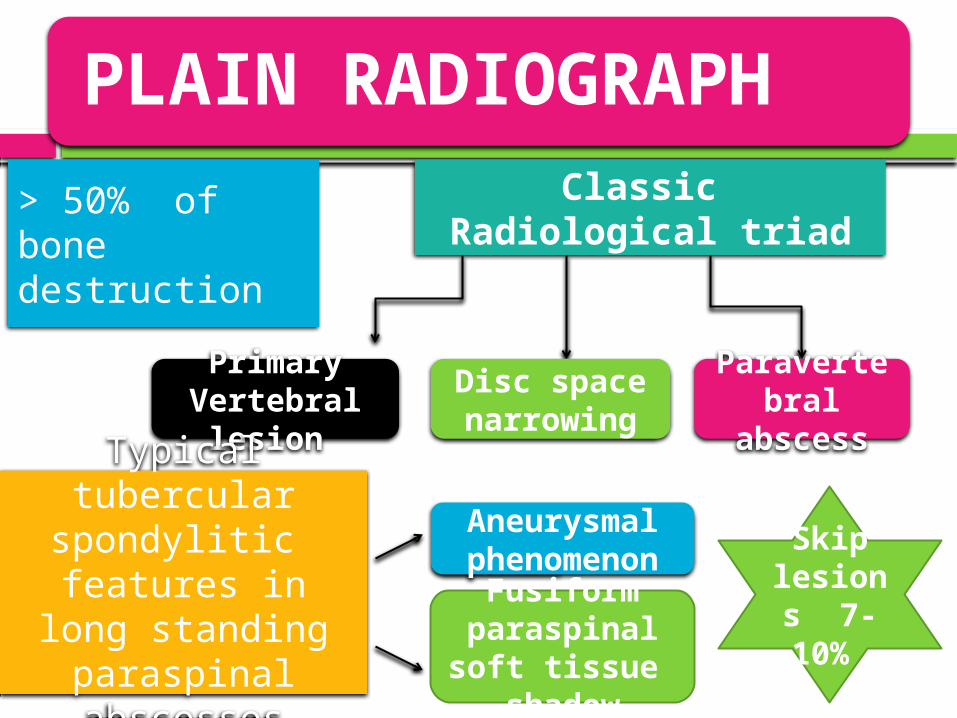
PLAIN RADIOGRAPH
> 50% of bone destruction
Classic Radiological triad
Primary Vertebral
lesion
Disc space narrowing
Paravertebral
abscessTypical tubercular
spondylitic features in long
standing paraspinal abscesses
Aneurysmal phenomenon
Fusiform paraspinal soft tissue
shadow
Skip lesions 7-10%
04/15/2023
31
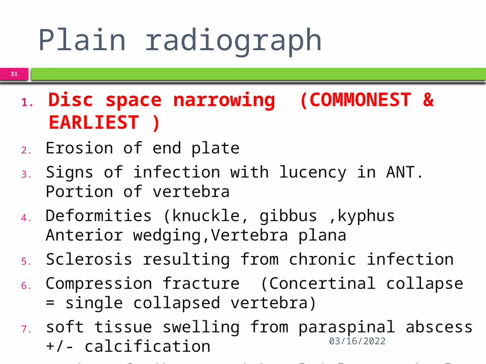
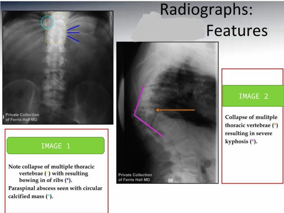
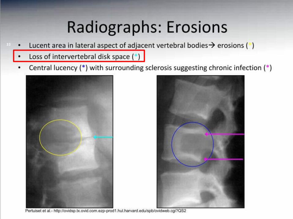
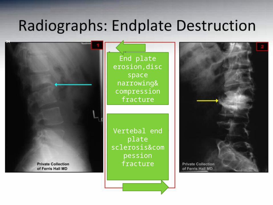
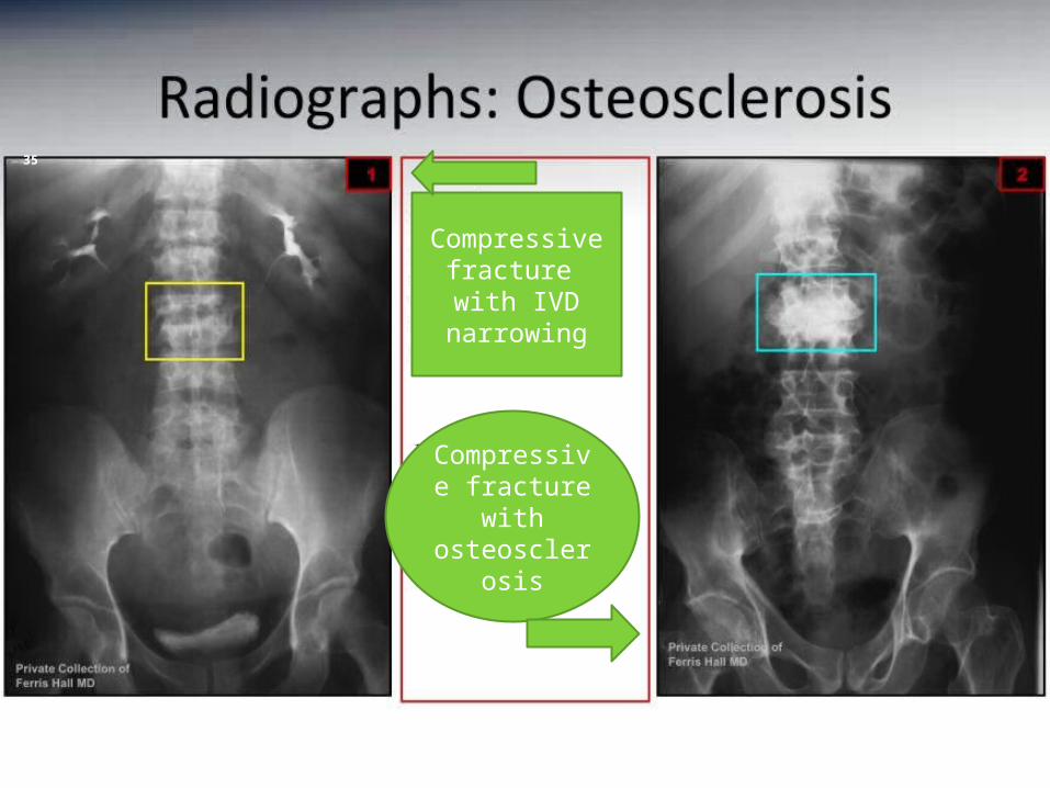
Plain radiograph
1. Disc space narrowing (COMMONEST & EARLIEST )
2. Erosion of end plate
3. Signs of infection with lucency in ANT. Portion of vertebra
4. Deformities (knuckle, gibbus ,kyphus Anterior wedging,Vertebra plana
5. Sclerosis resulting from chronic infection
6. Compression fracture (Concertinal collapse = single collapsed vertebra)
7. soft tissue swelling from paraspinal abscess +/- calcification
8. Bowing of rib cage with multiple vertebral fracture
04/15/2023
32
IMAGE 1
IMAGE 2
04/15/2023
33
04/15/2023
34
End plate erosion,disc
space narrowing&compression
fracture
Vertebal end plate
sclerosis&compession fracture
04/15/2023
35
Compressive fracture with IVD narrowing
Compressive fracture
with osteosclero
sis
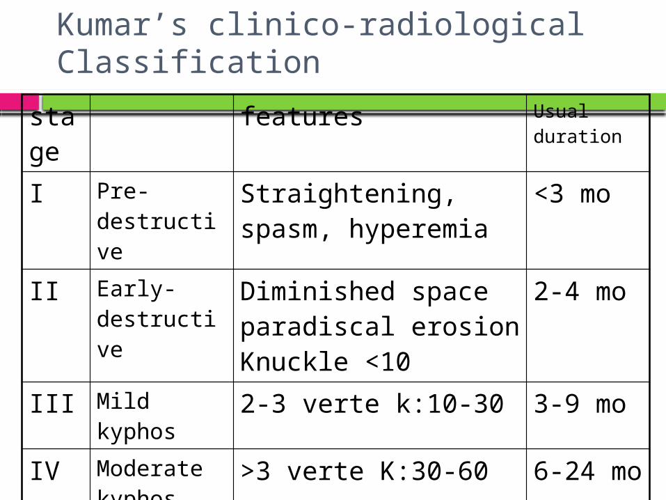
Kumar’s clinico-radiological Classification
stage features Usual duration
I Pre-destructive
Straightening, spasm, hyperemia
<3 mo
II Early-destructive
Diminished space paradiscal erosion Knuckle <10
2-4 mo
III Mild kyphos 2-3 verte k:10-30 3-9 mo
IV Moderate kyphos
>3 verte K:30-60 6-24 mo
V Severe kyphos
>3 verte K:>60 >2 years
04/15/2023
37
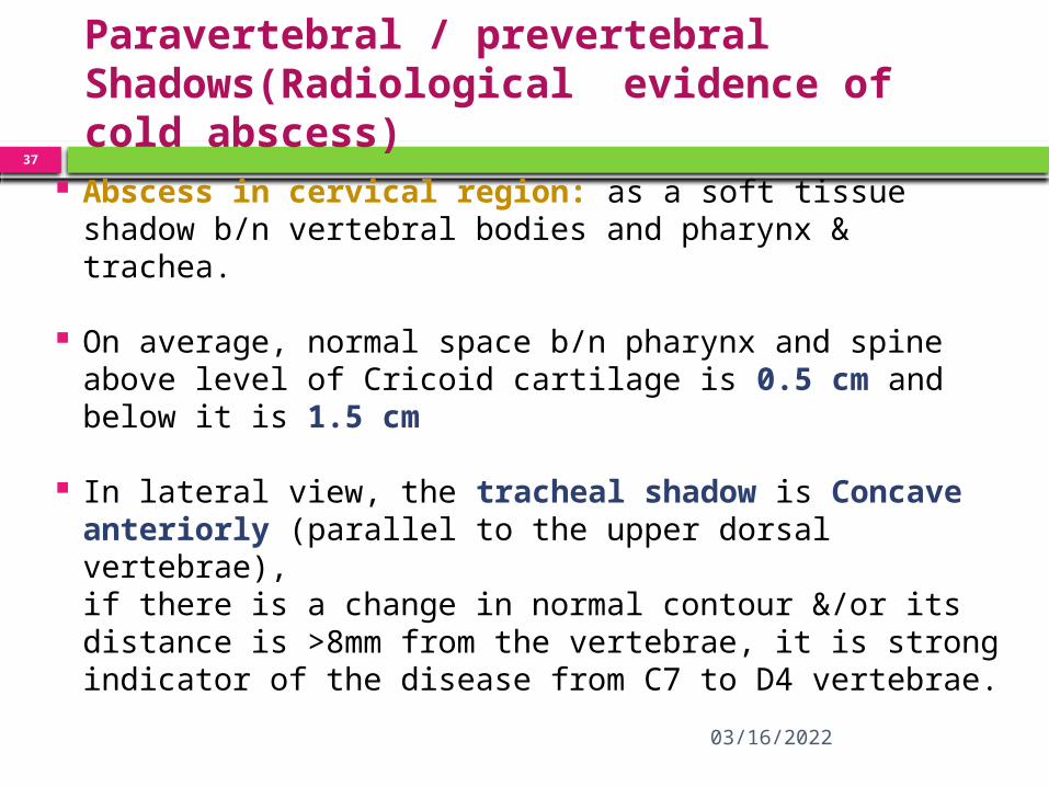
Paravertebral / prevertebral Shadows(Radiological evidence of cold abscess)
Abscess in cervical region: as a soft tissue shadow b/n vertebral bodies and pharynx & trachea.
On average, normal space b/n pharynx and spine above level of Cricoid cartilage is 0.5 cm and below it is 1.5 cm
In lateral view, the tracheal shadow is Concave anteriorly (parallel to the upper dorsal vertebrae),if there is a change in normal contour &/or its distance is >8mm from the vertebrae, it is strong indicator of the disease from C7 to D4 vertebrae.
04/15/2023
38
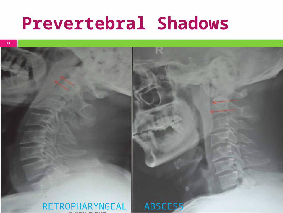
Prevertebral Shadows
RETROPHARYNGEAL ABSCESS

04/15/2023
39
Abscess below the level of D4 vertebrae – Fusiform shape (Bird nestappearance)An abscess under tension may produce- Globular shape
Paravertebral Shadows
04/15/2023
40
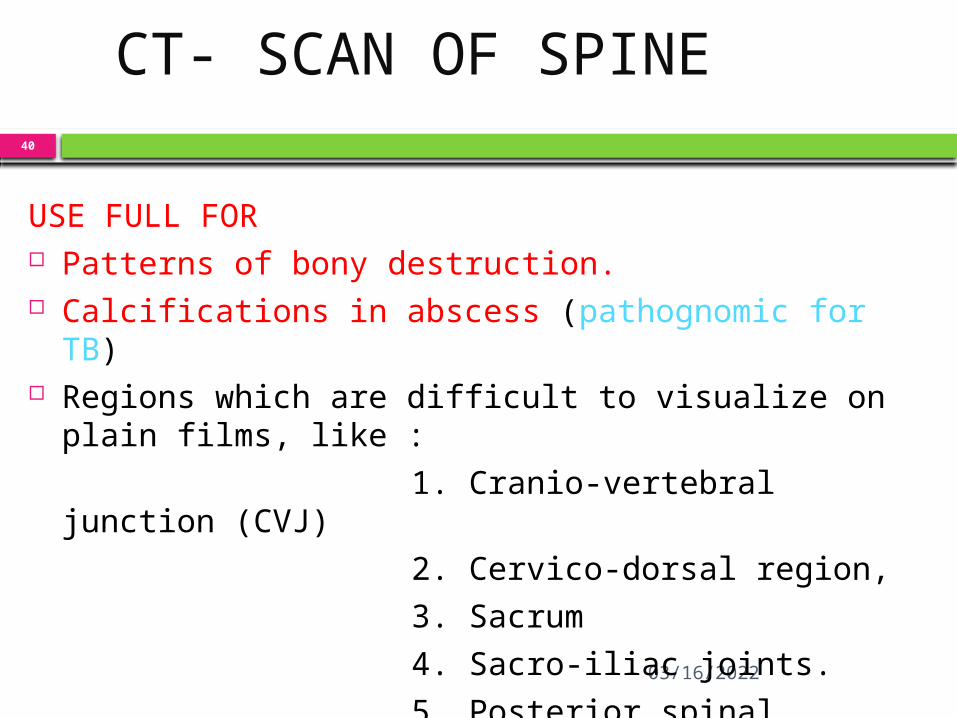
CT- SCAN OF SPINE
USE FULL FOR Patterns of bony destruction. Calcifications in abscess (pathognomic for TB) Regions which are difficult to visualize on plain films,
like :
1. Cranio-vertebral junction (CVJ)
2. Cervico-dorsal region,
3. Sacrum
4. Sacro-iliac joints.
5. Posterior spinal tuberculosis because lesions less than 1.5cm are usually missed due to overlapping of shadows on x rays.
04/15/2023
41
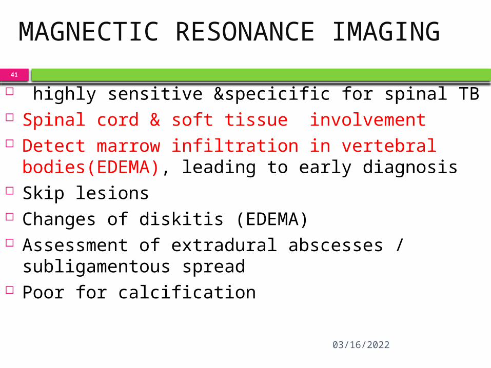
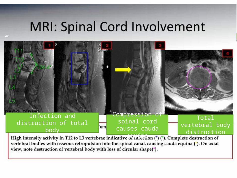
MAGNECTIC RESONANCE IMAGING
highly sensitive &specicific for spinal TB Spinal cord & soft tissue involvement Detect marrow infiltration in vertebral
bodies(EDEMA), leading to early diagnosis Skip lesions Changes of diskitis (EDEMA) Assessment of extradural abscesses /
subligamentous spread Poor for calcification
04/15/2023
42
Infection and distruction of total body
Compression of spinal cord causes
cauda equina
Total vertebral body distruction
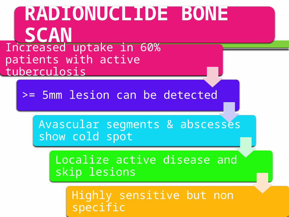
RADIONUCLIDE BONE SCAN
Increased uptake in 60% patients with active tuberculosis
>= 5mm lesion can be detected
Avascular segments & abscesses show cold spot
Localize active disease and skip lesions
Highly sensitive but non specific
04/15/2023
44
USG - to find out primary in abdomen - Detect cold abscess - Guided aspiration
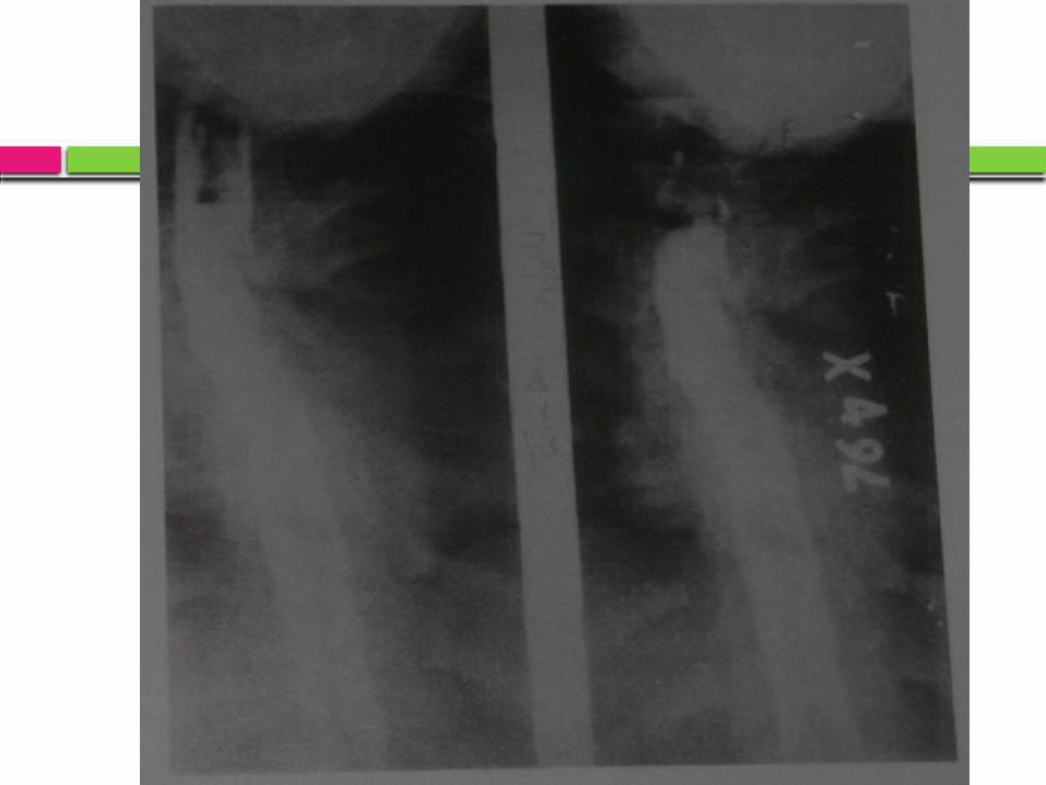
Myelography Spinal tumor syndrome Multiple vertebral lesions Patients not recovered after decompression 1.Block present : second decompression 2.Block not present : intrinsic damage
1.Ischemic infarction 2.Interstitial gliosis
3.atrophy 4. tuberculous myelitis 5.Myelomalacia
04/15/2023
47
DIFFERENTIAL DIAGNOSIS
Back pain 1. Traumatic 2. Secondaries to spine /myeloma/lymphoma 3. Prolapsed disc 4. Ankylosing spondylitisNeurological deficit 5. Spinal tumor 6. Traumatic 7. Secondaries to spine Radiologically SPINAL INFECTIONS : pyogenic, BRUCELLA SPONDYLITIS
NEUROPATHIC SPINE : Diabetes NEOPLASTIC : commonly lymphoma/
metastasis/primary DEGENERATIVE
04/15/2023
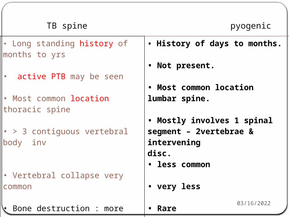
TB spine pyogenic
• Long standing history of months to yrs
• active PTB may be seen
• Most common location thoracic spine
• > 3 contiguous vertebral body inv
• Vertebral collapse very common
• Bone destruction : more
• Skip lesions common
• Pra vertebral abscesses-Commoncalcification if present is pathognomic.
• History of days to months.
• Not present.
• Most common location lumbar spine.
• Mostly involves 1 spinalsegment – 2vertebrae & interveningdisc.• less common
• very less
• Rare
• Rare
04/15/202349
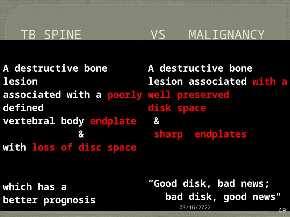
TB SPINE VS MALIGNANCY
A destructive bone lesionassociated with a poorly definedvertebral body endplate &with loss of disc space
which has abetter prognosis
A destructive bonelesion associated with a well preserveddisk space & sharp endplates
“Good disk, bad news; bad disk, good news"
04/15/2023
50
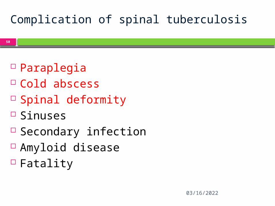
Complication of spinal tuberculosis
Paraplegia Cold abscess Spinal deformity Sinuses Secondary infection Amyloid disease Fatality
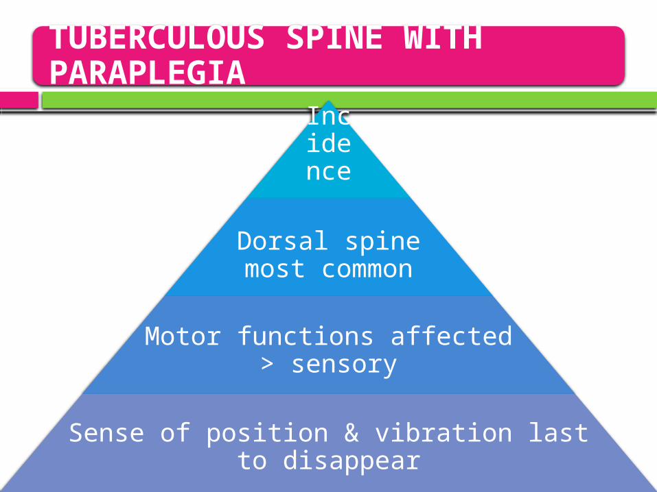
TUBERCULOUS SPINE WITH PARAPLEGIA
Incidence 10-30%
Dorsal spine most common
Motor functions affected > sensory
Sense of position & vibration last to disappear
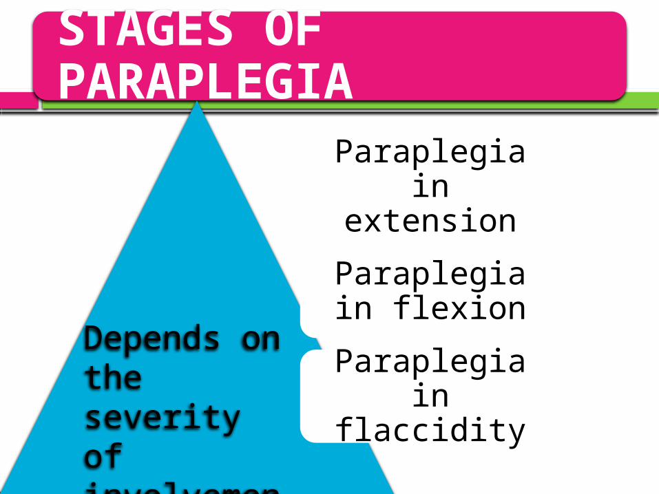
STAGES OF PARAPLEGIA
Paraplegia in extension
Paraplegia in flexion
Paraplegia in flaccidity
Depends on the severity of involvement of long tracts
04/15/2023
53
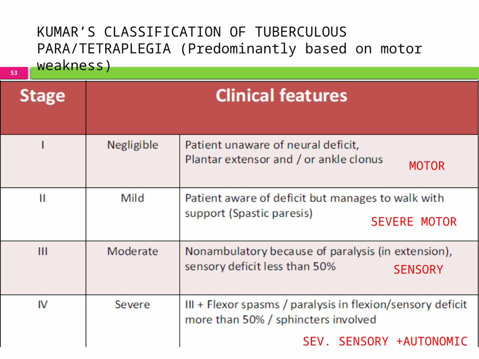
KUMAR’S CLASSIFICATION OF TUBERCULOUSPARA/TETRAPLEGIA (Predominantly based on motorweakness)
MOTOR
SEVERE MOTOR
SENSORY
SEV. SENSORY +AUTONOMIC
54
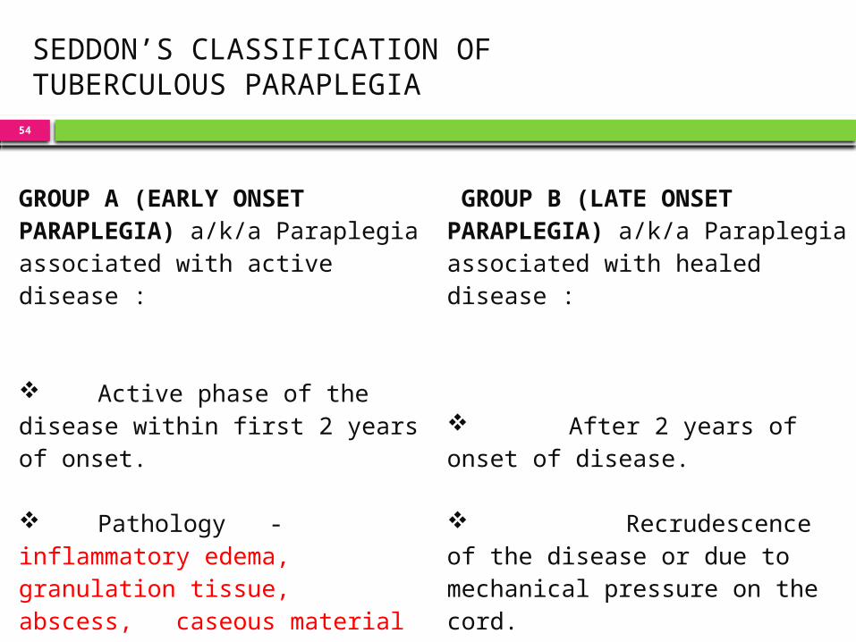
SEDDON’S CLASSIFICATION OFTUBERCULOUS PARAPLEGIA
10-09-2014
GROUP A (EARLY ONSET PARAPLEGIA) a/k/a Paraplegia associated with activedisease :
Active phase of the disease within first 2 years of onset.
Pathology - inflammatory edema, granulation tissue, abscess, caseous material or ischemia of cord.
GROUP B (LATE ONSET PARAPLEGIA) a/k/a Paraplegia associated with healed disease :
After 2 years of onset of disease.
Recrudescence of the disease or due to mechanical pressure on the cord.
Pathology can be sequestra, debris, internal gibbus or stenosis of the canal
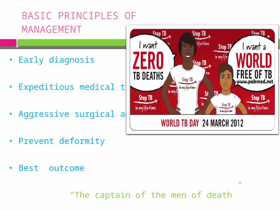
BASIC PRINCIPLES OFMANAGEMENT
• Early diagnosis
• Expeditious medical treatment
• Aggressive surgical approach
• Prevent deformity
• Best outcome “The captain of the men of death”
04/15/2023
56
Three approach
CONSERVATIVE PLAN MIDDLE PATH REGIME RADICAL SURGERY APPROACH
04/15/2023
61
MIDDLE PATH REGIME
Rest on hard bed Chemotherapy X-ray & ESR once in 3 months kyphosis
measurement MRI/ CT at 6 months interval for 2 years
Gradual mobilization is encouraged in absence of neural deficits with spinal braces & back extension exercises at 3 – 9 weeks.
Abscesses – aspirate when near surface & instil 1gm
Streptomycin +/- INH in solution
04/15/2023
62
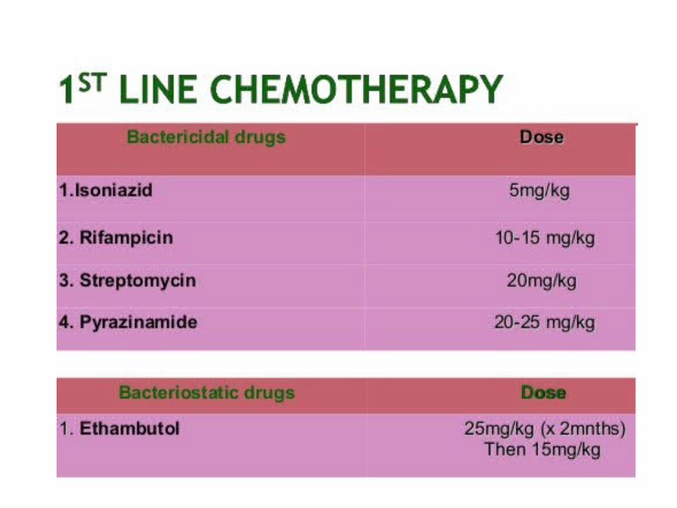
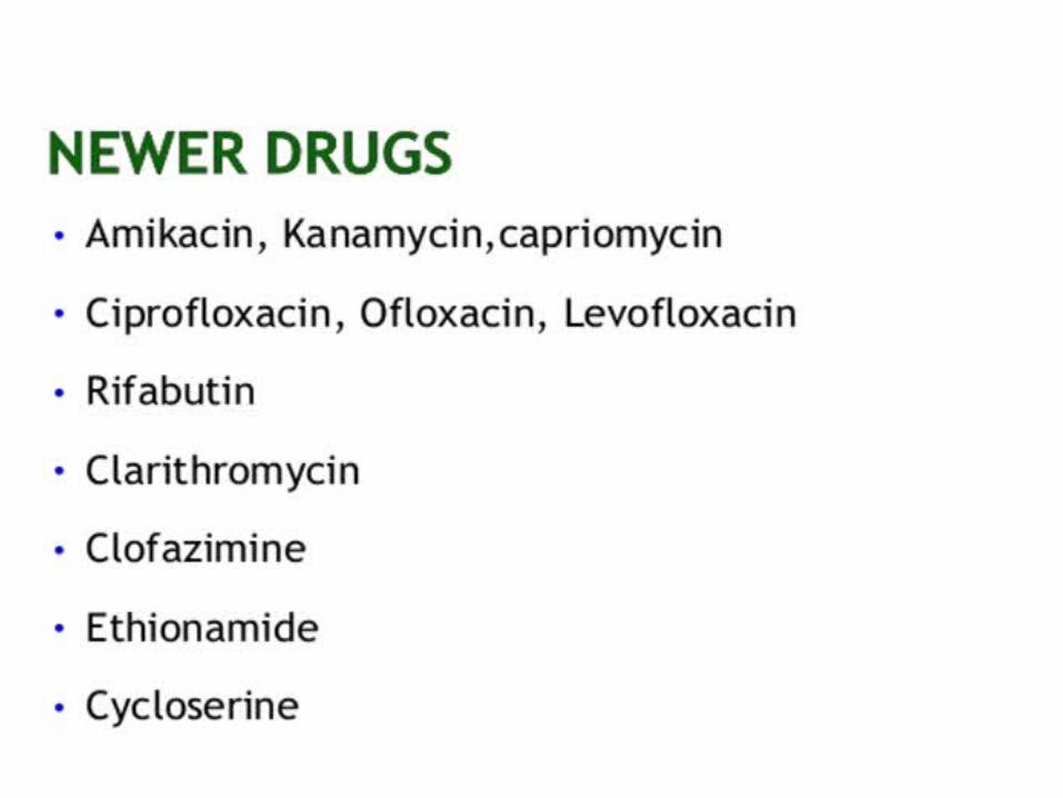
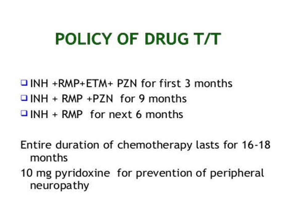
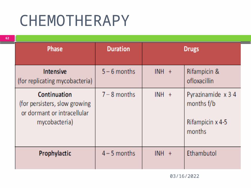
CHEMOTHERAPY
04/15/2023
63
MIDDLE PATH REGIME
Sinus heals 6-12 weeks Neural complications if showing progressive
recovery on ATT b/w 3-4 weeks :surgery unnecessary
IF NOT Excisional surgery for posterior spinal
disease associated with abscess / sinus formation +/- neural involvement.
Operative debridement–if no arrest of symptoms after 3-6 months of ATT / with recurrence of disease
Posterior spinal arthrodesis : symptomatic unstable lesion
Post op spinal brace→12 months-24

04/15/2023
64
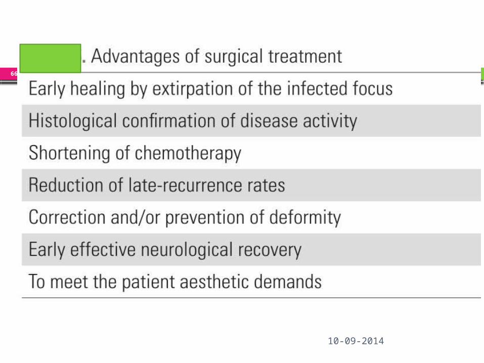
ABSOLUTE INDICATIONS FOR SURGERY:
Paraplegia during conservative treatment (6 weeks)
Paraplegia worsening during treatment (6 weeks)
Complete motor loss for 1 month despite of conservative treatment
Paraplegia with uncontrolled spasticity
Severe and rapid onset paraplegia
Severe flaccid paraplegia/ sensory loss
04/15/2023
65
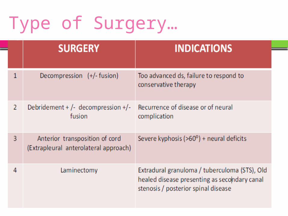
Other indications
Relative indications
1. Recurrent paraplegia 2. Paraplegia in
elderly 3. Painful and
spastic paraplegia 4. Paraplegia
with complications (UTI)
Rare indications1. Posterior elementdisease2. Spinal tumorsyndrome3. Severe cervicallesion c paraplegia4. Cauda equinopathy
66
10-09-2014
Type of Surgery…
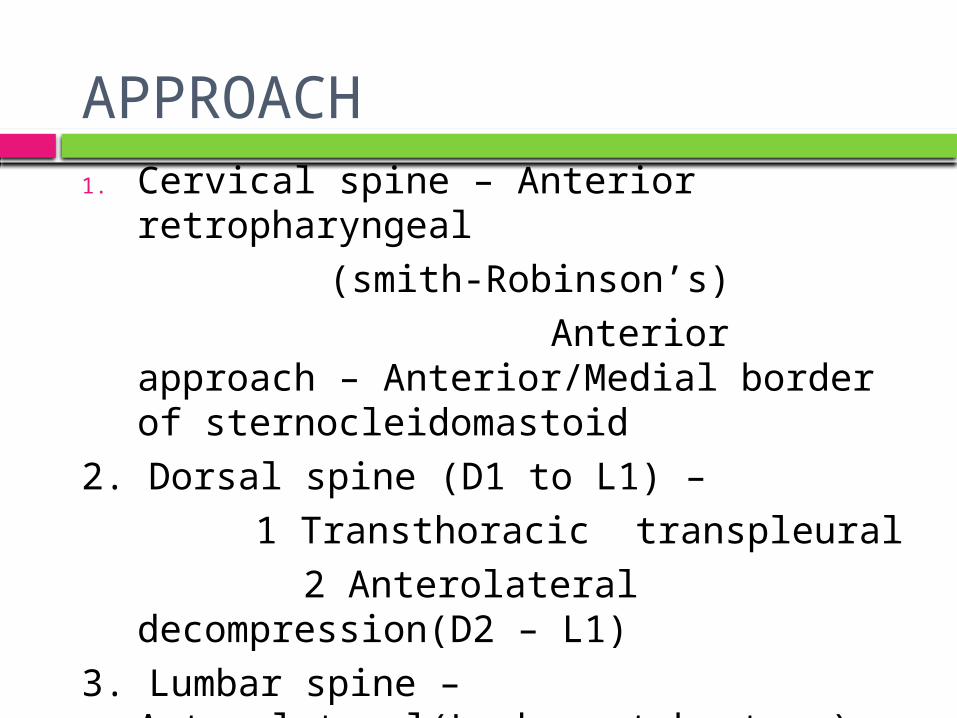
APPROACH
1. Cervical spine – Anterior retropharyngeal(smith-Robinson’s)
Anterior approach – Anterior/Medial border of sternocleidomastoid
2. Dorsal spine (D1 to L1) – 1 Transthoracic transpleural
2 Anterolateral decompression(D2 – L1)
3. Lumbar spine – Anterolateral(Lumbovertebrotomy)
Extraperitoneal Ant. approach
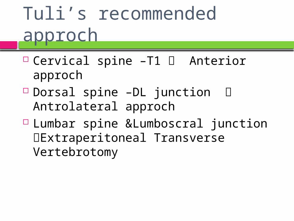
Tuli’s recommended approch Cervical spine –T1 Anterior approch Dorsal spine –DL junction Antrolateral
approch Lumbar spine &Lumboscral junction
Extraperitoneal Transverse Vertebrotomy
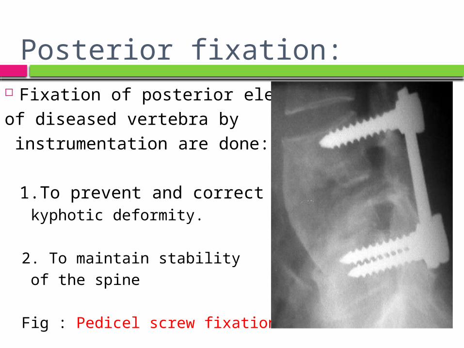
Posterior fixation: Fixation of posterior element of diseased vertebra by instrumentation are done:
1.To prevent and correct kyphotic deformity.
2. To maintain stability of the spine
Fig : Pedicel screw fixation
71
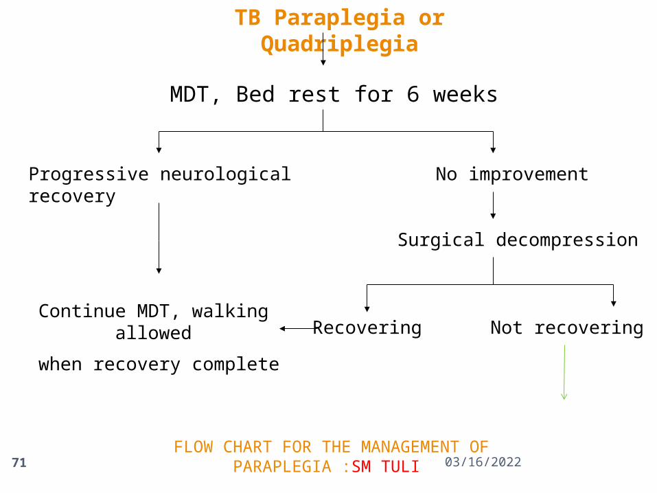
TB Paraplegia or Quadriplegia
MDT, Bed rest for 6 weeks
Progressive neurological recovery No improvement
Continue MDT, walking allowed
when recovery complete
Surgical decompression
Recovering Not recovering
FLOW CHART FOR THE MANAGEMENT OF PARAPLEGIA :SM TULI 04/15/2023
72
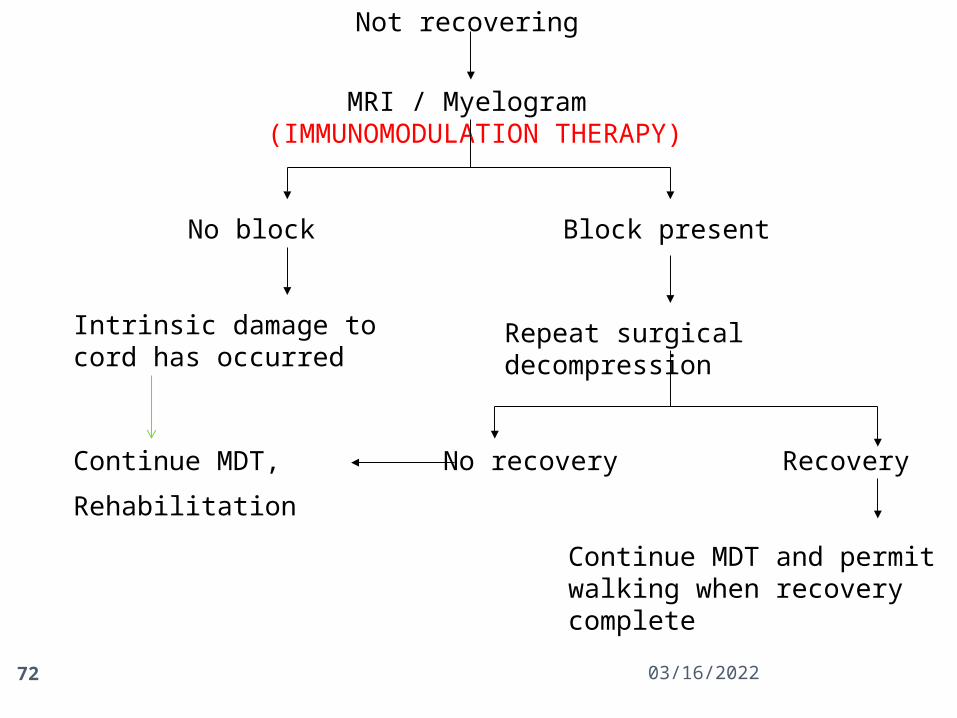
Not recovering
MRI / Myelogram (IMMUNOMODULATION THERAPY)
No block Block present
Intrinsic damage to cord has occurred
Repeat surgical decompression
No recovery Recovery Continue MDT,
Rehabilitation
Continue MDT and permit walking when recovery complete
04/15/2023
04/15/2023
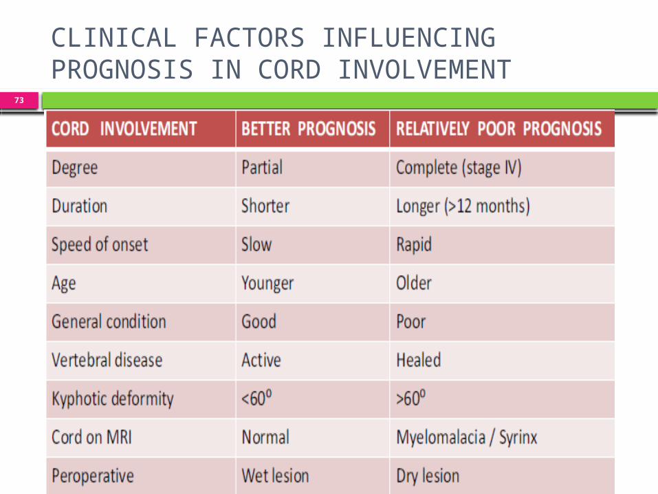
73
CLINICAL FACTORS INFLUENCINGPROGNOSIS IN CORD INVOLVEMENT
ANTERIOR APPROACH TO THECERVICAL SPINE (C2 to D1)
Smith & Robinson Oblique / transverse incision. Plane b/w SCM & carotid sheath laterally & T-O
medially. Longitudinal incision in ALL open a perivertebral
abscess, or the diseased vertebrae may be exposed by reflecting the ALL
& the longus colli muscles.
Hodgson approach via posterior triangle by retracting SCM,
Carotid sheath, T & O anteriorly & to the opposite side.
SURGICAL APPROACHES TODORSAL SPINE
Anterior transpleural transthoracic approach Anterolateral extrapleural approach Posterolateral approach
{Dura is exposed by hemilaminectomy first & then
extended laterally to remove the posterior ends of 2 – 4
ribs, corresponding transverse processes & the pedicles}.
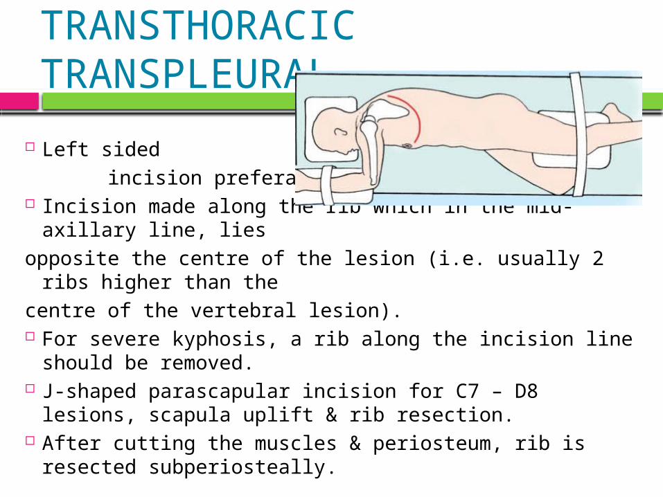
TRANSTHORACIC TRANSPLEURAL
Left sided incision preferable Incision made along the rib which in the mid-axillary line,
liesopposite the centre of the lesion (i.e. usually 2 ribs higher
than thecentre of the vertebral lesion). For severe kyphosis, a rib along the incision line should
be removed. J-shaped parascapular incision for C7 – D8 lesions,
scapula uplift & rib resection. After cutting the muscles & periosteum, rib is resected
subperiosteally.
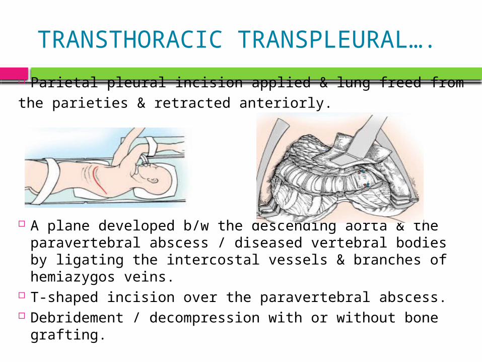
TRANSTHORACIC TRANSPLEURAL….
Parietal pleural incision applied & lung freed fromthe parieties & retracted anteriorly.
A plane developed b/w the descending aorta & the paravertebral abscess / diseased vertebral bodies by ligating the intercostal vessels & branches of hemiazygos veins.
T-shaped incision over the paravertebral abscess. Debridement / decompression with or without bone
grafting.
ANTEROLATERAL DECOMPRESSION
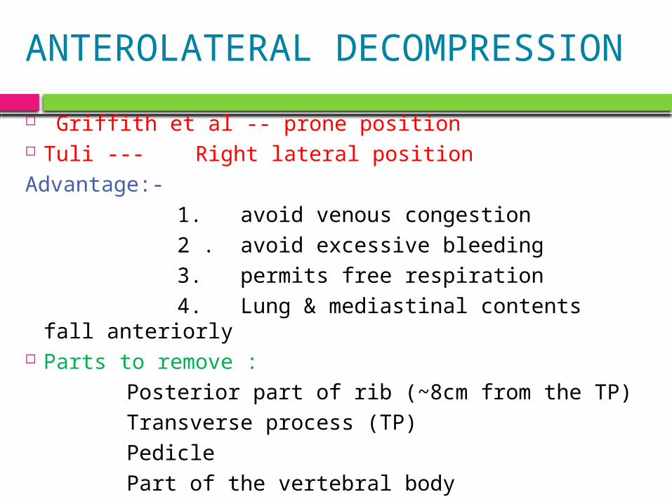
Griffith et al -- prone position Tuli --- Right lateral positionAdvantage:- 1. avoid venous congestion 2 . avoid excessive bleeding 3. permits free respiration 4. Lung & mediastinal contents fall anteriorly Parts to remove : Posterior part of rib (~8cm from the TP) Transverse process (TP) Pedicle Part of the vertebral body
ANTEROLATERAL DECOMPRESSION….
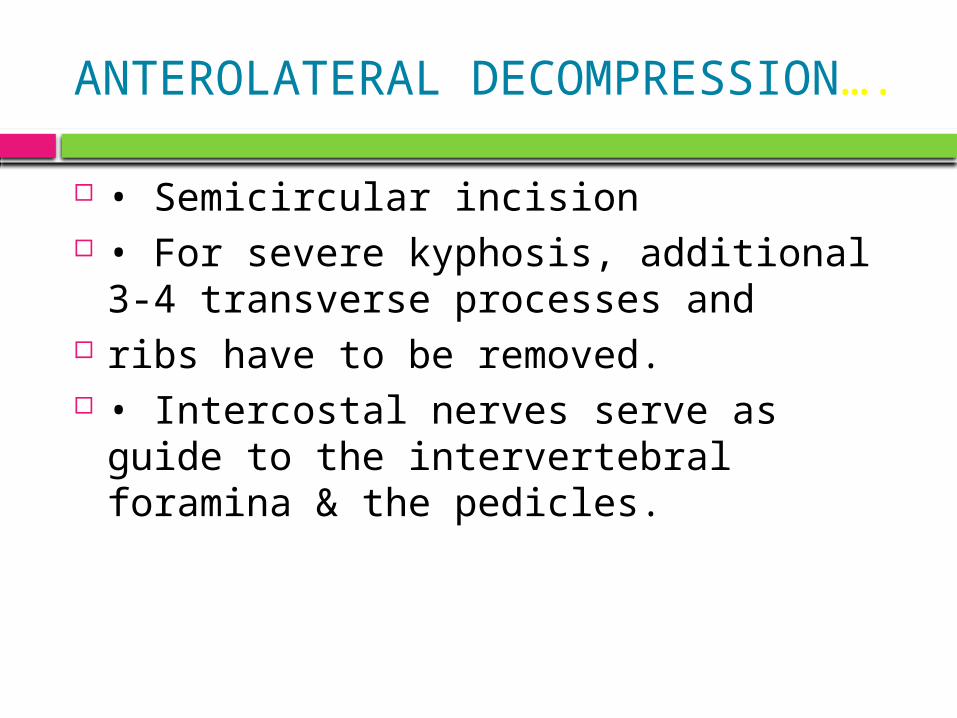
• Semicircular incision • For severe kyphosis, additional 3-4
transverse processes and ribs have to be removed. • Intercostal nerves serve as guide to
the intervertebral foramina & the pedicles.
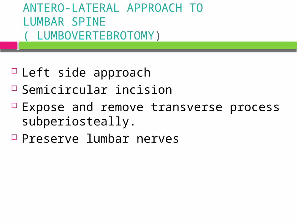
ANTERO-LATERAL APPROACH TOLUMBAR SPINE ( LUMBOVERTEBROTOMY)
Left side approach Semicircular incision Expose and remove transverse process
subperiosteally. Preserve lumbar nerves
CONT…
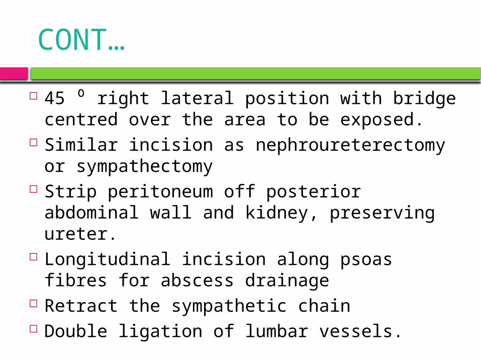
45 ⁰ right lateral position with bridge centred over the area to be exposed.
Similar incision as nephroureterectomy or sympathectomy
Strip peritoneum off posterior abdominal wall and kidney, preserving ureter.
Longitudinal incision along psoas fibres for abscess drainage
Retract the sympathetic chain Double ligation of lumbar vessels.
EXTRA PERITONEAL APPROACH TOLUMBO-SACRAL REGION
Left side preferred ( left Common iliac vessels longer & retracted easily).
Lazy “S” incision Strip & reflect the parietal peritoneum
along with ureter & spermatic vessels towards right side.
POSTERIOR SPINALARTHRODESIS
Albee– Tibial graft inserted longitudinally in to the split
spinous processes across the diseased site.
Hibbs– overlapping numerous small osseous flaps from contiguous laminae , spinous processes & articular facets
Indications– 1. Mechanical instability of spine in otherwise
healed disease. 2. To stabilize the craniovertebral region (in
certain cases of T.B.)
SURGERY IN SEVERE KYPHOSIS HIGH RISK PATIENTS: - Patients < 10 years - Dorsal lesions - Involvement of >= 3 vertebrae - Severe deformity in presence of active disease,
especially in children is an absolute indication for decompression , correction and stabilization.
Staged operations- 1. Anteriorly at the site of disease, 2. Osteotomy of the posterior elements at the
deformity & 3. Halopelvic or halofemoral tractions post-
operatively.
TREATMENT OF PARAPLEGIA INSEVERE KHYPHOSIS
Griffiths et al :anterior transposition of cord through
laminectomy Rajasekaran : posterior stabilization f/b Anterior debridement and bone grafting ( titanium cages) in active stage of disease and vice versa for healed disease. Antero-lateral (Preferred approach) .
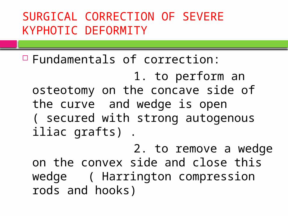
SURGICAL CORRECTION OF SEVEREKYPHOTIC DEFORMITY
Fundamentals of correction: 1. to perform an osteotomy on
the concave side of the curve and wedge is open ( secured with strong autogenous iliac grafts) .
2. to remove a wedge on the convex side and close this wedge ( Harrington compression rods and hooks)
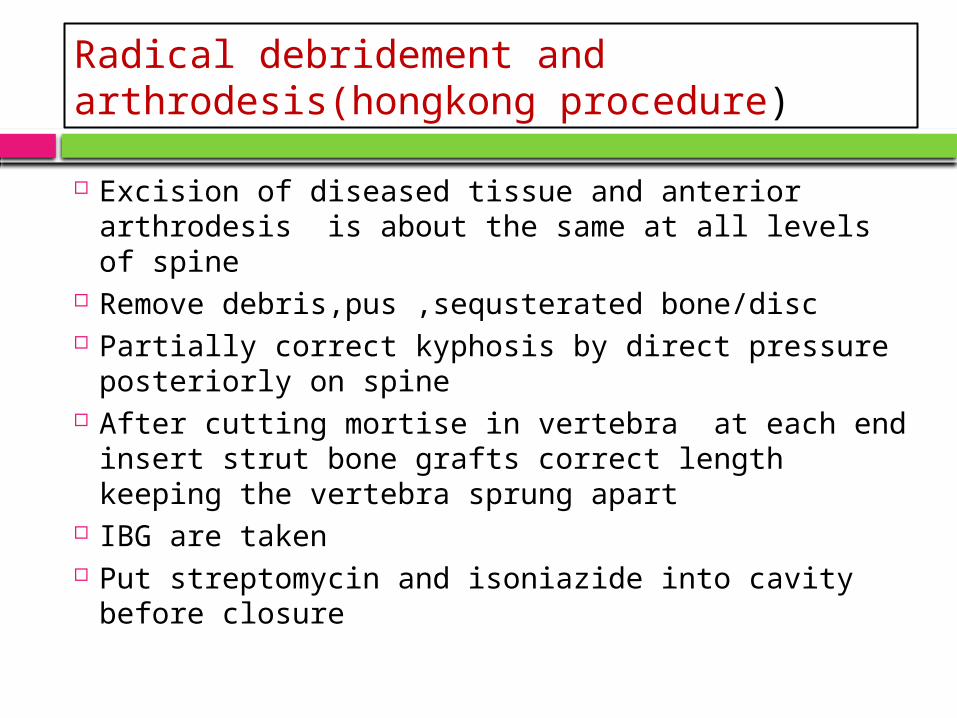
Radical debridement and arthrodesis(hongkong procedure)
Excision of diseased tissue and anterior arthrodesis is about the same at all levels of spine
Remove debris,pus ,sequsterated bone/disc Partially correct kyphosis by direct pressure
posteriorly on spine After cutting mortise in vertebra at each end
insert strut bone grafts correct length keeping the vertebra sprung apart
IBG are taken Put streptomycin and isoniazide into cavity before
closure
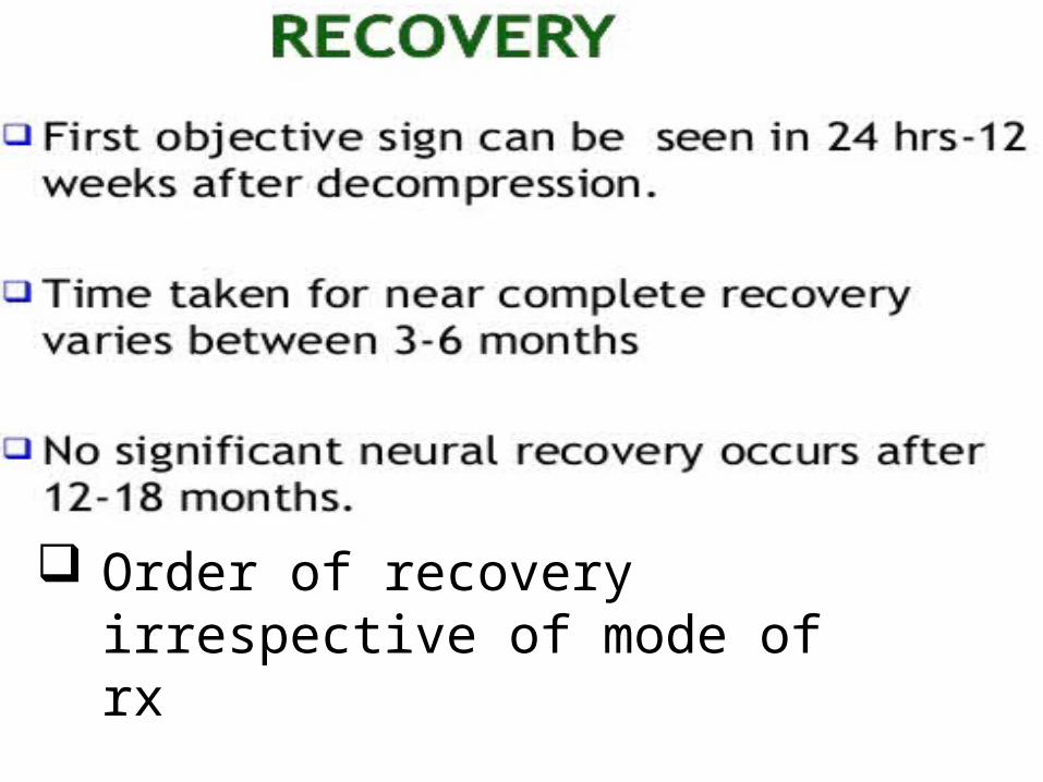
Order of recovery irrespective of mode of rx
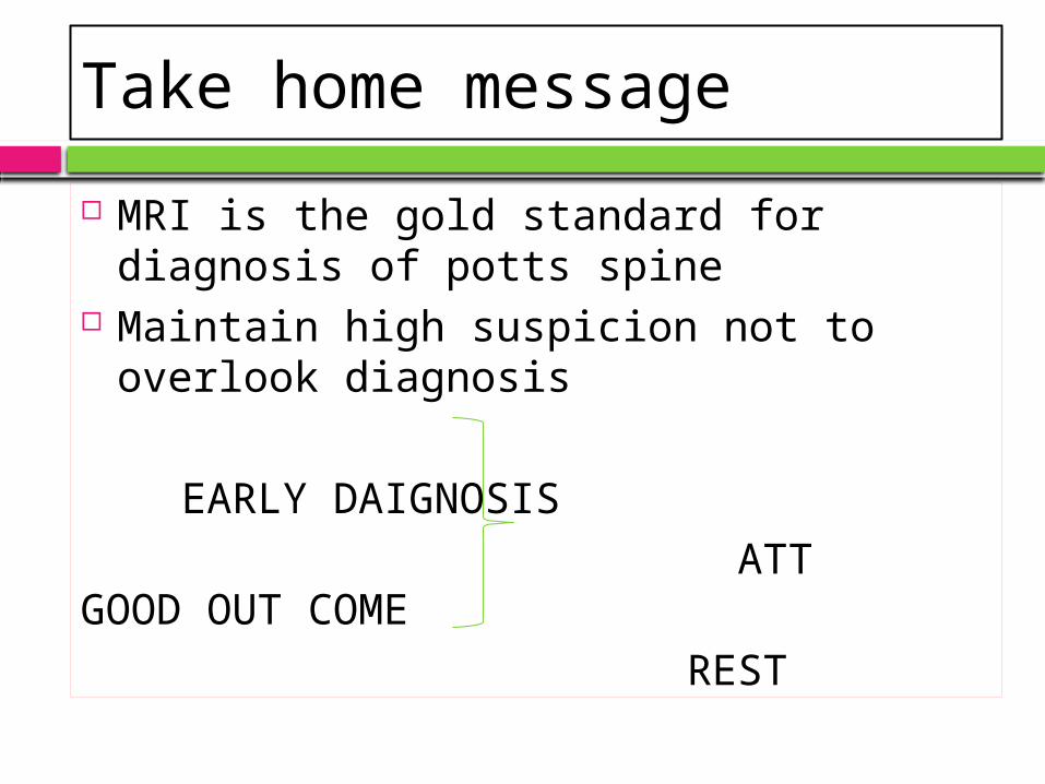
Take home message
MRI is the gold standard for diagnosis of potts spine
Maintain high suspicion not to overlook diagnosis
EARLY DAIGNOSIS ATT GOOD OUT COME REST

04/15/2023
91