poster vienne ecr
TRANSCRIPT
In vivo reduction of radiation exposure with a single-source coronary CT angiography
effects of optimal parameters settings in real life conditions
Alain Tavildari MD; François Vochelet MD; Luc Maillard MD PhDClinique AXIUM – Aix en Provence - FRANCE

FINANCIAL DISCLOSURES
None

BACKGROUND
Coronary computed tomographic angiography (CCTA) has become a common diagnostic test for evaluating patients with coronary artery disease
Radiation exposure has been deemed too high

OBJECTIVES
To assess the feasibility of in vivo radiation reduction only by modifying acquisition parameters in real life conditions with a single source 64-slice CT
Comparison of radiation exposure with conventional angiography
METHOD
Patients : Over 18 YO Co-morbidities including diabetes mellitus and
overweight were not excluded Coronary calcifications were not excluded All patients recieved sublingual nitrate Target heart rate was under 65 bpm at
acquisition time Atenolol intraveinously was given if needed Informed consent was obtained for all patients
METHOD
Acquisition parameters : General Electric VCT Xte Prospective acquisition if heart rate (HR) under 65 bpm
80 kV or 100 kV 120 mA to 400 mA Acquisition at 75% of R-R intervall No padding
Retrospective acquisition if HR between 65 and 75 bpm 80 kV or 100 kV Modulation limited to 400 mA (maximum around
phase 75%) Reconstruction every 10% from phase 0% to phase
90%
METHOD
Image quality : Evaluatued on a per-segment basis using the
American Heart Association coronary model Two independant observers gradded image quality
on a 4 point scale 1 : excellent : vessel fully evaluable, no artifact 2 : good : vessel fully evaluable, slight artifacts 3 : fair : evaluable concerning the presence of
stenosis, blurred vessel margins 4 : unevaluable
Average quotation was taken into account if in case of difference between the 2 observers
METHOD
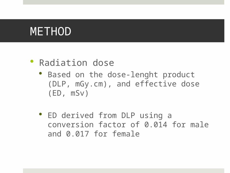
Radiation dose Based on the dose-lenght product (DLP,
mGy.cm), and effective dose (ED, mSv)
ED derived from DLP using a conversion factor of 0.014 for male and 0.017 for female
METHOD
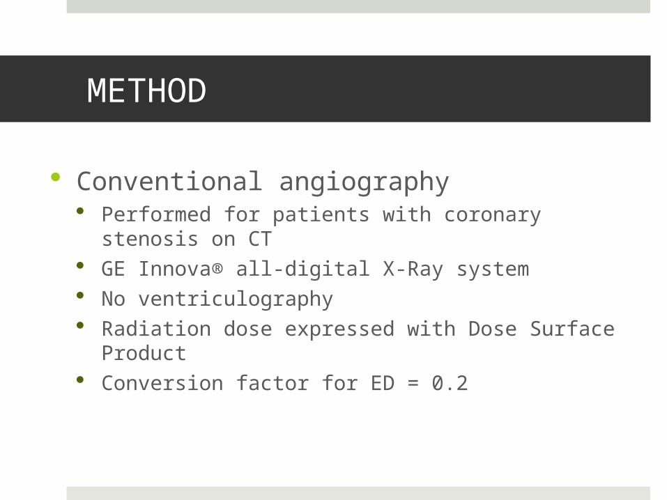
Conventional angiography Performed for patients with coronary stenosis on CT GE Innova® all-digital X-Ray system No ventriculography Radiation dose expressed with Dose Surface Product Conversion factor for ED = 0.2

IMAGE QUALITY = 1 (excellent)
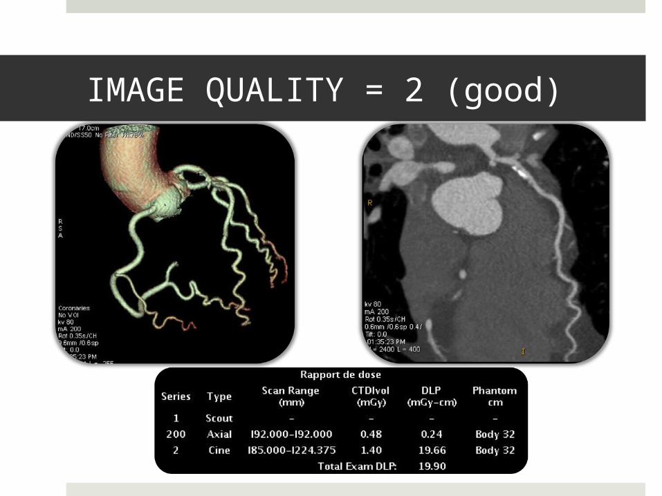
IMAGE QUALITY = 2 (good)
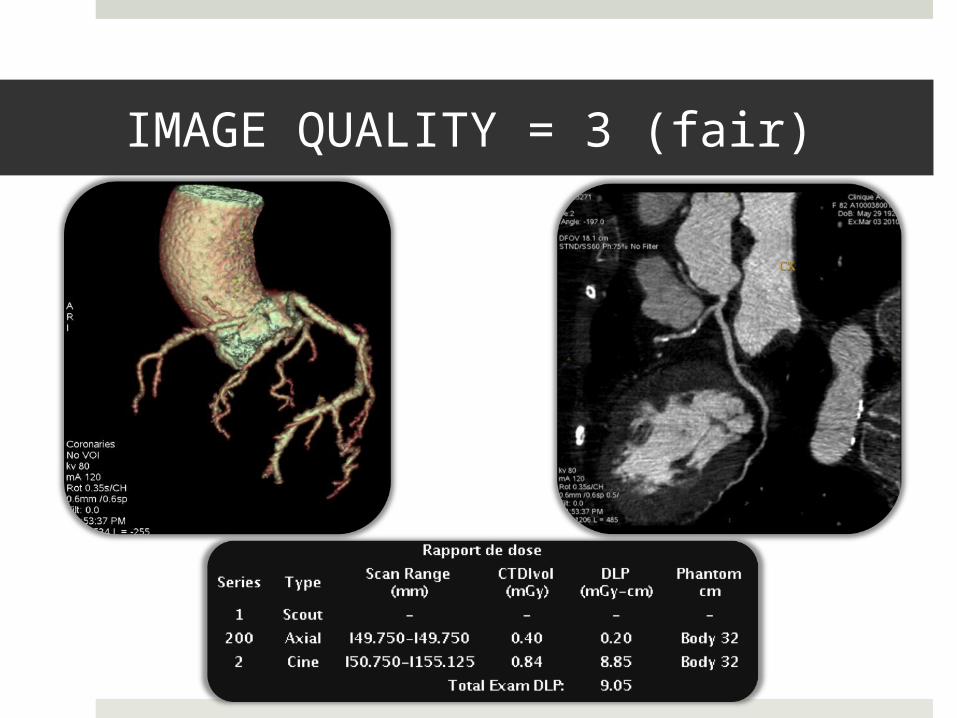
IMAGE QUALITY = 3 (fair)

IMAGE QUALITY = 4 (unevaluable)

RESULTS : patients characteristics (n=137)
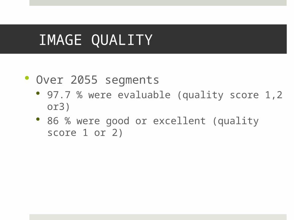
IMAGE QUALITY
Over 2055 segments 97.7 % were evaluable (quality score 1,2 or3) 86 % were good or excellent (quality score 1 or
2)
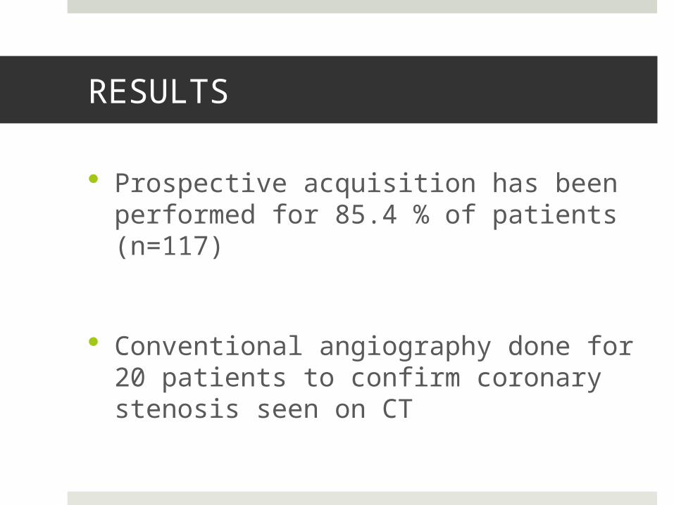
RESULTS
Prospective acquisition has been performed for 85.4 % of patients (n=117)
Conventional angiography done for 20 patients to confirm coronary stenosis seen on CT
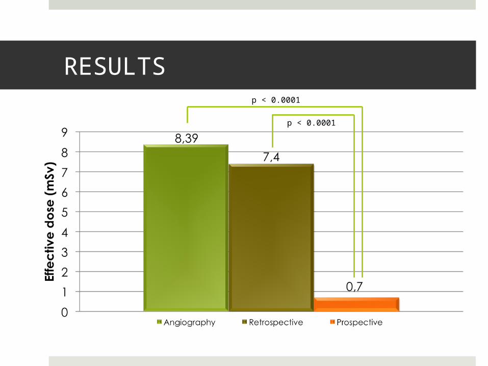
RESULTSp < 0.0001
p < 0.0001
SUMMARY
Our study was able to demonstrate that very low dose coronary CT is feasible with a standard 64 slice CT
Radiation dose was dramatically reduced in all acquisition modes compared to historical data and to conventional angiography
Prefered mode is prospective

STUDY LIMITATION
Non randomized study
Single center
Conventional angiography not made for all patients
Subjective image quality evaluation
CONCLUSION
These data may reconsider the decisional algorithm for coronary artery stenosis detection
Radiation dose for the whole studied population is nearly as low as a mammogram
CCTA could be proposed, with the parameter settings used in this study, for screening asymptomatic patients with high cardio-vascular risk factors