poster abstracts mcgowan retreat 2017 is in bold 2017 mcgowan retreat poster abstracts cellular and...
TRANSCRIPT
Presenter is in bold
2017 McGowan Retreat Poster Abstracts
Cellular and Gene Therapy 1. Faina Linkov, Sharon L. Goughnour, Shalkar
Adambekov, Anna Lokshin, Joseph Kelley, Paniti Sukumvanich, John Comerci, Kacey Marra, Lauren Kokai, Peter J. Rubin, Anda Vlad, Brian J. Philips, Robert P. Edwards. Exploration of inflammatory biomarkers in three depots of adipose tissue in women with endometrial cancer
2. Matthew B. Amdahl, Courtney E. Sparacino-Watkins, Paola Corti, Mark T. Gladwin and Jesus Tejero. Efficient reduction of vertebrate Cytoglobins by the Cytochrome b5 reducing system
3. Colin Beckwitt, Amanda M Clark, Katsu Warita, Zoltan Oltvai, Alan Wells. Adjuvant Statin Therapy Efficacy is dictated by Tumor Dormancy and Statin Lipophilicity in ex vivo and in vivo Models of Metastatic Breast Cancer
4. Kory J. Blose, Justin S. Weinbaum, and David A. Vorp. Computer Aided Design and Evaluation of a Novel Stem Cell Therapy for Small Abdominal Aortic Aneurysms
5. Peng, Y., Cárdenes, N., Huleihel, L., Álvarez, D., Sellarés, J., John, S., Chandler, C., Chen P., Rojas M. Different miroRNA expression in MSC-derived exosomes: IPF patients and age-matched normal individuals
6. Jacobo Sellarés, Luai Huleihel, Nayra Cárdenes, Diana Álvarez, Rosa Faner, Koji Sakamoto, Guoying Yu, Maria G. Kapetanaki, Naftali Kaminiski and Mauricio Rojas. Modified mesenchymal stem cells using miRNA transduction modify lung fibrosis progression
7. Nayra Cárdenes, Jonathan P. Carney, Diana Álvarez, Paola Aranda, Jacobo Sellarés, Scott Mason, Ergin Kocydirim, Luigi Lagazzi, Brian J. Lopresti, Julie A. Wolfram, Anthony E. Ting, Chandler Caufield, Ernesto Santos, Mauricio Rojas. Biodistribution of [F-18] FDG-labeled adult bone marrow-derived stem cells with PET/CT scan in an ARDS sheep model
8. Nayra Cárdenes, Diana Álvarez, Catherine Corey, John Sembrat, Sophie Wecht, Vidya Sagar Hanumanthu, Marta Bueno, Jeffrey Nine, Sruti Shiva, Mary Armanios, Ana Mora, Mauricio Rojas. Deficiencies in Mesenchymal Stem Cells from Idiopathic Pulmonary Fibrosis Patients Result in Lower Capacity to Protect the Lung from Injury
9. Nayra Cárdenes, Ergin Kocydirim, Paola Aranda, Diana Álvarez, Jacobo Sellarés, Andrea Elliot, John Sembrat, Pete Arlia, Luigi Lagazzi, Mauricio Rojas. The use of GMP-produced B-MSC Cells in Combination of Extracorporeal Membrane Oxygenation or Hemolung (Extracorporeal CO2 Removal) in ARDS in Combat Casualties
10. John Sembrat, Hannah D’Cunha, Julian Camilo Arango, Rebecca Vanderpool, Margaret Bennewitz, Prithu Sundd, Mauricio Rojas. Mesenchymal Stem Cells Drive Cell Repopulation In An In Vivo Model Of Lung Regeneration
11. Rojas M, McVerry B, Donahue M, Cardenes N and Ting A. MultiStem® Therapy in Subjects with Acute Respiratory Distress Syndrome
12. Roger Esteban-Vives, Myung Sun Choi, Matthew Young, Patrick Over, Jenny Ziembicki, Alain Corcos, J, Jörg C. Gerlach. Cell-spray autografting of deep partial-thickness burns: A critical review of 44 patient applications
13. Phillip Gallo, Mazen S Zenati, Latha Satish, Rachael M Kreft, Tianbing Yang, Fang Liu, Anne Argenta, Jennifer Ziembicki, Alain Corcos and Sandeep Kathju. PCR-Electrospray Ionization Mass Spectrometry in Identifying Microbial Infections in Burn Wounds
14. Dave Gau, William Veon, Teresa Capasso, Marion Joy, Beth Roman, David Koes and Partha Roy. Small molecule-mediated inhibitions of transcriptional cofactor MKL and its downstream target profilin impedes endothelial cell migration and angiogenesis
15. Bing Han, Junji Komori, Maria Giovanna Francipane, Fei Chen, Eric Lagasse. Omental Milky Spots as Sites for Ectopic Liver Development
16. Moriah Johngrass, Ryan Schroth, Lauren Kokai. Modulating Macrophage Dynamics to Improve Autologous Fat Grafting Outcomes
17. Philip M. Bauer, Elizabeth R. Kahle, Han Zheng, Michael T. Lotze, Timothy R. Billiar, Eileen M. Bauer. Chloroquine, a pharmacological inhibitor of autophagy, attenuates hypoxia-induced pulmonary hypertension
18. Mehwish Khaliq, Donghun Shin. Stat3 regulates hepatocyte and biliary epithelial cells proliferation and hepatocyte maturation in the zebrafish hepatic progenitor cell (HPC)-driven liver regeneration model
19. Chiaki Komatsu, Yolandi van der Merwe, Lin He, Maxine R. Miller, Katie A. Lucy, Huamin Tang, Ian Rosner, Wendy Chen, Jila Noori, Valeria Fu, Michael Steketee, Gadi Wollstein, Mario Solari, Joel S. Schuman, Kevin C. Chan, Kia M. Washington. Retinal and optic nerve viability evaluated with optical coherence tomography, manganese-enhanced MRI and electroretinography after whole eye transplantation
20. Yuan Liu, Travis Lear, Bill B. Chen. Small Molecule Therapeutics Targeting Ubiquitin E3 Ligase
21. Mark Murdock, Ilea Swinehart, Sherin David, Kathrin Gassei, Kyle Orwig, Stephen Badylak. Extracellular Matrix and Spermatogonial Stem Cell Culture
22. Franziska Nitzsche, Harmanvir Ghuman, Madeline Gerwig, Jeffrey Moorhead, Alex Poplawsky, Brendon Wahlberg, Fabrisia Ambrosio, Michel Modo. Establishing efficacy for a combination of physical and cell therapy for stroke
Presenter is in bold
23. Deborah Osakue, Yiqin Du. Stem Cell Resistance to Endoplasmic Reticulum Stress and its Implications for Glaucoma Treatment
24. Jacquelyn Russell, Hirohisa Okabe, Sucha Singh, Laura Molina, Minakshi Poddar, Satdarshan P. Monga. Lack of beta-catenin in hepatocytes impairs proliferation and promotes liver progenitor cell-mediated repair in response to the choline-deficient ethionine-supplemented diet
25. M. Asher Schusterman II, Isaac James, Debra Bourne, Sheri Wang, Mayara Silva, Kassandra Albright, Damian Grybowski, Liyong Zhang, Latha Satish, Kacey Marra, J. Peter Rubin. Platelet-Rich Plasma and Adipose Stem Cells Improve Burn Wound Healing in Yorkshire Pigs
26. Elizabeth C. Stahl, Bryan N. Brown. Accumulation of Bone Marrow Derived Macrophages in the Aged Murine Liver
27. Kyle Sylakowski, Austin Nuschke, Alan Wells. Characterization of Tenascin-C signaling on Mesenchymal Stem Cells for Chronic Wound Healing Therapies
28. Li Wen and Sohail Husain. Novel calcineurin inhibitor strategies to prevent radiocontrast-induced organ injury
29. Chaoming Zhou, Yeal Zeldin, Sandeep Kathju, Latha Satish. Pirfenidone Inhibits TGF-b1 stimulated Non-SMAD Signaling Pathways in Dupuytren’s-derived fibroblasts
30. Daniel A. Zuppo, Maria A. Missinato, Manush Sayd Mohammed and Michael K.W. Tsang. Cardiac transcriptome profiling during regeneration in zebrafish
Computation and Modeling 31. Emily E. Ackerman, Jason E. Shoemaker. Controllability
Analysis of Protein-Protein Interaction Networks for Antiviral Drug Development
32. Eric Lambert, Tristan Ford, Satya VVN Kothapalli, Hong Chen and Jelena M. Janjic. High intensity focused ultrasound-sensitive perfluorocarbon nanoemulsions in targeted drug delivery
33. Lisa Carey Lohmueller, Manreet Kanwar, Carmen Khoo, Robert Kormos, James Antaki. Predicting all cause mortality for LVAD patients using Bayesian Networks
34. Luke Ziegler, Salim Olia, Marina Kameneva. The Modified Mechanical Fragility Index: A New Tool for Clinical Measurement of Red Blood Cell Fragility
35. Elaine Soohoo, Lewis K Waldman, Dennis R Trumble. Computational Assessment of Design Parameters for a Torsional Ventricular Assist Device (tVAD)
Medical Devices 36. Moataz M. Abdulhafez, Moataz M. Elsisy, Rob Lewis,
Mohamed A. Zaazoue, Liliana C. Goumnerova and Mostafa Bedewy. Lowering the risk of injury in head immobilization devices by design
37. Dan Crompton, Salim E Olia, Trevor A Snyder, Peter D Wearden, Marina V Kameneva. In vitro and in vivo hemocompatibility assessment of the VADovations novel pediatric left ventricle assist device (LVAD)
38. Firuz Feturi, Hua Wang, Yevgeny Brudno, Vasil Erbas, Liwei Dong, Zhaoxiang Zhang, Huseyin Sahin, Wensheng Zhang, Mubin Ali Aral, Raman Venkatramanan, David Mooney, Vijay Gorantla. Ultrasound Triggered-Release Embedded Anti-Rejection Therapy (TREAT™) for Targeted Immunomodulation in Vascularized Composite Allotransplantation
39. Garrett Jeffries, Brian Frankowski, William Federspiel. High Efficiency Respiratory Dialysis at Ultra-Low Blood Flows
40. Alexander D. Malkin, William J. Federspiel, John A. Kellum and Kai Singbartl. An Extracorporeal Neutrophil Reprogramming Device for the Treatment of Acute Inflammatory Conditions
41. Jonquil R. Mau, Chak-Yuk Yu, Yajnesh Vedanaparti, Savio L-Y. Woo. Corrosion Characterization of Magensium Coated Via Micro-arc Oxidation
42. Salim E. Olia, Timothy M. Maul, Marina V. Kameneva. The Effect of Inlet Pressure on Gas Embolism and Hemolysis in Continuous-Flow Blood Pumps
43. Ryan A. Orizondo, Alex G. May, Shalv P. Madhani, Brian J. Frankowski, Greg W. Burgreen, Peter D. Wearden and William J. Federspiel. Pittsburgh Pediatric Ambulatory Lung (P-PAL)
44. Akhil Patel, Samer Zaky, Elia Beniash, Charles Sfeir, Hongshuai Li, Sachin Velankar, Yadong Wang, Shilpa Sant. RegenMatrix: Collagen-mimetic Bioactive Hydrogels for Growth Factor Free Approach for Bone Regeneration
45. Nathaniel T. Weberding, Richard A. Saladino, M. Beth Minnigh, Patrick J. Oberly, Samuel M. Poloyac, Mioara D. Manole. Traditional method small volume drug delivery method fails to provide a therapeutic dose in small infants with supraventricular tachycardia
46. Puneeth Shridhar, Yanfei Chen, Stephen Emery, Stephanie Greene, Youngjae Chun. Ventriculo Amniotic Shunt for Fetal Aqueductal Stenosis
47. Jingyao Wu, Boeun Lee, Abhijit Roy, Tianbing Yang, Patricia Hebda, Sandeep Kathju, Thomas Gilbert, David Chi, Prashant N. Kumta. Application of novel ultrahigh ductility (UHD) magnesium stents for the treatment of airway obstruction
48. Vesselin Shanov, Guangqi Zhang, Pravahan Salunke, Vibhor Chaswal, Savio Woo, Charles Sfeir, Prashant Kumta, Sergey Yarmolenko, Boyce Collins, Yeoheung Yun, Mark Schulz, Zhangzhang Yin, Zhongyun Dong, William Heineman, Sarah Pixley, Maren Pink. Magnesium Single Crystals for Medical Implant Applications
Presenter is in bold
Tissue Engineering 49. Qahtan AlQahtani, Samer Zaky, Herbert Ray and
Charles Sfeir. Dental Pulp Derived Extracellular Matrix as a Scaffold for Pulp Regenerative Therapy
50. Zhaoxiang Zhang, Ali Mubin Aral, Keewon Lee, Xiaozhou Fan, Kang Kim, Mario G. Solari, Vijay S. Gorantla, Yadong Wang. Arterial remodeling in autologous vein and cell-free, fast-degrading vascular grafts using a rat carotid artery interposition model
51. Catalina Ardila, David Maestas, Victoria Lundine, Marvin Slepian, David Harris, Jonathan Vande Geest. Surface Modification of Electrospun Gelatin/Fibrinogen Scaffolds to Encourage Endothelial Cell Function
52. Travis Armiger, Kris Dahl. Measuring Changes in Force Generation in Cell Monolayers via Intranuclear Kinetics
53. Andrew Bradshaw, Jelena Grahovac, Brian L. Hood, Linda G. Griffith, Alan Wells. Modeling Metastasis from Invasion to Colonization on a Human Physiomimetic Chip
54. Laura T. Beringer, Shaohua Li, Ethan J. Kallick, Kelly J. Shields, Erin M. Faight, Francis Cartieri, Ariel Aballay, Howard Edington and Saadyah Averick. Accelerating Wound Healing with a Collagen VI - Heparin Sulfate Coated Matrix
55. Patrick G. Chan, Alex Hall, Marie Billaud, Amrinder Nain, Julie Phillippi, Thomas G. Gleason. Measuring SMC Death and Adhesion Forces Via Tissue Engineered Models Simulating ECM Microarchitecture
56. Jingming Chen, Amalie E. Donius, Juan M. Taboas. Hydrogel Composition Regulates Chondrogenesis by Mesenchymal Stem Cells and Endochondral Ossification in Engineered Cartilaginous Interfacial Tissues
57. Martin Haschak, Siddhartha Dash and Bryan Brown. The effects of cardiac extracellular matrix aging on macrophage polarization
58. Darren Haskett, Kamiel Saleh, Katherine Lorentz, Jeffrey Krawiec, Justin Weinbaum, Antonio D’Amore, William Wagner, Lauren Kokai, Kacey Marra, J. Peter Rubin, David Vorp. Towards a “Same-Day” Autologous Tissue-Engineered Vascular Graft: Seeding and Implantation of an Elastomeric Scaffold with the Stromal Vascular Fraction
59. Thomas Hinton, Andrew Lee, Andrew Hudson, Adam Feinberg. 3D Bioprinting of Organ-Scale Type I Collagen Scaffolds
60. Jorge Jimenez, Michael A. Washington, Ken K. Nischal and Morgan V. Fedorchak. Development of a Topical Ophthalmic Biomaterial for the Controlled Release of Cysteamine
61. Irona Khandaker, Golnar Shojaati, Martha Funderburgh, Mary Mann, Moira Geary, Stephen F. Badylak, James Funderburgh. Extracellular matrix bioscaffolds reduce corneal scaring after traumatic wounding
62. Karis Kosar, Kari Nejak-Bowen. Wnt7b and Wnt10a, beta-catenin independent signaling regulates cholangiocyte proliferation and function during cholestasis
63. Chelsea D. Merkel and Adam V. Kwiatkowski. N-cadherin adhesion is coordinated with the actomyosin network to regulate adherens junction dynamics and mechanical coupling in cardiomyocytes
64. Yoojin Lee, Ula Zdanowicz, Joe Bartolacci, Jenna Dziki, Madeline Cramer, Scott Johnson, Stephen Badylak. Evaluation of a Novel Method of Treatment of Tendinopathy with the Use of Extracellular Matrix Hydrogel
65. Hang Lin, Angela Beck, Shimomura Kazunori, Peter Alexander, Madalyn Fritch, Evan Kilroy(2), Rocky Tuan. Development of Optimized Photocrosslinked Gelatin/Hyaluronic Acid Scaffold for Repair of Osteochondral Defect
66. Wen Liu, Ngoc Pham, Yiwei Wang, Nina Reger, Yong Fan, Nick Giannoukakis, Ellen S. Gawalt and Wilson S. Meng. Delivering Antibodies in Skin Transplant Hosts using IgG-Binding Injectable Coacervates
67. Hassan Awada, Daniel Long, Zhouguang Wang, Mintai Hwang, Kang Kim, Yadong Wang. A Single Injection of Protein-loaded Coacervate-Gel Significantly Improves Cardiac Function Post Infarction
68. Samuel LoPresti, Sharisse Victor, Bryan Brown. Effect of Age on Macrophage Response to Muscle Extracellular Matrix
69. Samuel K. Luketich, Giuseppe Raffa, Salim Olia, Xinzhu Gu, Michele Pilato, Marina V. Kameneva, Vinay Badhwar, William R. Wagner and Antonio D’Amore. Double component electrospun fibers deposition (DCD): heart valve fabrication with controlled mechanics, microstructure, and anatomy
70. Christopher Mahoney, Malik Snowden, J. Peter Rubin, Kacey Marra. Composite Adipose Derived Delivery System for Adipose Restoration
71. Chelsea D Merkel, Roisin M O'Dowd, Adam V Kwiatkowski. The Role of Vinculin in Cardiomyocyte Adhesion and Mechanical Continuity
72. Drake Pedersen, Marzio DiGiuseppe, Salvatore Pasta, Antonio D'Amore, James F. Antaki, William R. Wagner. Fluid Dynamics Assessment of Microfibrillar, Elastomeric Heart Valve Scaffolds
73. Alessandro Pirosa, Riccardo Gottardi, Peter G. Alexander, Dario Puppi, Federica Chiellini and Rocky S. Tuan. An in vitro Chondro-Osteo-Vascular Triphasic Model of the Osteochondral Complex
74. Travis A. Prest, Mara Palmer, Meghan Wyatt, Jonathan Cheetham, Bryan N. Brown. Peripheral Nerve-Specific Extracellular Matrix Hydrogel Supports Repair after Peripheral Nerve Injury
75. Aneesh K. Ramaswamy, Rachel E. Sides, Tamara S. Maihle, Victor O. Morell, David A. Vorp and Justin S. Weinbaum. Improved Elastogenesis for Pediatric Patients with Genetic Defects of the Aorta
Presenter is in bold
76. Chris Reyes, Li Mo, Danielle Guimaraes, Kelly Quesnelle, Yinna Wang and Sruti Shiva. Nitrite Regulates Mitochondrial Dynamics to Inhibit Vascular Smooth Muscle Cell Proliferation
77. Lindsey T. Saldin, Molly Klimak, Ryan C. Hill, Madeline C. Cramer, Luai Huleihel, Maria Quidgley-Martin, David Cardenas, Tim J. Keane, Ricardo Londono, George S. Hussey, Lori A. Kelly, Juliann E. Kosovec, Emily J. Lloyd, Ashten N. Omstead, Daisuke Matsui, Blair A. Jobe, Kirk C. Hansen, Ali H. Zaidi, Stephen F. Badylak. Tissue-specific Effects of Normal, Metaplastic, and Neoplastic Esophageal Extracellular Matrix Hydrogels
78. Benjamin Schilling, Kacey Marra. Construction of a Muscle-Derived Extracellular Scaffold intended to Promote Muscle Function after Nerve Injury
79. Joseph H Shawky, Uma Balakrishnan and Lance A. Davidson. Form and Mechanical Function of Germ Layer Architecture in the Developing Embryo
80. Chelsea E.T. Stowell, Diego Celdran-Bonafonte, Begona Campos, Aous Jarrouj, Sukit Raksasuk, Peter L. Jernigan, Yadong Wang and Prabir Roy-Chaudhury. Resorbable vascular grafts support early cell infiltration and endothelialization in a porcine vascular access model
81. Aaron X Sun, Rachel Brick, Kelsey M. Gloss, He Shen, Guang Yang, Pete G. Alexander, Michael DeHart, Rocky S. Tuan. Novel Conduits with Wall-Encapsulated Cells Improve Peripheral Nerve Regeneration
82. Ehab Tamimi, Jamie L. Hernandez, Corina MacIsaac, Catalina Ardila, Jonathan P. Vande Geest. Biomechanical evaluation of gelatin/fibrinogen electrospun cylindrical scaffolds seeded with 3T3 mouse fibroblasts and porcine smooth muscle cells
83. Deepthi S. Vijayraghavan, Lance Davidson. Biomechanical analysis of cell behaviors during neural plate convergent extension
84. Yuanheng Yang, Hang Lin, He Shen, Bing Wang, Rocky Tuan. Stem cell derived Extracellular Matrix Enhancement of Autologous Chondrocytes Implantation (ACI) for Articular Cartilage Repair
85. DiBernardo G, Bliley JM, Waldner M, Schroth RN, Mahoney C, Grybowski D, Kim D, Schusterman MA, Fadia N, McGovern V, Narayanan A, Bourne D, James I, Simpson T, Tompkins-Rhoades C, Taylor A, Dees A, Washington K, Spiess AM, Crammond DJ, Marra KG. Long Gap Median Nerve Regeneration Using Tissue Engineered Guides in a Non-Human Primate Model
Faina Linkov (1,2,4), Sharon L. Goughnour (1), Shalkar Adambekov (2), Anna Lokshin (3), Joseph Kelley (5),Paniti Sukumvanich (5), John Comerci (5), Kacey Marra (6), Lauren Kokai (6), Peter J. Rubin (6), Anda Vlad(1), Brian J. Philips (1), Robert P. Edwards (1,4)
(1) Magee-Womens Research Institute, Department of Obstetrics, Gynecology, and Reproductive Sciences, University ofPittsburgh, (2) Department of Epidemiology, University of Pittsburgh, (3) Department of Medicine, and the Luminex CoreLaboratory, University of Pittsburgh Cancer Institute, University of Pittsburgh, (4) University of Pittsburgh Cancer Institute,(5) Division of Gynecologic Oncology, Department of Obstetrics, Gynecology and Reproductive Sciences, University ofPittsburgh Medical Center, Magee-Womens Hospital, (6) Department of Plastic Surgery, School of Medicine, University ofPittsburgh
Exploration of inflammatory biomarkers in three depots of adipose tissue in women withendometrial cancer
Background: Obesity is characterized by a state of chronic, low-grade inflammation and is recognized as a primary riskfactor in the development of endometrial cancer (EC). Increased adipose tissue mass may promote carcinogenesisthrough the increased expression of pro-inflammatory biomarkers. We explored the inflammatory environment in the threeadipose tissue depots (omental, retroperitoneal, subcutaneous) in women diagnosed with EC by evaluating biomarkerssecreted by adipose-derived stem cells (ASC). ASC have been investigated in inflammation, aging, and obesity, but notexplored in the context of EC, the most common gynecologic malignancy.
Methods: Omental, retroperitoneal, and subcutaneous adipose tissue samples were collected from 22 women, aged 35-83 years, undergoing hysterectomy for EC. Conditioned media was generated from the growth of ASC from the threedepots. Angiopoietin-2, EGF, IL-8, leptin, VEGFA, VEGFC, and VEFGD levels in the conditioned media were analyzed byLuminex bead-based xMAP immunoassays. One-way ANOVA was used to compare the mean levels of biomarkersbetween the three depots.
Results: The 22 women had a mean age of 64.30 (12.35) years and a mean BMI of 37.48 (7.81) kg/m2. Angiopoietin-2,EGF, leptin, VEGFD and approximately 20% of VEGFC levels were below detection limits in the three depots. There wasa significant difference between the three depots for IL-8, with the highest levels of IL-8 in the omental depot and thelowest levels in the retroperitoneal depot (p-value <0.0001). VEGFA levels were highest in the retroperitoneal depot andlowest in the subcutaneous depot; however, these differences were not statistically significant.
Conclusions: This is one of the first studies to compare inflammatory biomarker expression in ASC conditioned mediafrom three depots of adipose tissue. Adipose tissue is a potentially attractive depot to evaluate biomarkers in relation tocancer risk and more research needs to be done.
Cellular and Gene TherapyPoster number 1 Category:
Matthew B. Amdahl (1,2), Courtney E. Sparacino-Watkins (1), Paola Corti (1), Mark T. Gladwin (1,3) andJesus Tejero (1,3)
(1) Heart, Lung, Blood, and Vascular Medicine Institute, (2) Department of Bioengineering, (3) Division of Pulmonary,Allergy, and Critical Care Medicine, University of Pittsburgh, Pittsburgh, PA
Efficient reduction of vertebrate Cytoglobins by the Cytochrome b5 reducing system
Cytoglobin (Cygb) is a heme-containing protein ubiquitous in mammalian tissues. Unlike the evolutionarily related proteinshemoglobin and myoglobin, Cygb shows a six-coordinated heme binding, with the heme-iron coordinated by two histidineside chains. Cygb is involved in cytoprotective and regenerative pathways through yet undefined mechanisms, and it hasrecently been demonstrated that cytoglobin has redox signaling properties via NO and nitrite metabolism.The reducedferrous molecule can bind oxygen and will react with NO in a dioxygenation reaction to form nitrate, which dampens NOsignaling. When deoxygenated cytoglobin can bind nirite and reduce it to NO. This oxido-reductase activity could becatalytic if an effective reduction system exists. The nature of the physiological Cygb reducing system is unknownalthough it has been proposed that ascorbate and cytochrome b5 could fulfill this role. Here we describe that physiologicalconcentraitons of cytochrome b5/cytochrome b5 reductase system can reduce human and fish Cygbs at rates up to 250-fold higher than their known physiological substrates, hemoglobin and myoglobin and many times faster, as well as morethoroughly, than even high levels of ascorbate. These data suggest that the cytochrome b5/cytochrome b5 reductasesystem is a viable reductant for Cygb in vivo, allowing for catalytic oxido-reductase activity.
Cellular and Gene TherapyPoster number 2 Category:
Colin Beckwitt (1), Amanda M Clark (1), Katsu Warita (2), Zoltan Oltvai (1,3), Alan Wells (1,3)
(1) Department of Pathology and (3) Department of Computational and Systems Biology, University of Pittsburgh, Schoolof Medicine, Pittsburgh, PA, (2) Department of Veterinary Anatomy, School of Veterinary Medicine, Tottori University,Japan
Adjuvant Statin Therapy Efficacy is dictated by Tumor Dormancy and Statin Lipophilicity in exvivo and in vivo Models of Metastatic Breast Cancer
Metastasis in breast cancer patients heralds mortality, as disseminated disease is generally chemoresistant. After tumorcells reach the ectopic tissue, they undergo an epithelial reversion to enter a period of quiescence, termed dormancy,which may last for decades before outgrowing again as mesenchymal/dedifferentiated masses. Thus, long-term, relativelynon-toxic interventions that prevent metastatic outgrowth are needed to treat this mortal stage of tumor progression.
Epidemiological analyses have suggested that statin usage, for cardiovascular indications, is correlated with a reductionin metastatic emergent (though not in incidence of primary) breast cancer. The goal of this study is to demonstrate this isdue to statins suppressing breast cancer cell proliferation, a hallmark of emergent outgrowth.
We have found that atorvastatin and simvastatin limit the growth of some cancer cell lines, but not others. The sensitivelines were marked by lacking surface E-cadherin, the hallmark of the mesenchymal phenotype. Furthermore, this is adirect effect, as we now have shown that hydrophilic statins are relatively ineffective compared to the membranepermeant lipophilic statins.
To determine whether the statins target emergent metastatic tumor cells, we are using an all human microphysiologicalsystem of the most common site for metastases, the liver. Briefly, a micro-hepatic tissue is established by seedingprimary human liver cells in a porous scaffold subject to a physiological flow. RFP-labeled breast cancer cells are seededinto these microtissues and examined weeks later. Initial studies suggest that statins suppress the emergence of dormanttumor cells when challenged by stressors that lead to outgrowth. Statins were further found in vivo to influence breastcancer metastasis and metastatic growth using a mouse model of spontaneous liver metastases. As 26% of adultscurrently take a statin for other medical conditions, these studies may suggest the best statin to use in the context ofmaintaining breast cancer dormancy long-term and delaying or avoiding the morbid emergence.
Cellular and Gene TherapyPoster number 3 Category:
Kory J. Blose (1), Justin S. Weinbaum (1,5), and David A. Vorp (1,2,3,4,5)
(1) Department of Bioengineering, University of Pittsburgh, Pittsburgh, PA, (2) Department of Cardiothoracic Surgery,University of Pittsburgh, Pittsburgh, PA, (3) Department of Surgery, University of Pittsburgh, Pittsburgh, PA, (4) Center forVascular Remodeling and Regeneration, University of Pittsburgh, Pittsburgh, PA, (5) McGowan Institute for RegenerativeMedicine, University of Pittsburgh, Pittsburgh, PA
Computer Aided Design and Evaluation of a Novel Stem Cell Therapy for Small AbdominalAortic Aneurysms
Exsanguination from abdominal aortic aneurysm (AAA) rupture is frequently fatal and is currently the 13th leading causeof death in the United States of America. AAAs can take years to progress to the point where surgical intervention isrecommended (> 5.5 cm diameter). Our goal is to develop a novel stem cell therapy for small AAAs. Approximately 90%of patients with AAA do not meet the size criterion for intervention and could benefit from this alternative therapy.
Our proposed strategy is delivery of adipose-derived mesenchymal stem cells (ADMSCs) to the external surface of theAAA. In this way autologous cells can be isolated from a patient, culture-expanded if necessary, mixed in a hydrogel, andinjected around that same patient’s aorta in a minimally invasive procedure.
Aspects of this treatment paradigm were evaluated in-silico, in-vitro, and in-vivo. We found that allowing for elastinproduction reduces the aneurysm enlargement rate using in-silico models of aneurysm growth and remodelling. We alsofound that adult smooth muscle cells produced a mature elastin network when co-cultured with ADMSCs in-vivo. Lastly,we found that delayed, periadventitial delivery of ADMSCs halted two aspects of aneurysm progression – expansion ofthe aortic diameter and fragmentation of the elastic lamella – in an elastase perfusion mouse model of AAA.
The results of these studies show promise for using ADMSCs as a possible AAA therapy by stimulating new elastinproduction by adult SMCs.
Cellular and Gene TherapyPoster number 4 Category:
Peng, Y. (1,2), Cárdenes, N. (1), Huleihel, L. (3), Álvarez, D. (1), Sellarés, J. (1,4), John, S. (1), Chandler, C.(1), Chen P. (2,*), Rojas M. (1,3,*)
(1) Simmons Center for Interstitial Lung Disease, Division of Pulmonary, Allergy & Critical Care Medicine, University ofPittsburgh, (2) Research Unit of Respiratory Diseases, Central South University, Changsha Hunan, 410011, China, (3)McGowan Institute of Regenerative Medicine University of Pittsburgh, Pittsburgh, PA, (4) Servei de Pneumologia,Hospital Clínic, IDIBAPS, Universitat de Barcelona, Barcelona, Spain, *Corresponding authors
Different miroRNA expression in MSC-derived exosomes: IPF patients and age-matchednormal individuals
Purpose: We aimed to observe the difference between exosomes isolated from mesenchymal stem cells (MSCs) fromboth healthy subjects and patients with Idiopathic Pulmonary Fibrosis(IPF).
Methods: Bone-marrow MSCs from vertebral bodies were isolated from IPF and age-match normal subjects. When thecells grow to 80-90% confluence, cell culture medium was replaced with MEM media without fetal bovine serum,differential centrifugation and 18 hours density gradient ultracentrifugation was used to isolate and purify the exosomes.The morphological features of exosomes were examined by Transmission Electron Microscope (TEM). MSCs derived-exosome RNAs were extracted by SeraMir Exosome RNA kit. MicroRNAarray were used to analyse different expressionof microRNAs in exosomes from IPF patient’s MSCs and normal subject’s MSCs.
Results: The density of the gradient after 18 hours of ultracentrifuge is 1.020, 1.030, 1.042, 1.053, 1.066, 1.088, 1.104,1.121, 1.182, 1.212, 1.251g/ml. Detected by their HSP70 expression, microvesicles were enriched in the Fraction 3, 4, 5,6, 7 and exosomes were enriched in Fraction 3, 4, 5, 6. The corresponding floating density of exosomes is from 1.042g/mlto 1.088g/ml. TEM showed the exosomes were cup-shaped and the diameters were around 30-100nm. Bioanalyzershowed that MSCs have intact 18S and 28S ribosomal RNA but MSCs derived-exosomes contain little or no ribosomalRNA, instead of a lot of microRNA and mRNA. ExosomesRNA from IPF patient’s MSCs express different microRNAscompared with normal subject’s MSCs.
Conclusion: IPF patient’s MSCs-derived exosomes differ both morphologically and in miroRNA expressions fromexosomes present in normal subject’s MSCs. Further studies need to be completed to elaborate functional differencesbetween IPF and normal subject’s MSCs.
Cellular and Gene TherapyPoster number 5 Category:
Jacobo Sellarés (1,2,3), Luai Huleihel (1,2,4,5), Nayra Cardenes (1,2), Diana Álvarez (1,2), Rosa Faner (6,7),Koji Sakamoto (8), Guoying Yu (8), Maria G. Kapetanaki (1), Naftali Kaminiski (8) and Mauricio Rojas (1,2,5)
(1) Dept. of Med., Div. of Pulmonary, Allergy, and Critical Care Med., UPMC, (2) The Dorothy P. and Richard P. SimmonsCtr. for Interstitial Lung Disease, UPMC, (3) Servei de Pneumologia, Hospital Clínic, IDIBAPS, Univ. de Barcelona,Barcelona, Spain, (4) The Shraga Segal Dept. of Microbiology, Immunology and Genetics, Faculty of Health Sci., BenGurion Univ. of the Negev; Beer-Sheva, Israel, (5) McGowan Institute for Reg. Med., UPMC, (6) Centro de InvestigaciónBiomédica en Red, Respiratory Diseases (CIBERES), Madrid, Spain, (7) Fundació Clínic per a la Recerca Biomédica,Barcelona, Spain, (8) Section of Pulmonary, Critical Care and Sleep Med., Dept. of Med., Yale Univ., New Haven, CT
Modified mesenchymal stem cells using miRNA transduction modify lung fibrosis progression
Background/Objectives: .Although different preclinical models have demonstrated a favorable role of bone marrowderived-mesenchymal stem cells (B-MSCs) in preventing fibrosis, this protective effect is not observed with lateadministration of B-MSCs, when fibrotic changes are consolidated. The possibility of modify B-MSCs to preventdeleterious effects or even enhancing their therapeuthic properties could be relevant in their future potential therapeuthicuse. We sought out to investigate if the modification of B-MSCs using miRNAs let-7d (antifibrotic) and miR-154(profibrotic) could modify their ability to alter lung fibrosis in a murine bleomycin model
Methods: Concentrated let-7d, miR-154 miRNAs or a control sequence lentiviral vector were transduced into human B-MSCs. These modified B-MSCs were intravenously administered to mice at day 7 day after bleomycin instillation. Micewere sacrificed at day 14. Different epithetial/mesenchymal markers in B-MSCs were assesed. In addition, the effect ofmodiefied B-MSCs in physical signs and diferent lung fibrosis markers on mice were also evaluated.
Results: B-MSCs were successfully modified and overexpressed let-7d and miR-154 after transfection. Although noreverse in lung fibrosis was observed, bleomycin-injured animals that were treated with let7d- B-MSCs were found tohave an improvement in weight and a reduction in collagen mRNA levels in lung tissue, what suggests a decrease inactivity of lung fibrosis progression. This positive effect was probably associated with the changes of B-MSCsepithelial/mesenchymal properties after miRNA modification. Treatment with miR154 modified-BMSCs not only did nothave a beneficial effect but miR-154 group had the worst survival.
Conclusions: Our results establish the use of modified B-MSCs with mi-RNAs as a potential future treatment in lungfibrosis.
Cellular and Gene TherapyPoster number 6 Category:
Nayra Cárdenes (1), Jonathan P. Carney (2), Diana Álvarez (1), Paola Aranda (1), Jacobo Sellarés (1), ScottMason (2), Ergin Kocydirim (3), Luigi Lagazzi (4), Brian J. Lopresti (2), Julie A. Wolfram (5), Anthony E. Ting(5), Chandler Caufield (1), Ernesto Santos (2), Mauricio Rojas (1,3)
(1) PACCM, (2) Radiology, (3) McGowan Institute, (4) Cardiotorathic Surgery, (5) Athersys Inc.
Biodistribution of [F-18] FDG-labeled adult bone marrow-derived stem cells with PET/CT scanin an ARDS sheep model
Acute respiratory distress syndrome (ARDS) is an acute, inflammatory lung injury with mortality rates around 40-60% incritically ill patients. To date, no specific treatment strategy exists. Human stem cells have emerged as a promisingtherapeutic strategy. Early results from our group and others have shown the efficacy and safety of the use of humanstem cells in LPS-induced ARDS in sheep when administered by endo-bronchial or intravascular route. As part of theunderstanding of their molecular mechanism by which they are therapeutically beneficial, the evaluation of biodistribution,migration and homing in vivo of stem cells are needed. Noninvasive tracking method using Positron EmissionTomography (PET) has been successfully used for in vivo tracking of [18F]-FDG labeled cells. Here we aimed to comparein vivo human stem cell trafficking, their tissue tropism and their engraftment to the targeted organ lung, using bonemarrow derived multipotent adult progenitor cells (MAPC®), comparing two methods of administration: endo-bronchial(EB) and intravenous (IV), in a sheep model of LPS-induced lung injury.
Sheep were kept under anesthesia with 100% oxygen. A dose of 5 μg/Kg E. coli endotoxin was infused over 21 minutesto induce lung injury. Arterial blood gases were measured before endotoxin, 1 hr, 2 hr, 4 hr, and 6 hr after endotoxininfusion to determine the onset of the acute phase of hypoxemia. MAPC cells were labeled with [18F]-FDG for an hourand delivered EB at a dose of 1 mill./Kg or IV at 10 mill./Kg one hour after the infusion of endotoxin. ComputedTomography (CT) and PET scans were acquired to evaluate injury and biodistribution of the cells. The free [18F]-FDGwas also evaluated in both routes. Plasma samples were collected to evaluate toxicity.
Arterial blood gases demonstrated a decrease in oxygen levels as a consequence of injury induced by LPS and completerecovery of pO2 to normal levels after administration of MAPC cells with no significant differences observed between thetwo routes used to administer the cells. Neither of the two groups showed a deleterious effect in kidney and liver function.The [18F]-FDG labeled MAPC administered by EB route were found mainly in the compartment in which the cells wereadministered with a minimal fraction remaining in the upper airway and no changes in the distribution over 5 hours wereobserved. After IV injection of [18F]-FDG labeled MAPC, the main organ of cell uptake was the lung with re-distributionafter 5 hours. Bio-distribution of [18F]-FDG labeled MAPC and free [18F]-FDG MAPC was administered IV in no-LPS andLPS-lung injury model, at 1 and 6 hours after LPS Injection. Administration of [18F]-FDG labeled MSCs resulted insignificantly higher accumulation of radioactivity in the lungs compared with free [18F]-FDG. A high amount of activity wasdetected in the kidneys and brain.
The endo-bronchial and intravenous routes of administration of MAPC cells are equally effective for treatment of theARDS sheep model. After endo-bronchial administration the MAPC cells do not migrate through the alveolar epitheliumand remain entrapped within the bronchiole after six hours, suggesting that their effect was not mediated by integrationinto the lung tissue or systemic bio-distribution. The bio-distribution of MAPC cells after intravascular instillation is similarto previous reports. However, the bio-distribution was heterogeneous among the lobes and it was further aggravated bythe variation in the lung volumes, structural alterations (hyperdensities).
Cellular and Gene TherapyPoster number 7 Category:
Nayra Cárdenes (1), Diana Álvarez (1), Catherine Corey (1,2), John Sembrat (1), Sophie Wecht (1), VidyaSagar Hanumanthu (3), Marta Bueno (1,2), Jeffrey Nine (4), Sruti Shiva (1,2), Mary Armanios (3), Ana Mora(1,2), Mauricio Rojas (1,2)
(1) PACCM Univeristy of Pittsburgh, (2) VMI University of Pittsburgh, (3) Department of Oncology, Johns HopkinsUniversity, (4) Department of Pathology University of Pittsburgh
Deficiencies in Mesenchymal Stem Cells from Idiopathic Pulmonary Fibrosis Patients Resultin Lower Capacity to Protect the Lung from Injury
RATIONALE: Aging is a natural process characterized by progressive functional impairment and reduced capacity torespond to environmental stimuli. The incidence of idiopathic pulmonary fibrosis (IPF) increases with age. It is plausible,therefore, that the abnormal regulation of the mechanisms of lung repair that characterizes aging contributes to thepathobiology of IPF.
We aimed to determine some of the differences in the biological and functional characteristics of bone marrow derivedmesenchymal stem cells (B-MSC) of normal individuals and IPF patients in the same age range.
METHODS: B-MSC were obtained from vertebral bodies, procured from normal controls and IPF patients. Mitochondriawere examined under transmission electron microscopy and their length and area were quantified and their respirationand glycolytic rates were measured by Seahorse XF Analyzer. Mitochondrial import receptor subunit Tom20 expressionwas determined by western blot and Mitotracker Deep Red staining was quantified to assess mitochondrial mass.Senescence was assessed by β-galactosidase assay and quantification of p21 mRNA expression which was determinedby qRT-PCR. Telomere length was measured by FISH assay. Cell number in the proliferation assays was quantified byDNA staining. These cells were used in a murine model of bleomycin-induced lung injury and their ability to protectagainst fibrosis was measured.
RESULTS: We demonstrate that cells obtained from IPF patients are functionally defective with changes in mitochondrialmorphology and function. IPF B-MSC mitochondria are smaller in size, exhibit decreased oxygen consumption rate andare less glycolytic than the B-MSC from age-matched healthy individuals. These data correlate with reduced total ATPproduction and lower ROS generation per mitochondrion, albeit a slightly higher mitochondrial content. In addition, IPF B-MSC show signs of accelerated senescence by β-Gal staining, with shorter telomere length, increased expression ofsenescence marker p21 and have longer doubling times. In functional studies, IPF B-MSC and their age matched controlswere less effective than B-MSC from young controls in preventing fibrotic changes observed after bleomycin-induced lunginjury in mice.
CONCLUSION: B-MSC from IPF patients have important differences in mitochondrial morphology and function that resultin defects in critical cell functions when compared to age-matched controls. IPF B-MSC show signs of acceleratedsenescence which could suggest a link between aging and the late onset of the disease. Impaired function of B-MSC mayhave a role in the pathobiology of IPF.
Cellular and Gene TherapyPoster number 8 Category:
Nayra Cárdenes (1), Ergin Kocydirim (2), Paola Aranda (1), Diana Álvarez (1), Jacobo Sellarés (1), AndreaElliot (3), John Sembrat (1), Pete Arlia (4), Luigi Lagazzi (5), Mauricio Rojas (1,2)
(1) Pulmonary, Allergy and Critical Care Medicine, University of Pittsburgh, Pittsburgh, PA, (2) McGowan Institute forRegenerative Medicine, University of Pittsburgh, Pittsburgh, PA, (3) Cardiology, University of Pittsburgh, Pittsburgh, PA,(4) Department of Perfusion, University of Pittsburgh, Pittsburgh, PA, (5) Cardiothoracic Surgery, University of Pittsburgh,Pittsburgh, PA
The use of GMP-produced B-MSC Cells in Combination of Extracorporeal MembraneOxygenation or Hemolung (Extracorporeal CO2 Removal) in ARDS in Combat Casualties
Transfer of injured service members from the Level 3 combat support hospital to level 4 and 5 medical facility increasestheir chance of survival from devastating injuries. Aeromedical evacuation of patients with Acute Respiratory DistressSyndrome (ARDS) is sometimes beyond possibility due to limitations in providing ventilator support in flight with apossible further deterioration in patient status. Cell based therapy with adult bone marrow-derived mesenchymal stromalcells (B-MSC) in experimental models of ARDS has been the focus of intense investigation. Data suggest thatadministered allogeneic B-MSCs can mitigate hypoxemia in ARDS and promote recovery. However, it is unknown howthis new form of therapy can be used as an adjunct to current supportive measures for lung failure.
Our objective is to complete a series of preclinical studies in a large animal model using extracorporeal membraneoxygenation (ECMO) or minimal invasive extracorporeal CO2 removal (Hemolung), alone or in combination with B-MSCin sheep with LPS-induced ARDS.
We will use a model to assess the efficacy of ECMO/Hemolung with B-MSCs and B-MSCs alone: A sheep model of LPS-induced ARDS (short-term support). Human B-MSCs will be generated from a single healthy normal adult donor byAthersys Co. We will utilize up to 50 sheep for the proposed two-year study.
Cellular and Gene TherapyPoster number 9 Category:
John Sembrat (1,2), Hannah D’Cunha (1), Julian Camilo Arango (3), Rebecca Vanderpool (1), MargaretBennewitz (2), Prithu Sundd (2), Mauricio Rojas (1,2)
(1) Dorothy P. & Richard P. Simmons Center for Interstitial Lung Disease, University of Pittsburgh School of Medicine,Pittsburgh, PA, (2) Division of Pulmonary, Allergy and Critical Care Medicine, University of Pittsburgh School of Medicine,Pittsburgh, PA, (3) Medical and Experimental Mycology Group CIB- University of Antioquia, Medellin – Colombia
Mesenchymal Stem Cells Drive Cell Repopulation In An In Vivo Model Of Lung Regeneration
Rationale: The use of bone marrow mesenchymal stem cells (B-MSCs) to promote the recruitment of endogenous cells toa decellularized scaffold provides a novel approach for the generation of a functional organ for clinical use. Lungtransplantation remains the only accepted treatment for end-stage lung, diseases however, long wait list times andscarcity of acceptable donor organs result in nearly 400,000 deaths per year for patients awaiting transplant. Thesereasons underscore the need for novel approaches to increase the number of organs suitable for transplant.
Methods: Lungs from C57BL/6 wild type mice were decellularized in situ by perfusion of the pulmonary vasculature. Inshort, the pulmonary artery was cannulated through the right ventricle and the vasculature perfused with PBS, water andSDS. The matrix was then seeded with GFP B-MSCs and heterotopically transplanted into the dorsum of wild type micefor 1 month. Lungs containing DMEM were implanted on the opposite side of the dorsum of the same mouse to serve asan internal control. Alternatively, lungs were seeded with fibroblasts and also placed in the dorsum of mice as a positivecontrol. Revascularization of implanted lungs was imaged using two-photon microscopy prior to tissue retrieval. Todetermine the cellular makeup of the recellularized tissue, histological and immunofluorescent staining, qPCR and flowcytometry were used.
Results: Lungs seeded with GFP B-MSCs and heterotopically placed in recipient mice exhibited macroscopic re-vascularization confirmed by two-photon microscopy compared to control lungs. Markers for CD45, CD4, CD8, CD19,GR1, CD11b, Cd73, CD44, CD106, Ter119, Cd31 and cytokeratin were used in both IF and flow cytometry to confirm thepresence of endothelial, epithelial and smooth muscle as well as immune cells in lungs seeded with GFP B-MSCscompared to control lungs. A lack of co-localized GFP signal with cells indicates cells where recruited to the matrix fromthe recipient mouse, not differentiated GFP B-MSCs.
Conclusions: These results indicate that decellularized lung matrix seeded with B-MSCs, serves as a viable scaffold forthe recruitment of specific types of cells that will generate a functional and viable organ for transplant. Lack of co-localization of the GFP signal with cell markers and flow cytometry data indicate that repopulation of the decellularizedmatrix is by mesenchymal stem cell mediated recruitment of endogenous cells. Further studies are needed to interrogatethe signaling pathways involved in this process.
Cellular and Gene TherapyPoster number 10 Category:
Rojas M (1,2), McVerry B (1), Donahue M (1), Cardenes N (1) and Ting A (3)
(1) PACCM, (2) McGowan, (3) Athersys
A Phase 1/2 Study to Assess the Safety and Efficacy of MultiStem® Therapy in Subjects withAcute Respiratory Distress Syndrome
ARDS is a common clinical condition and a major cause of morbidity and mortality in the critical care setting. Historically,ARDS has been associated with mortality ranging from 25% to 45%, with worse outcomes in the elderly population.According to the ARDS Foundation, the annual incidence of ARDS is 190,000. No drug treatment exists for ARDS andrecovery from ARDS is a slow process. Only 34% of ARDS survivors are well enough to be discharged directly homewhich means extended rehabilitation in skilled nursing facilities. The treatment of moderate to severe ARDS thereforerepresents an unmet medical need for effective therapies. The primary objective of this trial is to evaluate the acute safetyand tolerability of MultiStem therapy as a treatment for subjects with ARDS. A single dose of MultiStem Therapy (300 or900 million cells) will be administered by intravenous (i.v) infusion. and cohort 3, control, is the vehicle in which MultiStemtherapy is administered. Our primary objective of this trial is to evaluate the acute safety and tolerability of MultiStemtherapy as a treatment for subjects with ARDS. The secondary objectives of this trial are to evaluate in a longer term:safety, tolerability, efficacy, pulmonary function and mortality of MultiStem therapy as a treatment for subjects with ARDS.
Cellular and Gene TherapyPoster number 11 Category:
Roger Esteban-Vives (1), Myung Sun Choi (2), Matthew Young (1), Patrick Over (1), Jenny Ziembicki (3), AlainCorcos (3), J, Jörg C. Gerlach (1)
(1) Department of Surgery and Bioengineering, McGowan Institute for Regenerative Medicine, University of Pittsburgh,Pittsburgh, PA, (2) School of Medicine, University of Pittsburgh, Pittsburgh, PA, (3) The University of Pittsburgh MedicalCenter, UPMC Mercy Hospital Trauma and Burn Centers, Pittsburgh, PA
Cell-spray autografting of deep partial-thickness burns: A critical review of 44 patientapplications
Cell-spray autografting is an innovative treatment for the early treatment of deep- partial thickness burn wounds usingisolated, non-cultured, adult, and, epidermis/ dermis-derived stem cells. This technique offers an improvement over non-operative management, particularly with larger wounds, while avoiding the need for large donor sites (donor-site to burn-wound surface ratio of 1:100) known for mesh grafting to achieve rapid wound reepithelialization. Our clinical routineincludes donor area harvesting, cell isolation from the donor tissue, and spray grafting of single cells, including adult stemcells from the epidermis and dermis. We present data on 47 cell isolation procedures in 44 patients with deep partial-thickness burns performed over the last five years. The patients treated with cell-spray autografting presented withvarious etiologies and a wide range of TBSA. The average hospital length of stay following treatment was seven days.The focus of this review is to provide an analysis of technical problems, pitfalls, and solutions specific to the cell isolationprocedure. We hope that presenting our data here may help to plan future clinical studies.
Cellular and Gene TherapyPoster number 12 Category:
Phillip Gallo (1), Mazen S Zenati (2), Latha Satish (1), Rachael M Kreft (4), Tianbing Yang (1), Fang Liu (1),Anne Argenta (1), Jennifer Ziembicki (3), Alain Corcos (3) and Sandeep Kathju (1)
Departments of (1) Plastic Surgery, (2) Surgery, (3) Trauma Surgery, University of Pittsburgh, (4) Center for GenomicSciences, Allegheny-Singer Research Institute, Pittsburgh
PCR-Electrospray Ionization Mass Spectrometry in Identifying Microbial Infections in BurnWounds
Background: Infection remains the major complication associated with burn injury and remains a cause of death.Managing burn wound infection is challenging and would benefit from early detection of microbes so as to initiateappropriate therapy.
Hypothesis: We hypothesized that molecular examination of the microbial DNA contents of burn wounds may be able tobetter and more speedily identify burn wound bacteria/pathogens versus routine culture-based methods.
Methods: Tissue samples from burn wounds were obtained from 141 patients that underwent first time surgicaldebridement at more than one body site (n=316). Tissues were analyzed by standard microbiological culture andcompared to a novel culture-independent PCR/electrospray-ionization-mass spectrometric (PCR/ESI-MS) assay aftergenomic DNA isolation. Demographics, complications, and outcome data were prospectively collected during recruitmentand also from the electronic medical records. Parametric and non-parametric analyses were used along with logisticregression. All tests were two sided with α=0.05.
Results: PCR/ESI-MS analyses identified far greater numbers of microbial organisms resident in burn wounds comparedto standard culture methods. Of the 316 patient samples analyzed, 80 derived from sites with clinical evidence of burnwound infection, of which 10 showed microbiological concordance between PCR/ESI-MS and standard culture methods,approaching statistical significance (p=0.07). The result of multivariate logistic regression showed that a model with sixindependent variables was statistically significant P<0.0001. The strongest predictor of burn wound infection wasPCR/ESI-MS, OR=8.6, p=0.007, followed by concordance in microbiological identification between PCR/ESI-MS andculture. Wound culture alone is also a statistically significant predictor, along with degree of burn and high age-adjustedCharlson Comorbidity Index.
Conclusions: Our results indicate that using PCR/ESI-MS in identifying microbial pathogens could be a test of merit tocomplement standard microbiological wound culture that can be developed to identify patients that are at high risk forburn wound infection.
Cellular and Gene TherapyPoster number 13 Category:
Dave Gau (1), William Veon (1), Teresa Capasso (2), Marion Joy (1), Beth Roman (2), David Koes (3) andPartha Roy (1,4,5,6)
(1) Department of Bioengineering, (2) Department of Human Genetics, (3) Department of Computational and SystemsBiology, (4) Department of Cell Biology, (5) Department of Pathology, (6) Magee Women's Research Institute
Small molecule-mediated inhibitions of transcriptional cofactor MKL and its downstreamtarget profilin impedes endothelial cell migration and angiogenesis
Angiogenesis is a fundamental mechanism of neovascularization that when dysregulated contributes to progression ofmany diseases including cancer. Current anti-angiogenic therapies often have limited success in clinical settings due tofunctional compensation by alternative pro-angiogenic pathways after selective blocking of one pathway and/or toxicityeffects. Therefore, there is a critical need to discover new anti-angiogenesis targets and translational strategies. De novosynthesis of cytoskeleton-regulatory proteins triggered by the MKL (megakaryoblastic leukemia)/SRF (serum responsefactor) transcriptional system in response to pro-angiogenic growth factors lies at the heart of endothelial cell (EC)migration (a critical element of angiogenesis) and physiological/pathological neovascularization. In this study, wedemonstrated that a small molecule inhibitor of MKL inhibits migration and angiogenic ability of microvascular EC in vitroand developmental angiogenesis in vivo. Our next goal was to identify key cell motility- and angiogenesis-promotinggenes regulated by MKL pathway. To this end, we discovered that profilin (Pfn) family of actin-binding protein (anessential regulator of actin dynamics) is a major target of MKL, but surprisingly MKL-dependent regulation of Pfn does notrequire its traditional SRF-related activity. Through loss-of-function studies utilizing RNAi and conditional EC-specific genedeletion, we further demonstrated that Pfn is an essential molecular player of angiogenesis. Finally, we performedstructure-based virtual screening followed by biochemical assays to identify novel first-generation inhibitors of Pfn that areable to inhibit migration and angiogenic ability of EC mimicking the genetic loss-of-function phenotypes. In conclusion,these findings provide a conceptual foundation for novel anti-angiogenic strategies that involve functional inhibition of amajor transcription system and its downstream cytoskeletal target, setting a stage for further preclinical evaluation ofthese inhibitors in disease settings.
Cellular and Gene TherapyPoster number 14 Category:
Bing Han (1,2), Junji Komori (1,2), Maria Giovanna Francipane (1,2,3), Fei Chen (1,2), Eric Lagasse (1,2)
(1) McGowan Institute for Regenerative Medicine and (2) Department of Pathology, University of Pittsburgh, Pittsburgh,PA, (3) Ri.MED Foundation, Palermo, Italy
Omental Milky Spots as Sites for Ectopic Liver Development
Liver transplantation, currently the only curative treatment for patients with end-stage liver diseases, is greatly limited bythe shortage of available donors. Hepatocyte transplantation has been proposed to be an alternative approach to livertransplantation in treating end-stage liver diseases. However, most of the research has focused on cell engraftment in thediseased liver where hepatocyte survival and/or proliferation is limited due to fibrosis and cirrhosis. Therefore, hepatocytetransplantation at ectopic locations could facilitate the development of functional liver tissue.
We previously demonstrated that transplantation of hepatocytes in a lymph node generate an ectopic liver that rescuemice with lethal metabolic disease. Here we show that, besides lymph nodes, hepatocytes can engraft in omental milkyspots as well, where they proliferated and formed multiple ectopic liver nodules that rescued the mice with lethalmetabolic disease. How hepatocytes engineer a functional ectopic liver in vivo at lymphatic sites, and what is the cellularand molecular mechanism responsible for this liver organogenesis, is unknown. We hypothesize that hepatocytes borrowsome of the molecular mechanisms lymphocytes use to interact with the lymphatic system.
As a first step to understand the cellular mechanism involved, we transplanted FRGN mice (Fah-/-/Rag2-/-/Il2rγ-/-). InFRGN mice, omental milky spots are lacking due to the absence of γc expression. Transplanted hepatocytes in FRGNmice still engrafted in the omentum but failed to expand to form ectopic livers. Next, milky spots were restored in FRGNmice by infusion of wild type bone marrow cells. Ectopic livers formed in the restored omental milky spots that preventedmice from dying of liver failure. This result indicates the importance of the hematopoietic system, directly or indirectly,influence the development of ectopic liver in milky spots.
As another step to understand the molecular mechanism responsible of ectopic liver growth, we used the alymphoplasia(aly) mice. These mutant mice have a point mutation in the NF-κB inducing kinase (NIK) that disrupts non-canonical NF-κB signaling pathway and lymph node development. We show that even though there are omental milky spots in aly mice,the engrafted hepatocytes had very limited expansion and were not able to form ectopic livers to rescue the mice fromliver failure. This result suggested a role of the NIK pathway in ectopic liver development.
Collectively, omental milky spots are unique structures in the peritoneal cavity that facilitate liver development and weshow that both a cellular mechanism with hematopoietic cells in FRGN mice, and the NIK pathway in aly mice areinvolved in ectopic liver organogenesis.
Cellular and Gene TherapyPoster number 15 Category:
Moriah Johngrass (1), Ryan Schroth (1), Lauren Kokai (1,2)
(1) Department of Plastic and Reconstructive Surgery, (2) McGowan Institute for Regenerative Medicine
Modulating Macrophage Dynamics to Improve Autologous Fat Grafting Outcomes
Fat grafting has shown great potential for soft tissue repair for tumor resection, trauma, burn, and congenital tissue loss.Published surveys show that 62% of US Plastic Surgeons use fat grafting techniques for breast reconstruction despitecontinued variability in patient and physician satisfaction. We propose that inflammatory biomarkers can be measuredpreoperatively to predict patient capacity for macrophage polarization and likelihood of long term fat graft success. Weintend to test Calcitriol, a FDA-approved drug with known immune-modulatory capacity for improving long term fat graftoutcomes. To design a culture system for adipose particles, we assessed the impact of three experimental parameters onupregulation of hypoxia-related genes and proteins to determine optimal experimental conditions for future drug screeningstudies. Fat particle size (300mg, 600mg, or 900mg), hypoxic oxygen concentration (1% or 2.5%), and culture period (24or 48 hours) were assessed. Results showed that 600 mg and 900 mg particles depleted all the available free fatty acidsby 48 hours and therefore significant differences between the experimental groups were not detectable. There was asignificant difference in free fatty acid concentration of normoxic vs 1% hypoxic groups (p < 0.05, n = 5 per group) for300mg particles, therefore future studies will be conducted using fat particles in this size range. Adipose tissue cultured in1% oxygen has significantly increased CD68 macrophage gene expression and CA9 expression. Hypoxia-inducedapoptosis is significantly higher in samples cultured in 1% oxygen compared with 2.5% or 8% oxygen (p < 0.05). Our datasupports the hypothesis that adipose tissue has unique capacity to respond to hypoxic stress with variable apoptoticprotective gene expression. We conclude that additional patients should be tested in our culture system with theexperimental drug, Calcitriol.
Cellular and Gene TherapyPoster number 16 Category:
Philip M. Bauer (1,2,3), Elizabeth R. Kahle (1), Han Zheng (1), Michael T. Lotze (1,4,5), Timothy R. Billiar (1),Eileen M. Bauer (1,3)
(1) Department of Surgery, University of Pittsburgh School of Medicine, Pittsburgh, PA, (2) Department of Pharmacologyand Chemical Biology, University of Pittsburgh, PA, (3) Vascular Medicine Institute, University of Pittsburgh, PA, (4)Hillman Cancer Center, Pittsburgh, PA, (5) University of Pittsburgh Cancer Institute, Pittsburgh, PA
Chloroquine, a pharmacological inhibitor of autophagy, attenuates hypoxia-inducedpulmonary hypertension
Autophagy is an important biologic process involved in maintaining cellular energy homeostasis. While its importance indiseases, such as cancer, has been well established, its role in other proliferative diseases, such as pulmonary arterialhypertension (PH), is just emerging. We hypothesized that autophagy contributes to the development of PH in a chronichypoxia (CH) induced PH mouse model and that pharmacological inhibition of autophagy by chloroquine (CQ) willattenuate the disease. In this study, mice were exposed to 3-weeks of CH and treated daily with either chloroquine orsaline. At the end of exposure, PH was assessed by measuring right ventricular pressure, obtaining right ventricularhypertrophy, and determining pulmonary vascular remodeling. In vitro experiments included cytotoxicity and proliferationstudies in human pulmonary vascular cells in normoxia/hypoxia +/- CQ. Autophagy was assessed by Western blotanalysis, LC3-staining and electron microscopy.
During the course of CH, the lungs of the hypoxic control mice showed an increase in autophagic flux. Daily CQ treatmentof hypoxic mice, however, prevented the development of PH compared to mice receiving saline injections. The CQtreated mice displayed lower RV pressures, hypertrophy, and decreased pulmonary remodeling. We further determinedthat the increase in autophagy correlates with increased levels of the DAMP HMGB1 observed in hypoxic mice. In vitroexperiments showed that the beneficial effects of chloroquine on hypoxia-induced PH involved the potent inhibition ofhypoxia-induced cell proliferation with concurrent reduction of endothelin-1 release into cell media. In conclusion, wedemonstrate here that autophagy, at least partially induced by HMGB1, contributes to the development of PH and that itspharmacologic inhibition prevents it creating a potential new therapeutic target.
Cellular and Gene TherapyPoster number 17 Category:
Mehwish Khaliq (1), Donghun Shin (1,2)
(1) Department of Developmental Biology, University of Pittsburgh, Pittsburgh, PA, (2) McGowan Institute forRegenerative Medicine, University of Pittsburgh, Pittsburgh, PA
Stat3 regulates hepatocyte and biliary epithelial cells proliferation and hepatocyte maturationin the zebrafish hepatic progenitor cell (HPC)-driven liver regeneration model
The only definitive treatment for end-stage liver disease is liver transplantation. Unfortunately, the demand for livertransplants exceeds the availability of donor livers, underpinning the need for harnessing the liver’s innate regenerativecapacity. Following acute liver injury, regeneration manifests as either: (1) hepatocytes regenerating to restore the lostmass or (2) if hepatocyte proliferation is compromised, biliary cells (BECs) proliferating and contributing to theregenerating hepatocytes. To induce hepatic progenitor cell (HPC)-driven liver regeneration, our lab generated thezebrafish transgenic line, Tg(fabp10a:CFP-NTR), in which hepatocytes can be specifically and genetically ablated.Consequently, BECs de-differentiate into hepatoblast-like cells (HB-LCs), proliferate and then re-differentiate into newlygenerated hepatocytes, eventually proliferating to restore the liver mass. During this regenerative process, inflammatorypathways are considered important regulators of the HB-LC response as increased cytokine production is linked with HB-LC proliferation. One of the downstream mediators of inflammatory cytokine signaling is the evolutionarily conservedtranscription factor, signal transducer and activator of transcription 3 (Stat3). The binding of certain cytokines, such as IL-6, to its respective receptor activates JAKs, which subsequently phosphorylate and activate Stat3; once active, Stat3dimerize, translocate to the nucleus and activate downstream target genes. Using the zebrafish liver regeneration model,the purpose of this study is two-fold: (1) to elucidate the role of Stat3 and (2) its downstream targets in HPC-driven liverregeneration. In the regenerating liver, stat3 expression was upregulated. When Stat3 activation was blocked with achemical inhibitor, JSI-124, HB-LC induction occurred normally. However, hepatocyte maturation was delayed and thenumber of mature BECs was significantly reduced in inhibitor-treated regenerating livers, manifesting in a reduction ofliver size. The reduced liver size was due to diminished hepatocyte and BEC proliferation, as shown by EdU staining.Interestingly, JSI-124 treatment caused no significant difference in cell death. In addition, the defective intrahepatic biliarynetwork in JSI-124-treated livers resulted in the loss of proper liver function. Stat3 homozygous mutants also displayed alower number of BECs and aberrant hepatocyte maturation. For future studies, we will conduct qPCR analysis toelucidate downstream targets of Stat3 and examine mutants of socs3a, a Stat3 negative feedback regulator, in HPC-driven liver regeneration.
Cellular and Gene TherapyPoster number 18 Category:
Chiaki Komatsu (1), Yolandi van der Merwe (2,3,5), Lin He (1), Maxine R. Miller (1), Katie A. Lucy (4), HuaminTang (1), Ian Rosner (1), Wendy Chen (1), Jila Noori (1), Valeria Fu (3), Michael Steketee (3), Gadi Wollstein(4), Mario Solari (1), Joel S. Schuman (4), Kevin C. Chan (4), Kia M. Washington (1,6)
(1) Department of Plastic Surgery, University of Pittsburgh, Pittsburgh, PA, (2) Department of Bioengineering, Universityof Pittsburgh, Pittsburgh, PA, (3) Department of Ophthalmology, University of Pittsburgh, Pittsburgh, PA, (4) Departmentof Ophthalmology, New York University School of Medicine, New York, NY, (5) Neuroimaging Laboratory, University ofPittsburgh, Pittsburgh, PA, (6) Veterans Administration Pittsburgh Healthcare System, Pittsburgh, PA
Retinal and optic nerve viability evaluated with optical coherence tomography, manganese-enhanced MRI and electroretinography after whole eye transplantation
Purpose: The purpose of this study was to evaluate the viability and structural integrity of the visual system in a rodentwhole eye transplantation (WET) model with in vivo optical coherence tomography (OCT), manganese-enhanced MRI(MEMRI) and electroretinography (ERG).
Methods: We performed syngeneic orthotopic WET in 9 Lewis rats. 7 rats underwent OCT at 1 week and MEMRI at 3weeks after WET. ERG was performed at 7 weeks in 2 separate animals to evaluate retinal function.
Results: OCT revealed visualization of retinal layers in five of the seven transplanted eyes. Corneal opacities of the othertwo rats prevented visualization. Results from MEMRI showed significantly increased signal post-Mn injection at all thelevels of visual pathway, from the left, untreated eye to the right visual brain centers compared to pre-Mn injection. Mn-enhancement was also observed in the right, transplanted donor eye and intraorbital ON when compared to the eye andvisual system prior to Mn injections. No apparent signal difference was observed between naïve and transplantedintraorbital ON post-Mn injection. No apparent difference in Mn-enhancement was observed between pre- and post-Mninjection in the right recipient prechiasmatic ON, left lateral geniculate nucleus or left superior colliculus. ERG datarevealed the presence of small a- and b-waves in response to stimulating the retina with light in the transplanted eyes.
Conclusions: Qualitative analysis of OCT revealed the existence of retinal layers after WET. The presence of anterogradeMn transport in the donor eye to the site of transection at approximately three weeks after WET was seen in MEMRI.ERG data suggested a small response to light stimuli in the photoreceptors of the transplanted eyes after WET. Viabilityof retina and ON found in transplants allows the potential for future therapeutic strategies for neuroprotection, optic nerveregeneration and restoration of vision.
Cellular and Gene TherapyPoster number 19 Category:
Yuan Liu (1,2), Travis Lear (1,3), Bill B. Chen (1,4)
(1) Department of Medicine, (2) McGowan Institute of Regenerative Medicine, (3) Department of Environmental andOccupational Health, (4) Vascular Medicine Institute
Small Molecule Therapeutics Targeting Ubiquitin E3 Ligase
Cellular protein abundance is higly regulated by the ubiquitin proteasome system that eliminates proteins by selectivedegradation either by the proteasome or via the lysosome. The ubiquitin conjugation to the target protein is orchestratedby an enzymatic cascade involving an E1 ubiquitin activating enzyme, an E2 ubiquitin conjugating enzyme, and an E3ubiquitin ligase that generates an isopeptide bond between the C-terminus of ubiquitin and the substrate’s ε-amino lysine.There are more than 1,000 E3 ligases classified into three families: Complex type E3 ligase, HECT E3 ligase and RING-finger E3 ligase. These E3 ligases are gaining attentions through their diverse roles in cancer, inflammation and metabolicdisorders. Through unbiased screening, our laboratory identified a series of E3 ligases targeting substrates involving incell cycle progression, inflammatory response, mitochondria function and metabolism. We then performed computationalsimulation based screening and developed a series of E3 inhibitors interrupting the association between E3 ligase and itssubstrate. We further tested the efficacy of these small molecule compounds in mice acute lung injury model, micebleomycin induced idiopathic pulmonary fibrosis model and obese induced diabetic model. These small molecule inhibitorprojects are currently under further development.
Cellular and Gene TherapyPoster number 20 Category:
Mark Murdock (1), Ilea Swinehart (1), Sherin David (2), Kathrin Gassei (2), Kyle Orwig (2), Stephen Badylak(1)
(1) McGowan Institute for Regenerative Medicine and (2) Magee-Womens Research Institute, University of Pittsburgh,Pittsbugh, PA
Extracellular Matrix and Spermatogonial Stem Cell Culture
Successful human spermatogonial stem cell (SSC) culture could enable therapies restoring fertility in males subjected tochemotherapy or radiation therapy. Conditions for expanding rodent SSCs in culture are established and robust, buthuman SSC cultures are in early development and no human SSC culture system has yet been independently verified.Mammalian extracellular matrix (ECM) contains signaling molecules promoting mitogenesis, migration, and/ordifferentiation of various stem/progenitor cells which we hypothesize will improve human SSC cultures. Human testicularECM (htECM) and porcine small intestinal submucosa ECM (SIS) were prepared and solubilized as a media supplementfor culture experiments. SSCs were cultured on STO or C166 feeder cells, Matrigel, murine or human laminin, or humanlaminin with htECM or SIS. Cells were passaged at day 7 and stained for the SSC marker UTF-1 at 0, 7, and 14 days toidentify the percentage of UTF1+ cells relative to day 0. By 7 days the wells with feeder cells, Matrigel, or murine lamininshowed ~45% the number of starting UTF1+ cells, whereas wells with htECM or SIS showed 77% and 187%,respectively. At 14 days, wells with feeder cells, Matrigel, and murine or human laminin showed ~20% the number ofstarting UTF1+ cells, whereas wells with htECM or SIS showed 59% and 114%, respectively. ECM appears to improveSSC survival in culture and establishes a foundation for development of robust human SSC cultures that will be valuablefor fundamental investigations and may have application for treating some cases of male infertility.
Cellular and Gene TherapyPoster number 21 Category:
Franziska Nitzsche (1,2), Harmanvir Ghuman (1,3), Madeline Gerwig (1,4), Jeffrey Moorhead (1,5), AlexPoplawsky (2), Brendon Wahlberg (2), Fabrisia Ambrosio (1,5), Michel Modo (1,2,3)
(1) McGowan Institute for Regenerative Medicine, (2) Department of Radiology, (3) Department of Bioengineering, (4)Department of Neuroscience, (5) Department of Physical Medicine and Rehabilitation, University of Pittsburgh, PA
Establishing efficacy for a combination of physical and cell therapy for stroke
Stroke is caused by the occlusion of a cerebral artery and results in death or severe, long-lasting sequel. Treatmentoptions, however, are extremely limited, extensive, and cost-intensive without the guarantee to restore lost functions.Alternative approaches, such as stem-cell based therapies, are promising and can support tissue regeneration andfunctional recovery. Despite this, the restoration of sensory and motor deficits is still not complete and requires furtherenhancement. Of note, physical therapy (PT) is known to also improve behavioral functions. Thus, the aim of this studywas to evaluate the efficacy of a combination therapy (CoT) consisting of human Neural Stem Cell (NSC) transplantationand PT for improved recovery in a model of stroke.
Adult male Sprague-Dawley rats underwent transient middle cerebral artery occlusion (MCAO). Success of MCAO wasdetermined by T2-weighted magnetic resonance imaging (MRI) and animals were randomly assigned to groups (MCAOonly, MCAO + NSC, MCAO + PT, MCAO + CoT; 4-5 rats/group). Sham-operated animals served as healthy control. Ratssubjected to MCAO+NSC or MCAO+CoT groups received a peri-lesional NSC graft 2 weeks after MCAO. Experimentalgroups with PT underwent daily physical training. Functional deficits and improvements were followed over a time courseof 12 weeks using a battery of behavioral tests, including maximal capacity test, bilateral asymmetry test, foot fault test,rotameter, and grip strength test. Serial MRI was conducted throughout the experiment, including diffusion tensor imaging(DTI), cerebral blood volume (CBV), and functional MRI (fMRI) based on electrical forepaw-stimulation in the concludingimaging session. After animal perfusion, brains and upper forelimb muscles were collected for histology. Behavioralassessment revealed functional improvements after PT, NSCs and CoT. CoT is a promising approach to further enhancethe efficacy of NSCs and to improve the restoration of lost functions.
Cellular and Gene TherapyPoster number 22 Category:
Deborah Osakue (1), Yiqin Du (1,2,3,4)
(1) Department of Ophthalmology; (2) Department of Developmental Biology, (3) Louis J. Fox Center for VisionRestoration, (4) McGowan Institute for Regenerative Medicine, University of Pittsburgh, Pittsburgh, PA
Stem Cell Resistance to Endoplasmic Reticulum Stress and its Implications for GlaucomaTreatment
Glaucoma is an eye disease that can damage the optic nerve and result in vision loss. Elevated intraocular pressure isthe major risk factor of glaucoma and it is associated with reduced cellularity of the trabecular Meshwork (TM). Themechanism by which cellularity is reduced in the TM is unknown. In glaucoma patients, mutant myocilin is not secretedand it accumulates within the endoplasmic reticulum (ER). The research aim was to develop an open angle glaucomamodel demonstrating reduced cellularity of the TM due to ER stress with implications for stem cell therapies. It washypothesized that TM stem cells (TMSCs) would be more resistant to ER stress than TM cells so that TMSCs wouldsurvive in a glaucomatous environment. TM cells and TMSCs were cultured and exposed to ER stress inducers(Thapsigargin, Tunicamycin, Brefeldin A) for 72 hours. The expression of ER stress markers in the cells were evaluated.Transmission Electron Microscopy revealed morphological changes in individual cells such as swollen and fragmentedendoplasmic reticulum. Immonoflourescent staining revealed the increased presence of myocilin and GRP78 in ERstressed induced TM cells and TMSCs. Western blotting revealed the increased presence of the proteins BiP, GRP78and PDI in ER stress induced TM cells and TMSCs. Quantitative analysis showed that the increase in TM cells wasgreater than in TMSCs. qPCR revealed a larger increase in the relative transcript levels of sXBP1 and Chop for ER stressinduced TM cells than TMSCs. All of the data supports the idea that TMSCs are more resistant to ER stress than TMcells. The results from these in vitro experiments can be used to optimize the in vivo ER stressed induced glaucomamodel in mice and indicate that stem cell-based therapy might be feasible for glaucoma treatment.
Cellular and Gene TherapyPoster number 23 Category:
Jacquelyn Russell (1), Hirohisa Okabe (1), Sucha Singh (1), Laura Molina (1), Minakshi Poddar (1),Satdarshan P. Monga (1)
(1) University of Pittsburgh, School of Medicine, Pittsburgh, PA
Lack of beta-catenin in hepatocytes impairs proliferation and promotes liver progenitor cell-mediated repair in response to the choline-deficient ethionine-supplemented diet
Despite the liver’s capacity for regeneration, liver disease is the 12th leading cause of death in the United States.Treatments for chronic liver disease are limited due to incomplete understanding of liver regeneration mechanisms.Typically, liver regeneration is mediated by proliferation of hepatocytes. When hepatocyte proliferation is impaired, liverprogenitor cells (LPCs) are activated and are thought to mediate regeneration by differentiating into hepatocytes.However, the role and origin of LPCs remains controversial. The choline-deficient ethionine-supplemented (CDE) dietmodel of liver injury is known to induce proliferation of LPCs. However, since the CDE diet does not block hepatocyteproliferation, recent evidence has supported repair primarily driven by hepatocyte self-duplication in the CDE diet model.As a member of the WNT signaling pathway, beta-catenin plays an important role in liver regeneration by promotinghepatocyte proliferation. Therefore, we hypothesize that beta-catenin loss in hepatocytes would impair hepatocyteproliferation and lead to biliary-derived LPC-mediated hepatic repair in the CDE diet model. Mice that lack beta-catenin inboth hepatocytes and biliary epithelial cells (BECs) display increased morbidity, mortality, and defective hepatocyteproliferation when compared to wild-type (WT) littermates after 2 weeks of CDE diet. To investigate whether liverregeneration is mediated by LPCs in this model, we performed genetic fate tracing in mice by utilizing adeno-associatedvirus serotype 8 carrying thyroid binding globulin-driven Cre (AAV8-TBG-Cre) to simultaneously delete beta-catenin andpermanently label hepatocytes with EYFP (KO mice). Importantly, in this model BECs contain beta-catenin and do notexpress EYFP. When these mice were given two weeks of CDE diet they displayed increased liver injury and a lack ofhepatocyte proliferation compared to beta-catenin WT littermates. Notably, in KO mice given two weeks of CDE dietfollowed by a two week recovery on normal diet we detected clusters of hepatocytes which expressed beta-catenin anddid not express EYFP, indicating that they originated from the BEC compartment. We did not observe expansion ofEYFP-negative hepatocytes in control mice where hepatocytes retained beta-catenin expression. Our results demonstratethat loss of beta-catenin in hepatocytes impairs hepatocyte proliferation after CDE diet-induced liver injury and supportsthe hypothesis that LPCs mediate liver regeneration when hepatocyte proliferation is blocked.
Cellular and Gene TherapyPoster number 24 Category:
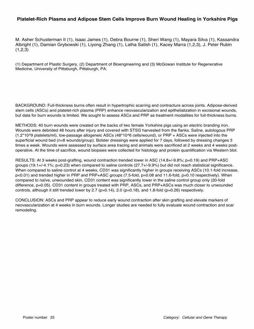
M. Asher Schusterman II (1), Isaac James (1), Debra Bourne (1), Sheri Wang (1), Mayara Silva (1), KassandraAlbright (1), Damian Grybowski (1), Liyong Zhang (1), Latha Satish (1), Kacey Marra (1,2,3), J. Peter Rubin(1,2,3)
(1) Department of Plastic Surgery, (2) Department of Bioengineering and (3) McGowan Institute for RegenerativeMedicine, University of Pittsburgh, Pittsburgh, PA.
Platelet-Rich Plasma and Adipose Stem Cells Improve Burn Wound Healing in Yorkshire Pigs
BACKGROUND: Full-thickness burns often result in hypertrophic scarring and contracture across joints. Adipose-derivedstem cells (ASCs) and platelet-rich plasma (PRP) enhance neovascularization and epithelialization in excisional wounds,but data for burn wounds is limited. We sought to assess ASCs and PRP as treatment modalities for full-thickness burns.
METHODS: 40 burn wounds were created on the backs of two female Yorkshire pigs using an electric branding iron.Wounds were debrided 48 hours after injury and covered with STSG harvested from the flanks. Saline, autologous PRP(1.2*10^9 platelets/ml), low-passage allogeneic ASCs (48*10^6 cells/wound), or PRP + ASCs were injected into thesuperficial wound bed (n=8 wounds/group). Bolster dressings were applied for 7 days, followed by dressing changes 3times a week. Wounds were assessed by surface area tracing and animals were sacrificed at 2 weeks and 4 weeks post-operative. At the time of sacrifice, wound biopsies were collected for histology and protein quantification via Western blot.
RESULTS: At 3 weeks post-grafting, wound contraction trended lower in ASC (14.8+/-9.8%; p=0.19) and PRP+ASCgroups (19.1+/-4.1%; p=0.23) when compared to saline controls (27.7+/-9.9%) but did not reach statistical significance.When compared to saline control at 4 weeks, CD31 was significantly higher in groups receiving ASCs (10.1-fold increase,p<0.01) and trended higher in PRP and PRP+ASC groups (7.5-fold, p=0.08 and 11.6-fold, p=0.10 respectively). Whencompared to naïve, unwounded skin, CD31 content was significantly lower in the saline control group only (20-folddifference, p=0.05). CD31 content in groups treated with PRP, ASCs, and PRP+ASCs was much closer to unwoundedcontrols, although it still trended lower by 2.7 (p=0.14), 2.0 (p=0.18), and 1.8-fold (p=0.26) respectively.
CONCLUSION: ASCs and PRP appear to reduce early wound contraction after skin grafting and elevate markers ofneovascularization at 4 weeks in burn wounds. Longer studies are needed to fully evaluate wound contraction and scarremodeling.
Cellular and Gene TherapyPoster number 25 Category:
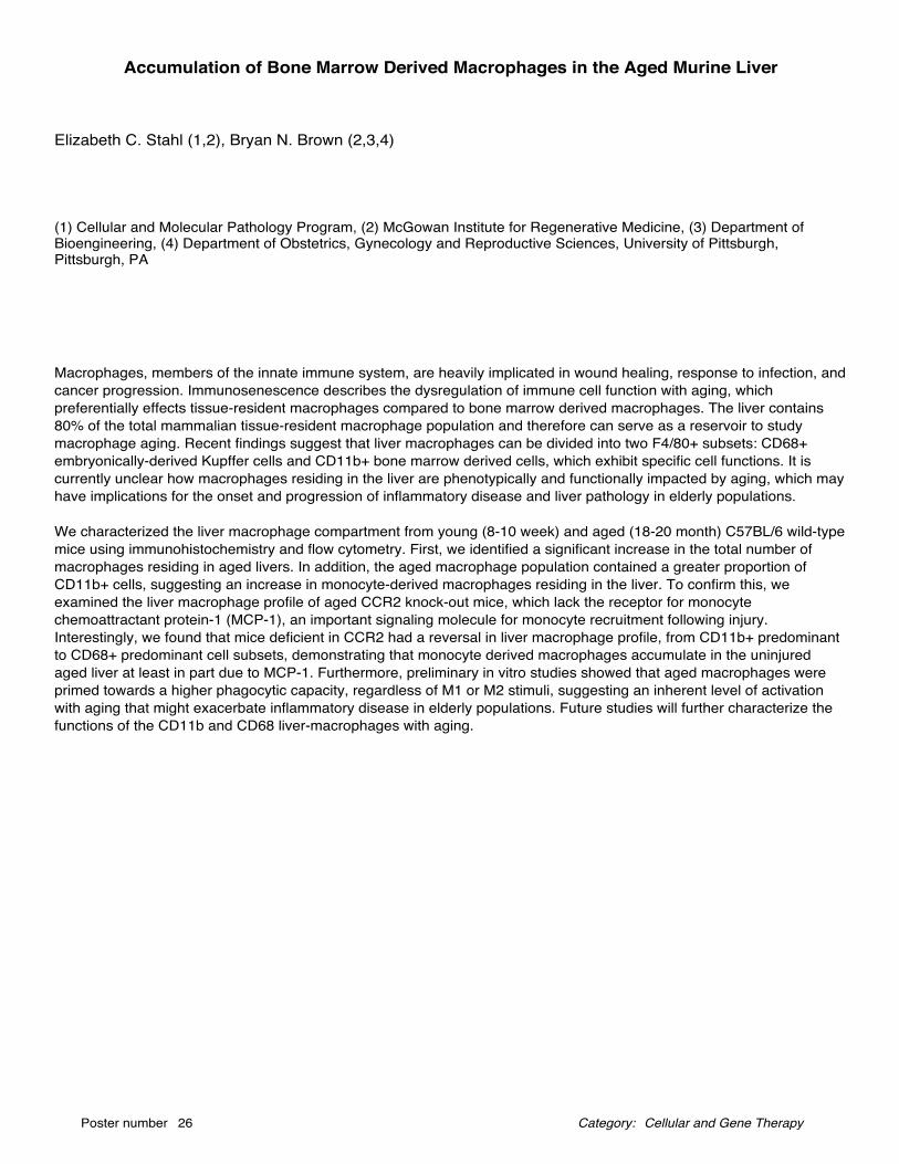
Elizabeth C. Stahl (1,2), Bryan N. Brown (2,3,4)
(1) Cellular and Molecular Pathology Program, (2) McGowan Institute for Regenerative Medicine, (3) Department ofBioengineering, (4) Department of Obstetrics, Gynecology and Reproductive Sciences, University of Pittsburgh,Pittsburgh, PA
Accumulation of Bone Marrow Derived Macrophages in the Aged Murine Liver
Macrophages, members of the innate immune system, are heavily implicated in wound healing, response to infection, andcancer progression. Immunosenescence describes the dysregulation of immune cell function with aging, whichpreferentially effects tissue-resident macrophages compared to bone marrow derived macrophages. The liver contains80% of the total mammalian tissue-resident macrophage population and therefore can serve as a reservoir to studymacrophage aging. Recent findings suggest that liver macrophages can be divided into two F4/80+ subsets: CD68+embryonically-derived Kupffer cells and CD11b+ bone marrow derived cells, which exhibit specific cell functions. It iscurrently unclear how macrophages residing in the liver are phenotypically and functionally impacted by aging, which mayhave implications for the onset and progression of inflammatory disease and liver pathology in elderly populations.
We characterized the liver macrophage compartment from young (8-10 week) and aged (18-20 month) C57BL/6 wild-typemice using immunohistochemistry and flow cytometry. First, we identified a significant increase in the total number ofmacrophages residing in aged livers. In addition, the aged macrophage population contained a greater proportion ofCD11b+ cells, suggesting an increase in monocyte-derived macrophages residing in the liver. To confirm this, weexamined the liver macrophage profile of aged CCR2 knock-out mice, which lack the receptor for monocytechemoattractant protein-1 (MCP-1), an important signaling molecule for monocyte recruitment following injury.Interestingly, we found that mice deficient in CCR2 had a reversal in liver macrophage profile, from CD11b+ predominantto CD68+ predominant cell subsets, demonstrating that monocyte derived macrophages accumulate in the uninjuredaged liver at least in part due to MCP-1. Furthermore, preliminary in vitro studies showed that aged macrophages wereprimed towards a higher phagocytic capacity, regardless of M1 or M2 stimuli, suggesting an inherent level of activationwith aging that might exacerbate inflammatory disease in elderly populations. Future studies will further characterize thefunctions of the CD11b and CD68 liver-macrophages with aging.
Cellular and Gene TherapyPoster number 26 Category:
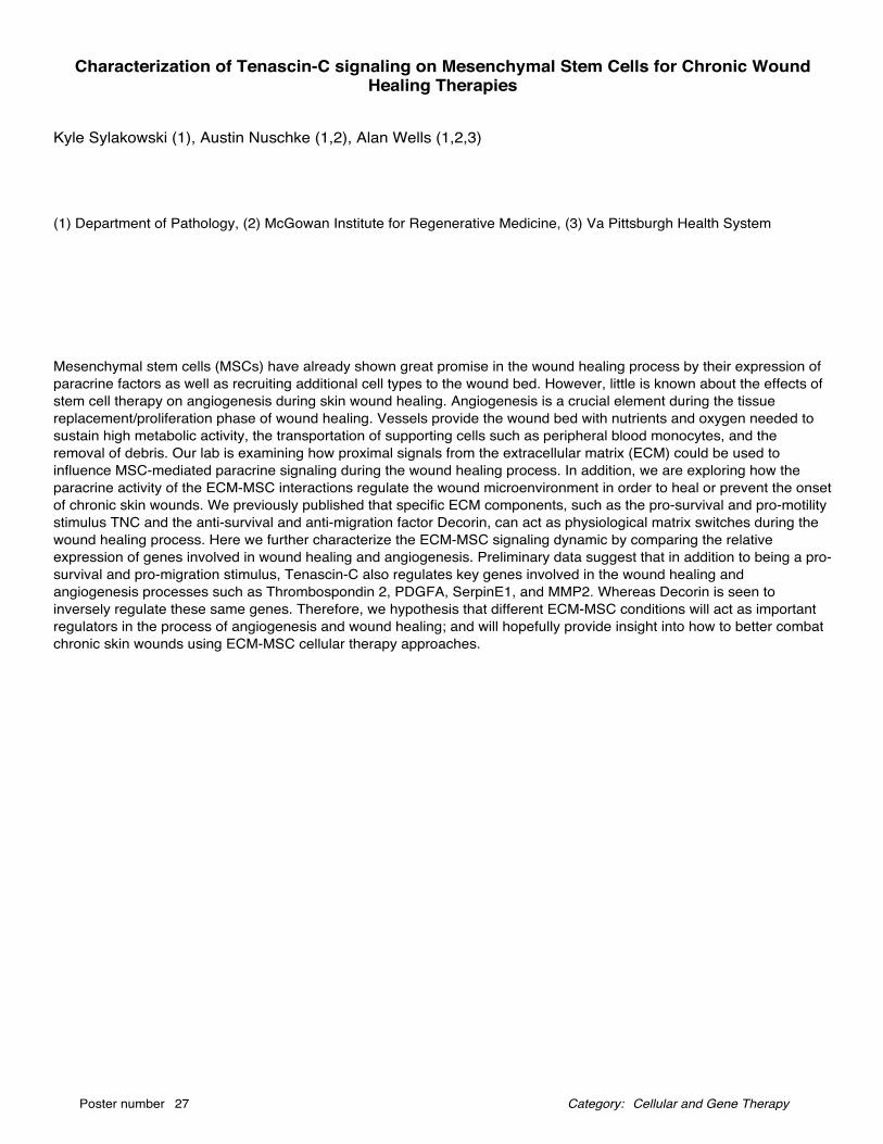
Kyle Sylakowski (1), Austin Nuschke (1,2), Alan Wells (1,2,3)
(1) Department of Pathology, (2) McGowan Institute for Regenerative Medicine, (3) Va Pittsburgh Health System
Characterization of Tenascin-C signaling on Mesenchymal Stem Cells for Chronic WoundHealing Therapies
Mesenchymal stem cells (MSCs) have already shown great promise in the wound healing process by their expression ofparacrine factors as well as recruiting additional cell types to the wound bed. However, little is known about the effects ofstem cell therapy on angiogenesis during skin wound healing. Angiogenesis is a crucial element during the tissuereplacement/proliferation phase of wound healing. Vessels provide the wound bed with nutrients and oxygen needed tosustain high metabolic activity, the transportation of supporting cells such as peripheral blood monocytes, and theremoval of debris. Our lab is examining how proximal signals from the extracellular matrix (ECM) could be used toinfluence MSC-mediated paracrine signaling during the wound healing process. In addition, we are exploring how theparacrine activity of the ECM-MSC interactions regulate the wound microenvironment in order to heal or prevent the onsetof chronic skin wounds. We previously published that specific ECM components, such as the pro-survival and pro-motilitystimulus TNC and the anti-survival and anti-migration factor Decorin, can act as physiological matrix switches during thewound healing process. Here we further characterize the ECM-MSC signaling dynamic by comparing the relativeexpression of genes involved in wound healing and angiogenesis. Preliminary data suggest that in addition to being a pro-survival and pro-migration stimulus, Tenascin-C also regulates key genes involved in the wound healing andangiogenesis processes such as Thrombospondin 2, PDGFA, SerpinE1, and MMP2. Whereas Decorin is seen toinversely regulate these same genes. Therefore, we hypothesis that different ECM-MSC conditions will act as importantregulators in the process of angiogenesis and wound healing; and will hopefully provide insight into how to better combatchronic skin wounds using ECM-MSC cellular therapy approaches.
Cellular and Gene TherapyPoster number 27 Category:
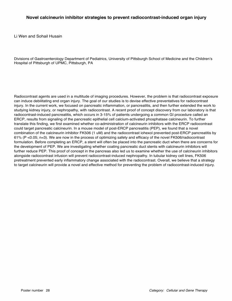
Li Wen and Sohail Husain
Divisions of Gastroenterology Department of Pediatrics, University of Pittsburgh School of Medicine and the Children’sHospital of Pittsburgh of UPMC, Pittsburgh, PA
Novel calcineurin inhibitor strategies to prevent radiocontrast-induced organ injury
Radiocontrast agents are used in a multitude of imaging procedures. However, the problem is that radiocontrast exposurecan induce debilitating end organ injury. The goal of our studies is to devise effective preventatives for radiocontrastinjury. In the current work, we focused on pancreatic inflammation, or pancreatitis, and then further extended the work tostudying kidney injury, or nephropathy, with radiocontrast. A recent proof of concept discovery from our laboratory is thatradiocontrast-induced pancreatitis, which occurs in 3-15% of patients undergoing a common GI procedure called anERCP, results from signaling of the pancreatic epithelial cell calcium-activated phosphatase calcineurin. To furthertranslate this finding, we first examined whether co-administration of calcineurin inhibitors with the ERCP radiocontrastcould target pancreatic calcineurin. In a mouse model of post-ERCP pancreatitis (PEP), we found that a novelcombination of the calcineurin inhibitor FK506 (1 uM) and the radiocontrast iohexol prevented post-ERCP pancreatitis by61% (P <0.05; n=3). We are now in the process of optimizing safety and efficacy of the novel FK506/radiocontrastformulation. Before completing an ERCP, a stent will often be placed into the pancreatic duct when there are concerns forthe development of PEP. We are investigating whether coating pancreatic duct stents with calcineurin inhibitors willfurther reduce PEP. This proof of concept in the pancreas also led us to examine whether the use of calcineurin inhibitorsalongside radiocontrast infusion will prevent radiocontrast-induced nephropathy. In tubular kidney cell lines, FK506pretreatment prevented early inflammatory change associated with the radiocontrast. Overall, we believe that a strategyto target calcineurin will provide a novel and effective method for preventing the problem of radiocontrast-induced injury.
Cellular and Gene TherapyPoster number 28 Category:
Chaoming Zhou (1), Yeal Zeldin (1), Sandeep Kathju (1,2), Latha Satish (1,2)
(1) Department of Plastic and Reconstructive Surgery, (2) McGowan Insitute for Regenerative Medicine
Pirfenidone Inhibits TGF-b1 stimulated Non-SMAD Signaling Pathways inDupuytren’s-derived fibroblasts
Background: Dupuytren’s disease (DD) is a complex fibro-proliferative disorder of the hand that is often progressive andeventually can cause contractures of the affected fingers. The increase in transforming growth factor (TGF-b1) expressionhas been implicated as a key stimulator of myofibroblast activity and palmar fascial contraction in DD. Pirfenidone (PFD),is an active small molecule with potential to inhibit TGF-b1-mediated action in fibrotic disorders. Our recent publishedfindings show that PFD reduced TGF-b1 mediated cellular functions leading to Dupuytren’s through SMAD signalingpathway. In the present study, the effect of PFD on TGF-b1 mediated non-SMAD signaling pathway was determined inboth CT- and DD-derived fibroblasts.
Materials and Methods: Fibroblasts harvested from Dupuytren’s disease (DD) and carpal tunnel (CT)-derived fibroblastswere treated with or without TGF-b1 (10 ng/ml) and/or PFD (800 μg/ml) and were subjected to Western blots analyses todetermine the phosphorylation levels of phosphatidylinositol-3 kinase (PI3K/AKT), extracellular regulated kinases(ERK1/2) and Rho family related myosin light chain (MLC).
Results: Our results show that the basal phosphorylation levels of ERK1/2, Akt and MLC were increased in DD- incomparison to CT-derived fibroblasts. Treatment with PFD led to the inhibition of both basal and TGF-b1-inducedphosphorylation of the above proteins in both CT-and DD-derived fibroblasts. PFD inhibits not only expression andactivation of downstream p-SMAD2/3 effectors but also non-SMAD signaling targets.
Discussion: These in-vitro results indicate for the first time that PFD has potential to inhibit TGF-b1 induced non-SMADsignaling pathways. Furthermore, these in vitro results suggest PFD as a promising candidate to inhibit the TGF-b1cellular signaling candidates that lead to fibrosis. Further, in vivo studies are required to determine the therapeutic efficacyof PFD in preventing the recurrence of DD.
Cellular and Gene TherapyPoster number 29 Category:
Daniel A. Zuppo (1), Maria A. Missinato (1), Manush Sayd Mohammed (1) and Michael K.W. Tsang (1)
(1) Department of Molecular Genetics and Developmental Biology, University of Pittsburgh, Pittsbugh, PA
Cardiac transcriptome profiling during regeneration in zebrafish
Heart disease is one of the leading causes of mortality worldwide. While therapeutics to ameliorate these diseases exist,they are poor at completely reversing sustained damage. Myocardial infarction represents a disease that results in a largeloss of cardiomyocytes (CMs) that results in the formation of scar tissue. In contrast to embryonic development, adultCMs are post-mitotic and do not proliferate. Understanding which genes that are involved during development thatregulate CM proliferation will offer insights into stimulating cell division in the adult heart. Zebrafish provide an excellentsystem to explore these pathways in adult CM proliferation. Previous research has revealed that zebrafish possess a highcapacity for cardiac regeneration, share considerable homology with mammalian genome, and can be manipulated easilyfor developmental/molecular studies. Current research has yielded a multitude of genes involved in cardiac regenerationwithin zebrafish, but their exact function is not completely understood. In this study, we performed RNA-seq on uninjuredand injured (3dpa) zebrafish hearts. We confirmed their induction after injury via RT-PCR. Among these genes, cenpf,errfi1, and foxm1 show high levels of expression at 3dpa and we sought to further analyze their role in heart regeneration.cenpf encodes a kinetochore-binding protein critical for mitosis, is highly expressed in developing mammalian CMs, butrapidly down-regulated 6 days after birth. foxm1 mediates G1-S/G2-M transitions, binds murine cenpf promoter andupregulates it, and has limited expression in adult CMs. errfi1 is a negative regulator of neuregulin 1 (Nrg1) signaling, andNrg1 administration was shown to induce re-entry of mammalian CMs into the cell cycle after injury. Currently, we arecharacterizing transcript localization in adult uninjured and 3dpa hearts via immunostaining with specific antibodies.Future studies will include analysis of zebrafish that harbor mutations in these genes to determine their role in CMproliferation and heart regeneration.
Cellular and Gene TherapyPoster number 30 Category:
Emily E. Ackerman (1), Jason E. Shoemaker (1,2,3)
(1) Department of Chemical and Petroleum Engineering, (2) McGowan Institute for Regenerative Medicine and (3)Department of Computational and Systems Biology, University of Pittsburgh, Pittsburgh, PA
Controllability Analysis of Protein-Protein Interaction Networks for Antiviral DrugDevelopment
Interaction networks are a tool for analyzing collections of biomolecular data, particularly protein interactions (PPInetworks) within a human host. The advantage of PPI networks lies in their ability to take a “whole-cell” approach in lieu ofrequiring detailed kinetic data to build a system of differential equations. In this method, large-scale protein interactiondata is represented in a network and analyzed by its position within the network and its interacting partners. Performing acontrollability analysis on a network can predict how the system can be driven to any desired state. In a cell undergoingviral attack, the goal of the virus is to seek control of the host machinery and carry out virus replication tasks, the “desiredstate” of infection. To better understand how the virus takes control of host functions, we completed a controllabilityanalysis of the proteins of the human PPI network in combination with Influenza A viral protein-host protein interactiondata. In the absence of proteins deemed “dispensable” by the analysis, it is easier for an external input (such as a virus)to influence and control total network behavior. We find that though 43.8% of proteins in the total human PPI network aredispensable, 61.5% of host proteins which directly interact with viral proteins are dispensable. This increased proportionsuggests that viruses are more likely to attack proteins which will make it easier to take control of the network andultimately hijack host cell function. Information about the types of proteins targeted by viruses can be used to identifyprotein candidates for further study and guide antiviral drug development.
Computation and ModelingPoster number 31 Category:
Eric Lambert (1), Tristan Ford (4), Satya VVN Kothapalli (5), Hong Chen (5) and Jelena M. Janjic (1,2,3)
(1) Graduate School of Pharmaceutical Science, Mylan School of Pharmacy and (2) Chronic Pain Research Consortium,Duquesne University, Pittsbugh, PA, (3) McGowan Research Institute for Regenerative Medicine, University of Pittsburgh,Pittsbugh, PA, (4) Department of Biomedical Engineering, University of Rochester, Rochester, NY, (5) Department ofBiomedical Engineering, Washington University, St. Louis, MS
High intensity focused ultrasound-sensitive perfluorocarbon nanoemulsions in targeted drugdelivery
Current cancer treatment advances focus on reducing systemic drug toxicity by targeting the tumor. This is accomplishedby using the enhanced permeability and retention (EPR) effect, wherein widened endothelium gaps near the tumor resultin elevated nanoparticle uptake. Stimulus-mediated drug delivery strategies take advantage of this by making use of light,pH, or other stimuli to cause drug release on demand (Rapoport 2007). Magnetic resonance imaging (MRI) -guided highintensity focused ultrasound (HIFU) has been used to induce drug delivery from nanoparticles (Zhang et al. 2016).Nanoemulsions (NEs) are suitable candidates for both (i) targeting via the EPR effect and (ii) undergoing stimulus-mediated drug delivery (Astafyeva et al. 2015; Rapoport et al. 2011). We have previously shown that perfluorocarbon(PFC) NEs are capable of selectively distributing into the tumor compared to normal tissue in a mouse tumor model(Balducci et al. 2013). Several studies demonstrate the utility of PFC NEs as participants in HIFU-mediated drug delivery(Zhang et al. 2016; Rapoport et al. 2011; Shiraishi et al. 2011) due to the 19F MRI capabilities of PFCs and acousticcavitation resulting from PFC droplets converting to gaseous microbubbles in response to HIFU. Triggered drug deliveryproduct development is limited by unstable excipients and hence short shelf life. Presented here, a careful selection of NEexcipients is conducted and statistical modeling is performed to explore what excipient attributes are critical for balancingacceptable colloidal stability with favorable drug delivery properties in the NE. It is of interest to elucidate physicochemicalproperties that explain the differences in colloidal behavior of hydrocarbon oil-based nanoemulsions and perfluorocarbonoil-based nanoemulsions. Perfluoro-15-crown-5-ether (PCE) demonstrates the most promising HIFU responsiveness, butlacks drug loading capabilities. A triphasic NE, which combines a hydrocarbon oil phase with PFC phase, allows forsimultaneous drug loading capacity and favorable drug release properties.
Computation and ModelingPoster number 32 Category:
Lisa Carey Lohmueller (1,2), Manreet Kanwar (3), Carmen Khoo (1), Robert Kormos (4), James Antaki (1,2)
(1) Department of Bioengineering, University of Pittsburgh, (2) Department of Biomedical Engineering, Carnegie MellonUniversity, (3) Cardiovascular Institute, Allegheny General Hospital, Pittsburgh, PA (4) Department of Cardiology,University of Pittsburgh Medical Center, Pittsburgh, PA
Predicting all cause mortality for LVAD patients using Bayesian Networks
The two main treatment options for end stage heart failure are heart transplant or implant of a left ventricular assist device(LVAD). Heart transplant is the gold standard, but the availability of viable hearts is limited and many patients are cannotreceive a transplant due to age or health status. LVADs allow patients to either bridge to transplant or can add years tothe life of a patient not eligible for transplant. However, LVAD implantation is a major surgery with risks for bleeding,infection, and stroke and requires a significant change in lifestyle. The goal of this work is to use pre-implant informationto calculate the risk of mortality in order to improve patient selection for LVAD implant.
Using data from Interagency Registry for Mechanically Assisted Circulatory Support (INTERMACS) we built predictivealgorithms for all-cause mortality at six timepoints (1, 3, 6, 12, 24, 36mo) using Bayesian Networks in Weka and GeNiesoftware packages. The number of patients used to construct the model for each timepoint ranged from N = 9568 to N =361. Patients were excluded at each timepoint for explant, transplant, death in a previous period, and lack of adequatefollow-up time. Models were validated using 10-fold cross validation.
Model performance ranged from c-statistic of 0.65 to 0.90 for 1mo to 36mo timepoints. Key variables that influencedoutcomes were hemodynamics, kidney and liver status for early timepoints, and then shifted to patient medical historyand quality of life for later timepoints.
This work will be used to build a personalized prognostic tool for both physicians and patients to improve decision makingfor LVAD implantation.
Computation and ModelingPoster number 33 Category:

Luke Ziegler (1,2), Salim Olia (1,2), Marina Kameneva (1,2,3)
(1) McGowan Institute for Regenerative Medicine, (2) Department of Bioengineering, (3) Department of Surgery
The Modified Mechanical Fragility Index: A New Tool for Clinical Measurement of Red BloodCell Fragility
Study: Red blood cell (RBC) susceptibility to mechanically induced hemolysis, or RBC mechanical fragility (MF), is animportant parameter in the characterization of erythrocyte membrane health. The rocker bead test and associatedcalculated mechanical fragility index (MFI) has proven to be a simple and reliable method for RBC MF measurement.While we have recently made progress towards making the rocker bead test clinically applicable by reducing the bloodvolume requirements from 15.5 ml to 6.5 ml, the MFI produces hematocrit (Ht) dependent results, making directmeasurement of blood obtained from patients impossible. In the interest of applying the reduced volume rocker bead testin the clinic, we have developed the modified mechanical fragility index (MMFI): a numerical estimation of MFI ifperformed at ‘standard’ Ht (40%). Methods: The reduced volume rocker bead test utilizes 3.0 ml plastic collection tubescontaining 2.0 ml of whole blood each. Each of two experimental tubes contains three 1/8” stainless steel beads while onecontrol tube does not, and all tubes are rocked using a platform mixer (18 cycles/min ±17° from horizontal) for one hour.MFI is calculated as a function of total hemoglobin and the difference in free hemoglobin between experimental andcontrol tubes. 96 tests were performed across hematocrit ranges of 25-50% using blood from 4 human donors. Results: Astrong non-linear relationship was observed between sample Ht and the resultant MFI. After linearization, multivariatelinear regression analysis yielded a highly correlated fit (r=0.965). The proposed correction factor accurately predicted (R2=0.923) MFI at 40% Ht from native hematocrit. With the MMFI equation, the reduced volume rocker bead test may beused to compare blood samples between 25–50% Ht without normalizing for hematocrit.
Computation and ModelingPoster number 34 Category:
Elaine Soohoo (1), Lewis K Waldman (2), Dennis R Trumble (1)
(1) Department of Biomedical Engineering, Carnegie Mellon University, (2) Insilicomed, Inc.
Computational Assessment of Design Parameters for a Torsional Ventricular Assist Device(tVAD)
PurposeThe goal of this study is to quantify changes in hemodynamic function with the application of apical torsion as a means ofcardiac assist. These results will help identify optimum parameters for the design of a torsional ventricular assist device(tVAD).
MethodsParametric computational simulations were performed using ContinuityPro software (Insilicomed, Inc., La Jolla, CA) andused to determine design parameters for a second-generation tVAD prototype. These simulations utilized beating heartmodels of both ventricles attached to a closed-loop circulatory system. Ventricular size, shape and dimensions werebased on anatomic measurements of a dysynchronous heart failure patient with dilated cardiomyopathy. Biomechanicand circulatory model parameters were taken from literature values. Varying degrees of applied apical torsion weresimulated by altering coverage areas (12.5%, 18.2% and 24%) and rotation angles (0, 45, 55, 65, and 75 degrees) todetermine the effects on global cardiac function. Results were compared to baseline hemodynamic values of the clinicalheart failure model (No VAD) before torsion was applied.
ResultsIncreasing the parameters of tVAD coverage areas and applied angles of rotation produced pronounced increases inejection fraction (EF), peak systolic pressure (PSP), and stroke work (SW) while simultaneously lowering end-systolicvolume (ESV) in both ventricles. The simulation representing the most aggressive level of tVAD assist, with a large tVADcoverage area (24%) and 75 degrees of applied rotation, produced left ventricular EF of 30.4%, PSP of 164 mmHg, ESVof 145 mL, and SW of 9145 mmHg*mL. When compared to our baseline No VAD hemodynamics - left ventricular EF of20.3%, PSP of 123 mmHg, ESV of 200 mL, and SW of 5321 mmHg*mL - we observed increasing returns of 49%, 34%,28% and 72% for EF, PSP, ESV, and SW, respectively. Large coverage and 75 degree rotation angle yielded rightventricular EF of 69.2%, PSP of 54.6 mmHg, ESV of 26.6 mL, and SW of 2673 mmHg*mL. When compared to the NoVAD hemodynamics - right ventricular EF of 43.9%, PSP of 44.3 mmHg, ESV of 64.5 mL, and SW of 1793 mmHg*mL -we observed increasing returns of 58%, 23%, 58% and 49% for EF, PSP, ESV, and SW, respectively.
ConclusionThese initial results have helped us to identify the optimal working parameters for tVAD coverage area and rotation angle.Based on these findings, we believe that applied apical torsion has the potential to help restore cardiovascularhemodynamics toward healthier values for both ventricles.
Computation and ModelingPoster number 35 Category:
Moataz M. Abdulhafez (1), Moataz M. Elsisy (1), Rob Lewis (1), Mohamed A. Zaazoue (2), Liliana C.Goumnerova (2) and Mostafa Bedewy (1)
(1) Department of Industrial Engineering, University of Pittsburgh, Pittsburgh, PA, (2) Department of Neurosurgery,Boston Children’s Hospital, Boston, MA
Lowering the risk of injury in head immobilization devices by design
Precise and firm cranium fixation is critical in craniotomy and neurosurgery, because small head motion could disrupt theoperation, cause complications, and may even lead to fatalities, especially in high risk patients. Hence, headimmobilization devices (HIDs) have become a staple instrument in neurosurgery. Despite their abundance over the pastfew decades, serious injuries are still being reported today for HIDs which motivates our detailed analysis of existing HIDdesigns. This work is a design-based approach aimed at reducing the risk of injuries arising from the use of HIDs,especially in pediatric neurosurgery.
The Mayfield head clamp is one of the earliest designs of HID and is one of the most widely used instruments today.However, complications were reported in case studies published since 1984, including skull fractures, depressed skulls,and epidural hematoma. These findings were recently supported by an FDA safety communication issued in 2016, whichreports more than 700 injuries in relation to skull clamp complications before or during surgeries in the period from 2009to 2016. Although design modification to HID were proposed, such as combining the clamp with a headrest, themechanics of pin-bone contact have not been systematically studied.
This work applies finite element modeling (FEM) techniques to analyze the mechanical loading of HID pins on the skullduring clamping. Our findings reveal a detailed understanding of the indentation caused by pushing pins against skullbone, highlighting the shortcomings of current pin designs. We simulate skull failure by exploring the effect of pin material,geometry and loading on bone deformation and stresses. Using the developed FEM model, we show the tradeoffbetween maximizing slip-resistance and compressive stresses. We also propose and evaluate new pin geometries withoptimized designs to reduce the risk of skull fracture, while adequately preventing slippage.
Medical DevicesPoster number 36 Category:
Dan Crompton (1,2), Salim E Olia (1,2), Trevor A Snyder (3), Peter D Wearden (4,5), Marina V Kameneva(1,2,4)
(1) Department of Bioengineering, University of Pittsburgh, PA, (2) McGowan Institute for Regenerative Medicine,University of Pittsburgh, PA, (3) VADovations Inc., Oklahoma City, OK, (4) Department of Surgery, University ofPittsburgh, PA, (5) Nemours Children’s Hospital, Orlando, FL
In vitro and in vivo hemocompatibility assessment of the VADovations novel pediatric leftventricle assist device (LVAD)
VADovations is developing a platform of miniaturized continuous flow ventricular assist devices (VADs), which permitexchanging 2 hydraulic components during manufacture to meet the hemodynamic requirements of different patientpopulations. In this early stage effort, adult RVAD hydraulic designs were evaluated for use as pediatric LVADs in vitroand in sheep implants at the McGowan Institute Center for Preclinical Studies. Hemolysis was measured in a mock flowloop at a flow rate of 2.0 L/min against 45-75 mmHg. Two hydraulic designs were compared using the same pumphousing and blood pool. The average blood parameters were hematocrit (29.7±0.6%), total plasma protein (7.15±0.05g/dL), and pH (7.10±0.03). The Normalized Index of Hemolysis (NIH) values for the two rotors were 0.103±0.031 and0.212±0.095 g/100L, respectively, acceptable for pediatric LVAD use.
In vivo, pumps with small variations of hydraulic designs were implanted through the apex of the left ventricle with a10mm graft anastomosed to the ascending aorta. Pump speeds were adjusted from 17,500–24,000 RPM producingaverage flows of 0.8-3.8 L/min. Four studies reached or exceeded planned duration (1-3 months), two studies wereelectively ended early due to outflow graft kinking, two studies were terminated peri-operatively, and one study due toright ventricular thrombus from an indwelling i.v. line. Plasma free hemoglobin (pfHb), total hemoglobin/hematocrit, totalplasma protein, and fibrinogen returned to baseline pre-op values by the end of the second week of the study. Post-operative hypertension produced intermittent retrograde flow in some studies, but renal infarcts were only observed in oneof 10 chronic implants.
The results demonstrated acceptable hemocompatibility as a pediatric LVAD, but presented opportunities for improvedhemodynamic and hemolysis performance. Based on these findings, three pediatric specific hydraulics have beendeveloped, Infant 0.5-1.5 L/min, Toddler 1.0-3.5 L/min, and Child 3+ L/min, which will be undergoing testing in 2017.
Medical DevicesPoster number 37 Category:

Firuz Feturi (1), Hua Wang (2), Yevgeny Brudno (2), Vasil Erbas (3), Liwei Dong (3), Zhaoxiang Zhang (3),Huseyin Sahin (3), Wensheng Zhang (3), Mubin Ali Aral (3), Raman Venkatramanan (1), David Mooney (2),Vijay Gorantla (3)
(1) Department of Pharmaceutical Science, University of Pittsburgh School of Pharmacy, Pittsburgh, PA, (2) WyssInstitute for Biologically Inspired Engineering, Harvard University, Boston, MA, (3) Department of Plastic andReconstructive Surgery, University of Pittsburgh School of Medicine, Pittsburgh, PA
Ultrasound Triggered-Release Embedded Anti-Rejection Therapy (TREAT™) for TargetedImmunomodulation in Vascularized Composite Allotransplantation
PurposeMore than 30,000 people receive organ transplants every year in the US. Vascularized composite allotransplantation(VCA) is the newest realm of solid organ transplantation. The skin component of VCA is highly antigenic and mandateshigh doses of systemic immunosuppressive drugs. Oral dosing of immunosuppressive drugs such as tacrolimus (TAC),rapamycin (Rapa), and mycophenolic acid (MPA) leads to fluctuating, erratic, or unpredictable blood levels risking toxicityor lack of efficacy . We propose a drug delivery platform that can not only provide sustained drug release but also on-cuetriggered drug release upon ultrasound stimulation (USS) in graft tissues with stable, low blood levels, minimizing overalldrug exposure and facilitating long-term VCA survival with no systemic complications.
MethodAn injectable, re-loadable, biocompatible drug eluting hydrogel was prepared. We characterized the in vitro releasekinetics of the drugs from alginate gels in absence and /or presence of USS. We evaluated feasibility and efficacy of thesystem in vivo in absence of USS. Brown Norway to Lewis rats received fully mismatched Brown Norway rat hind limbVCA (4/group) and a single dose of gel subcutaneously injected into the allograft. The gel was loaded with either TAC(10mg), Rapa (10mg), or TAC+Rapa (10mg) each in 1 ml. Drug levels in blood and VCA tissues were analyzed by LC–MS/MS. Flow cytometry was performed to detect expression of regulatory marker, FOXP3. In addition to allograft survival,systemic toxicity was evaluated using percent change in body weight (BW) and creatinine clearance (CrCL).
ResultsIn vitro, TAC and Rapa exhibited a low baseline level (without fluctuation) of release from alginate gels in the absence ofUSS. Pulsatile USS triggered drug release, leading to increased drug levels after each pulse. Sustained drug releaseoccurred from alginate gels in the absence of ultrasound with blood levels within the therapeutic range (5-10ng/ml). Drugconcentration in allograft tissues was higher than in blood and contralateral limb (P<0.05). In the first 2 weeks post gelinjection, there was a ≤15% change in BW which stabilized with time. BW gradually increased over time. No significantchange in CrCL occurred post gel injection over time (>0.05). Rats receiving Rapa developed Banff grade 3 rejection onday 21, while rats receiving TAC or TAC+Rapa showed allograft survival (>100 days). Expression of the regulatorymarker FOXP3 was observed which may indicate peripheral immunomodulation.
ConclusionWe successfully developed, for the first time, a smart hydrogel drug delivery system with sustained baseline and on-demand release of drugs upon USS for use in VCA. The TREAT ™ system provides stable, low drug levels in the bloodwith preferential drug concentration in VCA tissues facilitating long-term VCA survival/outcomes with no systemic adverseeffects. Further efforts are being made to use USS to optimize the on-cue drug release. We developeddibenzocyclooctyne (DBCO)-immunosuppressant prodrugs that can be reloaded into the azido-modified alginate gels viaClick chemistry, with the goal of refilling the alginate scaffolds with immunosuppressive drugs once the encapsulateddrugs are running out and thus maintaining the immunosuppressive effect for a long term without replacing the gelscaffolds via surgery method.
Medical DevicesPoster number 38 Category:
Garrett Jeffries (1,4), Brian Frankowski (4), William Federspiel (1,2,3,4)
(1) Department of Bioengineering, (2) Department of Chemical Engineering, (3) Department of Critical Care Medicine, (4)McGowan Institute for Regenerative Medicine, University of Pittsburgh, Pittsburgh, PA
High Efficiency Respiratory Dialysis at Ultra-Low Blood Flows
Clinical adoption of new extracorporeal carbon dioxide removal (ECCO2R) systems for management of hypercapnicrespiratory failure is hindered by invasive connections strategies and complex blood circuitry, limiting use to specializedmedical centers. We have developed a high efficiency ECCO2R device capable of treatment analogous to renalhemodialysis to close the logistical gap between respiratory and renal dialysis.
The Ultra-Low-Flow ECCO2R Device (ULFED) uses an array of rotating impellers within a gas exchange bundle togenerate an “active mixing” effect. Impeller mixing dramatically enhances gas transfer rates and fiber washing to easeanticoagulation requirements. Significant improvements in CO2 removal efficiency versus available systems enableoperation at blood flow rates comparable to renal hemodialysis (250 mL/min), facilitating potential for adoption of commondialysis cannulation strategies and use with existing dialysis circuitry.
Three ULFED prototypes (fiber surface area = 0.42 m2) were fabricated with varying bundle aspect ratios (length versuswidth) and two impeller arrangements (impeller length effects) to evaluate the effects of bundle and impeller design in theactive mixing system. CO2 removal rates up to 75 mL/min from blood were achieved during in vitro gas exchange testing(4000 impeller RPM, inlet blood pCO2 = 45 mmHg, blood flow 250 mL/min), matching performance of leading approvedECCO2R systems at half the necessary blood flow rate. Subsequent in vitro hemolysis testing in a clinical circuit(including pump and cannula) revealed with no significant difference versus a control circuit using time-of-therapynormalized rates of cell trauma (ULFED Therapeutic Index of Hemolysis (TIH) = 0.190 ± 0.041 g/100min versus ControlTIH = 0.123 ± 0.013 g/100min; p>0.05).
Performance of the ULFED matches or exceeds the abilities of all available ECCO2R devices, but does so at half theblood flow rate of leading systems, and 30% lower fiber surface area. Under these operating conditions, the ULFED couldbe connected to patients using dramatically simplified methods directly adopted from routine renal hemodialysis, withpotential to be integrated directly into existing dialysis circuitry.
Medical DevicesPoster number 39 Category:
Alexander D. Malkin (1,2), William J. Federspiel (1,2,3,4), John A. Kellum (2,4) and Kai Singbartl (5)
(1) Department of Bioengineering, (2) McGowan Institute for Regenerative Medicine, (3) Chemical Engineering, (4)Critical Care Medicine, University of Pittsburgh, (5) Critical Care Medicine, Mayo Clinic Arizona
An Extracorporeal Neutrophil Reprogramming Device for the Treatment of Acute InflammatoryConditions
Sepsis is a systemic inflammatory response due to an infection. This condition affects 900,000 Americans per year and itsincidence is expected to increase over the next 10-20 years as the population ages. As one of the first responders to sitesof inflammation, neutrophils play a critical role in innate immune response and sepsis. High quantities of neutrophilsmigrating to remote organs such as the lungs has been associated with increased morbidity and mortality by initiatingmultiple organ dysfunction or immune paralysis in septic patients. The migration and activation of neutrophils is regulatedby CXCR-1 and CXCR-2, g-protein coupled receptors that bind to interleukin-8 (IL-8).
An extracorporeal device is being developed to attenuate neutrophil chemotactic response in inflammatory conditionssuch as sepsis. This device achieves cell phenotype reprogramming by exposing neutrophils to immobilized IL-8 within anartificial microcirculation. First IL-8 was covalently immobilized on the inner lumen of amine functionalized fibers [BaxterIntl.] using a bifunctional polyethylene glycol spacer. After perfusion of human whole blood through modules constructedfrom modified fibers, neutrophil surface receptor expression was quantified using flow cytometry. In scaled devicessurface receptor expression of CXCR-1 and CXCR-2 decreased by 19% and 70% respectively when compared tobaseline blood. When hAlbumin was immobilized in place of IL-8, downregulation closely resembled baseline blood,indicating IL-8 was responsible for receptor downregulation. IL-8 ELISAs (Thermo Fisher) were completed to determinethe extent of nonspecifically bound IL-8 leaching from the modules. Tests determined IL-8 concentrations of 662 ± 31pg/ml after 1 hour 5% BSA recirculation; potentially enough to induce partial downregulation. Future tests will evaluatechemotaxis of neutrophils treated with the extracorporeal device. Beyond sepsis, a similar device platform may be appliedto alternative disease states such as ischemia reperfusion and solid organ rejection, which are regulated by cell-cellinteractions.
Medical DevicesPoster number 40 Category:
Jonquil R. Mau (1), Chak-Yuk Yu (1), Yajnesh Vedanaparti (1), Savio L-Y. Woo (1)
(1) Musculoskeletal Research Center, Department of Bioengineering, University of Pittsburgh
Corrosion Characterization of Magensium Coated Via Micro-arc Oxidation
INTRODUCTION: Devices used for orthopaedic applications have been made using inert metallic alloys such as titaniumand stainless steel as well as bioresorbable polymeric materials like poly(lactic-co-glycolic acid) (PLGA). Recently, therehas been a renewed interest in the use of magnesium (Mg). While Mg has been used experimentally for development ofbone plates and screws, it has not been used for soft tissue applications such as healing of ligaments and tendons. In ourresearch center, we have developed a Mg device to facilitate the healing of the anterior cruciate ligament (ACL). We havechosen this material because it has suitable mechanical properties, controllable degradation, and biocompatibility thatmakes it acceptable for ACL healing. Specifically, Mg is more ductile than resorbable polymeric materials and the rate ofdegradation of Mg can be controlled through surface coatings without compromising its mechanical properties. Hence, Mgcan be engineered to degrade away over time being replaced by the native tissue. Thus, the goal of this study was tocharacterize the corrosion rate of Mg with a surface coating to identify the appropriate coating time for use in the intendedapplication of ACL healing.
METHODS: Pure magnesium (99.9%) were machined into 10 mm diameter by 1 mm thick disks. The samples were thenrinsed with acetone and polished with 1200 grit SiC abrasive paper. After being polished, the samples were rinsed inmethanol and allowed to air dry. In order to apply an micro-arc oxidation (MAO)surface coating, the samples wereimmersed in an electrolytic solution (6 M Na3PO4 + 8 M Na2SiO3•5 H20 + 4 M KF). An adjustable DC pulse source wasused to apply a constant potential of 300 V through titanium wire for 2 and 5 minutes. Uncoated samples were used as acontrol. The corrosion rate was determined by in vitro dorrosion testing which involves submerging the samples intoHanks’ Balanced Salt solution and monitoring the change in mass, change in pH, and the release of Mg ions.Measurements were taken daily for 1 week and weekly for 4 weeks.
RESULTS: The uncoated samples degraded faster than the samples coated for 2 minutes and 5 minutes. After 1 week,the uncoated, 2 minute, and 5 minute samples had an increased in mass after which the change in mass decreased up to1% at 4 weeks. In terms of the pH measurements, the Hanks’ solution became more alkaline after immersion of thesamples. For the uncoated samples, there was a sharper increase in the pH value at the initial stage and an increase inpH of 3 by 4 weeks. Lastly, after measurement of the Mg ions remaining in the Hanks’, a similar trend was seen as withthe change in pH. For the uncoated, 2 minute, and 5 minute samples, there was a faster increase in Mg concentrationwithin the first week which slowed over the remaining 3 weeks with a 4 times increase by 4 weeks.
DISCUSSION: These results confirm that the Mg samples would begin to degrade when immersed in Hanks’ BalancedSalt Solution within the 4 week time period. Also, the initial increase of change in mass can be attributed to theaccumulation of corrosion products on the surface of the sample. However, as the sample continues to undergocorrosion, there was more mass loss. The trends of pH and Mg ion concentration indicate that the uncoated samplesdegraded faster than the 2 minute and 5 minute samples as expected because the MAO coating slows the corrosion atinitial time points. However, there was no significant difference for the corrosion rate between the groups. Thus, thecoating times of 2 minutes and 5 minutes would allow a device for ACL healing to have a surface favorable to cellinteractions, initially resist degradation as the ligament heals, and ultimately degrade away with the healed ligamentremaining. With these promising results, we have used MAO coated Mg in subsequent animal studies for ACL healingwith successful results.
Medical DevicesPoster number 41 Category:
Salim E. Olia (1,2,3), Timothy M. Maul (2,4), Marina V. Kameneva (1,2,5)
(1) McGowan Institute for Regenerative Medicine, UoP, (2) Department of Bioengineering, UoP, (3) Artificial HeartProgram, UPMC, (4) Nemours Children's Hospital, Orlando, FL, (5) Department of Surgery, UoP
The Effect of Inlet Pressure on Gas Embolism and Hemolysis in Continuous-Flow BloodPumps
Purpose: While there is an increased enthusiasm in the development of small, high speed, continuous-flow ventricularassist devices, in clinical use these pumps can encounter negative inlet pressures. To date, in-vitro tests have notquantified the effect of low preload on hemolysis.
Methods: Two preliminary in-vitro studies examined the effect of inlet pressure on hemolysis and gas embolism in amagnetically levitated, pediatric, centrifugal blood pump: (1) gaseous emboli exiting the pump in a saline filled mock loopwere measured with the EDAC Quantifier at variable inlet pressures (-40-10 mmHg) and speeds (2500-4000 RPM); and(2) hemolysis testing was performed at 0.5 and 1.5 L/min using a mock loop for four hours with three inlet pressures (-40-0 mmHg) while holding speed, flow and pressure drop constant.
Results: In saline, significant numbers of gas emboli (>50 μm) exited the pump at negative inlet pressures. Hemolysistesting revealed a strong correlation between the NIH and negative inlet pressure at both flow rates with foam generatedat all negative pressures. On-going studies combining the two experiments will enable the quantification of the gaseousemboli size in blood and further elucidate the effect of inlet pressure on blood trauma in continuous-flow pumps.
Medical DevicesPoster number 42 Category:
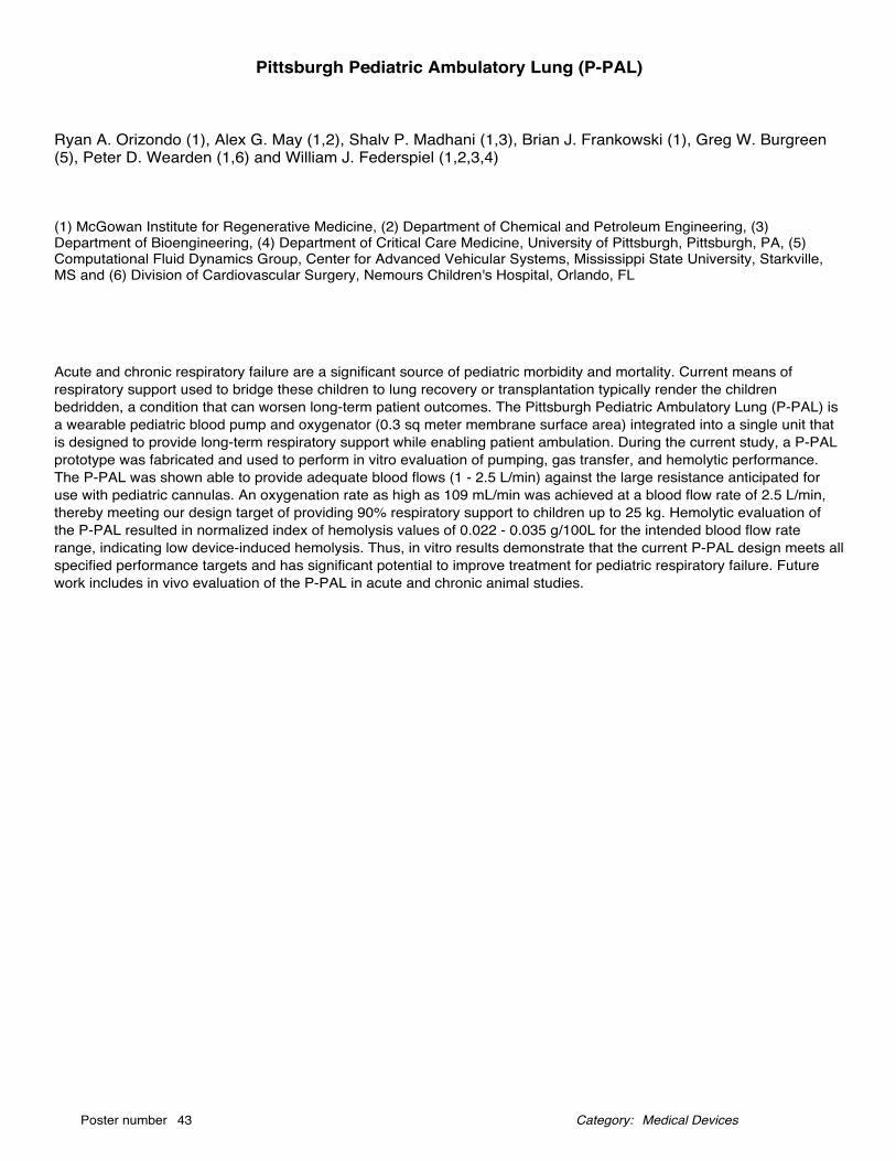
Ryan A. Orizondo (1), Alex G. May (1,2), Shalv P. Madhani (1,3), Brian J. Frankowski (1), Greg W. Burgreen(5), Peter D. Wearden (1,6) and William J. Federspiel (1,2,3,4)
(1) McGowan Institute for Regenerative Medicine, (2) Department of Chemical and Petroleum Engineering, (3)Department of Bioengineering, (4) Department of Critical Care Medicine, University of Pittsburgh, Pittsburgh, PA, (5)Computational Fluid Dynamics Group, Center for Advanced Vehicular Systems, Mississippi State University, Starkville,MS and (6) Division of Cardiovascular Surgery, Nemours Children's Hospital, Orlando, FL
Pittsburgh Pediatric Ambulatory Lung (P-PAL)
Acute and chronic respiratory failure are a significant source of pediatric morbidity and mortality. Current means ofrespiratory support used to bridge these children to lung recovery or transplantation typically render the childrenbedridden, a condition that can worsen long-term patient outcomes. The Pittsburgh Pediatric Ambulatory Lung (P-PAL) isa wearable pediatric blood pump and oxygenator (0.3 sq meter membrane surface area) integrated into a single unit thatis designed to provide long-term respiratory support while enabling patient ambulation. During the current study, a P-PALprototype was fabricated and used to perform in vitro evaluation of pumping, gas transfer, and hemolytic performance.The P-PAL was shown able to provide adequate blood flows (1 - 2.5 L/min) against the large resistance anticipated foruse with pediatric cannulas. An oxygenation rate as high as 109 mL/min was achieved at a blood flow rate of 2.5 L/min,thereby meeting our design target of providing 90% respiratory support to children up to 25 kg. Hemolytic evaluation ofthe P-PAL resulted in normalized index of hemolysis values of 0.022 - 0.035 g/100L for the intended blood flow raterange, indicating low device-induced hemolysis. Thus, in vitro results demonstrate that the current P-PAL design meets allspecified performance targets and has significant potential to improve treatment for pediatric respiratory failure. Futurework includes in vivo evaluation of the P-PAL in acute and chronic animal studies.
Medical DevicesPoster number 43 Category:
Akhil Patel (1), Samer Zaky (2,3), Elia Beniash (2,3,4,5), Charles Sfeir (2,3,4,5), Hongshuai Li (5), SachinVelankar (6), Yadong Wang (4,5,6), Shilpa Sant (1,4,5)
(1) Department of Pharmaceutical Sciences, School of Pharmacy, (2) Center for Craniofacial Regeneration, (3)Department of Oral Biology, (4) Department of Bioengineering (5) McGowan Institute for Regenerative Medicine, (6)Chemical/Petroleum Engineering, University of Pittsburgh; Pittsburgh, PA
RegenMatrix: Collagen-mimetic Bioactive Hydrogels for Growth Factor Free Approach forBone Regeneration
Bone and teeth are two major hard tissues in human body. These hard tissues are formed by a natural process of mineraldeposition called biomineralization. In this process, collagen acts as an organic template to guide mineral deposition boneextracellular matrix (ECM) and apatite minerals act as inorganic phase, both of which interact at molecular level forminghierarchical nanocomposites. However, most of the currently explored materials are just physical blends of organic andinorganic phases. In this study, we focused on re-creating biomimetic regenerative microenvironment for guidingbiomineralization and bone formation by employing fibrous hydrogel scaffolds (RegenMatrix) with collagen-mimetic multi-scale hierarchy and mineral sequestration. We assessed their potential for biomineralization in vitro and in vivo.
Two different RegenMatrix were fabricated from pairs of oppositely charged natural polymers. In vitro mineralization insimulated body fluid was assessed by fourier transform infrared spectroscopy (FTIR) and x-ray diffraction (XRD),scanning electron microscopy (SEM) and transmission electron microscopy (TEM). In vivo mineralization potential ofRegenMatrix was assessed in a critical sized mouse calvarial defect model. Regenerated bone was quantified as bonevolume using micro computed tomography (µCT). Histology was employed to assess progenitor and other native cellinfiltration in RegenMatrix and the injury site.
Both RegenMatrix showed biomimetic mineralization by promoting amorphous calcium phosphate inside the matrix andapatite minerals on the surface when tested in vitro in SBF. RegenMatrix was proven to be safe in mice calvarial defectmodel. A quantitative comparison of regenerated bone volume from µCT analysis showed significantly higher boneregeneration with one of the two RegenMatrix compared to empty defect group. It is to our knowledge, the first materialthat could promote osteogenesis without any growth factors and exogenous synthetic minerals. Moreover, µCT imagesshow evidence of guided bone regeneration indicated by the directionality of the regenerated bone with one of the twoRegenMatrix.
Medical DevicesPoster number 44 Category:
Nathaniel T. Weberding (1), Richard A. Saladino (1), M. Beth Minnigh (2), Patrick J. Oberly (2), Samuel M.Poloyac (2), Mioara D. Manole (1)
(1) Department of Pediatrics, Division of Pediatric Emergency Medicine, University of Pittsburgh School of Medicine,Children’s Hospital of Pittsburgh, (2) University of Pittsburgh School of Pharmacy
Traditional method small volume drug delivery method fails to provide a therapeutic dose insmall infants with supraventricular tachycardia
Study Objective: Adenosine administration using a traditional three-way stopcock mechanism is the recommendedtreatment for pediatric patients with acute supraventricular tachycardia. Recent reports suggest that many infants do notrespond to the first dose of adenosine as recommended by the American Heart Association. We observed that a portionof the adenosine remains in the stopcock and is not delivered to the patient. Our aim was to determine whetheradministration of adenosine using a stopcock delivers lower than expected drug doses in patients weighing less than 10kg.
Methods: We developed an in vitro model of adenosine delivery. Doses of adenosine corresponding to weights 2-25 kgwere calculated using a dose of 0.1 mg/kg and administered through one port of a stopcock. Water was administeredthrough the second port. The adenosine concentration of the output was measured using mass spectrometry and resultswere confirmed using spectrophotometry of Evans blue.
Results: The mean doses of adenosine delivered through the stopcock increased as weights increased. The mean dosesdelivered were 0.07±0.01, 0.09±0.01, and 0.1±0.01 mg/kg for weights 2-5, 6-9, and 10-25 kg, respectively (p<0.05 for 2-5vs. 10-25 kg). The percentage of the intended adenosine doses delivered via the stopcock method were 67±10%, 87±8%and 98±15%, for weights 2-5, 6-9 and 10-25 kg, respectively. Similar results were obtained using spectrophotometry.
Conclusion: Administration of adenosine via a stopcock delivers doses lower than intended in infants, which may accountfor decreased responsiveness of infants to the first dose of adenosine. Engineering of a delivery system for small volumemedications that mitigates a finite volume of “drug left behind” may improve the accuracy of the intended dose. Such asmall volume drug delivery mechanism may then improve patient responsiveness to the initial dose of a medication.
Medical DevicesPoster number 45 Category:
Puneeth Shridhar (1), Yanfei Chen (2), Stephen Emery (3), Stephanie Greene (4), Youngjae Chun (1,2,5)
(1) Department of Bioengineering, (2) Department of Industrial Engineering, (3) Department of Obstetrics andGynecology, (4) Department of Neurosurgery (5) McGowan Institute for Regenerative Medicine
Ventriculo Amniotic Shunt for Fetal Aqueductal Stenosis
Neonatal hydrocephalus due to aqueductal stenosis is a debilitating disease characterized by an excessive accumulationof fluid within the brain. Current treatment consists of a postnatal shunt, but this does not prevent poor neurologicaloutcomes. VASFAS, a prenatal shunt which can be placed during pregnancy to prevent the buildup of pressure from fluidin the brain, may drastically decrease the morbidity, mortality and cost of overall care of these children. Fetal aqueductalstenosis affects an estimated 25-60,000 infants annually on a global basis. The application of our device can be widenedto treat conditions such as lower urinary tract obstruction and pressure hydrothorax, both of which are of similar incidenceto aqueductal stenosis.
Medical DevicesPoster number 46 Category:
Jingyao Wu (1,8), Boeun Lee (1), Abhijit Roy (1,8), Tianbing Yang (2), Patricia Hebda (2,8), Sandeep Kathju(2,8), Thomas Gilbert (6), David Chi (7), Prashant N. Kumta (1,3,4,5,7,8)
(1) Department of Bioengineering, (2) Department of Plastic Surgery, (3) Department of Chemical and PetroleumEngineering, (4) Department of Mechanical Engineering and Materials Science, University of Pittsburgh, Pittsburgh, PA,(5) Center for Complex Engineered Multifunctional Materials, University of Pittsburgh, Pittsburgh, PA, (6) ACell, LLC,Columbus, Maryland, (7) Children's Hospital of Pittsburgh, University of Pittsburgh School of Medicine, (8) McGowanInstitute of Regenerative Medicine, University of Pittsburgh School of Medicine
Application of novel ultrahigh ductility (UHD) magnesium stents for the treatment of airwayobstruction
Airway obstruction is rare, but could be extremely difficult and challenging to handle. Standard treatment is open surgery.However, these reconstructive surgical procedures are usually associated with serious complications. Stent basedinterventional pulmonology has raised great interest by providing immediate relief with reduced morbidity and risk. Due tothe permanent nature of current non-degradable stents, no stent provides satisfactory long-term effectiveness. Abiodegradable stent could potentially solve this problem. In this study, we have developed proprietary ultrahigh ductility(UHD) magnesium based absorbable stents and evaluated the biocompatibility and in vivo degradation in rat airwaybypass model and rabbit tracheal model. First generation magnesium stents were made from commercially obtained pureMg and AZ31 alloys and Mg-Y alloys with an outer diameter of 2.25mm, inner diameter of 1.25mm and length of 5mm.Donor female Lewis rats were sacrificed and the tracheas harvested to provide a bypass graft for stent evaluation. Thestent was placed intra-luminally in the donor trachea, which was anastomosed to recipient rats in and end to side fashion.The animals were euthanized at 1, 8, 16, and 24 weeks for post evaluation by µCT and standard histologic analysis.Second generation stents were machined from ERC-P6 alloy based on the in vivo results of the first generation stents.The size of the stents was scaled up to fit the trachea size of rabbit. Stents were directly delivered inside the trachea ofrabbits without a bypass. Animals were euthanized at 4 weeks for post evaluation by endoscopy and standard histologicalanalysis. First generation stents showed the potential of Mg alloys for use as airway stents. A ciliated epithelium wasmaintained even after 24 weeks in vivo. The in vivo corrosion results showed that Mg-Y alloys appeared to looseconfiguration at 24 weeks. For the second generation stents, novel ERC-P6 alloys showed excellent cyto-compatibilitiesin vitro. Airway lumen was well maintained after 4 weeks of implantation as shown in the endoscopy image. Mg stentswere covered by degradation product; on top of which healthy epithelium layers were observed. Results of these studieswill be presented and discussed.
Medical DevicesPoster number 47 Category:
Vesselin Shanov (1), Guangqi Zhang (2), Pravahan Salunke (2), Vibhor Chaswal (2), Savio Woo (3), CharlesSfeir (4), Prashant Kumta (4,5), Sergey Yarmolenko (6), Boyce Collins (7), Yeoheung Yun (8), Mark Schulz(2), Zhangzhang Yin (2), Zhongyun Dong (9), William Heineman (10), Sarah Pixley (11), Maren Pink (12)
(1) Dept. of Biomedical, Chemical and Environmental Eng., Univ. of Cincinnati, (2) Dept. of Mechanical and MaterialsScience Eng., Univ. of Cincinnati, (3) Musculoskeletal Research Center, Univ. of Pittsburgh, (4) School of DentalMedicine, Univ. of Pittsburgh, (5) Swanson School of Eng., Univ. of Pittsburgh, (6) Center for Advanced Materials andSmart Structures, NC A&T State Univ., (7) Dept. of Mechanical and Chemical Eng., NC A&T State Univ., (8) Dept. ofBioEng., NC A&T State Univ., (9) Internal Medicine, Hematology-Oncology Division, Univ. of Cincinnati, (10) Dept. ofChemistry, Univ. of Cincinnati, (11) Dept. of Molecular and Cellular Physiology, Univ. of Cincinnati, (12) Dept. ofChemistry, Indiana Univ.
Magnesium Single Crystals for Medical Implant Applications
Magnesium is a promising material for orthopedic implants due to its biocompatibility and the ability to resorb in body.Single crystal of Mg is expected to reveal absence of grain boundaries which will result in high strength, non-catastrophicfailures, high purity and increased corrosion resistance. Mg single crystals were grown by Bridgman method. The singlecrystal structure was confirmed by Laue XRD, pole diffraction figures, X-Ray tomography, electron backscatter diffraction,metallography, and synchrotron characterization at the Argonne National Labs. Magnesium single crystals revealedmechanical properties close to those of the natural bone with an ultimate tensile strength of 60-70 MPa and a remarkabletendency for higher ductility of 50-60%. Thanks to the absence of grain boundaries and the applied proprietary nanometerthin coating, the corrosion of Mg single crystal was controlled and significantly suppressed in vitro and in vivo. Orthopedicdevices were machined from Mg single crystals and tested in 3 animal models such as mouse, rabbit and goat. Implantedin a rabbit ulna fracture model, single crystal Mg plates & screws showed promising Mg resorption and bone overgrowtharound the device. Anterior Cruciate Ligament (ACL) rings implanted in goats successfully repaired the damagedligament and have fully degraded in 12 weeks. No abnormal inflammatory response was observed. The conductedexperiments confirmed that Mg single crystal is a promising material for biodegradable implant applications. Currentefforts are reported on fabricating stents from Mg single crystal by EDM machining and laser cutting.
Medical DevicesPoster number 48 Category:
Qahtan AlQahtani (1,2), Samer Zaky (1,2), Herbert Ray (3,4) and Charles Sfeir (1,2,3)
(1) Department of Oral Biology, 598 Salk Hall, 3501 Terrace Street, Pittsburgh, PA, (2) The Center for CraniofacialRegeneration, 598 Salk Hall, 3501 Terrace Street, Pittsburgh, PA, (3) The McGowan Institute for Regenerative Medicine,Suite 300, 450 Technology Drive, Pittsburgh, PA, (4) Department of Endodontics, 364 Salk Hall, 3501 Terrace Street,Pittsburgh, PA
Dental Pulp Derived Extracellular Matrix as a Scaffold for Pulp Regenerative Therapy
In the theme of dental pulp regeneration, two main concepts have emerged: cell-based and cell-free approaches. Theseare the current models for pulp regenerative treatment. While attempts have been made using cell-based approachutilizing cells from dental populations alone or combined with a scaffold as a carrier. The cell-free approach offers minimalmanipulation and eliminates the need for cells to be harvested from the host, since it depends on the scaffolds ability tosupport cellular infiltration and proliferation. As scaffolds gained attention in this field, biologically inspired and syntheticscaffolds were developed to mimic the natural environment of dental pulp. The extracellular matrix (ECM) represents anatural scaffold material resembling the native tissue chemical and mechanical components. In the past few years, ECM-based scaffolds showed promising results in terms of recruiting progenitor cells, promoting constructive remodeling andmodulation of host response. The aforementioned properties make ECM-based scaffolds an ideal candidate for pulpregenerative therapy.
Development of strategies for clinically relevant tissue engineering using dental pulp extracellular matrix (DP-ECM) canprovide an alternative to conventional root canal treatment. This alternative could serve our goal to provide a suitableenvironment for pulp regeneration. In this work, we successfully decellularized ECM derived from porcine dental pulp. Theresulting scaffold was characterized using immunostaining for extracellular proteins. To determine the effect of thedecellularization process on growth factors, we performed ELISA for TGF beta, VEGF and bFGF. Furthermore, the matrixwas implanted orthotopically for 8 weeks in canine model. In-vivo, the ECM supported cellular infiltration and remodelingof the scaffold. The resulting tissue had higher expression of DSP and CD31 when compared to control. Herein, wevalidated the concept of using ECM based scaffolds for pulp regeneration. To fully investigate the ECM potential inendodontics, development of clinical protocols and further work is needed.
Tissue EngineeringPoster number 49 Category:
Zhaoxiang Zhang (1), Ali Mubin Aral (1), Keewon Lee (2), Xiaozhou Fan (3), Kang Kim (2,3,7), Mario G. Solari(1), Vijay S. Gorantla (1), Yadong Wang (2,4,5,6)
(1) Department of Plastic Surgery, (2) Department of Bioengineering, (3) Center for Ultrasound Molecular Imaging andTherapeutics, Department of Medicine and Heart and Vascular Institute, (4) Departments of Surgery, Chemical andPetroleum Engineering, Mechanical Engineering and Material Science, (5) Vascular Medicine Institute, (6) Clinial &Translational Science Institute, (7) McGowan Institute of Regenerative Medicine, University of Pittsburgh and UPMC,Pittsburgh, PA
Arterial remodeling in autologous vein and cell-free, fast-degrading vascular grafts using a ratcarotid artery interposition model
BackgroundThe clinical need for vascular grafts is increasing with the high incidence of cardiac and peripheral vascular diseases.Autologous grafts remain as a gold standard, but are not always feasible and need an additional surgery to obtain. Wehave developed cell-free, biodegradable vascular grafts, which have demonstrated accelerated cell infiltration and hostremodeling in a rat carotid artery interposition model.
MethodsStudy included 2 groups. Group 1 (n=21) consisted of vascular grafts and group 2 (n=18) consisted of vein grafts. The leftcommon carotid artery of 2-month old male Sprague-Dawley rats (body weight = 225-250 g) was exposed, clamped, andtransected. In group 1 bi-layered composite vascular grafts with a fast-degrading elastomer, polyglycerol sebacate (PGS)core and an electrospun polycaprolactone (PCL) outer sheath grafts were used. In group 2 left external jugular vein washarvested (10mm long). Both synthetic and naïve vein grafts (10 mm long) were interpositioned by end-to-endanastomosis with 10-0 suture. Graft patency was checked with flow Doppler at 0, 5, 10, 30 min after anastomosis andultrasound scanning was done at 2, 4, 8, 12-week post-implantation. Grafts were explanted at 2-week (n=7 for synthetic,n=6 for vein), 4-week (n=7 for synthetic, n=6 for vein) and 12-week (n=6 for both) post-implantation. Histology andimmunofluorescence staining was done.
ResultsOverall patency of composite and vein grafts were 90.5 and 100%, respectively. Ultrasound scanning confirmedunobstructed lumen and blood flow in both grafts without narrowing and dilation. Histology and immunofluorescencestaining demonstrated artery-like structures: endothelial monolayer in lumen and organized smooth muscle cells in mediallayer.
ConclusionOur results showed successful neoartery remodeling in the cell-free, fast-degrading vascular grafts following rat carotidartery interposition. Our vascular graft can be ideally suited for small-diameter arterial regeneration.
Tissue EngineeringPoster number 50 Category:
Catalina Ardila (1), David Maestas (2), Victoria Lundine (2), Marvin Slepian (3,4,5,6), David Harris (7),Jonathan Vande Geest (1,8,9)
(1) Bioengineering, University of Pittsburgh, (2) Biomedical Engineering, University of Arizona, (3) InterventionalCardiology, University of Ariona, (4) Sarver Heart Center, University of Arizona, (5) SACABI, The Arizona Center forAccelerated BioMedical Innovation, (6) BIO5 Institute, University of Arizona, (7) Department of Immunobiology, Universityof Arizona, (8) McGowan Institute for regenerative medicine, (9) Vascular Medicine Institute, University of Pittsburgh
Surface Modification of Electrospun Gelatin/Fibrinogen Scaffolds to Encourage EndothelialCell Function
Surface modification of biomaterials is a popular method in tissue engineering for increasing the biocompatibility of apolymer intended for graft design. In the case of tissue engineered vascular grafts (TEVG), they need to have the abilityto promote the formation of a healthy endothelial cell monolayer in the inner lumen of the graft. In this study,gelatin/fibrinogen and gelatin/fibrinogen/PCL scaffolds were electrospun to create flat scaffolds. In order to increase thebiocompatibility of these scaffolds with endothelial cells, the surface of each sample was modified using a thermoformingprocess and then coated with a blend of collagen IV and fibronectin. Human umbilical cord blood derived endothelial cells(hCB-ECs) were seeded onto the electrospun grafts and the important characteristics of a healthy endothelium wereevaluated under static conditions. According to our findings, electrospun scaffolds composed of 50% PCL, 40% gelatin,and 10% fibrinogen, that were surface modified by thermoforming and coating with a mixture of collagen IV andfibronectin, are the most suitable for endothelial cell growth. hCB-ECs growing on these constructs have the ability toproliferate and form a stable monolayer on the surface of the planar scaffold, produce eNOS, respond to the addition of IL-1β by the upregulation of VCAM-1 and ICAM-1, and reduce platelet deposition and activation rate.
Tissue EngineeringPoster number 51 Category:
Travis Armiger (1), Kris Dahl (1,2)
(1) Department of Chemical Engineering, Carnegie Mellon University, Pittsburgh, PA, (2) Department of BiomedicalEngineering, Carnegie Mellon University, Pittsburgh, PA
Measuring Changes in Force Generation in Cell Monolayers via Intranuclear Kinetics
Intracellular force generation is critical to a cell’s function particularly in areas such as wound healing, and embryonicdevelopment. We have developed a technique, which uses the nucleus as a force sensor within cells to determinechanges in intracellular force propagation within cell monolayers. This technique is termed Sensors from IntraNuclearKinetics (SINK), in which fluorescent, chromatin bound, proteins within the nucleus are tracked over time and meansquare displacement of these particles is calculated. Upon fitting these displacements to a power law equation, we showthat the exponent serves as a relative measure of intracellular force. Here we demonstrate this by decreasing cellularforce generation, with the chemical Y-27632, and by physical decoupling of the nucleus from the cytoskeleton. In bothcases we see a reduction in intranuclear motion, compared to control cells. Next, we investigate the changes in forcepropagation through cells in a monolayer, compared to isolated cells, which have different cytoskeletal structures. Then,we investigate the extent that monolayer cells respond to the stiffness of their underlying substrate, and the extent thatthis propagates to the cell nucleus. Finally, we investigate the role that a mechanically compromised cell in a monolayer,has on its surrounding cells. This demonstrates the power of the SINK technique for investigating force propagationthrough cell monolayers as it relates to diseases, which often stem from point defects in a monolayer.
Tissue EngineeringPoster number 52 Category:
Andrew Bradshaw (1), Jelena Grahovac (1,2), Brian L. Hood (1), Linda G. Griffith (3), Alan Wells (1)
(1) Department of Pathology, University of Pittsburgh, and Pittsburgh VA Healthcare System, Pittsburgh, PA, (2) Edwin L.Steele Laboratory for Tumor Biology, Massachusetts General Hospital, Boston, MA, (3) Department of BiologicalEngineering, Massachusetts Institute of Technology, Boston, MA
Modeling Metastasis from Invasion to Colonization on a Human Physiomimetic Chip
Melanoma progression from curable primary tumors to distant metastases heralds morbidity and mortality. The transitorystate involves invasion from the epidermis through the dermis and dissemination to metastatic sites, where generallyincurable micrometastases form. The newly seeded tumor clusters hone to canonical sites, namely liver (48% of cases,median survival [ms] = 4 mo.), brain (29%, ms = 4 mo.), and bone (23%), with an overall year fatality rate of 85%, [ms <12months mo.]. Metastatic melanoma (MM) is extremely aggressive due to clinical resistance to a spectrum ofchemotherapeutic, small molecule, and antibody targeted therapeutics. Effective treatments for MM remain elusive due toa lack of vital information regarding early seeding of MM, dormancy, and re-emergence at the site of metastasis. Severalstudies have partially elucidated mechanisms for MM progression using 2D models and xenograft animal models. Despitethese advances, these models are limited in recapitulating the microenvironments in primary and distant sites, and invariations among species. We use an all-human organotypic skin organ culture system (SOCs) and human livermicrophysiological system (MPS-LiverChip) to model early MM and key mechanisms for MM driven remodeling at distantsites. Previous data has implicated Tenascin-C (TNC), an extracellular matrix protein present in development and woundhealing, as a key driver of MM invasion. TNC limits cell death through pro-survival signaling from epidermal-like growthfactor (EGF-like) repeats present in the N-terminal domain. Accordingly, aberrant re-expression of TNC in MM isassociated with poor clinical outcome. Preliminary data indicates that MM is capable of integrating with hepatocytes andnon-parenchymal cells in the LiverChip, in both 2D and 3D culture. It is hypothesized that MM-TNC both “protects” MM innewly established metastatic sites and reciprocal interactions between MM-TNC and innate immune cells altersstroma/endothelial cell function to establish new tumor microenvironments
Tissue EngineeringPoster number 53 Category:
Laura T. Beringer (1), Shaohua Li (1), Ethan J. Kallick (2), Kelly J. Shields (3), Erin M. Faight (3), FrancisCartieri (2), Ariel Aballay (4), Howard Edington (2) and Saadyah Averick (1)
(1) Neuroscience Disruptive Research Lab, Allegheny Health Network Research Institute, Pittsbugh, PA, (2) Departmentof Surgery, Allegheny Health Network, Pittsbugh, PA, (3) Lupus Center of Excellence, Autoimmunity Institute, Departmentof Medicine, Allegheny Health Network, PA, (4) West Penn Burn Center, Allegheny Health Network, Pittsbugh, PA
Accelerating Wound Healing with a Collagen VI - Heparin Sulfate Coated Matrix
Treating burn related injuries remains challenging due to donor site morbidity, bacterial infection, and major scarring.Recent advances have led to the development of promising natural and synthetic polymer scaffolds, which aim toregenerate skin using a “top-down” approach in which the top layers of the skin populated by fibroblasts and keratinocytesare regenerated first. An often overlooked component of the integumentary system is the subcutaneous adipose tissue,which has been implicated in a variety of activities, including wound healing. We have created a novel coating composedof collagen VI (col VI) and heparin sulfate (HS) that promotes adipocyte attachment and adipogenesis at a ratesignificantly faster at both early and late timepoints (5 and 23 days) compared to a non-coated control. Additionally,Vicryl®, a biodegradable surgical implant mesh, was coated with the optimized col VI-HS ratio. Coated Vicryl® had anincreased number of attached adipocytes with more pronounced growth when compared to uncoated Vicryl ®. Thesefindings suggest that the combination of optimized col VI-HS coating and the fibrous woven Vicryl® may have excellentpotential in developing skin graft applications using a paradigm-shifting ‘bottom-up’ approach. The next stage of thisresearch will move beyond synthetic woven mesh and combine natural scaffolding materials with Collagen-Heparincoating to create a bio-absorbable felt-like adipomesh that mimics the extracellular matrix of dermal tissue.
Tissue EngineeringPoster number 54 Category:
Patrick G. Chan (1), Alex Hall (2), Marie Billaud (1), Amrinder Nain (2), Julie Phillippi (1), Thomas G. Gleason(1)
(1) Department of Cardiothoracic Surgery, University of Pittsburgh, Pittsbugh, PA, (2) Department of MechanicalEngineering, Virginia Tech, Blacksburg, VA
Measuring SMC Death and Adhesion Forces Via Tissue Engineered Models Simulating ECMMicroarchitecture
Introduction: Bicuspid aortic valve (BAV) is the most common congenital cardiac malformation and is associated withaneurysmal disease which predisposes patients to aortic catastrophe. (1) Our prior studies have uncovered collagenremodeling in extracellular matrix (ECM) and decreased oxidative stress defense of the smooth muscle cells in aorticspecimens from BAV patients compared to specimens from patients with the morphologically normal tricuspid aortic valve(TAV). (2, 3) These alternations in ECM and SMC biology contribute to cystic medial degeneration (CMD) in aneurysmaldisease, which are highlighted by cellular death and impaired cellular adhesion. To measure these two key components,we employ two tissue-engineered models to elucidate the interplay between the ROS-induced SMC changes and ECM.
Methods: Ascending aortic SMCs were harvested using previously established protocols. SMC populations isolated fromBAV (n=9) and TAV (n=10) patients were seeded at a density of 5.0x10³/well in a 96 well plate on tissue culturepolystyrene (TCP) in the presence or absence of type 1 collagen substrate. Once 80-100% confluent, each well wastreated with 0-4800µM of tert-butyl hydroperoxide (tBHP). The cells were stained with propidium iodide and read on aplate reader for cellular death. TAV SMCs (n=3) were seeded at 35µL of a 1.0x10⁴ cells/mL onto polystyrene nanofiberscaffolds. After cells are allowed to attach onto the fibers, cells are visualized unto 20x magnification. A 20-30 minute timelapse is taken of each cell that fit the criteria, with pictures taken at 1-minute intervals. Fiber deflection is measured usinga custom MAT LAB program. (4)
Results: At 4800µM tBHP, there was 86.7% BAV SMC death compared to 77.7% TAV SMC, p=.048. When seeded oncollagen coated plates, TAV SMCs have significantly increased survival compared to BAV SMCs when treated with tBHPat 1200, 2400 and 4800µM, (57.9% vs 79.2%, p=.036; 59.4% vs. 78.1%, p=.01; 67.5% vs 87.5%, p=.001, respectively) .The average adhesion forces to the STEP fibers of the SMCs were found to be 12.4±2.4, 14.5±1.4, and 12.9±0.8 nN. Theaverage IO force for the three cell populations was 12.9±1.0 nN. The calculated IO force in cells exposed to hydrogenperoxide (500µM) was decreased by about one-half that of SMCs under normal culture conditions (5.6±0.7 nN vs. 12.9±1.0 nN, p< 0.01).
Conclusion: The results of this study suggests that oxidative affects SMC interactions with the substrate, likely via integrinmediators. With these models, we can modulate the orientation and anisotropy of the collagen substrates via processeslike macromolecular crowding to simulate ECM of BAV. Also, nanofiber scaffolds can also reproduce the orientation seenin the ECM of BAV. These models will allow us to further investigate ROS effects on ECM-cell interactions in futurestudies.
References1. Keane MG, Wiegers SE, Plappert T, Pochettino A, Bavaria JE, Sutton MG. Bicuspid aortic valves are associated withaortic dilatation out of proportion to coexistent valvular lesions. Circulation. 2000;102(19 Suppl 3):Iii35-9.2. Phillippi JA, Klyachko EA, Kenny JPt, Eskay MA, Gorman RC, Gleason TG. Basal and oxidative stress-inducedexpression of metallothionein is decreased in ascending aortic aneurysms of bicuspid aortic valve patients. Circulation.2009;119(18):2498-506.3. Tsamis A, Phillippi JA, Koch RG, Chan PG, Krawiec JT, D'Amore A, et al. Extracellular matrix fiber microarchitecture isregion-specific in bicuspid aortic valve-associated ascending aortopathy. The Journal of thoracic and cardiovascular
Tissue EngineeringPoster number 55 Category:
Jingming Chen, Amalie E. Donius, Juan M. Taboas
University of Pittsburgh
Hydrogel Composition Regulates Chondrogenesis by Mesenchymal Stem Cells andEndochondral Ossification in Engineered Cartilaginous Interfacial Tissues
Introduction: The physis is the cartilaginous interfacial tissue that drives appendicular skeletal growth in children. Physealcartilage exhibits a stratified cellular architecture with chondrocytes in zones of distinct differentiation states, includingpolymorphic, proliferative, and hypertrophic zones [1]. To successfully regenerate the physis, a tissue engineeringapproach is needed that supports the physeal chondrocyte states and proper interzonal signaling.
Materials and Methods: We evaluated the effects of two hydrogel formulations on osteochondrogenesis of humanmesenchymal stem cells ( hMSCs) and on progression of chondrocytes through their differentiation states (chick cellsisolated from the sternae) using a subcutaneous model in mice. We encapsulated the cells and transforming growth factorβ3 in methacrylated gelatin hydrogels (GEL, 10% w/v) and a blend hydrogel (PGH, 10% w/v) composed of GEL,methacrylated polyethylene-glycol, and methacrylated heparin.
Results: Over 8 weeks, the PGH hydrogel promoted only chondrogenic differentiation of hMSCs and augmentedmaintenance of chick sternal chondrocytes in a glycosaminoglycan (GAG) producing state while promoting timelydevelopment of the hypertrophic phenotype. The GEL hydrogel favored osteogenesis of hMSCs and led to decreasedGAG synthesis, accelerated hypertrophy, and ECM mineralization in chick chondrocytes.
Discussion and Conclusions: The PGH hydrogel is ideal for physeal engineering because it supports endochondralossification while inhibiting direct osteogenesis by progenitor cells. Biomaterials that can control cell phenotypeprogression are essential for regenerating cartilaginous tissue interfaces throughout the skeleton.
Reference:1. Chung, Recent research on the growth plate. J Mol Endocrinol 53,2014.
Acknowlegement: The authors gratefully acknowledge Jessica Liao for tissue harvest and tissue culture expertise. Thiswork supported by K01AR062598 (JMT) and P30DE020740 and the University of Pittsburgh School of Dental Medicine.
Disclosures: no relevant financial or nonfinancial relationships to disclose
Tissue EngineeringPoster number 56 Category:
Martin Haschak (1,2), Siddhartha Dash (2,3) and Bryan Brown (1,2,4)
(1) Department of Bioengineering, (2) McGowan Institute for Regenerative Medicine, (3) Department of BiologicalSciences and (4) Department of Obstetrics, Gynecology, and Reproductive Sciences
The effects of cardiac extracellular matrix aging on macrophage polarization
Historically, the mammalian heart was thought to be a post-mitotic organ that possessed little to no regenerative capacityfollowing cardiomyocyte depletion[1]. However, investigators have recently demonstrated potent cardiac regeneration inneonates and limited cardiomyocyte proliferation in aged individuals following myocardial infarction[2,3]. Of particularnote, the observed neonatal cardiac regeneration was not holistically dependent on non-terminally differentiatedcardiomyocytes but was instead dependent on the macrophage populations present in the tissue[3]. Recently, a studyconducted by Lavine et. al. demonstrated the ability to recapitulate this neonatal regenerative response in adult cardiactissue through the selective recruitment of extra-embryonic yolk sac-derived macrophages to the infarct site[4]. However,the factors governing the maintenance and recruitment of these macrophages in aging individuals remains unclear.Cardiac extracellular matrix (ECM) has been shown to undergo compositional changes with increasing age that canimpact cardiovascular functionality[5,6]. Additionally, aging extracellular matrix has been shown to differentially regulatemacrophage phenotype and function[7]. Thus, this study sought to examine the potential role cardiac extracellular matrixaging plays in altering macrophage phenotype and functionality. A skeletal muscle decellularization protocol was adaptedand optimized to remove cellular antigens from cardiac ECM. Histology and DNA quantification assays confirmed thedecellularization protocol sufficiently removed antigenic materials from the cardiac ECM without significant removal ofECM proteins. The decellularized scaffold was then pepsin-digested and macrophages were exposed to the cardiac ECMdegradation products. Following cardiac ECM and cytokine polarization, we found enhanced nitric oxide concentrations inmacrophage populations exposed to aged ECM compared to macrophages exposed to young ECM degradationproducts. Thus, the preliminary results of this study demonstrate the ability of cardiac ECM to modulate macrophagefunctionality in vitro.
Works Cited:[1]. Soonpaa MH, Field LJ. Survey of studies examining mammalian cardiomyocyte DNA synthesis. Circ Res. 1998 Jul13;83(1):15-26.[2]. Beltrami AP, Urbanek K, Kajstura J, et. al. Evidence that human cardiac mocytes divide after myocardial infarction. NEngl J Med. 2001 Jun 7;344(23):1750-7.[3]. Aurora AB, Porrello ER, Tan W, et. al. Macrophages are required for neonatal heart regeneration. J Clin Invest. 2014Mar;124(3):1382-92.[4]. Lavine KJ, Epelman S, Uchida K, et. al. Distinct macrophage lineages contribute to disparate patterns of cardiacrecovery and remodeling in the neonatal and adult heart. Proc Natl Acad Sci USA. 2014 Nov 11;111(45):16029-34.[5]. Spadaccio C, Mozetic P, Nappi F, et. al. Cells and extracellular matrix interplay in cardiac valve disease: because agematters. Basic Res Cardiol. 2016 Mar;111(2):16.[6]. Dworatzek E, Baczko I, Kararigas G. Effects of aging on cardiac extracellular matrix in men and women. ProteomicsClin Appl. 2016 Jan;10(1):84-91.[7]. Sicari BM, Johnson SA, Siu BF, et. al. The effect of source animal age upon the in vivo remodeling characteristics ofan extracellular matrix scaffold. Biomaterials. 2012 Aug;33(22):5524-33.
Tissue EngineeringPoster number 57 Category:
Darren Haskett (1,6), Kamiel Saleh (2), Katherine Lorentz (2), Jeffrey Krawiec (2,5), Justin Weinbaum (2,5),Antonio D’Amore(1,2,5), William Wagner (1,2,5), Lauren Kokai (3,5), Kacey Marra (3,5), J. Peter Rubin (3,5),David Vorp (1,2,4,5,6)
(1) Department of Surgery, (2) Department of Bioengineering, (3) Department of Plastic Surgery, (4) Department ofCardiothoracic Surgery, (5) McGowan Institute for Regenerative Medicine, (6) Center for Vascular Remodeling andRegeneration, University of Pittsburgh
Towards a “Same-Day” Autologous Tissue-Engineered Vascular Graft: Seeding andImplantation of an Elastomeric Scaffold with the Stromal Vascular Fraction
Significance: Tissue engineered vascular grafts (TEVGs) containing mesenchymal stem cells offer a possible treatmentsolution for those suffering from cardiovascular disease, however the time needed to culture remains a large hurdle forTEVGs as a treatment option. An alternative TEVG treatment option bypasses the time needed for culture making use ofcells taken directly from the stromal vascular fraction (SVF) and obtained directly after liposuction or panniculectomyprocedures. However, the ability of an uncultured, “same day” SVF seeded TEVG to remain patent and initiateremodeling in vivo and cell fate after implantation remain unknown.
Objective: The purpose of this study was to determine if SVF seeded TEVGs would remain patent in vivo and remodelafter a “same day” implantation.
Methods: SVF, obtained from adult adipose tissue, was seeded within 4 hours after acquisition from the patient onto poly(ester urethane)urea bilayered scaffolds using a customized rotational vacuum seeding device. Constructs, after a shorttime in culture to allow for cell incorporation into the scaffold, were then surgically implanted as abdominal aorticinterposition grafts in Lewis rats for 8 weeks. Patency was documented using angiography and gross inspection, whilevascular components, along with the presence of any remaining implanted cells, were detected using immunofluorescentchemistry.
Results and Discussion: Initial findings show patency through both angiography and gross inspection after a period (i.e. 8weeks) in vivo. IFC analysis was positive for von Willebrand Factor indicating the presence of an endothelium, andsmooth muscle alpha actin and calponin suggesting the presence of smooth muscle cells in the neotissue. Such findingsdemonstrate that a “same day” SVF-seeded TEVG remains patent after implantation in vivo, with remodeling and neo-tissue formation occurring by 8 weeks.
Conclusion: Establishing a “same-day” implementation of a cell based TEVG will advance the technology towards forclinical use and greatly enhance its appeal.
Tissue EngineeringPoster number 58 Category:
Thomas Hinton (1), Andrew Lee (1), Andrew Hudson (1), Adam Feinberg (1,2)
(1) Department of Biomedical Engineering, (2) Department of Materials Science and Engineering
3D Bioprinting of Organ-Scale Type I Collagen Scaffolds
Collagen has numerous applications in tissue engineering because it is the primary constituent of extracellular matrix inthe human body. However, as a material for 3D bioprinting, collagen type I and similar hydrogels cannot be assembledlayer-by-layer without extensive support because each fluid must be held in place during gelation. In order to 3D printsuch fluids, we developed an approach termed Freeform Reversible Embedding of Suspended Hydrogels (FRESH),wherein fluids are crosslinked in a supporting medium and released after gelation.(1) A 3D printed tissue requiresdimensional accuracy, manifoldness, and porosity. We demonstrate the simultaneous creation of these by 3D printingintricate objects from collagen solutions using an open-source 3D printing platform and FRESH printing. A cube with aperfusable conduit (designed in CAD software), a mammary epithelial network (generated from Optical Tomographydata), and a human heart model (generated from MRI data) were FRESH printed and characterized for fidelity.
Collagen FRESH prints displayed manifold surfaces and details consistent with the small diameter nozzles used. Aparametric cuboid with an internal, horseshoe-shaped conduit was designed and the resulting print contained an intactlumen with well-defined entrance and exit. A mammary epithelial network was imaged and converted to a model, and theprint consisted of a hollow, dendritic tree that was manifold with well-defined internal and external surfaces. An open-source human heart model created using MRI data, was printed, released and preserved in formaldehyde and displayedintact vasculature as well as pectinate muscle.
Assembling hydrogels with this level of detail represents a first step toward complete control of biopolymer scaffoldarchitecture. Through this method, it is possible to fully render 3D models as hydrogel, regardless of geometriccomplexity.
Tissue EngineeringPoster number 59 Category:
Jorge Jimenez (1), Michael A. Washington (2), Ken K. Nischal (4,5,6) and Morgan V. Fedorchak (3,4,5)
(1) Department of Bioengineering, (2) Department of Chemistry, (3) Department of Chemical Engineering, University ofPittsburgh, Pittsburgh, PA, (4) Department of Ophthalmology, University of Pittsburgh School of Medicine, Pittsburgh, PA,(5) Louis J. Fox Center for Vision Restoration and (6) Children’s Hospital of Pittsburgh, Pittsburgh, PA
Development of a Topical Ophthalmic Biomaterial for the Controlled Release of Cysteamine
Nephropathic cystinosis is an autosomal recessive disease characterized by the mutation of the CTNS gene and itsprotein product cystinosin. Cystinosin functions to transport cystine out of cell lysosomes. Lack of cystinosin functioncauses intracellular accumulation of cystine which crystallizes and damages organ tissues including the kidney, pancreas,thyroid and eyes. Cystine accumulates in all the ocular tissues but is most easily evident as crystal desposits in thecornea that lead to debilitating photophobia, corneal erosion, and can lead to blindness. Corneal cystine crystals aretreated by hourly administration of topical cysteamine eye drops. The eye drop formulation requires a high concentrationof cysteamine per drop to account for its instability as it is easily oxidized to inactive cystamine. The strict dosing regimenand high concentration of drug per drop make this treatment inconvenient and painful for patients leading to almostuniversal non-compliance. We hypothesize that an appropriately designed controlled release formulation may decreasethe number of eye drops and prolong the effect of treatment, while decreasing unwanted side effects. The purpose of thisstudy is to explore development of a thermoresponsive gel-based eye drop that contains cysteamine-encapsulatedmicrospheres. The thermoresponsive hydrogel matrix is administered as a liquid drop and is retained within theconjunctival cul-de-sac. The target release kinetics for cysteamine is zero-order for a minimum of one day to a maximumof one week, providing patients with an alternative treatment for corneal cystinosis.
Tissue EngineeringPoster number 60 Category:
Irona Khandaker (1), Golnar Shojaati (1), Martha Funderburgh (1), Mary Mann (1), Moira Geary (1), StephenF. Badylak (3,4,5), James Funderburgh (1,2,3)
(1) Department of Ophthalmology University of Pittsburgh, Pittsburgh, PA, (2) Louis J Fox Center for Vision Restoration,(3) McGowan Institute for Regenerative Medicine, (4) Department of Surgery, University of Pittsburgh, Pittsburgh, PA, (5)Department of Bioengineering, University of Pittsburgh, Pittsburgh, PA
Extracellular matrix bioscaffolds reduce corneal scaring after traumatic wounding
Biologic scaffold materials composed of mammalian extracellular matrix (ECM) have been used for a variety surgicalrepair reconstruction. They also exhibit a regenerative function in damaged tissues. Here we examined whether ECMbioscaffolds can suppress of scarring in a manner similar to that of the mesenchymal stem cells isolated from humancorneal stroma (CSSC) cells. We used pepsin-soluble urinary bladder matrix (UBM) as reported by Huleihel et al. Sci.Adv. 2016. Suppression of scarring was examined in a mouse model of corneal wound healing with UBM applied in fibringel at the time of wounding. Scar area 14 days after wounding was calculated in masked fashion from ex-vivo lightmicroscope images. 14 days after wounding corneal scar area was significantly reduced compared to untreated wounds(p<0.001, n=16). Expression of genes producing fibrotic proteins (tenascin C, smooth muscle actin, collagen III, andSPARC) was significantly reduced in UBM treated wounds in comparison with that of wounded tissue. In conclusion, UBMexerts a high regenerative potential in healing corneal wounds in vivo. It reduced corneal scarring and reduced fibroticgene expression. These results suggest that UBM may allow a non-cell based therapy for corneal scars in the future.
Tissue EngineeringPoster number 61 Category:

Karis Kosar (1), Kari Nejak-Bowen (1)
(1) Department of Pathology
Wnt7b and Wnt10a, beta-catenin independent signaling regulates cholangiocyte proliferationand function during cholestasis
Cells found in bile ducts are believed to be bipotential, capable of differentiating into both cholangiocytes andhepatocytes, making them resemble stem cells for both the liver and biliary tract. Wnt/beta-catenin signaling has beenimplicated as an important factor in hepatoblast atypical ductular reaction and in the liver stem cells of rats and in avariety of liver pathologies. We identified upregulation of specific Wnt proteins in the cholangiocytes during cholestaticliver injury and found that mice lacking Wnt secretion from hepatocytes and cholangiocytes (Alb-cre Wls KO) showedfewer proliferating cholangiocytes and high mortality in response to DDC diet. Therefore, we hypothesize that Wntinduces proliferation of cholangiocytes, which may alleviate some complications caused by cholangiopathies. We havehave shown Wnt7B and Wnt10A, via vitro studies, to promote proliferation of cholangiocytes in an autocrine manner.These Wnts are specific for cholestasis, as wild-type (WT) mice show normal expression of Wnt7B and Wnt10A after both6 and 14 days of choline deficient, ethionine-supplemented diet, which induces hepatocyte injury and ductal hyperplasiaunrelated to cholestasis. Proliferation and cell viability were unimpaired in a small cholangiocyte (sm-cc) cell line after β-catenin suppression in vitro under normal growth conditions. To confirm these findings, sm-cc cells were transfected witheither Wnt7B or Wnt10A, in combination with control or β-catenin siRNA. The absence of β-catenin did not inhibit Wnt 7B-and 10A-induced increase in cholangiocyte proliferation. Media from sm-cc cells transfected with Wnt7B or Wnt10A wasanalyzed for cytokine production. Compared to control cells, Wnt7B and Wnt10A induced a significant increase in theexpression of cytokines and growth factors, while expression of anti-inflammatory IL-2 decreases in the transfected cells.Thus, β-catenin independent Wnt signaling promotes ductular reaction during cholestasis and may also alter thephenotypic response of proliferating cholangiocytes.
Tissue EngineeringPoster number 62 Category:
Chelsea D. Merkel (1) and Adam V. Kwiatkowski (1)
(1) Department of Cell Biology, University of Pittsburgh School of Medicine, Pittsburgh, PA
N-cadherin adhesion is coordinated with the actomyosin network to regulate adherensjunction dynamics and mechanical coupling in cardiomyocytes
The junctional complexes that couple cardiomyocytes must transmit mechanical force while maintaining adhesivehomeostasis. How these complexes are assembled and regulated to withstand the mechanical forces of contraction isunknown. The adherens junction (AJ) links the actin networks of adjoining cardiomyocytes. The core of the AJ is thecadherin-catenin complex, and N-cadherin is the primary classical cadherin in cardiomyocytes. Cadherins dimerizethrough a trans interface – forming an intermediate X-dimer and mature strand-swap dimer – and interact through a cisinterface. In epithelia, mechanical tension is thought to regulate cadherin trans interactions and stability at AJs. Weinvestigated the adhesive properties of N-cadherin in contracting cardiomyocytes. We measured N-cadherin-EGFPmobility by fluorescence recovery after photobleaching (FRAP) and found that N-cadherin was stabilized at sites ofmyofibril integration along the membrane. The N-cadherin mobile fraction in cardiomyocytes was similar to E-cadherin inepithelia (26% vs. 23%), whereas the recovery rate was significantly slower (t1/2=154 sec vs. 32 sec), which could reflectthe unique topology of cardiomyocyte junctions and/or differential regulation of cadherin engagement. We then testedhow trans or cis interactions affected cadherin dynamics. Strand-swap dimerization and cis interactions regulated N-cadherin stability at AJs, whereas X-dimer formation was not required. Our data show that, in cardiomyocytes, N-cadherinis stabilized by the actomyosin network and requires both trans and cis interactions to form stable adhesion complexes.We postulate that N-cadherin adhesive properties are coordinated with actomyosin tension to promote AJ assembly andmechanical coupling in cardiomyocytes.
Tissue EngineeringPoster number 63 Category:
Yoojin Lee (1,2), Ula Zdanowicz (3), Joe Bartolacci (2), Jenna Dziki (1,2), Madeline Cramer (1,2), ScottJohnson (2), Stephen Badylak (1,2,4)
(1) Department of Bioengineering, University of Pittsburgh, Pittsburgh, PA, (2) McGowan Institute for RegenerativeMedicine, University of Pittsburgh, Pittsburgh, PA, (3) Carolina Medical Center, Warsaw, Poland, (4) Department ofSurgery, University of Pittsburgh, Pittsburgh, PA
Evaluation of a Novel Method of Treatment of Tendinopathy with the Use of ExtracellularMatrix Hydrogel
Tendinopathy is an overuse type of injury that is an inflammatory and degenerative disease process affecting over 32million patients in the U.S. alone [1], and its incidence is rising each year. Although the Achilles tendon is the largest andthickest tendon in the human body, it is one of the most frequently affected. Achilles tendinopathy can range from mildand acute to chronic and severe and requires physical rehabilitation. Achilles tendon rupture associated with tendinopathyis not uncommon. Surgical repair of Achilles tendon rupture associated with tendinopathy is typically required but isassociated with high patient morbidity and even re-rupture of the tendon. Ultimately, there is no consensus on the bestmethod of treatment of the Achilles tendon pathologies.
The present study aims to investigate the use of a hydrogel form of extracellular matrix (ECM) as a non-invasiveapproach to treat tendinopathy. The ECM provides essential cues to govern tissue-specific function by “dynamicreciprocity”: the bidirectional crosstalk between a cell and its surrounding matrix to dictate cell behavior [2]. Herein, theeffectiveness of ECM hydrogels to promote tendon repair were evaluated in a rat model of collagenase-inducedtendinopathy. Experimental groups include a control group of sterile saline, hydrogels derived from tendon ECM, urinarybladder matrix (UBM), and matrix bound nanovesicles (MBVs) [3] isolated from UBM. Animals were sacrificed at 1 and 6week time points to evaluate histologic and mechanical remodeling. Histologic evaluation and uniaxial mechanical testingresults show that ECM hydrogel treatment promoted restoration of tendon mechanical strength and evidence of siteappropriate tissue remodeling via hematoxylin & eosin staining. Future work will investigate the respective abilities of (1)homologous versus heterologous sources of ECM (i.e. tendon vs non-tendon), (2) ECM concentration, and (3) dosingregimens to promote efficient tendinopathy repair.
References:1. Omae, H., et al., Multilayer tendon slices seeded with bone marrow stromal cells: A novel composite for tendonengineering. Journal of Orthopaedic Research, 2009. 27(7): p. 937-942.2. Bissell, M.J., H.G. Hall, and G. Parry, How does the extracellular matrix direct gene expression? Journal of theoreticalbiology, 1982.3. Huleihel, L., G.S. Hussey, and J.D. Naranjo, Matrix-bound nanovesicles within ECM bioscaffolds. Science …, 2016.
Tissue EngineeringPoster number 64 Category:
Hang Lin (1), Angela Beck (2), Shimomura Kazunori (1), Peter Alexander (1), Madalyn Fritch (2), Evan Kilroy(2), Rocky Tuan (1,2,3)
(1) Department of Orthopaedic Surgery, (2) Department of Bioengineering, (3) McGowan Institute for RegenerativeMedicine
Development of Optimized Photocrosslinked Gelatin/Hyaluronic Acid Scaffold for Repair ofOsteochondral Defect
Objectives: Osteoarthritis (OA) affects about 60% of men and 70% of women above 65 years of age, and represents aglobal growing health and economic problem. Our laboratory has recently developed a visible light-activatedmethacrylated gelain/hyaluronic acid (mGL/mHA) hydrogel, which is biocompatible and biodegradable, maintaining highcell viability and promoting the encapsulated human mesenchymal stem cells (hMSCs) to undergo chondral or osseousdifferentiation with high efficiency by chondrogenic or osteogenic stimulation. The applicability of this novel scaffold forosteochondral repair in vivo was further examined using a rabbit model.
Methodology: A cylindrical defect of 4 mm in diameter and 3 mm in depth were surgically created in the young (5-7weeks) or aging (13-15 weeks) rabbit femoral condyle and a 3-dimensional (3D) predifferentiated hMSC-biomaterialconstruct was fabricated in situ to fully fill the lesion site. The efficacy of the in situ formed neo-cartilage to repair thelesion site was estimated at 12 weeks post implantation.
Results: Young rabbits showed strong self healing ability 12 weeks post-surgery. In all groups including defect withouttreatment, the osteochondral lesion appeared completely covered. Tissue regeneration variation was significant amongaging rabbits. The application of mGL/mHA scaffold generally improved filling of the defect and osteochondralregeneration in rabbits. However, no apparent histological difference was observed between the cell-free and cell-encapsulated groups. Tracking of transplanted cells is ongoing.
Tissue EngineeringPoster number 65 Category:
Wen Liu (1), Ngoc Pham (1), Yiwei Wang (1), Nina Reger (2), Yong Fan (3,4), Nick Giannoukakis (3,4), EllenS. Gawalt (2,5) and Wilson S. Meng (1,5)
(1) Graduate School of Pharmaceutical Sciences and (2) Department of Chemistry and Biochemistry, DuquesneUniversity, Pittsburgh, PA, (3) Institute of Cellular Therapeutics, Allegheny Health Network, Pittsburgh, PA, (4)Department of Biological Sciences, Carnegie Mellon University, Pittsburgh, PA, (5) McGowan Institute for RegenerativeMedicine, University of Pittsburgh, PA
Delivering Antibodies in Skin Transplant Hosts using IgG-Binding Injectable Coacervates
We have designed and characterized peptidic composites formed by coassembly of EAK16-II (amino acid sequence:AEAEAKAKAEAEAKAK) with novel Fc-binding domains for concentrating IgG antibodies subcutaneously in vivo. Mixturesof EAK16-II with the domains in low salt buffers undergo sol-gel phase transition upon exposure to physiological ionicstrength. The coassembly approach allows mutual optimization of the Fc-binding domain concentration and gellingpropensity because the two are decoupled structurally. Antibodies injected subcutaneously in these systems remainstable locally in vivo for up to 12 days, compared to less than 24 h with IgG injected in saline (Biomaterials. 2014,35:5196; Acta Biomater. 2014, 10:4759; Mol Pharm. 2013, 10:1035; J Control Release. 2016, 28:230:1)
In the current study we characterized a second generation design in which antibodies are displayed on Z34c, a 4.18 kDamimetic of Staphylococcal protein A (Mw~42 kDa). Z34c is a cyclic peptide, analogous to domain B in the native proteinA. Z34c is estimated to have low immunogenicity, based on frequency and relative binding affinities of predicted humanMHC-II ligands. We have engineered a tryptophan-inserted Z34c fused with EAK16-II into a plasmid vector. The resultantfusion protein (Mw = 9.02 kDa), named wZ34c_EAK2, was expressed in E.coli and purified. We hypothesized that thecoacervates formed by intermixing wZ34c_EAK2 and molar excess of EAK16-II can be used to display IgG at theinterface of skin graft and host cutaneous tissue for interdicting leukocytes and cytokines. The wZ34c_EAK2-EAK16-IIcoacervates were shown to bind IgG in vitro. Retention of IgG conjugated with near infrared dye injected subcutaneouslyin C57BL/6 mice was extended by the coacervates. The coacervate-IgG composite was found to remain stableunderneath skin grafts in vivo for at least 48 h. These data indicate that the materials system can be developed as local ortopical antibody delivery modules for suppressing inflammation.
Tissue EngineeringPoster number 66 Category:
Hassan Awada (1,5), Daniel Long (1,5), Zhouguang Wang (1), Mintai Hwang (1,5), Kang Kim (1,5,8,9),Yadong Wang (1,2,3,4,5,6,7)
(1) Department of Bioengineering, (2) Department of Chemical and Petroleum Engineering, (3) Department of Surgery,(4) Department of Mechanical Engineering and Materials Science, (5) McGowan Institute for Regenerative Medicine, (6)Vascular Medicine Institute, (7) Clinical and Translational Science Institute, (8) Heart and Vascular Institute, (9) Center forUltrasound Molecular Imaging and Therapeutics, University of Pittsburgh, Pittsburgh, PA
A Single Injection of Protein-loaded Coacervate-Gel Significantly Improves Cardiac FunctionPost Infarction
After myocardial infarction (MI), the heart undergoes fibrotic pathological remodeling instead of repair and regeneration.With multiple pathologies developing after MI, treatment using several proteins is expected to address this range ofpathologies more effectively than a single-agent therapy. A factorial design of experiments study guided us to combinethree complementary factors in one injection: tissue inhibitor of metalloproteinases-3 (TIMP-3) was embedded in a fibringel for signaling in the initial phase of the treatment, while basic fibroblast growth factor (FGF-2) and stromal cell-derivedfactor 1-alpha (SDF-1α) were embedded in heparin-based coacervates for sustained release and distributed within thesame fibrin gel to exert their effects over a longer period. The gel was then tested in a rat model of myocardial infarction.Contractility of rat hearts treated with the protein coacervate-gel composite stabilized and slightly improved after the firstweek while contractility continued to decrease in rats treated with free proteins or saline over the 8 week study period.Hearts receiving the protein coacervate-gel composite treatment also exhibited reduced ventricular dilation, inflammation,fibrosis, and extracellular matrix (ECM) degradation. Revascularization, cardiomyocyte preservation, stem cell homing,and increased myocardial strain likely all contributed to the repair. This study demonstrates the potential of a multifactorialtherapeutic approach in MI, using three complementary proteins delivered sequentially for comprehensive healing. Thestudy also shows the necessity of controlled delivery for growth factors and cytokines to be an effective treatment.
Tissue EngineeringPoster number 67 Category:
Samuel LoPresti (1,2), Sharisse Victor (2), Bryan Brown (1,2,3)
(1) Department of Bioengineering, (2) McGowan Institute for Regenerative Medicine, (3) Department of Obstetrics,Gynecology and Reproductive Sciences, University of Pittsburgh, Pittsburgh, PA
Effect of Age on Macrophage Response to Muscle Extracellular Matrix
Extracellular matrix acts as the supporting structure of a tissue and dynamically changes in a reciprocal relationship withcells of that tissue. Skeletal muscle strength and healing potential are known to decrease with aging. Macrophagepolarization, which is dysfunctional with age, has been shown to be necessary for appropriate skeletal muscle healing.This study uses decellularized skeletal muscle ECM from young and aged mice to determine these microenvironmentaleffects on macrophage phenotype. Skeletal muscle ECM was decellularized from 4 mo and 20 mo old C57BL/6 miceabdominal wall muscle. ECM scaffolds were characterized with H&E, DAPI, Masson’s Trichrome, Picrosirius Red,Collagen I IHC, Advanced Glycation End-Products fluorescence and IHC, PicoGreen DNA quantification, DNA agarosegel electrophoresis, SDS-Page protein gel electrophoresis, hydroxyproline, sulfated GAG and fatty acid quantification.Bone marrow macrophages were harvested from 4 mo old C57BL/6 mice. ECM scaffolds were digested in pepsin HClbuffer for 48 hours and used to treat macrophages for 24 hours with or without subsequent 24 hour IFN-y/LPS or IL-4stimulation. Macrophage phenotype was assessed with immunofluorescent staining for iNOS and arginase. Macrophagefunction was assessed using Greiss reagent system for nitric oxide production and Vybrant phagocytosis beads forphagocytosis activity. Increased ECM age resulted in a non-significant trend toward decreased sulfated GAG content andsignificant decreases in hydroxyproline content and fluorescent AGEs. Staining of native and decellularized ECMscaffolds showed a reduction in collagen staining with aged ECM. Treatment of bone marrow-derived macrophages withIFN-y/LPS showed expected increases in iNOS, nitric oxide production and phagocytosis. IL-4 stimulation ofmacrophages resulted in increases in arginase staining and decreases in phagocytosis. ECM from aged skeletal musclepromoted elevated levels of iNOS labeling with and without IFN-y/LPS post-stimulation. Aged skeletal muscle ECM alsopromoted a decrease in arginase staining with and without IL-4 post-stimulation. ECM from aged skeletal muscle ECMresulted in enhanced nitric oxide production and phagocytosis with and without IFN-y/LPS post-stimulation. Aged skeletalmuscle ECM promotes an increase in macrophage pro-inflammatory phenotype and function which proves that age-related ECM changes are a contributor to altered macrophage responses.
Tissue EngineeringPoster number 68 Category:
Samuel K. Luketich (1), Giuseppe Raffa (2), Salim Olia (3), Xinzhu Gu (1,3,4), Michele Pilato (2), Marina V.Kameneva (3,4), Vinay Badhwar (5), William R. Wagner (1,3,4) and Antonio D’Amore (3,4,6)
(1) Department of Chemical Engineering, University of Pittsburgh, Pittsburgh, PA, (2) Istituto mediterraneo trapianti eterapie ad alta specializzazione (ISMETT), UPMC Italy, (3) Department of Bioengineering, University of Pittsburgh,Pittsburgh, PA, (4) Department of Surgery, University of Pittsburgh, Pittsburgh, PA, (5) Division of Cardiothoracic Surgery,West Virginia University, Morgantown, WV, and (6) Fondazione RiMED, Italy
Double component electrospun fibers deposition (DCD): heart valve fabrication withcontrolled mechanics, microstructure, and anatomy
Valvular heart disease (VHD) is a major source of morbidity and mortality, requiring approximately 280,000 replacementsurgeries worldwide each year [1]. For patients with VHD, current surgical replacement options include: (I) mechanicalheart valves made of synthetic materials such as polymers and metals; (II) bioprosthetic heart valves derived from bovineor porcine tissue, or homograft valves. Mechanical heart valves are durable and can last for more than 20 years, but arelimited by potential thromboembolic complications which require lifelong anticoagulation therapy. Bioprosthetic heartvalves have reduced thromboembolic effects and do not require anticoagulation therapy, but durability is affected bycalcification and leaflet failure. Tissue engineering has the potential to overcome major drawbacks such as the need foranticoagulation therapy and the risk of calcification [2, 3]. Electrospun processed woven meshes have shown the ability tofine-tune structure and mechanics. This work introduces a novel processing method for electrospun valves: doublecomponent electrospun fibers deposition (DCD). DCD principle is based on a combination of a conductive collectingelectrode and a non-conductive shield, which allows for microfibers deposition onto complex geometries, whilemaintaining the ability to control fabrication thickness, mechanics, and microstructure. Results documented control overin-plane and out-of-plane mechanics as well as microarchitecture and anatomy. Static and dynamic valve function hasbeen assessed for the tricuspid indication, showing performance comparable with commercially available bioprostheticvalves.
1. Pibarot, P. and J.G. Dumesnil, Prosthetic heart valves. Circulation, 2009. 119(7): p. 1034-1048.2. Hoerstrup, S.P., et al., Functional living trileaflet heart valves grown in vitro. Circulation, 2000. 102(suppl 3): p. Iii-44-Iii-49.3. Mol, A., et al., Autologous human tissue-engineered heart valves. Circulation, 2006. 114(1 suppl): p. I-152-I-158.
Tissue EngineeringPoster number 69 Category:
Christopher Mahoney (1), Malik Snowden (1), J. Peter Rubin (1,2,3), Kacey Marra (1,2,3)
(1) Department of Bioengineering, (2) Department of Plastic and Reconstructive Surgery and (3) McGowan Institute forRegenerative Medicine, University of Pittsburgh, Pittsburgh, PA
Composite Adipose Derived Delivery System for Adipose Restoration
Musculoskeletal injuries inflicted by wars, congenital deformities, tumor resection, and general traumatic injuries oftenrequire soft tissue reconstruction. Damages to soft tissue can not only affect cosmetic appearance, but also hinderfunction and emotional well-being. Autologous adipose grafting using processed lipoaspirate is a safe, resourceful, andminimally invasive option gaining tremendous momentum in clinical practice due to potential applications in trauma andreconstructive surgery, especially for breast cancer reconstruction. However, results can be unpredictable due to graftresorption rates reaching as high as 90%. These limitations serve as motivation for the development of new therapies toregenerate adipose tissue. The goal of this study was to evaluate quality and efficacy of a composite adipose deriveddelivery system (CADDS) containing adipogenic factors to increase adipogenesis in human adipose-derived stem cells(ASC) culture before examining volume retention in an immunocompetent rat model. Abdominal fat was obtained from anon-diabetic female undergoing elective panniculectomy at the University of Pittsburgh Medical Center, approved byInstitutional Review Board. Decellularization consisted of alcohol rinses, delipidization, and disinfection of adipose matrix.Adipose derived matrix was frozen using liquid nitrogen and lyophilized. Room temperature milling reduces the matrix to apowder for pepsin digest. The pH of adipose extracellular matrix-pepsin solution was balanced to 7.4 and raised to 37 Cto gel. Poly(lactic-co-glycolic) Acid (PLGA) (50:50) was used as the base polymer to encapsulate the adipogenic factor,dexamethasone, into microspheres being the second component of the composite delivery system. The decellularizationprotocol, confirmed by Quant-IT PicoGreen, resulted in DNA content less then 20 ng/mg while preserving total proteincontent of 23.67 ug/mg and 6.5 ug/mg of sGAG content in the matrix. After 58 days in degradation buffer, remainingCADDS mass percentage was 5% in total. Images of Oil Red O stained lipids from the 14-day in vitro study of ASCdifferentiation demonstrated increased adipogenesis when comparing the control sample to the CADDS experimentalcondition. Preliminary animal studies will be conducted using an immunocompetent rat animal model to observe volumeretention and tissue remodeling up to 6 weeks.
Tissue EngineeringPoster number 70 Category:
Chelsea D Merkel (1), Roisin M O'Dowd (1), Adam V Kwiatkowski (1,2)
(1) Department of Cell Biology, (2) McGowan Institute for Regenerative Medicine, University of Pittsburgh, Pittsbugh, PA
The Role of Vinculin in Cardiomyocyte Adhesion and Mechanical Continuity
Cardiomyocytes are joined end-to-end by a complex adhesive structure known as the intercalated disc (ICD) to create afunctional syncytium. The ICD is composed of mechanical and electrical junctions including the adherens junction (AJ),whose core is the cadherin/catenin complex. Myofibrils – contractile machinery – are coupled to the ICD; however, thismechanical linkage is poorly defined. The force produced by a contracting cardiomyocyte is greater than epithelia, raisingthe question of how AJs are capable of maintaining adhesion through cyclical contractions. In epithelia, tension applied tothe AJ is sensed in part through alpha-catenin, which connects the AJ to actin. Mechanical force alters alpha-catenin toreveal a binding site for vinculin, thus strengthening the connection. We investigated the role of alpha-catenin and vinculinin linking the AJ to actin in cardiomyocytes. Using a FRET tension sensor in N-cadherin, we found that N-cadherin isunder tension at cell-cell contacts. We then measured protein mobility of EGFP-N-cadherin by FRAP and found that N-cadherin dynamics were reduced at sites of myofibril integration, suggesting that myofibrils stabilize N-cadherin. Toexamine the organization of the myofibril-AJ interface, we used immunogold-labeling platinum replica EM and found bothvinculin and alpha-catenin decorating actin filaments at cell-cell adhesions. To further delineate the individual andcombined roles of alpha-catenin and vinculin in regulating cadherin stability, we built a series of fluorescently-tagged N-cadherin:alpha-catenin fusion constructs. A constitutively active alpha-catenin increased vinculin recruitment anddecreased the mobile fraction of the N-cadherin fusion, suggesting that vinculin recruitment stabilizes the AJ. In contrast,blocking vinculin binding and restricting actin binding to alpha-catenin increased the dynamics of the N-cadherin fusion,suggesting that vinculin binding is required to stabilize the AJ in cardiomyocytes. Together, our data support a model inwhich alpha-catenin and vinculin cooperate to anchor myofibrils at the ICD to provide mechanical continuity betweencardiomyocytes.
Tissue EngineeringPoster number 71 Category:
Drake Pedersen (1,2), Marzio DiGiuseppe (3), Salvatore Pasta (3,4,5), Antonio D'Amore (1,2,3,4,5), James F.Antaki (6), William R. Wagner (1,2,7)
(1) Department of Bioengineering, (2) McGowan Institute for Regenerative Medicine, University of Pittsburgh, Pittsburgh,PA, USA, (3) DICGIM Università degli Studi di Palermo, (4) Fondazione RiMED, (5) IRCCS Istituto Mediterraneo per iTrapianti e Terapie ad Alta Specializzazione, Palermo, Italy, (6) Department of Biomedical Engineering, Carnegie MellonUniversity, (7) Department of Surgery, University of Pittsburgh, Pittsburgh, PA, USA
Fluid Dynamics Assessment of Microfibrillar, Elastomeric Heart Valve Scaffolds
Approximately 90,000 heart valve replacement procedures are performed each year in the United States alone. The twomost common clinically used solutions, mechanical and bioprosthetic valves, have shortcomings that limit quality of life oftheir recipients and require further treatment, often reoperation, to ensure proper device function. For mechanical valves,the major drawback is device-initiated thrombosis and the need for anticoagulation therapy. Bioprosthetic valves do notrequire anticoagulants, yet still may fail due to calcification or structural deterioration. No current therapy would reduce thedrawbacks of both bioprosthetic and mechanical valves and improve long-term patient quality of life. Recentadvancements in tissue engineering have revealed valve prostheses that promote tissue growth and remodeling.Thermoplastic, fibrillar elastomeric scaffolds stand out for their capacity to be manipulated into complex geometries.Specifically, polyurethanes are noted for superior durability, biocompatibility, and support of endogenous tissue growth.Electrospinning, a technique by which a positively charged polymer is directed through a metal spinneret to a groundedtarget mandrel, has been identified as a potential method of producing polymeric scaffolds useful for valve replacement.Manipulation of fabrication parameters enables production of polymer scaffolds with any specified degree of fiberalignment and mechanical anisotropy. Relevant parameters in valve function are the mechanical response of the valvedue to physiological stress conditions and the fluid mechanics environment surrounding the valve. I aim to mechanicallycharacterize microfibrillar elastomeric heart valve scaffolds via biaxial and flexural mechanical testing and analyze theresulting fluid mechanical environment via particle image velocimetry and valve leaflet strain mapping. The goal of thiswork is to demonstrate and document that electrospun valve prostheses do not share the same limitations of currentclinically utilized valves.
Tissue EngineeringPoster number 72 Category:

Alessandro Pirosa (1), Riccardo Gottardi (1,2), Peter G. Alexander (1), Dario Puppi (3), Federica Chiellini (3)and Rocky S. Tuan (1)
(1) Center for Cellular and Molecular Engineering, University of Pittsburgh, Pittsburgh, PA, (2) Ri.MED Foundation,Palermo, Italy, (3) BIOlab Research Group, University of Pisa, Italy
An in vitro Chondro-Osteo-Vascular Triphasic Model of the Osteochondral Complex
The development of veritable in vitro models of the osteochondral (OC) interface tissue is essential to understand thebiology of cartilage/bone and to develop high throughput screenings for the study of degenerative joint diseases, such asosteoarthritis. Tissue engineering approaches have exploited the combination of biodegradable polymers, cells andbioactive molecules in a variety of composite structures; however, there are as yet no satisfactory in vitro models of theosteochondral complex, mainly because of the inability to produce a stable vasculature in the bone compartment. Herewe report the development, within a microphysiological system bioreactor, of a novel triphasic model of vascularizedosteochondral interface tissue. The chondral construct is based on photocrosslinkable methacrylated gelatin (gelMA)seeded with human bone marrow mesenchymal stem cells (MSCs); the vascularized bone construct is based onadditively manufactured poly(epsilon-caprolactone) (PCL) or PCL/hydroxyapatite (HA) (PCL/HA) porous scaffold seededwith MSCs and endothelial cells (HUVECs). Histology (alcian blue/alizarin red/H&E staining) revealed MSCschondrogenic/osteogenic differentiation on the gelMA/PCL-PCL/HA constructs, respectively. qRT-PCR analysis showedup-regulation of chondral and osseous gene expression in the gelMA and PCL-PCL/HA construct, respectively.Constitutive GFP-expressing HUVECs formed interconnected networks of capillary-like structures in the bonecompartment clearly detectable by epifluorescence microscopy. Presence of the HUVECs enhanced MSCs osteogenesisand chondrogenesis in both PCL-PCL/HA scaffolds and gelMA phases compared to HUVEC-free controls, based onhistology and gene expression. These results indicate that our construct mimics native OC tissue in terms of structure andgene expression, in particular tissue-tissue communication, suggested by the effect of subchondral HUVECs on MSCs ofthe upper chondral compartment. The triphasic microtissue model recapitulates three different tissue types of the OC unit,and represents a key step towards the development of an effective, in vitro analog of this composite tissue, potentiallyapplicable as a biomimetic platform for drug screening.
Tissue EngineeringPoster number 73 Category:
Travis A. Prest (1,2), Mara Palmer (1), Meghan Wyatt (1), Jonathan Cheetham (3), Bryan N. Brown (1,2,4)
(1) Department of Bioengineering, University of Pittsburgh, Pittsburgh, PA, (2) McGowan Institute for RegenerativeMedicine, University of Pittsburgh, Pittsburgh, PA, (3) Department of Clinical Sciences, Cornell University, Ithaca, NY, (4)Department of Biomedical Engineering, Cornell University, Ithaca, NY
Peripheral Nerve-Specific Extracellular Matrix Hydrogel Supports Repair after PeripheralNerve Injury
Peripheral nerve injury commonly results in loss of neuromuscular function, often resulting in significant impact upon bothquality of life and cost of care for patients. In moderate and severe cases of peripheral nerve injury, such as thoseinvolving nerve transection, long-term outcomes are poor even with state-of-the-art microsurgical repair techniques andproducts. One promising target for improving patient outcomes is the use of a peripheral nerve specific extracellularmatrix hydrogel (PNS-ECM). PNS-ECM is an injectable gel generated from the extracellular matrix of healthy porcinenerve tissue. PNS-ECM provides a tissue-specific microenvironment which is conducive to nerve repair, including: nervespecific growth factors that are chemotactic signals for Schwann cells, promote neurite outgrown, as well as factors thatmodulate the macrophage inflammatory response to injury. PNS-ECM was created by decellularizing porcine sciaticnerve by a previously described method. The PNS-ECM was validated for removal of immunogenic cellular components,retention of beneficial nerve specific proteins, and tested for bioactivity in a series of in vitro and in vivo experiments. Wefound that PNS-ECM significantly enhanced Schwann cell migration and axon extension in vitro and in vivo using a criticallength nerve defect rodent model. We also confirmed that exposure to PNS-ECM generates a unique macrophagephenotype and a greater M2:M1 ratio than negative controls. Rodents treated with PNS-ECM were also seen toexperience about 15% less muscle atrophy and similarly significant improvement in function locomotion return whencompared to conduit alone. PNS-ECM is now being compared to the standard of care or autograft repair using the sciaticfunctional index in a critical length sciatic nerve gap injury model. As an easily injectable material which promotesrecruitment of alternately activated, M2 macrophages, Schwann cell migration, and axon extension, we believe that PNS-ECM would significantly improve quality of life for affected patients.
Tissue EngineeringPoster number 74 Category:

Aneesh K. Ramaswamy (1), Rachel E. Sides (1), Tamara S. Maihle (5), Victor O. Morell (5), David A. Vorp(1,2,3,4) and Justin S. Weinbaum (1,4)
(1) Department of Bioengineering, (2) Department of Surgery, (3) Department of Cardiothoracic Surgery, (4) McGowanInstitute for Regenerative Medicine, University of Pittsburgh, Pittsburgh, PA, (5) Division of Pediatric CardiothoracicSurgery, Children's Hospital of Pittsburgh, Pittsburgh, PA
Improved Elastogenesis for Pediatric Patients with Genetic Defects of the Aorta
Aortic aneurysms are enlargements of the ascending thoracic or infrarenal abdominal aorta that occur in the agingpopulation (primarily smokers), and within pediatric patients with inherited connective tissue genetic disorders. Mediatedby inflammatory damage to elastic fibers, these aortic aneurysms undergo dilation, reduction in wall strength, andultimately rupture.
A regenerative therapy, wherein functional elastic fibers are restored, could offer a non-surgical therapeutic option forboth adults below the “critical dilation diameter” necessitating surgical intervention, and for children with geneticconnective tissue disorders as an aortic maintenance combination therapy with beta blockers and ACE inhibitors.
Recent work from our group has shown that adipose-derived mesenchymal stem cell (MSC) delivery to a growingelastase-induced mouse aneurysm slows growth and preserves elastic lamellae. While multiple mechanisms couldexplain the MSC effect, in this study we tested the hypothesis that MSCs secrete factors that lead to elevated elastic fiberproduction by vascular smooth muscle cells (SMC). The central method for this study was 3D fibrin gel culture of healthyadult human SMCs, in the presence or absence of MSC-conditioned media (CM). Total elastin content was measured bybiochemical assay and elastic fiber morphology was analyzed by indirect immunofluorescence. Aspects of fibermicroarchitecture were quantified by a custom Matlab image analysis script.
After 3 weeks of incubation within 3D fibrin gels, while SMCs produced substantial amounts of insoluble elastin (1% totalprotein) without exogenous stimulation, CM induced a 3-fold elastin deposition increase. Imaging the elastic matrixrevealed increased continuous, thick elastic fibers when CM was used. Image quantification revealed a higherintersection density with CM, potentially reflecting more intrafiber elastin crosslinks.
We conclude that secreted factors from MSC are capable of stimulating elastic fiber assembly by SMCs, perhapsmediated through the activity of elastin accessory proteins. We are currently planning to integrate explanted aortic SMCsfrom pediatric patients with connective tissue disorders to further assess this pro-elastogenic CM therapy.
Tissue EngineeringPoster number 75 Category:
Chris Reyes (1,2), Li Mo (2,3), Danielle Guimaraes (2), Kelly Quesnelle (2), Yinna Wang (2) and Sruti Shiva(2,3,4)
(1) University of Pittsburgh Department of Bioengineering, (2) Pittsburgh Heart, Lung and Blood Vascular MedicineInstitute, (3) University of Pittsburgh Department of Pharmacology & Chemical Biology, (4) Center for Metabolism &Mitochondrial Medicine
Nitrite Regulates Mitochondrial Dynamics to Inhibit Vascular Smooth Muscle Cell Proliferation
Nitrite, a dietary constituent and endogenous signaling molecule previously thought to be a biologically inert product ofendogenous nitric oxide oxidation, has recently been shown to regulate a myriad of biological processes. For example,nitrite has been shown to inhibit smooth muscle proliferation and attenuate restenosis after vascular injury. However, themechanism of nitrite-dependent inhibition of smooth muscle proliferation remains elusive. Nitrite is an establishedregulator of mitochondrial morphology and function, and mitochondrial dynamics have been previously shown to regulatecell cycle progression. Thus, we hypothesized that nitrite modulates mitochondrial dynamics and function to inhibit cellcycle progression and attenuate smooth muscle cell proliferation. Using rat aortic smooth muscle cells (RASMCs) wedemonstrate that nitrite inhibits RASMC proliferation induced by platelet derived growth factor (PDGF) in a concentrationdependent manner. This phenomenon is associated with mitochondrial fusion dependent on the upregulation of mitofusin-1 (Mfn1). Further, nitrite treatment upregulates the cyclin dependent kinase inhibitor p21, an effect that is abolished inMfn1 deficient RASMCs. Ongoing studies are focused on determining the mechanism by which nitrite upregulates Mfn1and stimulates mitochondrial fusion. These data have important implications for dietary and pharmacological modulationof vascular health and uncover a novel potential physiological mechanism for the regulation of smooth muscle cellnumber.
Tissue EngineeringPoster number 76 Category:
Lindsey T. Saldin (1,2), Molly Klimak (2), Ryan C. Hill (3), Madeline C. Cramer (1,2), Luai Huleihel (2), MariaQuidgley-Martin (2,4), David Cardenas (2), Tim J. Keane (1,2), Ricardo Londono (2,4), George S. Hussey (2),Lori A. Kelly (5), Juliann E. Kosovec (5), Emily J. Lloyd (5), Ashten N. Omstead (5), Daisuke Matsui (5), BlairA. Jobe (5), Kirk C. Hansen (3), Ali H. Zaidi (5), Stephen F. Badylak (1,2,6)
(1) Department of Bioengineering, University of Pittsburgh, (2) McGowan Institute for Regenerative Medicine, Universityof Pittsburgh, (3) Department of Biochemistry and Molecular Genetics, University of Colorado Denver, (4) School ofMedicine, University of Pittsburgh, (5) Esophageal and Thoracic Research Laboratories, West Penn Hospital, AlleghenyHealth Network, (6) Department of Surgery, University of Pittsburgh
Tissue-specific Effects of Normal, Metaplastic, and Neoplastic Esophageal Extracellular MatrixHydrogels
Cancer biology tools to recapitulate the microenvironment have largely remained unchanged: collagen gels and Matrigelhave been the gold standard for the past 50 and 33 years respectively. One stimulus for the present study was to developa new cancer biology technology: disease-specific extracellular matrix (ECM) hydrogels.
A novel approach was used to develop ECM hydrogels from decellularized normal, metaplastic, and neoplasticadenocarcinoma (EAC) esophageal tissue and the distinctive effect of these hydrogels upon macrophage activation statewas investigated. Matrix-bound nanovesicles (MBVs) were isolated from the ECM materials, which have been shown torapidly and markedly affect cell phenotype. Important unanswered questions are: 1) What is the profile of extracellularmiRNA contained within the ECM via MBVs to drive EAC progression? 2) How does diseased ECM activate an importantcell type in an inflammatory driven cancer, the macrophage, via dynamic reciprocity?
The three types of ECM showed distinctive fiber networks by SEM and showed distinctive protein profiles by SilverStain/SDS-PAGE and targeted mass spectroscopy. Metaplastic and neoplastic ECM activate macrophages (humanTHP1) to a dual ``pro-inflammatory" (TNFalpha+) and ``immunomodulatory" (IL1RN+) state, with increasing expression asECM tumorigenicity increased by qPCR/ELISA and corroborated by immunolabeling. Neoplastic ECM modulates themacrophage secretome to increase migration of normal esophageal epithelial cells, characteristic of migratory tumor cells.Top differentially regulated MBV miRNAs were notably related to epithelial-mesenchymal transition, a known mechanismof EAC progression; gastrointestinal cancer; and macrophage activation by small RNA sequencing/ pathway analysis,suggesting the direct role of the ECM in dynamically instructing cell behavior.
In conclusion, a novel ECM hydrogel “progression series” was developed. A better understanding of diseased ECM MBVmiRNA cargo and disease-specific macrophage activation will guide EAC regenerative medicine strategies.
Tissue EngineeringPoster number 77 Category:
Benjamin Schilling (1), Kacey Marra (1,2,3)
(1) Department of Bioengineering, (2) Department of Plastic and Reconstructive Surgery and (3) McGowan Institute forRegenerative Medicine; University of Pittsburgh, Pittsburgh, PA
Construction of a Muscle-Derived Extracellular Scaffold intended to Promote Muscle Functionafter Nerve Injury
Nerve injury is estimated to affect 22 million individuals in the United States with annual medical spending in the tens ofbillions of dollars. A disproportionate number of young adults suffer from these conditions due to higher incidence oftrauma and debilitating battlefield injuries. Additionally, nerve damage of moderate severity promotes atrophy to the nearmuscles. Despite interventions having the primary goal of reinnervating target tissues, the nerve has proven to benotoriously difficult to therapeutically reinnervate after trauma, and in turn perpetuates an atrophic, denervatingenvironment for the local muscular structures. Numerous studies have investigated muscle-derived ECM, amongstothers, to regenerate muscle power and function in volumetric muscle loss (VML) models; poor muscle function and itsregeneration as they pertain to nerve injury however, is a relatively unexplored niche with prospective solutions well-translatable into the VML arena. Using LabVIEW, Arduino, and various 3D-printed components, a small-scale bioreactorwas developed allowing a scaffold to be in constant fluidic free fall while being exposed to a constant source of freshsolution. Using this system, a decellularized muscular scaffold was generated using skeletal muscle from the hind limbs ofLewis rats. Decellularization was performed using exchanges of sodium chloride and a surfactant cocktail solution, beingsodium dodecyl sulfate and Triton X-100 in ethanol; the decellularized scaffold was then disinfected using peracetic acid(2% w/v). The scaffold was freeze-dried and digested into a hydrogel using a pepsin solution such that flow through a26G needle was achievable. The protein concentration was able to be modulated using various centrifugal forces, alsoeffecting the dispersive properties of the gel. These dispersive properties will translate rheologically in situ, relating to howthe gel can be interspersed within a native tissue construct. It is our intention to use this muscle-derived scaffold toregenerate atrophied muscle with a process and material able to be readily translated to the clinical setting.
Tissue EngineeringPoster number 78 Category:
Joseph H Shawky (1), Uma Balakrishnan (1) and Lance A. Davidson (1,2,3)
(1) Department of Bioengineering, (2) Department of Computational and Systems Biology and (3) Department ofDevelopmental Biology, University of Pittsburgh, Pittsburgh, PA
Form and Mechanical Function of Germ Layer Architecture in the Developing Embryo
To understand the connection between genetics, the environment, and birth defects we are particularly interested inphysical and genetic factors that regulate tissue mechanics in the developing embryo. Tissues within embryos of the frogXenopus laevis increase in modulus six-fold during early development; however, the origin of this increase is not wellunderstood. This stiffening happens at a time where the tissue is changing shape to form complex architectures.
To investigate the contribution of tissue architecture to bulk tissue modulus, we microsurgically isolate dorsal embryonictissues from early neural (stage 14) and late neurula (stage 21) embryos, gently dissociate the tissues, and reaggregatecells into a novel, 'scrambled' tissue that is similar in shape and cell density to the original. Fibronectinimmunofluorescence reveals only minor loss of ECM as local cell domains remain surrounded by fibrillar fibronectin.However, fibrils reveal that the overall laminar germ layer structure is effectively disrupted. Remarkably, mechanicaltesting of stage-matched scrambled tissues showed they retain the moduli of natively structured dorsal tissues. Thissuggests that the source of stiffness is inherent to individual cell stiffness, rather than the laminar architecture in which thecells are arranged. [Note: the laminar architecture plays a role in morphogenesis but not in differentiation since scrambledtissues no longer converge and extend as native dorsal isolates but are able to differentiate to somites and remodel theirlaminin and fibrillin ECM comparable to later stage native tissues.] We next tested whether tissues scrambled at stage 14had the capacity to stiffen to equivalent stage 21. We found scrambled tissues stiffened by significantly by 45% but werenot able to match the stiffening (250%) observed in either native or tissues scrambled at stage 21. Taken together, thissuggested to us that tissue architecture is not important for maintaining tissue modulus in early and late stage dorsaltissues; however native architecture is required for maturation of tissue mechanical properties.
To elucidate the developmental regulators of tissue mechanics we are now focusing on dynamic changes in the F-actincytoskeleton. Studies are underway to quantify how F-actin assembly and stabilization contribute to stiffening of thedorsal axis. Understanding how tissue architecture and cytoskeletal regulation affect embryonic tissue mechanics isimperative to uncovering the programs that control development, regeneration, and disease.
Tissue EngineeringPoster number 79 Category:
Chelsea E.T. Stowell (1), Diego Celdran-Bonafonte (2), Begona Campos (3), Aous Jarrouj (2), Sukit Raksasuk(2), Peter L. Jernigan (4), Yadong Wang (1) and Prabir Roy-Chaudhury (2)
(1) Department of Bioengineering, University of Pittsburgh, (2) Division of Nephrology and Arizona Kidney and VascularCenter, University of Arizona Health Sciences and SAVAHCS, (3) Division of Nephrology, Department of InternalMedicine and (4) Department of Surgery, University of Cincinnati
Resorbable vascular grafts support early cell infiltration and endothelialization in a porcinevascular access model
50% of prosthetic vascular grafts will require reintervention by 1 yr when used as dialysis access conduits. Only 15% ofnative vessels will. We are developing a synthetic dialysis graft that remodels into a living vessel in situ. Grafts werefabricated from a tubular core of electrospun poly(glycerol sebacate) (PGS), a wrap of electrospun polycaprolactone(PCL), and an adhesive/sealant layer of poly(L-lactide-co-caprolactone) (PLCL). 2-5 cm long grafts were implanted ascarotid-jugular or femoral-femoral shunts in a pilot set of pigs for 14 or 28 d. The external PCL/PLCL reinforcementimproved the elastic modulus of the graft from 130±40 kPa to 2500±800 kPa. The composite graft had a compliance of1.6±0.4%/100 mmHg and a burst pressure of 1605±298 mmHg. The suture retention load was 287±86 gf. In vitrocannulation testing confirmed that composite grafts demonstrated more complete needle hole closure than standardePTFE grafts. By day 14 in pigs, staining indicated cells had infiltrated 2/3 of the graft wall in spite of the nonporousablumenal PLCL coating. New ECM had been deposited on the lumenal surface, and CD31+ cells lined the lumen. Graftswere successfully cannulated with a dialysis needle at 14 d in vivo. However, grafts maintained past 14 d occluded.Explants and histology suggested degradation of the compression-resistant PGS core had enabled collapse of the stiffwrap/coating layer. Future work will utilize uncoated grafts and further study infiltrating cell phenotypes, neotissuestructure, and medium-term patency rates.
Tissue EngineeringPoster number 80 Category:
Aaron X Sun (1,2,3,6), Rachel Brick (1,6), Kelsey M. Gloss (1,3), He Shen (1,5), Guang Yang (1), Pete G.Alexander (1), Michael DeHart (4), Rocky S. Tuan (1,3)
(1) Center for Cellular and Molecular Engineering, Department of Orthopaedic Surgery, (2) Medical Scientist TrainingProgram, University of Pittsburgh School of Medicine, (3) Department of Bioengineering, University of PittsburghSwanson School of Engineering, (4) Department of Biology, University of Pittsburgh Dietrich School of Arts and Sciences,Pittsburgh, PA, USA, (5) Key Laboratory of Nano-Bio Interface, Division of Nanobiomedicine, Suzhou Institute of Nano-tech and Nano-bionics, Chinese Academy of Sciences, China, (6) These two authors contributed equally to this work
Novel Conduits with Wall-Encapsulated Cells Improve Peripheral Nerve Regeneration
Cell incorporation within biomaterial nerve conduits has been shown to confer a myriad of beneficial effects thought toderive from neurotrophic factor secretion. However, the application of cells lacks control and is generally limited toinjection into the conduit lumen or adsorption to the surfaces of the conduit after fabrication. In addition, the cell sourcestypically used are limited in both quantity and immediate availability. We report here the application of a readily availableand expandable cell type, induced mesenchymal progenitor cells (iMPCs), for peripheral nerve regeneration that exhibitsneurotrophic potential similar to bone marrow stem cells (BMSCs). In addition, a method to immediately encapsulate cellsinto the walls of a nanofiber conduit during fabrication has been developed, allowing for tight control of cell number andspatial distribution away from the lumen. The resulting nerve conduit demonstrates flexibility and mechanical strengthsnecessary for suture retention. Immunofluorescence staining shows that the encapsulated cells respond to nanofibertopography with concentric distribution of cells visualized within the conduit. Chick embryonic dorsal root ganglia culturedon constructs with encapsulated human BMSCs or iMPCs both demonstrate remarkably enhanced neurite extensions upto twice the length of controls. This novel strategy of conduit fabrication incorporating a new cell type presents apromising new approach to enhance and optimize the potential of cell application in peripheral nerve repair. (Support: NIHCATER and DoD W81XWH-10-2-0084)
Tissue EngineeringPoster number 81 Category:
Ehab Tamimi (1), Jamie L. Hernandez (2), Corina MacIsaac (2), Catalina Ardila (1), Jonathan P. Vande Geest(1,3,4)
(1) Department of Bioengineering, University of Pittsburgh Pittsburgh, PA, (2) Department of Biomedical Engineering,College of Engineering, The University of Arizona, Tucson, AZ, (3) McGowan Institute for Regenerative Medicine,University of Pittsburgh, Pittsburgh, PA, (4) Louis J. Fox Center for Vision Restoration University of Pittsburgh, Pittsburgh,PA
Biomechanical evaluation of gelatin/fibrinogen electrospun cylindrical scaffolds seeded with3T3 mouse fibroblasts and porcine smooth muscle cells
Heart disease is the major cause of death in the United States, with an estimated 83.6 million Americans with one or moretype of cardiovascular disease (CVD). Mortality data from 2010 shows that CVD is the underlying cause of death for 1 outof every 3 Americans. There is an increasing demand for affordable, compatible and more easily accessible coronaryvascular grafts. Tissue-engineered vascular grafts (TEVGs) offer a possible solution. The purpose of this study was tobetter understand the mechanisms of biomechanical changes that non-synthetic TEVGs undergo when seeded with 3T3mouse fibroblasts (3T3FB) and porcine smooth muscle cells (SMCs) cultured ex vivo in a custom bioreactor.
Gelatin/fibrinogen cylindrical constructs were fabricated by electrospinning a 10% (w/v) of gelatin/fibrinogen (80:20) inHFP onto a rotating translating mandrel and crosslinked with glutaraldehyde vapor. The constructs were seeded with anormalized concentration of either cultured 3T3MF or a mixture of 3T3MF and PSMCs by using a custom-built vacuumlumen seeding device and by pipetting cellularized media onto the constructs. The seeded constructs were thentransferred into custom-built bioreactors for culturing.
The cell proliferation assay results also suggested that constructs that were seeded with only 3T3FB cells proliferatedbetter than those of the mixed culture. Biaxial mechanical testing indicated that there were some experimental groups thatbecame slightly more compliant than the control. In addition, many of the construct experimental groups were able toachieve higher axial strains than that of the controls, suggesting more mechanical integrity and higher deformability.These results support the idea that it may be possible to further modulate the mechanical properties of non-syntheticconduits, which would allow more custom development of durable and biocompatible TEVGs. Furthermore, the current exvivo bioreactor study may provide information related to the in vivo durability and biocompatibility of these TEVGs.
Tissue EngineeringPoster number 82 Category:
Deepthi S. Vijayraghavan (1), Lance Davidson (1,2,3)
(1) Department of Bioengineering, (2) Department of Developmental Biology, (3) Department of Systems andComputational Biology, University of Pittsburgh, Pittsburgh, PA
Biomechanical analysis of cell behaviors during neural plate convergent extension
The formation of the neural tube, the primordial structure of the central nervous system, is one of the earliest embryonicmorphogenetic events. At the start of neurulation, the dorsal ectoderm epithelium of the embryo, comprised of the neuralplate and non-neural tissue, undergoes convergent extension (CE). The lateral edges of the neural plate convergetowards the dorsal midline while the tissue extends in the perpendicular anterior posterior (AP) direction. To achieve thisanisotropic tissue deformation, dorsal ectodermal cells must coordinate and orient their behaviors. Increasing evidencesuggests tissue tension may direct cell behaviors. We seek to elucidate whether the tissue mechanical environmentregulates the cell behaviors that facilitate neural plate CE in Xenopus laevis embryos.
From high-resolution confocal time-lapse sequences we performed a kinematic analysis of the apical dorsal surface ofXenopus laevis embryonic ectoderm as the neural plate is shaped. We observed distinct topological and cell shapingpatterns between the neural and non-neural ectoderm during tissue CE. Within the neural plate, cells undergo directedrearrangement into narrow AP elongated multicellular arrays. Cells at the lateral border of the neural plate alter theirapical shape becoming AP elongated. To understand if these behavioral programs are cell autonomous, we performedgrafting experiments to introduce cells to different external tissue environments. When the neural plate is grafted intoisotropically spreading non-neural explants neural cells continue to rearrange. Alternatively, non-neural cells grafted intothe center of the converging and extending neural plate stretch in the AP direction and mirror the tissue level deformation.Grafted cells had similar phenotypes to neural plate border cells. We hypothesized that border cells may passively deformdue to anisotropic tissue tensions generated during CE. To test this, we used dorsal tissue explants that maintain theirability to converge and extend in isolation. Physically constrained explants whose AP extension was blocked exhibitedaltered the tissue strains. Rearrangement persists in the neural plate in both constrained and freely elongating controlexplants. Border cell elongation and orientation in constrained explants significantly differ from control free explants.These results give insights into the multiscale mechanics of neural plate convergent extension and suggest processesthat link cell behaviors to tissue level deformations. Further exploration of this relationship may reveal how aberrantmechanics cause defective morphogenesis.
Tissue EngineeringPoster number 83 Category:
Yuanheng Yang (1,2), Hang Lin (1), He Shen (1), Bing Wang (1), Rocky Tuan (1,3)
(1) Department of Orthopeadics Surgery, University of Pittsburgh, Pittsburgh, PA, (2) Xiangya Hospital, Central SouthUniversity, Changsha China, (3) McGowan Institute for Regenerative Medicine, University of Pittsburgh, Pittsburgh, PA
Stem cell derived Extracellular Matrix Enhancement of Autologous Chondrocytes Implantation(ACI) for Articular Cartilage Repair
Objectives: Mesenchymal stem cell derived extracellular matrix (MSC-ECM), a natural material with good biocompatibilityand bioactivity, have been used as scaffold for tissue engineering. In this study, we explored the applicability of MSC-ECM as the culture substrate for chondrocytes expansion, and tested the capacity of chondrocytes impregnated MSC-ECM, without cell detachment after expansion, in forming cartilage tissue. We hypothesized that MSC-ECM wouldsignificantly enhance chondrocyte proliferation, maintain chondrocytic phenotype and promote robust in vivo articularcartilage tissue formation.
Methodology: Human bone marrow derived MSCs (hBMSCs) and human chondrocytes were obtained with IRB approval.hBMSC-ECM was collected after 10 days of hBMSCs culture. Chondrocytes were cultured on hBMSC-ECM after MSCswere devitalized. Cell proliferation, gene expression and chondrogenic capacity were analyzed. In vivo cartilage formationwas examined by implanting constructs subcutaneously into SCID mice, with chondrocyte cell sheets as the control.
Results: Comparing to those on tissue culture plastic (TCP), chondrocytes grown on hBMSC-ECM showed increasedproliferation rate as well as less dedifferentiation when reaching same cell number. In micromass culture, higherexpression of chondrogenic genes and cartilage matrix deposition were observed in ECM-expanded chondrocytes, whichwere further supported by higher pSMAD2/3 expression with transforming growth factor-β (TGF-β3) stimulation. Afterbeing implanted into mice, chondrocytes impregnated hBMSC-ECMs produced significant amount of cartilage matrix,revealed by safranin O staining. Similar phenomenon was however not observed in chondrocyte cell sheets group, whichwere slowly absorbed without obvious new tissue formation.
Acknowledgements: we acknowledge the financial support of US Dept. of Defense
Tissue EngineeringPoster number 84 Category:
DiBernardo G (1), Bliley JM (1), Waldner M (1), Schroth RN (1), Mahoney C (2), Grybowski D (1), Kim D (1),Schusterman MA (1), Fadia N (2), McGovern V (2), Narayanan A (2), Bourne D (1), James I (1), Simpson T(2), Tompkins-Rhoades C (2), Taylor A (2), Dees A (2), Washington K (1), Spiess AM (1), Crammond DJ (4),Marra KG (1,2,3)
(1) Department of Plastic Surgery, University of Pittsburgh, Pittsburgh, PA, (2) Department Bioengineering, University ofPittsburgh, Pittsburgh, PA, (3) McGowan Institute for Regenerative Medicine, Pittsburgh, PA, (4) Department ofNeurological Surgery, University of Pittsburgh, Pittsburgh, PA
Long Gap Median Nerve Regeneration Using Tissue Engineered Guides in a Non-HumanPrimate Model
Long gap (>3cm) peripheral nerve injuries are common among military and civilian patients. The gold standard of care forthese injuries is autografting. The disadvantages of this are long operative times, sensation loss, multiple incisions, andnerve damage. Autografting may not be possible when multiple injuries have occurred because of insufficient donortissue. To remedy this, we are investigating a biodegradable poly(caprolactone) (PCL) conduit with glial cell line- derivedneurotrophic factor (GDNF) to actively support nerve regeneration in non-human primates (NHPs).
A 5cm defect was created on the median nerve and repaired with an autograft, decellularized nerve allograft, empty PCLconduit, or PCL/GDNF conduit. NHPs were trained using a modified Klüver board to assess functional return out to 1-yearpostoperatively. Baseline and one-year postoperative electrophysiology was recorded to assess nerve conduction velocity(NCV), motor evoked potentials (MEPs), compound nerve action potentials (CNAPs), and sensory nerve action potentials(SNAPs). Histology was performed to evaluate Schwann cell density and neurofilament.
NHPs demonstrated 70-80% successful precision grip at baseline. At POD 60, baseline pinch retrieval was observed. AtPOD 100 baseline time retrieval was observed. PCL/GDNF group had an average functional return of 58% at 1-year postoperatively which is comparable to autograft. PCL empty had a 27% return of function at 1-year. NCV and MEPs wereobserved at one year in PCL/GDNF and decellularized nerve groups meaning the nerves were able to regenerate andreach the target muscle. No significant difference was found in Schwann cell density between PCL/GDNF, decellularized,or autograft at one year. PCL/GDNF had less nerve fibers and differences in fascicular structure at 1-year. Data suggestsPCL/GDNF and decellularized nerve groups are able to regenerate the nerve across a 5-cm gap and supportreinnervation of the target muscle. Future work will focus on completing the study and clinical translation.
Tissue EngineeringPoster number 85 Category: